Abnormal diastolic function underlies the different
beneficial effects of cardiac resynchronization
ther-apy on ischemic and non-ischemic cardiomyopathy
Qi Wang,I,#Kang-Yu Chen,I,#Fei Yu,IHao Su,IChun-Sheng An,IYang Hu,IDong-Mei Yang,IJian Xu,IJi YanI,II,*
IDepartment of Cardiology, Provincial Hospital Affiliated to Anhui Medical University, Hefei, Anhui, China.IICardiovascular Institute of Anhui, Hefei, China.
OBJECTIVES:To investigate the association between diastolic function and the different beneficial effects of cardiac resynchronization therapy in patients with heart failure due to different causes.
METHODS:The 104 enrolled patients were divided into an ischemic cardiomyopathy group (n=27) and a non-ischemic cardiomyopathy group (n=77) according to the cause of heart failure. Before implantation, left ventricular diastolic function was evaluated in all patients using echocardiography. After six months of follow-up, the beneficial effects of cardiac resynchronization therapy were evaluated using a combination of clinical symptoms and echocardiography parameters.
RESULTS:The ischemic cardiomyopathy group included significantly more patients with restrictive filling than the non-ischemic cardiomyopathy group. The response rate after the implantation procedure was significantly higher in the non-ischemic cardiomyopathy group than in the ischemic cardiomyopathy group. Degrees of improvement in echocardiography parameters were significantly greater in the non-ischemic cardiomyopathy group than in the ischemic cardiomyopathy group. Multivariate regression analysis showed that a restrictive filling pattern was an independent factor that influenced responses to cardiac resynchronization therapy. CONCLUSIONS:This study again confirmed that the etiology of heart failure affects the beneficial effects of cardiac resynchronization therapy and a lower degree of improvement in ventricular systolic function and remodelling was observed in ischemic cardiomyopathy patients than in non-ischemic cardiomyopathy patients. In addition, systolic heart failure patients with severe diastolic dysfunction had poor responses to cardiac resynchronization therapy. Ischemic cardiomyopathy patients exhibited more severe diastolic dysfunction than non-ischemic cardiomyopathy patients, which may be a reason for the reduced beneficial effect of cardiac resynchronization therapy.
KEYWORDS: Heart Failure; Cardiac Resynchronization Therapy; Diastole.
Wang Q, Chen KY, Yu F, Su H, An CS, Hu Y, et al. Abnormal diastolic function underlies the different beneficial effects of cardiac resynchronization therapy on ischemic and non-ischemic cardiomyopathy. Clinics. 2017;72(7):432-437
Received for publication onJanuary 24, 2017;First review completed onMarch 20, 2017;Accepted for publication onMay 17, 2017 *Corresponding author. E-mail: yanji111111@yeah.net
#These authors contributed equally.
’ INTRODUCTION
Cardiac resynchronization therapy (CRT) can improve symptoms, increase quality of life, reduce the risk of hospital readmission, and decrease the mortality of chronic heart failure patients (1). However, approximately 30% of patients present non-response to CRT based on current standard guidelines (2). Substantial evidence-based medical data indicate that the beneficial effect of CRT depends on the
etiology of heart failure. The beneficial effects of CRT are smaller in patients with ischemic cardiomyopathy (ICM) than in patients with non-ischemic cardiomyopathy (NICM); therefore, ICM is a predictive factor for CRT non-response (3,4). Given this difference in the beneficial effects of CRT for different patients, prior studies have examined quantity of viable myocardium, myocardial scar burden, degree of scar transmurality, scar location, and left ventricular (LV) pacing to assess the potential relevance of these factors (5-7). However, the specific mechanism of these different effects remains unclear. Diastolic function is an important compo-nent of overall cardiac function (8). The process and degree of diastolic dysfunction differ in different etiologies. Based on previous findings, we hypothesized that ICM patients who meet CRT indications exhibit more serious diastolic dysfunction than NICM patients, which may be a reason for the differing beneficial effects of CRT.
DOI:10.6061/clinics/2017(07)08
Copyright&2017CLINICS–This is an Open Access article distributed under the terms of the Creative Commons License (http://creativecommons.org/licenses/by/ 4.0/) which permits unrestricted use, distribution, and reproduction in any medium or format, provided the original work is properly cited.
’ METHODS
Study subjects
Consecutive patients who were scheduled to receive CRT pacemaker/defibrillator (CRT-P/D) treatment for chronic heart failure at Provincial Hospital Affiliated with Anhui Medical University from April 2013 to January 2015 were selected. Patients who met the following criteria were enrolled in the study (9): (1) grade III-IV on the New York Heart Association (NYHA) functional classification after treatment with standard drugs for heart failure; (2) sinus rhythm, left bundle branch block (LBBB), and QRS duration X120 ms; and (3) left ventricular ejection fraction (LVEF)
p0.35. The exclusion criteria consisted of (1) atrial
fibrilla-tion; (2) acute myocardial infarction within three months before implantation; and (3) primary organic valvular heart disease. A total of 106 patients were enrolled. Because two patients suffered LV lead implantation failure, 104 patients were included in the statistical analysis. The NICM group consisted of 77 patients, and the ICM group comprised 27 patients. ICM was defined as X70% stenosis of at least one major coronary artery and its large branch on coronary angiography or a history of myocardial infarction.
Clinical evaluation
The NYHA functional classification of patients was deter-mined before implantation, and the disease course of heart failure, medications, and serum creatinine values were recorded. All patients underwent 12-lead electrocardiogra-phy (ECG) to determine heart rhythm, QRS duration, and morphology as well as echocardiography to evaluate cardiac structure and systolic and diastolic function. Six months after implantation, the NYHA functional classification was re-evaluated, and ECG and echocardiography were performed to assess the changes.
Echocardiography
Each patient underwent conventional echocardiography before implantation and at six months after implantation. A Philips iE33 ultrasound system with an S5-1 probe was used (1.0-5.0 MHz).
Left atrial as well as LV end-systolic and end-diastolic diameters (LAD, LVESD and LVEDD, respectively) were obtained in M mode. LV volumes and the ejection fraction (LVESV, LVEDV and LVEF) were assessed using the biplane Simpson’s rule. The LV outflow tract-velocity time integral (LVOT-VTI) was measured within 0.5-1 cm below the aortic valve, and the systolic pulmonary artery pressure (SPAP) was estimated based on the tricuspid regurgitation velocity. LV dyssynchrony was assessed using the standard deviation of the time to peak systolic velocity in 12 LV segments (Ts-SD) derived from tissue Doppler images (10).
The degree of mitral regurgitation (MR) was evalua-ted according to the ratio between the maximal mitral regurgitant jet area and the left atrial area as follows: mild (Grade 1) o20%, moderate (Grade 2) 20-40%, and severe
(Grade 3)440%.
Diastolic function was assessed by transmitral Doppler and tissue Doppler. Pulse Doppler was used to acquire the diastolic mitral valve flow spectrum and record peak early diastolic velocity (E), peak late diastolic velocity (A), and diastolic early filling deceleration time (DT) and to calculate the E/A ratio. Tissue Doppler was used to acquire mitral annular velocity curves corresponding to the lateral wall
and interventricular septum, to record mitral annular early diastolic peak velocity (Em lateral and Em septal), and to calculate the mean Em value and E/Em ratio. According to the mitral valve flow spectrum and the tissue Doppler spectrum of the mitral annulus, patients were divided into restrictive filling pattern (RFP) (E/AX2, DTo160 ms, and
E/EmX13) and non-RFP (E/Ao2, DTX160 ms, and E/ Emo13) groups (11).
Placement and optimization of CRT
CRT was placed using the transvenous approach. Sub-clavian vein puncture was performed. After coronary sinus intubation, retrograde angiography was performed to fully display all branches of the coronary veins. The LV electrode lead was placed in the lateral or posterolateral cardiac vein to ensure pacing capture, with high voltage (10 V) used to demonstrate a lack of phrenic nerve stimulation. The right ventricular electrode lead was placed in the right ventri-cular apex or septum. The right atrial electrode lead was conventionally placed in the right atrial appendage. After the pacing tests were completed, the electrode leads were connected to a pulse generator and embedded in a sub-cutaneous or a deep to pectoralis major pouch in the chest wall. The incision was sutured in layers.
Within one week of implantation, atrioventricular (AV) delay and interventricular (VV) delay optimization were per-formed under echocardiographic guidance. AV delay opti-mization was conducted using the iterative method. In particular, the optimal AV delay was determined based on the maximum left ventricular filling times at 6 selected AV delays: 180, 160, 140, 120, 100, and 80 ms. VV delay optimi-zation was performed after AV delay optimioptimi-zation; the optimal VV delay was determined using the maximum left ventricular outflow tract velocity time integral at peak velo-city (LVOT-VTImax) (12).
Six months after implantation, a X15% reduction in LVESV compared with the preoperative value was defined as a CRT response.
Statistical analysis
The statistical analysis was performed using SPSS 19.0 software. Continuous variables are presented as the mean±
standard deviation. Categorical variables are presented as frequencies and percentages. Comparisons of data within groups were performed with paired Student’s t-tests (con-tinuous variables) and Wilcoxon signed-rank tests (NYHA classification, MR grade). Comparisons of data between groups were performed with unpaired Student’s t-tests (continuous variables) and Mann-Whitney U tests (NYHA classification, MR grade). Comparisons of gender and CRT response rates between groups were performed withx2tests. Influencing factors on CRT responses were analyzed using univariate and multivariate logistic regression models. A value ofpo0.05 indicated a statistically significant difference.
’ RESULTS
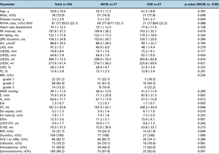
1.Baseline information(Table 1). In this study, 104 patients
different between these two groups. Compared with the NICM group, the ICM group exhibited higher values of E/A and E/ Em and shorter DT values (po0.05 for all comparisons). The
RFP was significantly higher in patients in the ICM group than in patients in the NICM group (51.9% vs. 24.7%, x2=6.816, p=0.009). One week after implantation, the AV delay optimiza-tion was not significantly different between the ICM group (119.3±19.6 ms) and the NICM group (123.1±17.2 ms).
2.CRT response rate. Six months after the implantation
procedure, 66 out of the 104 patients (63.5%) exhibited responses to CRT. The CRT response rate was significantly higher in the NICM group than in the ICM group (70.1% vs. 44.4%, x2=5.688,p=0.017).
3. Clinical beneficial effects of CRT. NYHA functional
classification, LAD, LVEDV, LVESV, LVEF, FS, and LVOT-VTI were improved six months after the implantation procedure relative to before implantation (po0.001 for all comparisons).
The degree of MR was lower than before implantation (po0.05). The SPAP did not change significantly. A
compar-ison of parameters before and after CRT is presented in Table 2. In the NICM group, the postoperative NYHA functional classification, LAD, LVEDV, LVESV, LVEF, FS, and LVOT-VTI were all significantly improved compared with the values recorded before implantation (all po0.001); the degrees of
SPAP and MR were both lower than those recorded before
implantation (both po0.05). In the ICM group, the
post-operative NYHA functional classification, LVESV, LVEF, FS, and LVOT-VTI were all significantly improved (allpo0.05),
whereas the LAD, LVEDV, SPAP, and MR did not exhibit significant changes.
Six months after implantation, the differences in echocar-diography parameters (LVESV, LVEF, FS, SPAP, and LVOT-VTI) between the two groups were significant (allpo0.05). A
com-parison of parameters between the NICM and ICM groups is shown in Table 3.
4.Factors that influence the CRT response(Table 4). The
univariate logistic regression analysis results suggested that Table 2-Clinical and echocardiographic characteristics of the overall population at baseline and follow-up.
Parameter Baseline Follow-up p-value
NYHA class, I/II/III/IV 0/0/81/23 15/58/23/8 o0.001
LAD, mm 47.2±5.7 45.9±6.5 o0.001
LVEDV, ml 300.7±73.3 279.8±84.9 o0.001 LVESV, ml 217.6±61.4 183.0±72.9 o0.001
LVEF, % 28.2±4.9 36.0±8.9 o0.001
FS, % 13.4±2.8 17.6±4.7 o0.001
SPAP, mmHg 39.1±11.9 37.8±12.8 NS
MR grade, 1/2/3 22/68/14 42/43/19 0.043 LVOT-VTI, cm 9.9±1.7 12.9±3.8 o0.001 Table 1-Basic Patient Data.
Parameter Total n=104 NICM n=77 ICM n=27 p-value (NICM vs. ICM)
Age, y 59.8±10.6 59.3±11.2 61.3±8.8 0.392
Male, n(%) 78 (75.0) 57 (74.0) 21 (77.8) 0.698
Disease course, y 5.2±2.9 5.3±3.0 5.0±2.7 0.600
NYHA class, n(%) III/IV 81 (77.9)/23 (22.1) 60 (77.9)/17 (22.1) 21 (77.8)/6 (22.2) 0.988
Heart rate (beats/min) 74.1±12.2 73.1±12.3 77.0±11.9 0.158
PR interval, ms 197.8±37.3 199.4±38.2 193.3±35.1 0.474
AV delay, ms 122.1±17.8 123.1±17.2 119.3±19.6 0.336
QRS duration, ms 154.3±24.8 155.6±24.7 150.7±25.0 0.384
SCr,mmol/l 88.8±29.5 88.3±28.5 90.1±32.7 0.794
LAD, mm 47.2±5.7 46.9±6.0 48.1±4.4 0.279
LVEDD, mm 74.8±8.0 74.7±7.6 75.2±9.1 0.771
LVESD, mm 64.8±7.8 64.4±7.4 65.7±9.0 0.454
LVEDV, ml 300.7±73.3 298.9±70.2 305.8±82.8 0.676
LVESV, ml 217.6±61.4 214.7±58.3 225.8±69.9 0.421
LVEF, % 28.2±4.9 28.4±4.7 27.4±5.4 0.332
FS, % 13.4±2.8 13.7±2.5 12.8±3.4 0.241
MR, n(%) 0.256
grade 1 22 (21.2) 17 (22.1) 5 (18.5)
grade 2 68 (65.4) 52 (67.5) 16 (59.3)
grade 3 14 (13.5) 8 (10.4) 6 (22.2)
SPAP, mmHg 39.1±11.9 38.4±12.0 41.2±11.4 0.290
E, cm/s 75.9±25.6 71.7±25.8 87.8±21.3 0.005
A, cm/s 64.6±17.1 67.1±17.0 57.5±15.8 0.012
E/A 1.3±0.7 1.2±0.7 1.7±0.7 0.003
DT, ms 187.3±43.8 193.9±42.1 168.3±43.9 0.008
Em septal, cm/s 5.2±1.3 5.4±1.4 4.7±1.0 0.010
Em lateral, cm/s 7.8±1.7 7.9±1.6 7.5±2.0 0.253
E/Em 12.3±5.6 11.2±5.1 15.4±6.1 0.001
LVOT-VTI, cm 9.9±1.7 10.0±1.7 9.6±1.5 0.242
Ts-SD, ms 70.3±31.5 72.6±30.6 63.8±33.7 0.215
RFP, n(%) 33 (31.7) 19 (24.7) 14 (51.9) 0.009
Diuretics, n(%) 104 (100) 77 (100) 27 (100) 1.000
ACE-I or ARB, n(%) 86 (82.7) 66 (85.7) 20 (74.1) 0.280
b-Blocker, n(%) 73 (70.2) 54 (70.1) 19 (70.4) 0.981
Amiodarone, n(%) 51 (49.0) 34 (44.2) 17 (63.0) 0.093
Spironolactone, n(%) 100 (96.2) 75 (97.4) 25 (92.6) 0.591
male gender, an etiology of ischemia, a disease course of heart failure, serum creatinine level, QRS duration, LAD, SPAP, severe MR, and RFP influenced the CRT response. When these factors were included in the multivariate logistic regression analysis, the results showed that QRS duration, LAD, and RFP were independent factors that influenced the CRT response.
’ DISCUSSION
This study showed that (1) the improvement of left ventricular systolic function and remodeling was redu-ced in ischemic cardiomyopathy patients compared with
non-ischemic cardiomyopathy patients; (2) compared with non-ischemic cardiomyopathy patients, ischemic cardiomyo-pathy patients exhibited more severe diastolic dysfunction; and (3) severe diastolic dysfunction (restrictive filling) was an independent factor influencing the CRT response.
Many randomized controlled trials and observational studies (3,4,13-15) have shown that CRT results in less impro-vement in LV systolic function and remodeling in ICM patients than in NICM patients. This study showed that the ICM group had lower CRT response rates six months after implantation; improvements in LVEF and LVOT-VTI, which reflect LV function, and improvements in LVESV, which reflect remodeling, were smaller in the ICM group than in Table 4-Univariate and multivariate logistic regression analyses: estimates of correlations between baseline clinical and
echocardiographic characteristics and response to CRT.
Parameter Univariate Multivariate
HR (95% CI) p-value HR (95% CI) p-value
Age, y 0.986 (0.949-1.025) 0.487
Male sex 0.261 (0.082-0.831) 0.023
Ischemic etiology 0.341 (0.138-0.840) 0.019
Disease course, y 0.815 (0.701-0.948) 0.008
NYHA class IV 0.487 (0.189-1.252) 0.135
SCr,mmol/l 0.979 (0.963-0.994) 0.007
QRS duration, ms 1.038 (1.016-1.060) 0.001 1.045 (1.013-1.077) 0.006
LAD, mm 0.821 (0.749-0.901) o0.001 0.846 (0.730-0.980) 0.026
LVEDV baseline, ml 0.997 (0.992-1.003) 0.297
LVEF baseline, % 1.019 (0.938-1.108) 0.653
SPAP baseline, mmHg 0.919 (0.879-0.961) o0.001
MR baseline grade 3 0.221 (0.052-0.944) 0.042
Ts-SD, ms 1.006 (0.993-1.020) 0.361
RFP 0.123 (0.048-0.311) o0.001 0.100 (0.025-0.397) 0.001
HR, indicates hazard ratio; CI, confidence interval.
Table 3-Clinical and echocardiographic characteristics of patients with ICM and NICM.
Parameter NICM ICM p-value
n=77 n=27
NYHA class, I/II/III/IV
Baseline 0/0/60/17 0/0/21/6 NS
Follow-up 12/45/16/4* 3/13/7/4# NS
LAD, mm
Baseline 46.9±6.0 48.1±4.4 NS
Follow-up 45.5±6.6* 47.0±6.2 NS
LVEDV, ml
Baseline 298.9±70.2 305.8±82.8 NS
Follow-up 272.6±79.9* 300.4±96.5 NS
LVESV, ml
Baseline 214.7±58.3 225.8±69.9 NS
Follow-up 174.5±67.2* 207.2±84.0# 0.045
LVEF, %
Baseline 28.4±4.7 27.4±5.4 NS
Follow-up 37.3±8.4* 32.4±9.3# 0.013
FS, %
Baseline 13.7±2.5 12.8±3.4 NS
Follow-up 18.3±4.3* 15.6±5.2# 0.008
SPAP, mmHg
Baseline 38.4±12.0 41.2±11.4 NS
Follow-up 36.0±12.1# 42.9±13.7 0.015
MR grade, 1/2/3
Baseline 17/52/8 5/16/6 NS
Follow-up 33/34/10# 9/9/9 NS
LVOT-VTI, cm
Baseline 10.0±1.7 9.6±1.5 NS
Follow-up 13.5±3.5* 11.1±3.9# 0.003
*po0.001;#
the NICM group. These results are not surprising. Several groups reported similar findings as long as a decade ago (16-18). Hummel et al. proposed that their similar findings were associated with the amount of viable myocardium (19). Because the ICM group had a smaller amount of viable myocardium, the improvement in LV function due to CRT could not be maintained. Other studies have shown that myocardial scars are associated with CRT nonresponse (5,6,20), which has been described in relation to the myo-cardial scar burden, degree of transmurality, and locations of the scars and LV lead. Chalil et al. further proposed that a scar size X33%, transmuralityX51%, and pacing over a posterolateral scar in ICM patients were associated with suboptimal CRT responses (7). However, the specific mecha-nism that causes different CRT curative effects in heart failure patients with different etiologies remains unclear. Waggoner et al. reported that even when ICM patients exhibited impro-ved LV systolic function, their diastolic function was not improved compared with NICM patients, and this result was attributed to the presence of relatively restrictive LV filling before CRT in ICM patients and a lack of improvement of end-diastolic volume and diastolic synchrony after CRT (21). The present study also showed that the ICM group presented with elevated LV filling pressures (i.e., elevated mitral E/A and E/Em) before CRT and that LVEDV was not improved six months after CRT.
Although diastolic function is an important component of overall cardiac function, it has long received much less attention than systolic function. Abnormal diastolic function is important for the development of heart failure symptoms and signs. Even during systolic heart failure, increased LV filling pressure is closely associated with exercise limitation and is independent of the degree of systolic dysfunction (8). Studies have increasingly indicated that severe diastolic dysfunction (restrictive filling) suggests a worse hemody-namic and clinical status of heart failure as well as increased mortality and heart transplantation rates (22), which is also true for patients undergoing drug treatment (23,24), surgical ventricular restoration (25,26), and CRT (27,28). Using multi-variate regression analysis, this study confirmed that severe diastolic dysfunction was an independent factor that influenced the CRT response.
Diastolic dysfunction is a typical presentation of ICM. Abnormal diastolic function occurs at the early stage of myocardial ischemia, even earlier than systolic dysfunc-tion (29,30). During myocardial ischemia, ATP produc-tion, ATP-dependent Ca2+pump activity, Ca2+uptake into
the sarcoplasmic reticulum, and Ca2+outflow decrease, and
myosin-actin complex dissociation disorder occurs, resulting in a delayed and incomplete active diastole (31). Long-term recurrent attacks of ischemia and infarction, myocardial necrosis, advanced interstitial fibrosis, scar formation, increases in ventricular stiffness, and reduction in compliance will cause passive diastolic dysfunction, sustained increase in ventricular filling pressure, and eventually, development of restrictive filling (32). This phenomenon is different from dilated cardio-myopathy, which mainly causes ventricular enlargement and systolic dysfunction. In this study, the ICM group exhibited more severe diastolic dysfunction than the NICM group. Therefore, we propose that in addition to a lack of viable myocardium and the presence of myocardial scars, the secondary severe diastolic dysfunction in ICM patients might also cause suboptimal improvement in LV function and structure after CRT.
Limitations
This study is subject to the following limitations. (1) This was a single-center observational study with no randomized control group. (2) The composition of the underlying etio-logies was not balanced; fewer cases of ICM were included. (3) Invasive measurements (such as left ventricle end dia-stolic pressure (LVEDP)) were not performed for the evalua-tion of diastolic funcevalua-tion. Therefore, it is necessary to increase the sample size and validate invasive measurements to fur-ther clarify the reason for the different curative effects of CRT for different disease etiologies.
In summary, this study again confirmed that the etiology of heart failure affected the curative effects of CRT and that a lower degree of improvement of ventricular systolic func-tion and remodeling was observed in ICM patients than in NICM patients. In addition, systolic heart failure patients with severe diastolic dysfunction showed poor CRT responses. ICM patients exhibited more severe diastolic dysfunction than NICM patients, which may be a reason for the reduced bene-ficial effect of CRT. The findings of our study are similar to those published as long as a decade ago, but the intervening period has seen little attention to this important aspect of the CRT response. We feel that it may now be reasonable to refocus on this aspect of CRT and that a study from an Asian country may reignite interest.
’ ACKNOWLEDGMENTS
The authors thank Kai Hu and Zhi-Quan Liu for statistical guidance and Zhi-Wei Du and Cui-Ping Xie for valuable discussions.
’ AUTHOR CONTRIBUTIONS
Wang Q, Chen KY and Yan J conceived and designed the study. Wang Q and Chen KY drafted the manuscript. Wang Q, Hu Y and Yang DM were responsible for the acquisition of data. Chen KY, Yu F, An CS and Su H were responsible for the analysis and interpretation of data. Yan J was responsible for the administrative support. Xu J, Su H and Yang DM were responsible for the critical revision of the manuscript for important intellectual content. All authors have read and approved thefinal version of this manuscript.
’ REFERENCES
1. Cleland JG, Daubert JC, Erdmann E, Freemantle N, Gras D, Kappenberger L, et al. The effect of cardiac resynchronization on morbidity and mor-tality in heart failure. N Engl J Med. 2005;352(15):1539-49, http://dx.doi. org/10.1056/NEJMoa050496.
2. Bax JJ, Bleeker GB, Marwick TH, Molhoek SG, Boersma E, Steendijk P, et al. Left ventricular dyssynchrony predicts response and prognosis after cardiac resynchronization therapy. J Am Coll Cardiol. 2004;44(9):1834-40, http://dx.doi.org/10.1016/j.jacc.2004.08.016.
3. Marsan NA, Bleeker GB, van Bommel RJ, Ypenburg C, Delgado V, Borleffs CJ, et al. Comparison of time course of response to cardiac resynchroni-zation therapy in patients with ischemic versus non-ischemic cardio-myopathy. Am J Cardiol. 2009;103(5):690-4, http://dx.doi.org/10.1016/ j.amjcard.2008.11.008.
4. Díaz-Infante E, Mont L, Leal J, García-Bolao I, Fernández-Lozano I, Hernández-Madrid A, et al. Predictors of lack of response to resynchro-nization therapy. Am J Cardiol. 2005;95(12):1436-40, http://dx.doi.org/ 10.1016/j.amjcard.2005.02.009.
5. Ypenburg C, Schalij MJ, Bleeker GB, Steendijk P, Boersma E, Dibbets-Schneider P, et al. Impact of viability and scar tissue on response to car-diac resynchronization therapy in ischaemic heart failure patients. Eur Heart J. 2007;28(1):33-41, http://dx.doi.org/10.1093/eurheartj/ehl379. 6. Delgado V, van Bommel RJ, Bertini M, Borleffs CJ, Marsan NA, Arnold
7. Chalil S, Foley PW, Muyhaldeen SA, Patel KC, Yousef ZR, Smith RE, et al. Late gadolinium enhancement-cardiovascular magnetic resonance as a predictor of response to cardiac resynchronization therapy in patients with ischaemic cardiomyopathy. Europace. 2007;9(11):1031-7, http://dx. doi.org/10.1093/europace/eum133.
8. Nishimura RA, Tajik AJ. Evaluation of diastolic filling of left ventricle in health and disease: Doppler echocardiography is the clinician’s Rosetta Stone. J Am Coll Cardiol. 1997;30(1):8-18, http://dx.doi.org/10.1016/ S0735-1097(97)00144-7.
9. McMurray JJ, Adamopoulos S, Anker SD, Auricchio A, Böhm M, Dick-stein K, et al. ESC guidelines for the diagnosis and treatment of acute and chronic heart failure 2012: The Task Force for the Diagnosis and Treatment of Acute and Chronic Heart Failure 2012 of the European Society of Cardiology. Developed in collaboration with the Heart Failure Associa-tion (HFA) of the ESC. Eur J Heart Fail. 2012;14(8):803-69, http://dx.doi. org/10.1093/eurjhf/hfs105.
10. Yu CM, Fung WH, Lin H, Zhang Q, Sanderson JE, Lau CP. Predictors of left ventricular reverse remodeling after cardiac resynchronization ther-apy for heart failure secondary to idiopathic dilated or ischemic cardio-myopathy. Am J Cardiol. 2003;91(6):684-8, http://dx.doi.org/10.1016/ S0002-9149(02)03404-5.
11. Nagueh SF, Appleton CP, Gillebert TC, Marino PN, Oh JK, Smiseth OA, et al. Recommendations for the evaluation of left ventricular diastolic function by echocardiography. J Am Soc Echocardiogr. 2009;22(2):107-33, http://dx.doi.org/10.1016/j.echo.2008.11.023.
12. Whinnett ZI, Sohaib SM, Jones S, Kyriacou A, March K, Coady E, et al. British randomised controlled trial of AV and VV optimization (‘‘BRAVO’’) study: rationale, design, and endpoints. BMC Cardiovasc Disord. 2014;14:42, http://dx.doi.org/10.1186/1471-2261-14-42.
13. Barsheshet A, Goldenberg I, Moss AJ, Eldar M, Huang DT, McNitt S, et al. Response to preventive cardiac resynchronization therapy in patients with ischaemic and nonischaemic cardiomyopathy in MADIT-CRT. Eur Heart J. 2011;32(13):1622-30, http://dx.doi.org/10.1093/eur-heartj/ehq407.
14. Chen Y, Duan C, Liu F, Shen S, Chen P, Bin J. Impact of etiology on the outcomes in heart failure patients treated with cardiac resynchronization therapy: a meta-analysis. PLoS One. 2014;9(4):e94614, http://dx.doi.org/ 10.1371/journal.pone.0094614.
15. Ghio S, Freemantle N, Scelsi L, Serio A, Magrini G, Pasotti M, et al. Long-term left ventricular reverse remodelling with cardiac resynchronization therapy: results from the CARE-HF trial. Eur J Heart Fail. 2009;11(5):480-8, http://dx.doi.org/10.1093/eurjhf/hfp034.
16. Salukhe TV, Francis DP, Clague JR, Sutton R, Poole-Wilson P, Henein MY. Chronic heart failure patients with restrictive LV filling pattern have significantly less benefit from cardiac resynchronization therapy than patients with late LV filling pattern. Int J Cardiol. 2005;100(1):5-12, http:// dx.doi.org/10.1016/j.ijcard.2005.01.010.
17. Porciani MC, Valsecchi S, Demarchi G, Colella A, Michelucci A, Pieragnoli P, et al. Evolution and prognostic significance of diastolic filling pattern in cardiac resynchronization therapy. Int J Cardiol. 2006;112(3):322-8, http:// dx.doi.org/10.1016/j.ijcard.2005.10.002.
18. Gradaus R, Stuckenborg V, Löher A, Köbe J, Reinke F, Gunia S, et al. Diastolic filling pattern and left ventricular diameter predict response and prognosis after cardiac resynchronisation therapy. Heart. 2008;94(8): 1026-31, http://dx.doi.org/10.1136/hrt.2007.126193.
19. Hummel JP, Lindner JR, Belcik JT, Ferguson JD, Mangrum JM, Bergin JD, et al. Extent of myocardial viability predicts response to biventricular
pacing in ischemic cardiomyopathy. Heart Rhythm. 2005;2(11):1211-7, http://dx.doi.org/10.1016/j.hrthm.2005.07.027.
20. Ypenburg C, Roes SD, Bleeker GB, Kaandorp TA, de Roos A, Schalij MJ, et al. Effect of total scar burden on contrast-enhanced magnetic resonance imaging on response to cardiac resynchronization therapy. Am J Cardiol. 2007;99(5):657-60, http://dx.doi.org/10.1016/j.amjcard.2006.09.115. 21. Waggoner AD, Rovner A, de las Fuentes L, Faddis MN, Gleva MJ,
Sawhney N, et al. Clinical outcomes after cardiac resynchronization therapy: importance of left ventricular diastolic function and origin of heart failure. J Am Soc Echocardiogr. 2006;19(3):307-13, http://dx.doi. org/10.1016/j.echo.2005.10.014.
22. Xie GY, Berk MR, Smith MD, Gurley JC, DeMaria AN. Prognostic value of Doppler transmitral flow patterns in patients with congestive heart fail-ure. J Am Coll Cardiol. 1994;24(1):132-9, http://dx.doi.org/10.1016/0735-1097(94)90553-3.
23. Ghio S, Alessandrino G, Albertini R, Klersy C, Girardi B, Maggi G, et al. Prognostic stratification of patients with chronic systolic heart failure using biomarkers and Doppler echocardiography. J Cardiovasc Med (Hagerstown). 2014;15(6):470-5.
24. Bajraktari G, Miccoli M, Buralli S, Fontanive P, Elezi S, Metelli MR, et al. Plasma metalloproteinase-9 and restrictive filling pattern as major predictors of outcome in patients with ischemic cardiomyopathy. Eur J Intern Med. 2012;23(7):616-20, http://dx.doi.org/10.1016/j.ejim.2012. 04.013.
25. Shudo Y, Matsumiya G, Sakaguchi T, Miyagawa S, Yamauchi T, Takeda K, et al. Impact of surgical ventricular reconstruction for ischemic dilated cardiomyopathy on restrictive filling pattern. Gen Thorac Cardiovasc Surg. 2010;58(8):399-404, http://dx.doi.org/10.1007/s11748-010-0597-8. 26. Marui A, Nishina T, Saji Y, Yamazaki K, Shimamoto T, Ikeda T, et al.
Significance of left ventricular diastolic function on outcomes after sur-gical ventricular restoration. Ann Thorac Surg. 2010;89(5):1524-31, http:// dx.doi.org/10.1016/j.athoracsur.2010.01.067.
27. Ciampi Q, Pratali L, Citro R, Villari B, Picano E, Sicari R. Additive value of severe diastolic dysfunction and contractile reserve in the identification of responders to cardiac resynchronization therapy. Eur J Heart Fail. 2011; 13(12):1323-30, http://dx.doi.org/10.1093/eurjhf/hfr132.
28. Yamamoto M, Seo Y, Ishizu T, Kawamatsu N, Sato K, Sugano A, et al. Prognostic significance of persistent restrictive filling pattern after cardiac resynchronization therapy. J Echocardiogr. 2015;13(1):20-6, http://dx.doi. org/10.1007/s12574-014-0234-0.
29. Nesto RW, Kowalchuk GJ. The ischemic cascade: temporal sequence of hemodynamic, electrocardiographic and symptomatic expressions of ischemia. Am J Cardiol. 1987;59(7):23C-30C, http://dx.doi.org/10.1016/ 0002-9149(87)90192-5.
30. Carluccio E, Biagioli P, Alunni G, Murrone A, Leonelli V, Pantano P, et al. Effect of revascularizing viable myocardium on left ventricular diastolic function in patients with ischaemic cardiomyopathy. Eur Heart J. 2009; 30(12):1501-9, http://dx.doi.org/10.1093/eurheartj/ehp125.
31. Bonow RO, Udelson JE. Left ventricular diastolic dysfunction as a cause of congestive heart failure. Mechanisms and management. Ann Intern Med. 1992;117(6):502-10, http://dx.doi.org/10.7326/0003-4819-117-6-502.