J Bras Pneumol. 2010;36(3):386-389
to face new challenges related to containing its rapid dissemination and delivering the appropriate treatment to the affected patients.
Although, to date, this disease has not proven to be more lethal than is seasonal influenza, it is a different disease, with different characteristics: it affects apparently healthy young individuals; transmission occurs very quickly; and its course can be severe.
It is the duty of epidemiological surveillance to keep case registries updated, especially regarding severe cases, and to identify potential risk factors for H1N1 infection.(2,3) Public and private health care facilities should be prepared to confront the situation, showing organizational capacity and allowing timely access to appropriate treatment. In addition, it is essential that an H1N1 vaccine be developed.
Introduction
The World Health Organization monitors diseases capable of spreading rapidly from one country to another, including infections that can result in severe acute respiratory syndrome (SARS).(1)
Among the principal etiologic agents that result in SARS are viruses (influenza A virus, dengue virus, respiratory syncytial virus, adenovirus, hantavirus and coronavirus) and other agents (pneumococci, other bacteria, Legionella sp., leptospirosis, etc.) The diagnosis of viral pneumonia is a differential diagnosis and is based on the absence of sputum production, failure to detect bacteria on culture and normal or slightly elevated leukocyte counts.(1)
The emergence of the flu pandemic caused by a new subtype of the influenza A virus (H1N1) has forced populations and health professionals
Severe acute respiratory syndrome caused
by the influenza A (H1N1) virus*
Síndrome respiratória aguda grave causada por influenza A (subtipo H1N1)
Sandra Aparecida Ribeiro, Graziela Sgreccia Brasileiro,
Luciana Novaes Campello Soleiman, Cristiano Cruz Silva, Cláudio Shoki Kavaguti
Abstract
In view of the pandemic caused by a new virus, influenza A (H1N1), we report the case of a 56-year-old patient without relevant risk factors and with severe acute respiratory syndrome resulting from infection with this virus. We present the results of laboratory tests and the imaging findings (chest X-ray and CT scans). The evolution was favorable, and the patient was discharged after 14 days.
Keywords: Influenza A virus, H1N1 subtype; Severe acute respiratory syndrome; Patient care.
Resumo
Frente à pandemia causada por um novo vírus, influenza A (H1N1), descrevemos o caso de um paciente de 56 anos com síndrome respiratória aguda grave causada por influenza A (H1N1) sem fatores de risco importantes. Os resultados dos exames laboratoriais e de imagem (radiografia e TC de tórax) são apresentados aqui. O paciente teve boa evolução e recebeu alta hospitalar em 14 dias.
Descritores: Vírus da influenza A subtipo H1N1; Síndrome respiratória aguda grave; Assistência ao paciente.
* Study carried out in the Department of Preventive Medicine, Federal University of São Paulo, São Paulo, Brazil.
Correspondence to: Sandra Aparecida Ribeiro. Departamento de Medicina Preventiva, Universidade Federal de São Paulo, Rua Borges Lagoa, 1341, Vila Clementino, CEP 04038-034, São Paulo, SP, Brasil.
Tel/Fax: 55 11 5571-5000. E-mail: sandrarib@uol.com.br Financial support: None.
Submitted: 9 September 2009. Accepted, after review: 11 March 2010.
Severe acute respiratory syndrome caused by the influenza A (H1N1) virus
J Bras Pneumol. 2010;36(3):386-389 387
Doppler echocardiogram and X-ray of the paranasal sinuses were normal.
Blood and urine cultures were negative for aerobic and anaerobic microorganisms. Testing (real-time PCR) of oropharyngeal secretion aspirate was positive for the influenza A (H1N1) virus.
The patient was admitted to the ICU and treated with oseltamivir, ceftriaxone, azithromycin and a corticosteroid. He received ventilatory support with intermittent positive pressure breathing, which was maintained at 50% via a Venturi mask, at a resting SpO2 of 92%. The patient progressively improved and was discharged from the ICU ten days later, presenting an SpO2 of 94% on room air, together with radiological improvement (Figure 3). He developed no fever or hemodynamic instability during the hospital stay.
Discussion
According to the influenza surveillance protocol, cases of SARS are defined as those
Case report
A 56-year-old nonsmoking businessman from São Paulo, Brazil, presented with mild hypertension controlled with candesartan (8 mg/day). The patient reported no diabetes or other comorbidities.
In the previous week, the patient had developed the flu, characterized by fever (38.5°C), generalized pain, rhinorrhea and nasal obstruction. Three days later, he began to present extreme fatigue and malaise and sought treatment in the emergency room, where he was submitted to chest X-ray and was diagnosed with pneumonia. On that same date, the blood workup revealed the following: hemoglobin, 14.8 g/dL; hematocrit, 41.9%; leukocytes, 4,600/mm3 (3-85-0-0-11-1); platelet count, 132,000/mm3; and ESR, 16 mm/h. The patient was treated with oral moxifloxacin and was instructed to return if his condition worsened. Since the patient felt increasingly tired, he returned to the emergency room two days later. He reported no contact with suspected cases of infection with influenza A (H1N1) virus.
Physical examination revealed that the patient was in satisfactory general health, was well hydrated and with good color, had no jaundice or lymph node enlargement and had +/4 cyanosis and +++/4 dyspnea. In addition, physical examination revealed the following: arterial pressure, 130-90 mmHg; body temperature, 36.8°C; weight, 68 kg; height, 1.65 m; BMI, 25.0 kg/m2; and RR, 30 breaths/min. Pulmonary auscultation revealed diffuse crackles in both lungs and normal, rhythmic heart sounds without heart murmurs. There were no organomegalies in his abdomen, there was no edema in his limbs, and his SpO2 was 87% on room air and 94% when receiving oxygen via a mask at 5 L/min.
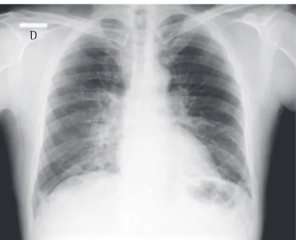
A chest X-ray showed worsening of the bilateral alveolar infiltrate, and a chest CT scan showed multifocal ground-glass infiltrate (Figures 1 and 2).
A second blood workup revealed the following: hemoglobin, 15 g/dL; hematocrit, 44.5%; leukocytes, 3,300/mm3 (10-79-0-0-8-1); platelet count, 131,000/mm3; and C-reactive protein, 12.36 mg/dL. The results of the arterial blood gas analysis were as follows: pH 7.45; PaCO2, 35 mmHg; PaO2, 50 mmHg; bicarbonate, 24 mEq/L; base excess, +0.5 mEq/L; and SaO2, 87.2%.
Figure 1 - Chest X-ray at admission.
388 Ribeiro SA, Brasileiro GS, Soleiman LNC, Silva CC, Kavaguti CS
J Bras Pneumol. 2010;36(3):386-389
respiratory diseases and cardiovascular diseases. Of the patients evaluated, 1,538 (7%) died. The concomitance of risk factors in the same individual results in higher mortality.(4)
The clinical and radiological manifestations caused by the influenza virus are not specific. The differential diagnosis includes infection with other viruses and bacteria, such as respiratory syncytial virus, coronavirus, parainfluenza, rhinovirus, adenovirus and Mycoplasma sp.(1)
In times of an influenza A pandemic, a high level of suspicion should be maintained, and, in all severe cases, the diagnosis should be confirmed by testing (real-time PCR) of nasopharyngeal or oropharyngeal aspirates for influenza A.(4) In addition, the use of antiviral agents should be initiated, if possible, in the first 48 h after the onset of symptoms.(5) The early use of antiviral agents reduces the severity of the disease and of the symptoms.(5)
In general, SARS has an early onset, is accompanied by severe hypoxemia and has a prolonged course, with extensive tissue damage caused by the virus, characterized by necrotizing bronchiolitis, neutrophilic infiltrate, diffuse alveolar damage and formation of hyaline membranes.(6)
The most common radiological
manifestations of influenza pneumonia consist of reticulonodular opacities, with or without superimposed areas of consolidation. Less commonly, patients with influenza pneumonia can present with focal areas of consolidation, typically in the lower lobes, without apparent reticular or reticulonodular opacities. The radiological abnormalities typically resolve in approximately three weeks. Consolidations can occur due to secondary bacterial pneumonia.(7,8)
On chest CT scans, various patterns are possible. However, the ones most commonly reported are those showing lobular areas, typically bilateral, of ground-glass attenuation. Other possible patterns are the presence of focal areas of consolidation, centrilobular nodules and the tree-in-bud pattern.(9)
The case reported here was a case of SARS caused by the influenza A (H1N1) virus and presenting a prolonged clinical and radiological course. Although there were no characteristics of associated bacterial infection, treatment with bactericidal antibiotics was initiated early. presenting with fever > 38°C, cough and
dyspnea, with or without gastrointestinal manifestations or other signs of severity, such as tachypnea (RR > 25 breaths/min), hypotension or clinical, laboratory and radiological findings consistent with bronchopneumonia, as well as those resulting in death.(2,3)
Of the 76,639 cases of SARS that had been added to the Case Registry Database as of November 7, 2009, 24,729 (32.3%) were confirmed for infection with the influenza virus by laboratory testing, 22,565 (91.0%) of which were cases of infection with the influenza A (H1N1) virus. There was a slight preponderance of cases in females (57%), the median age of the confirmed cases was 25 years, and the greatest proportion of cases occurred among those in the 15-49 year age bracket.(4)
In Brazil, the incidence of SARS caused by the influenza A (H1N1) virus is 12/100,000 population, this incidence being of note in the following states: Paraná, 109/100,000; Santa Catarina, 15/100,000; and São Paulo, 14/100,000. The nationwide mortality rate is 0.8/100,000 population.(4)
Of the patients with SARS caused by the influenza A (H1N1) virus, 13,316/24,729 (54%) had at least one risk factor (such as pregnancy) that could lead to complications. Among the associated risk factors, in addition to pregnancy, are (in ascending order of frequency) obesity, hemoglobinopathies, diabetes mellitus, kidney disease, age under 2 years, age over 60 years, metabolic diseases, immunosuppressive diseases,
Severe acute respiratory syndrome caused by the influenza A (H1N1) virus
J Bras Pneumol. 2010;36(3):386-389 389
epidemiológica da Influenza Pandêmica (H1N1) 2009 no Mundo e no Brasil, até a Semana Epidemiológica 44 de 2009. [Adobe Acrobat document, 14p.] Available from: http://portal.saude.gov.br/portal/arquivos/pdf/ boletim_influenza_se_44.pdf
5. Pan American Health Organization – PAHO [homepage on the Internet]. Washington: World Health Organization [cited 2009 Apr 1]. Guias de la OMS para el uso de vacunas y antiviricos en las pandemias de influenza. [Adobe Acrobat document, 67p.] Available from: http:// www.paho.org/Spanish/AD/DPC/CD/vir-flu-guia-oms-uso-vac-antivirales.pdf
6. Perez-Padilla R, de la Rosa-Zamboni D, Ponce de Leon S, Hernandez M, Quiñones-Falconi F, Bautista E, et al. Pneumonia and respiratory failure from swine-origin influenza A (H1N1) in Mexico. N Engl J Med. 2009;361(7):680-9.
7. Kim EA, Lee KS, Primack SL, Yoon HK, Byun HS, Kim TS, et al. Viral pneumonias in adults: radiologic and pathologic findings. Radiographics. 2002 Spec No:S137-49.
8. Greenberg SB. Respiratory viral infections in adults. Curr Opin Pulm Med. 2002;8(3):201-8.
9. Muller NL, Silva CI. Imaging of the Chest. Expert radiology series. Philadelphia: Saunders/Elsevier; 2008.
References
1. Pan American Health Organization – PAHO [homepage on the Internet]. Washington: World Health Organization [cited 2009 Apr 1]. Health Establishments Preparation for Unusual or Unexpected Cases or Clusters of Severe Acute Respiratory Infection (SARI). Available from: http://new.paho.org/hq/index.php?option=com_conten t&task=view&id=1265&Itemid=569
2. Portal da Saúde [homepage on the Internet]. Brasília: Ministério da Saúde [cited 2009 Apr 1]. Informe Epidemiológico - Influenza A (H1N1). Situação epidemiológica da nova influenza A (H1N1) no Brasil, até semana epidemiológica 33 de 2009. [Adobe Acrobat document, 10p.] Available from: http://portal. saude.gov.br/portal/arquivos/pdf/informe_influenza_ se_33_25_08_2009.pdf
3. Portal da Saúde [homepage on the Internet]. Brasília: Ministério da Saúde [cited 2009 Apr 1]. Diretrizes para o enfrentamento à pandemia de influenza A (H1N1): ações da atenção primária à saúde. [Adobe Acrobat document, 36p.] Available from: http://portal.saude.gov.br/portal/ arquivos/pdf/protocolo_influenzaa_aps_atualizado.pdf 4. Portal da Saúde [homepage on the Internet]. Brasília:
Ministério da Saúde [cited 2009 Apr 1]. Situação
About the authors
Sandra Aparecida Ribeiro
Head of the Department of Clinical Preventive Medicine. Federal University of São Paulo, São Paulo, Brazil.
Graziela Sgreccia Brasileiro
Pulmonologist. São Paulo Hospital for State Civil Servants, São Paulo, Brazil.
Luciana Novaes Campello Soleiman
Pulmonologist. Tatuapé Municipal Hospital, São Paulo, Brazil.
Cristiano Cruz Silva
Intensivist and Pulmonologist. Diadema City Hall, Diadema, Brazil.
Cláudio Shoki Kavaguti