J Bras Pneumol. 2007;33(5):612-615
Case Report
* Study carried out at the Faculdade de Ciências Médicas da Santa Casa de São Paulo – FCMSCSP, Santa Casa School of Medical Sciences in São Paulo – São Paulo (SP) Brazil.
1. Third-year Medical Resident in the Thoracic Surgery Department. Faculdade de Ciências Médicas da Santa Casa de São Paulo – FCMSCSP, Santa Casa School of Medical Sciences in São Paulo – São Paulo (SP) Brazil.
2. Fourth-year Medical Resident in the Thoracic Surgery Department. Faculdade de Ciências Médicas da Santa Casa de São Paulo – FCMSCSP - Santa Casa School of Medical Sciences in São Paulo – São Paulo (SP) Brazil.
3. Full Professor in the Thoracic Surgery Department. Faculdade de Ciências Médicas da Santa Casa de São Paulo – FCMSCSP, Santa Casa School of Medical Sciences in São Paulo – São Paulo (SP) Brazil.
4. PhD Professor in the Surgery Department. Faculdade de Ciências Médicas da Santa Casa de São Paulo – FCMSCSP, Santa Casa School of Medical Sciences in São Paulo – São Paulo (SP) Brazil.
5. PhD Professor in the Surgery Department. Faculdade de Ciências Médicas da Santa Casa de São Paulo – FCMSCSP, Santa Casa School of Medical Sciences in São Paulo – São Paulo (SP) Brazil.
6. Graduate Student in the Pathology Department. Faculdade de Medicina da Universidade de São Paulo – FMUSP, University of São Paulo School of Medicine – São Paulo (SP) Brazil.
Correspondence to: Ricardo Alexandre Faria. Rua Paracatu, 398, apto. 3B; Pq. Imperial (Saúde), CEP-04302-021, São Paulo, SP, Brasil. Tel 55 11 2176-7000. E-mail: rickpreto@zipmail.com.br
Submitted: 17 July 2006. Accepted, after review: 17 October 2006.
Intrapulmonary teratoma*
Ricardo Alexandre Faria1, José Alexandre Bizon2, Roberto Saad Junior3,
Vicente Dorgan Neto4, Marcio Botter5, Mauro Ajaj Saieg6
Abstract
Case report of a 49-year-old man, presenting chest pain and bloody sputum for six months. Chest X-ray and computed tomography scan showed opacification in the left upper lobe. The bronchoscopy showed bronchial hemorrhage in the lingular bronchial segment. Due to diagnostic and therapeutic needs, this patient underwent a left inframamillary thoracotomy. The anatomopathological analysis of the surgical sample revealed an intrapulmonary teratoma. The patient presented favorable evolution and is now under outpatient follow-up treatment.
Keywords: Teratoma; Lung; Neoplasms.
Introduction
Teratoma can be found in various organs. In decreasing order of location, it is seen in the ovaries, testicles, sacro-coccygeal region, mediastinum, and other sites.(1-4) In the
international literature, 43 cases of intrapulmonary teratoma have been described, the first in 1939.(3)
Intrapulmonary teratoma originates from totipotential cells of one or more of the three germinative layers, which can differentiate themselves into any type of tissue.(2,3) There
are no specific clinical or radiological characteristics, and the anatomopathological study is the definitive diagnostic method.(1,3,4) Curative treatment consists of complete
resec-tion of the tumor.(3,4)
Case report
We report the case of a 49-year-old male Caucasian who was a businessman from Italy and a resident of São Paulo. The patient sought emergency treatment, presenting with bloody sputum and chest pain in the left anterolateral region for six months. The epidemiology was negative for tuberculosis. He was a 20-pack-year smoker. He presented good general health status. The physical examination revealed discrete wheezing in the middle third of the left hemithorax, with no other alterations. The body mass index was 36.3 kg/m2.
meas-Intrapulmonary teratoma
J Bras Pneumol. 2007;33(5):612-615
613
Discussion
Teratomas present slow growth and are typi-cally benign.(3,4) When in the lung, they are most
often found in the upper left lobe, as in the case reported.(1-6) Symptomatology varies according to
the location and size of the tumor, as well as to the histological components.(3) In this case, the patient
reported bloody sputum and thoracic pain which, together with cough, are the most commonly reported symptoms. Other symptoms reported in the literature are trichoptysis, bronchiectasis, fever, abscess, repeated pneumonia, signs of compression of thoracic structures, and fistulas, with or without infection of the tumor.(1-6)
Teratoma is an extremely rare disease that affects both genders and all age brackets equally.(1-6) The
size of the tumor has not relationship with malig-nancy.(1,3,4) On chest X-rays, teratoma can present
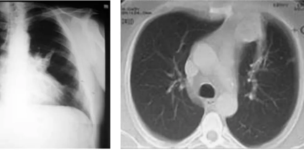
as lobulated upper lung lobe tumors or intrapa-renchymal opacities, corresponding to the exam shown above. On tomography scans of the chest, homogeneous or heterogeneous tumors can be observed.(1-3,5,7)
In anatomopathology, these tumors present endodermic components (pancreatic acinar tissue, with or without islets of Langerhans and respiratory epithelium), thus differing those found at extratho-racic sites. Intrapulmonary teratoma originates in cells that can differentiate into any tissue type. In the case reported, microscopy of the resected surgical sample revealed pancreatic tissue, carti-uring approximately five centimeters in diameter
(Figure 1), confirmed through a tomography scan of the chest (Figure 2). Sputum culture for Koch’s bacillus was negative, and the patient presented no reaction on the purified protein derivative test. Bronchoscopy revealed hematogenous remnants in the lingular bronchial segment, without further alterations. A transbronchial biopsy revealed a nonspecific inflammatory process. Pulmonary func-tion test results showed mild obstructive ventilatory disorder, with a forced expiratory volume in one second of 82%.
In view of the imperative diagnostic and thera-peutic conditions, surgery was recommended. A left inframamillary thoracotomy was performed, revealing a tumor in the lingular region, and we opted for a segmentectomy. The frozen section biopsy findings were negative for neoplasia, and the sample remnants were sent for anatomopathological analysis. The patient presented favorable evolution and was discharged on postoperative day 3.
The anatomopathological examination showed that the sample was nodular, weighed 25 g, and measured 5 × 4.5 × 3.5 cm, with a smooth, brownish surface, and that there were pulmonary and adipose tissue adherences. The distal pulmonary margin was normal. Microscopy showed mature epithelial tissues, such as pancreatic, epidermis, sebaceous glands, and mature mesenchymal tissues, such as bone and cartilage (Figure 3). Therefore, we concluded that it was a primary intrapulmonary mature teratoma.
614 Faria RA, Bizon JA, Saad Junior R, Dorgan Neto V, Botter M, Saieg MA
J Bras Pneumol. 2007;33(5):612-615
Mixed cell types
b) With non-germ cell tumor areas
Carcinoma - Sarcoma - Malignant embry-onal tumor
Mixed cell type
c) Malignant immature teratoma(8)
The prognosis of malignant teratoma is poor. In a study comprising eleven patients with malig-nant teratoma, seven patients (63.6%) died after a six-month follow-up period. None of these patients underwent surgery, or the tumor was nonresectable by thoracotomy. Metastases were reported in three of these patients.(4)
In patients with benign teratoma, the recom-mended procedure is complete resection of the tumor, upon which the patient is considered totally cured.(3) Patients who did not undergo surgery can
present massive hemoptysis or increased tumor growth and can die.(3) In the case reported, the
patient evolved without complications and did not require any further therapeutic measures, as has been reported in the literature, and, after two years of outpatient follow-up treatment, presented no signs of relapse.(8)
In conclusion, intrapulmonary teratoma is a rare tumor, and surgery is the definitive treatment. Definitive confirmation of the diagnosis is made through anatomopathological examination, and prognosis is good when the condition is properly treated.
lage, bone, epidermis and sebaceous glands.(2,3) It
has been suggested that intrapulmonary teratoma derives from the thymic tissue of the third branchial arch.(2-6) However, mediastinal teratoma results from
the dislocation and early separation of the thymic tissue, during embryogenesis, with capture and migration of thymic primordia during the develop-ment of the respiratory system.(2)
The classification system used in order to cate-gorize teratomas as benign or malignant is that proposed by Gonzales-Crussi in 1982, which has proven practical to date.(8)
1) Benign teratomas: a) Mature teratoma
Grade 0 - all components are well differentiated
Grade 1 - non-undifferentiated compo-nents do not exceed 10%
b) Immature teratoma
Grade 2 - immature tissues among 10 to 50% of components
Grade 3 - undifferentiated components over 50% and probable metastatic potential.
Benign evolution is still possible. 2) Malignant teratomas:
a) With germ cell tumor areas
Germinoma (seminoma - dysgerminoma) Embryonal carcinoma - Coriocarcinoma Yolk sac tumor
Figure 3 - a) Sebaceous glands and hair follicle; and b) bone parenchyma.
Intrapulmonary teratoma
J Bras Pneumol. 2007;33(5):612-615
615
5. Gomes NA, Medeiros GA. Tumores Benignos do Pulmão. In: Saad Jr R, Carvalho WR, Ximenes-Neto M, Forte V. Cirurgia Torácica Geral. São Paulo: Atheneu; 2005. p. 355-62. 6. Eren MN, Balci AE, Eren S. Benign intrapulmonary
teratoma: report of a case. J Thorac Cardiovasc Surg. 2003;126(3):855-7.
7. Fatureto MC. Tumores Benignos. In: Saad Jr R, Carvalho WR, Ximenes-Neto M, Forte V. Cirurgia Torácica Geral. São Paulo: Atheneu; 2005. p. 869-88.
8. Thomson JC. Tumores de Células Germinativas. In: Saad Jr R, Carvalho WR, Ximenes-Neto M, Forte V. Cirurgia Torácica Geral. São Paulo: Atheneu; 2005. p. 893-8.
References
1. Asano S, Hoshikawa Y, Yamane Y, Ikeda M, Wakasa H. An intrapulmonary teratoma associated with bronchiectasia containing various kinds of primordium: a case report and review of the literature. Virchows Arch. 2000;436(4):384-8. 2. Joo M, Kang YK, Lee HK, Lee HS, Yum HK, Bang SW, et al.
Intrapulmonary and gastric teratoma: report of two cases. J Korean Med Sci. 1999;14(3):330-4.
3. Medeiros CWL, Kondo W, Dyckyj MT, Suzuki N. Teratoma Intrapulmonar. J Pneumol. 2001;27(5):272-4.