AR
TIGO ORIGINAL / ORIGINAL AR
TICLE
INTRODUCTION
Upper gastrointestinal bleeding (UGB) is deined as the intraluminal bleeding proximal to the Treitz ligament(14). Regarding the etiology, can be classiied as nonvariceal and variceal UGB. Hematemesis and melena are signs and/or symptoms of UGB. The cause of UGB may be established in approximately 80% of cases, and the most frequent diagnoses are peptic ulcers (37%-55%), gastroduodenal erosions (6%-24%), esophageal varices (10%-23%), esophagitis (4%-6%)(14).
UGB is the most common emergency in Gastroenterology, with signiicant clinical and eco-nomic implications. It presents incidence of 0,7 to 1,5 cases every 1,000 inhabitants of the general popula-tion, annually, in the United States of America (USA). It is cause of 250,000 to 300,000 hospitalizations each year, and estimated total spending at more than 2.5 billion dollars per year, by the American health care system(43). Mortality is 3.5 to 10% in cases of non-variceal UGB(31), and 15% to 20% in 6 weeks after
PROPOSAL OF A CLINICAL CARE PATHWAY
FOR THE MANAGEMENT OF ACUTE UPPER
GASTROINTESTINAL BLEEDING
Matheus Cavalcante
FRANCO
1, Frank Shigueo
NAKAO
1,2, Rodrigo
RODRIGUES
2,
Fauze
MALUF-FILHO
3, Gustavo Andrade de
PAULO
4and Ermelindo Della
LIBERA
1,2Received 29/6/2015 Accepted 25/8/2015
ABSTRACT - Background - Upper gastrointestinal bleeding implies signiicant clinical and economic repercussions. The correct establish-ment of the latest therapies for the upper gastrointestinal bleeding is associated with reduced in-hospital mortality. The use of clinical pathways for the upper gastrointestinal bleeding is associated with shorter hospital stay and lower hospital costs. Objective- The primary objective is the development of a clinical care pathway for the management of patients with upper gastrointestinal bleeding, to be used in tertiary hospital. Methods- It was conducted an extensive literature review on the management of upper gastrointestinal bleeding, contained in the primary and secondary information sources. Results- The result is a clinical care pathway for the upper gastrointestinal bleeding in patients with evidence of recent bleeding, diagnosed by melena or hematemesis in the last 12 hours, who are admitted in the emergency rooms and intensive care units of tertiary hospitals. In this compact and understandable pathway, it is well demonstrated the management since the admission, with deinition of the inclusion and exclusion criteria, passing through the initial clinical treatment, posterior guidance for endoscopic therapy, and referral to rescue therapies in cases of persistent or rebleeding. It was also included the care that must be taken before hospital discharge for all patients who recover from an episode of bleeding. Conclusion- The introduction of a clinical care pathway for patients with upper gastrointestinal bleeding may contribute to standardization of medical practices, decrease in waiting time for medications and services, length of hospital stay and costs. HEADINGS- Gastrointestinal hemorrhage. Hematemesis. Melena. Endoscopy. Critical pathways. Clinical protocols.
Declared conflict of interest of all authors: none Disclosure of funding: no funding received
Research performed at: Departamento de Gastroenterologia, Universidade Federal de São Paulo, São Paulo, Brasil.
1 Departamento de Gastroenterologia e Unidade de Endoscopia do Hospital Universitário, Universidade Federal de São Paulo, São Paulo, SP, Brasil; 2 Departamento de Endoscopia, Fleury Medicina e Saúde, São Paulo, SP, Brasil; 3 Departamento de Endoscopia, Instituto de Câncer de São Paulo, São Paulo, SP, Brasil; 4 Departamento de Endoscopia, Hospital Israelita Albert Einstein, São Paulo, SP, Brasil.
variceal UGB, but it may reach 30% in patients with advanced cirrhosis(26).
Brazil data on UGB are not known, but is expected to be comparable to global statistics. In addition, the low socioeconomic condition is associated with increased incidence of UGB(12).
The correct establishment of support measures and speciic therapy for the UGB is associated with decreased mortality. Study demonstrated reduction of up to 28% on in-hospital mortality with the use of the latest therapies on variceal UGB(15).
Clinical pathways in urgent and emergency services are extensively used guides, which aim to provide a simpliied view to the diagnostic and therapeutic approach of frequent and relevant diseases of daily medical practice. For these characteristics, the critical use of clinical pathways is an important tool for physi-cians, especially for those under training, because it enables simpliied, standardized and effective service.
shorter hospital stay and lower hospital costs. With regard speciically to the treatment of UGB, studies have shown reduced cost and length of hospital stay after introduction and use of clinical pathways(37, 43).
OBJECTIVE
The objective of this study was the development of a clinical care pathway for patients with UGB, who are as-sisted at the emergency rooms and intensive care units (ICUs) of tertiary hospitals. This clinical pathway was originally designed to be used at the University Hospital of Federal University of São Paulo.
METHODS
For the development of an updated clinical pathway, we conducted an extensive review about the management of UGB. Initially with the analysis of the latest consen-sus opinion of experts, contained both in national and international databases, and then the data contained in the secondary information sources like Cochrane Review, UptoDate, Dynamed, ACP Journal Club and Evidence Based Medicine. Subsequently, we searched for evidences in the primary information sources of SciELO, Medline, Lilacs and Embase.
We use a structured way to elaborate a question, that is synthesized by the acronym P.I.C.O., where P corresponds to the patient or population, I of intervention or indicator, C for control or comparison, and O of outcome(10). From the structured question, we identiied the keywords in the Virtual Health Library and the MeSH Database. Finally, the keywords were organized for research with the addition of the booleans AND, OR or NOT.
Articles were selected initially from the title, and its relevance was then conirmed by the review of the corre-sponding abstract. We excluded publications with content that was considered irrelevant. Additional articles were iden-tiied by manual search of references lists of selected articles. Only the class I and II recommendations were included for the development of this clinical pathway, in accordance with the classiication of recommendations and levels of evidence of the American College of Cardiology Foundation and the American Heart Association Practice Guidelines(4)(Figure 1).
This study was approved by the Ethics and Research Com-mittee, from Federal University of São Paulo (#115.913).
LITERATURE REVIEW
Initial assessment
The evaluation of patients with suspected UGB must contain data about the current and past medical histories, physical examination and laboratory tests. The objectives are to early assess the severity of bleeding, identify a possible etiology, and provide initial therapy.
Blood tests with complete blood count, coagulation proile, electrolytes (sodium, potassium), urea and creatinine
must be requested on admission. The blood typing should also be ordered by the possibility of blood transfusions(14).
Risk stratification
Risk stratiication of cases with UGB enables to iden-tify the high-risk group with increased chance for worse outcomes, and also may select the low-risk group that can be discharged early. For that, it is used prognostic scales take into consideration clinical, endoscopic, and laboratory indings(13, 44).
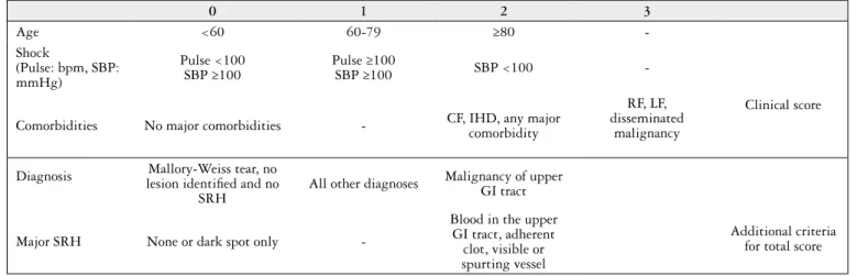
The Rockall score (RS) was primarily developed for assessment of mortality after 30 days of UGB (Figure 2). Rebleeding and mortality directly increase with the value of the RS(44).
Patients with total RS ≤2 are considered low-risk, and can receive early hospital discharge. The clinical RS (pre-endoscopic) equal to zero identiies about 15% of cases. These patients can be submitted to outpatient endoscopy, given the mortality and rebleeding low risks(44).
Glasgow Blatchford score (GBS) was developed to pre-dict mortality and medical intervention (blood transfusion, endoscopic or surgical treatment) after UGB episode(13). The GBS only takes into account laboratory tests and clini-cal indings to its clini-calculation, and can be easily used prior endoscopy (Table 1).
Patients with GBS ≥1 are considered high-risk for medi-cal intervention and mortality, with >99% sensitivity. On the other hand, patients with EGB=0 are considered low-risk, and therefore can be early discharged, with subsequent out-patient endoscopy(42).
Grade of recommendation
Class I There is evidence and/or general agreement that a given procedure or treatment is useful and effective
Class II
There is conlicting evidence and/or a divergence of opinion about the usefulness/eficacy of a procedure or treatment
Class IIa Weight of evidence/opinion is in favor of usefulness/ eficacy
Class IIb Usefulness/eficacy is less well established by evidence/ opinion
Class III
There is evidence and/or general agreement that the procedure/treatment is not useful/effective and in some cases may be harmful
Level of evidence
Level A Data derived from multiple randomized clinical trials or meta-analyses
Level B Data derived from a single randomized clinical trial, or nonrandomized studies
Level C Consensus opinion of experts, cases studies, or standard of care
Study conducted by Pang et al.(42) reported higher sensi-tivity, speciicity, and accuracy of GBS in comparison with both clinical and total RS, for the selection of patients for early discharge, but also to identify high-risk group that may present worse outcomes.
Publications from developed countries have shown that only half of the patients returned for outpatient endoscopy after discharge(47). On the current reality of Brazil’s public health system, where we imagine that adherence to medical treatment is worse, it seems to be most safety and effec-tive to recommend to all patients admitted with UGB to perform endoscopy before discharge, or at least schedule outpatient endoscopy for the next day in cases of low-risk group (GBS=0).
Endoscopic classification of Forrest
In patients with UGB due to ulcers the presence of stig-mata of active or recent hemorrhage is correlated with the risk of recurrence of bleeding (Table 2)(34).
Endoscopic indings of active hemorrhage (Forrest IA and IB), nonbleeding visible vessel (Forrest IIA), and adherent clot (Forrest IIB), are grouped as high-risk of rebleeding. The indings of haematin covered lat spot (Forrest IIC) and clean bed of ulcer (Forrest III) are, in turn, grouped as low-risk.
Initial clinical treatment
It is recommended that treatment of patients with UGB be performed in an ICU when dealing with elderly, with comorbidities, suspicion of variceal hemorrhage, initial presentation with active bleeding and/or hemodynamic instability(14, 26). (Class I, Level B).
The hemodynamic parameters of pulse oximetry, car-diac monitoring, blood pressure, and urine output, must be obtained regularly in all patients. Fasting should be maintained due to the need of endoscopy in the irst hours of admission(14).
Patients with massive bleeding, hematemesis, respiratory failure, or altered level of consciousness, should be evaluated
TABLE 1. Glasgow blatchford score
Risk factors Findings Score
Urea (mg/dL)
≥39 and <48 ≥48 and <60 ≥60 and <150
≥150
2 3 4 6
Hemoglobin (g/dL)
Men ≥12 and <13 Men ≥10 and <12 Women ≥10 and <12
Men or Women <10
1 3 1 6
Systolic blood pressure (mmHg)
100-109 90-99
<90
1 2 3
Pulse ≥100 bpm 1
Presentation with melena 1
Presenting with syncope 2
Hepatic disease 2
Cardiac failure 2
TABLE 2. Forrest classiication
Classiication Prevalence (%)
Rebleeding (%)
Active hemorrhage
IA – spurting 10 90
IB – oozing 10 10 to 20
Signs of recent hemorrhage
IIA – nonbleeding
visible vessel 25 50
IIB – adherent clot 10 25 to 30
IIC – haematin
covered lat spot 10 7 to 10
III – clean bed of
ulcer 35 3 to 5
0 1 2 3
Age <60 60-79 ≥80
-Clinical score Shock
(Pulse: bpm, SBP: mmHg)
Pulse <100 SBP ≥100
Pulse ≥100
SBP ≥100 SBP <100
-Comorbidities No major comorbidities - CF, IHD, any major comorbidity
RF, LF, disseminated
malignancy
Diagnosis
Mallory-Weiss tear, no lesion identiied and no
SRH
All other diagnoses Malignancy of upper GI tract
Additional criteria for total score
Major SRH None or dark spot only
-Blood in the upper GI tract, adherent clot, visible or spurting vessel
FIGURE 2. Rockall score
for orotracheal intubation, for airway protection, and to ensure adequate tissue oxygenation(14, 19) (Class I, Level B).
Fluid resuscitation
The hemodynamic stabilization before endoscopy is a priority, and modiies the natural history of UGB with signiicant reduction of mortality(6). It is recommended to obtain two peripheral venous access for crystalloids infusion, with goal of achieving a systolic blood pressure of 90 to 100 mmHg, and heart rate below 100 bpm(11, 14).(Class I, Level B).
Blood transfusion and coagulation disorders
Blood transfusion indication should take into considera-tion the presence of comorbidities, hemodynamic condiconsidera-tion and markers of tissue hypoxia, rather than a ixed level of hemoglobin.
It is recommended red blood cell (RBC) transfusion for maintenance of serum hemoglobin between 7 and 8 g/dL(7, 20, 26). (Class I, level B) Elderly patients or with heart disease may require higher levels of hemoglobin(11).
Platelets transfusion and correction of coagulation disor-ders, with use of fresh-frozen plasma, vitamin K or protamine sulfate should be considered in patients with severe bleeding and using antiplatelet drugs and/or blood thinners. However, these therapeutic measures should not delay endoscopy(7).
There is no consensus for the correction of coagulopathy and thrombocytopenia of patients with UGB and advanced cirrhosis(11, 20). However, cirrhotic patients with active bleeding and severe thrombocytopenia (50,000</µL) or coagulopathy (RNI>1.5) should be evaluated for platelet and/or and/or plasma transfusion(26). (Class IIb, Level B).
Gastric lavage
Gastric lavage with nasogastric tube in patients with UGB remains controversial. Study with patients admitted because of UGB showed that gastric lavage was associated with shorter time to endoscopy. However, there was no reduction in mortality, costs, blood transfusion or surgery in patients treated with gastric lavage(30).
Another publication reported that nasogastric aspirate without evidence of bleeding presented a negative predictive value of 85% for the diagnosis of high-risk lesions(2). How-ever, currently, there is no study to show that those patients with negative indings do not require endoscopy, or even that they can wait safely for later endoscopic evaluation.
Therefore, there is no consensus for routine use of gastric lavage with nasogastric tube. In our opinion, this procedure may be reserved if gastric chamber cleaning is needed, for removal of blood and clots in order to facilitate endoscopic examination.
Prokinetic agents
The use of prokinetic agents on UGB aims to empty the stomach before endoscopy, which can improve the evaluation of gastric mucosa, the identiication of bleeding lesions, and may reduce the need for a second endoscopy.
Recent meta-analysis comparing erythromycin with pla-cebo demonstrated a signiicant increase in the incidence of
gastric emptying using erythromycin, with signiicant reduc-tion of the need for a second endoscopy, amount of blood transfusion, and length of hospital stay(5).
Therefore, we recommend the use of erythromycin IV (intravenous route) at a dose of 250 mg, diluted in 100 mL of physiological saline, with infusion in 30 min and about 30 to 60 minutes before endoscopy, in patients with UGB and suspected of having signiicant amount of blood and clots inside stomach. (Class I, Level A).
Erythromycin IV is an approved drug in Brazil, and avail-able for use as the erythromycin lactobionate (Tramoxil®, Opem Pharmaceuticals, Brazil).
Proton pump inhibitors
Meta-analysis by Laine et al.(35) demonstrated that intrave-nous infusion of proton pump inhibitors (PPIs), in high doses (bolus of 80 mg, followed by 8 mg/h for 72 h), in patients with UGB by peptic ulcers with high-risk indings, compared with placebo, signiicantly reduced rebleeding, surgery and mortality. (Class I, Level A). In this same study, lower doses of PPIs also reduced rebleeding, but had no signiicant inlu-ence on need for surgery and mortality.
Another recent meta-analysis reported that there is insuficient data for concluding superiority, inferiority or equivalence of high dose PPIs infusion over lower doses in UGB secondary to peptic ulcers(41). In conclusion, the authors recommended the use of high doses PPIs infusion, based on the existence of a larger amount of evidence with this treatment.
An important issue is the introduction of high doses PPIs treatment before endoscopy in patients with suspected bleed-ing ulcer. This practice proved to be a cost-effective strategy, in comparison with placebo and the use of high doses PPIs only after endoscopy, with reduction of cases with ulcers and high-risk stigmata and decreasing the need for endoscopic therapy(3, 7). (Class IIa, Level A).
UGB and Helicobacter pylori
Helicobacter pylori (HP) is the main etiological factor of peptic ulcers. The HP infection rate in patients with bleeding ulcer is about 80%. Meta-analysis of Gisbert et al.(27) demon-strated that the eradication of HP, compared with antisecretory non-eradication therapy, signiicantly decreased rebleeding.
Therefore, all patients with UGB secondary to peptic ulcer must be tested for HP infection, and if the infection is detected the eradication therapy should be offered. (Class I, Level A). Among the tests available for HP infection, the rapid urease test performed during endoscopy is the most widely used, because of low cost, easy execution and fast result. However, diagnostic tests for HP infection have low negative predictive value during acute bleeding, with 25% to 55% of false-negative results(7). These indings suggest caution in interpreting of an initial negative test, as well as it is recom-mended to repeat a negative test during follow-up(7).
patients with HP infection before the oral route can be safely established.
Antibiotic prophylaxis
Gastrointestinal bleeding is an independent risk factor for the development of bacterial infection, and the presence of infection in cirrhotic patients with UGB results in higher rates of rebleeding and mortality(9).
Meta-analysis showed that the use of prophylactic an-tibiotics, for a short period in patients with cirrhosis and UGB, with or without ascites, compared with the absence of prophylactic antibiotic therapy, signiicantly reduced the incidence of bacterial infections, length of hospital stay, rebleeding and mortality(16). (Class I, Level A).
The prophylactic therapy with norloxacin 400 mg orally every 12 hours for 7 days is the most widely used and recom-mended in medical practice, and should be started during hospital admission, even before endoscopy(11, 26). Other similar antibiotic, such as ciproloxacin (500 mg, every 12 hours), can be used. Quinolones may also be administered intravenously, when the oral route is not possible(20, 26).
Study conducted by Fernández et al.(22) compared the use of intravenous ceftriaxone (1 g/day) with norloxacin oral (400 mg every 12 hours), both therapies for 7 days, in patients with advanced liver cirrhosis and UGB. The infec-tion rate was signiicantly higher in the group that received oral norloxacin. (Class IIa, B Level).
Vasoactive drugs
Vasoactive drugs act decreasing variceal blood low by mesenteric and splenic veins constriction. The vasoactive drugs most used in Brazil are: somatostatin, octreotide, vasopressin, and terlipressin.
Vasoactive drugs should be started immediately upon suspicion of variceal UGB, even in pre-hospital setting and before endoscopy(11, 20, 26). (Class I, Level A).
Vasopressin was the irst vasoactive drug used for treat-ment of variceal UGB, but studies have shown that its use is associated with serious adverse events(26). Somatostatin and octreotide have a good safety proile, with rare serious adverse effects and can be used continuously for up to 5 days(26).
Terlipressin is a synthetic derivative of vasopressin with long action, which features signiicantly reduced frequency and severity of side effects when compared to vasopressin. Terlipressin was the only drug that produced mortality reduc-tion in patients with cirrhosis with UGB(33). Thus, terlipressin has been recommended as the vasoactive drug of choice in variceal UGB.
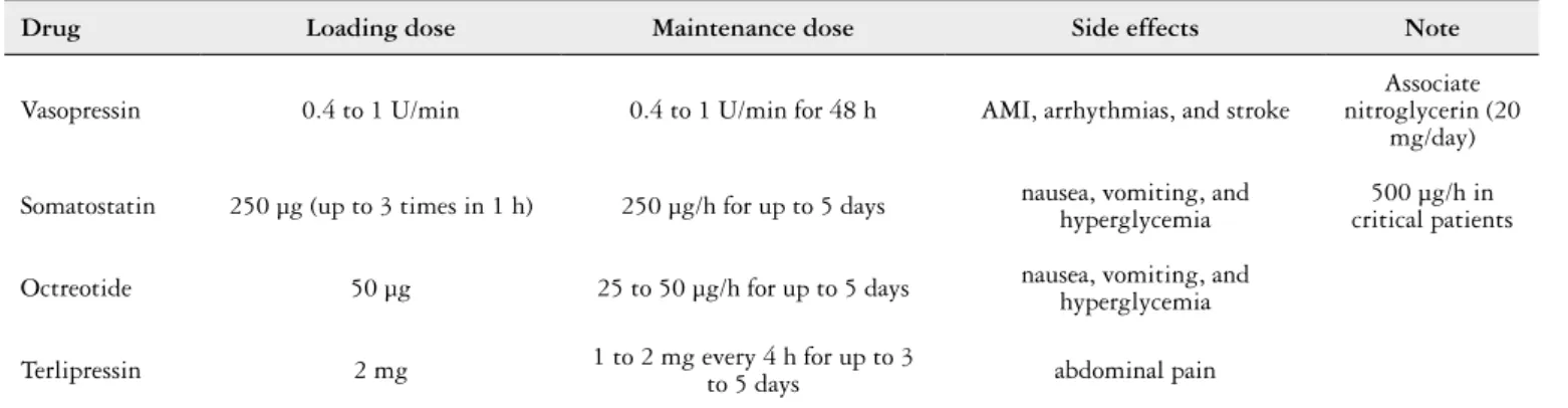
Terlipressin (Glypressin®, Ferring Pharmaceuticals, Brazil) is an approved and available drug in Brazil. It is suggested a loading dose of 2 mg, with maintenance dose every 4 hours according to body weight: 1.0 mg for patients with up to 50 kg; 1.5 mg for weight between 50 and 70 kg; and 2.0 mg for more than 70 kg(24). The therapy should be maintained until the bleeding has been controlled for 12 hours. In addition, duration of drug therapy can be extend for up to 3 to 5 days, if necessary(20, 24).
Table 3 summarizes the vasoactive drugs available in Brazil, with its doses and major adverse effects.
Endoscopic treatment
Endoscopy is the method of choice for diagnosis and treatment in the UGB, and should be performed as soon as possible after admission.
1. Endoscopy on the management of nonvariceal UGB
Endoscopy should be performed in the irst 24 hours in patients with suspected nonvariceal UGB, because it enables early discharge in the low-risk group, and in the high-risk group is associated with reduction of blood transfusion, rebleeding, surgery and length of hospital stay(18). (Class I, Level A).
Study of Lim et al.(38) showed that endoscopy performed within 13 hours of presentation, in selected high-risk patients, deined by GBS ≥12, was associated with lower mortality.
Endoscopic therapy is indicated for patients with peptic ulcers and high-risk indings, such as active bleeding (Forrest I) or a visible vessel (Forrest IIA)(7). Endoscopic therapy for ulcers with adherent clot (Forrest IIB) is controversial, since there are conlicting results in literature. It is recommended to consider endoscopic hemostatic therapy especially in patients with high-risk of rebleeding, through the RS and/or GBS(7). Patients with peptic ulcers and low-risk stigmata, such as haematin covered lat spot (Forrest IIC) or clean base (For-rest III), do not require endoscopic treatment(7).
Several meta-analyses have shown that, in cases of UGB secondary to ulcers with high-risk stigmata, combination therapy with epinephrine in association to a second endoscopic method, such as cautery, clips or injection of a second agent, compared to monotherapy with epinephrine injection, showed lower risk of rebleeding, surgery and mortality(8). (Class I, Level A).
We recommended the combination therapy, or mono-therapy with cautery or clips, for endoscopic hemostatic therapy of ulcers with high-risk stigmata.
In patients with nonvariceal UGB due to Mallory-Weiss syndrome, esophagitis, Dieulafoy lesion, or gastric antral vascular ectasia, the endoscopic treatment can be realized with cautery or clips. Cases of UGB secondary to upper gastrointestinal tract cancers can be treated endoscopically with cautery or injection method. However, the rebleeding rate is high. Surgical treatment, radiotherapy and hemostatic embolization have better results, with lower rebleeding(32).
• Second-look endoscopy
Second-look endoscopy is deined as scheduled second endoscopy performed 16 to 24 hours after initial endoscopic hemostatic therapy. Its goals is repetition of endoscopic treat-ment in cases with persistent high-risk lesions(7).
Additionally, the strategy of second-look endoscopy, in selected patients at high-risk of rebleeding, was most effective and with lower costs than the strategy of second-look for all patients(7). (Class IIa, B Level).
2. Endoscopy on the management of variceal UGB
Endoscopy on suspicion of variceal UGB should be performed in irst 12 hours after admission, since delayed endoscopy is an independent risk factor for in-hospital mortality(29). (Class I, Level A).
• Endoscopic therapy for esophageal varices
Methods of endoscopic hemostatic therapy for bleeding esophageal varices are endoscopic sclerotherapy (EST) and the endoscopic variceal ligation (EVL).
• Endoscopic sclerotherapy
The EST may be done by direct intravariceal injection of the sclerosant, or by paravariceal injection adjacent to variceal veins. Multiple sclerosants agents can be used (so-dium tetradecyl sulfate, so(so-dium morrhuate, ethanolamine oleate, polidocanol, ethanol, and cyanoacrylate), with good results and similar eficacies.
The EST is an effective treatment during acute variceal hemorrhage, with immediate control of bleeding in more than 90% of cases, and with signiicant reduction of rebleeding(32).
• Endoscopic variceal ligation
Majority of studies reported superiority of EVL com-pared to ES, regarding to lower complications, rebleeding and mortality(32). We recommended EVL as the treatment of choice in acute hemorrhage of esophageal varices. However, the ES should remain as an option when the EVL is not available, or when this is not possible due to technical dificulties(11, 26). (Class I, Level A).
• Endoscopic therapy for gastric varices
Gastric varices occur in up to 25% of patients with portal hypertension, and mostly associated with esophageal varices. Endoscopic treatment of choice in acute bleeding of fundal varices (GOV2 and IGV1) is the cyanoacrylate injection(11, 20). (Class I, Level B).
The hemostasis rate with cyanoacrylate injection is about 90%, and the comparison with other endoscopic methods, such as EVL, showed better rates of hemostasis, rebleeding and obliteration with cyanoacrylate(39).
• Secondary prophylaxis of variceal UGB
Patients who have recovered from an episode of variceal UGB should receive secondary prophylaxis with endoscopic erradication of variceal veins, once bleeding recurrence without the treatment is approximately 60% in 1 to 2 years, with 33% mortality(26).
In secondary prevention of esophageal variceal bleeding, combination of endoscopic treatment with nonselective beta-blocker (NSBB) reduced the risk of new bleeding, when compared with endoscopic therapy or NSBB alone (propranolol or nadolol)(28). (Class I, level A).
Propranalol is a widely available NSBB in Brazil. Its recommended its introduction after hemodynamic compen-sation, usually on the sixth day after bleeding episode. The initial dose is 20 mg, orally, every 12 hours, and should be adjusted to the maximum dose tolerated by patient(11, 20, 26).
Regarding to the endoscopic therapy of choice in secondary prophylaxis of esophageal varices, studies reported superiority of EVL compared to EST, because the eradication was obtained with less complications, shorter time and fewer number of sessions with EVL(26). Endoscopic therapy with EVL must be performed every 1 to 3 weeks until the esophageal varices are eradicated, which typically occurs after 2 to 4 sessions(11, 26).
In the secondary prophylaxis of bleeding fundal varices, the cyanoacrylate injection signiicantly reduced rebleeding and mortality, when compared with propranolol(40).
Persistent bleeding and rebleeding
Although majority of cases with UGB is controlled with endoscopic therapy, until 20% of cases failed to treatment or present rebleeding(7, 26). Persistent bleeding and rebleeding dei-nitions vary in literature. However, the concept of persistent bleeding, or failure to endoscopic treatment, refers to bleeding which is not controlled after endoscopy. Rebleeding is deined as a new bleeding episode after successful endoscopic therapy, within the irst 30 days after the hemorrhage(36).
TABLE 3. Vasoactive drugs used in variceal UGB
Drug Loading dose Maintenance dose Side effects Note
Vasopressin 0.4 to 1 U/min 0.4 to 1 U/min for 48 h AMI, arrhythmias, and stroke
Associate nitroglycerin (20
mg/day)
Somatostatin 250 µg (up to 3 times in 1 h) 250 µg/h for up to 5 days nausea, vomiting, and hyperglycemia critical patients500 µg/h in
Octreotide 50 µg 25 to 50 µg/h for up to 5 days nausea, vomiting, and hyperglycemia
Terlipressin 2 mg 1 to 2 mg every 4 h for up to 3
to 5 days abdominal pain
Cases of persistent bleeding or rebleeding usually pre-sent with the following indings(20, 36): new hematemesis or bloody nasogastric aspirate; new episode of melena; signs of hemodynamic instability (tachycardia, hypotension); and/ or hemoglobin drop ≥2 g/dL, or need of RBC transfusion ≥2 units, in 24 hours.
A second endoscopic attempt to control hemorrhage can be performed in cases of persistent bleeding or rebleeding(7, 11). (Class IIa, B level). A different endoscopic method from the previously used should be considered for retreatment.
Patients with persistent bleeding or rebleeding, when the hemorrhage is not stopped quickly and effectively with a second endoscopic attempt, should be referred for rescue therapies.
Rescue therapies on nonvariceal bleeding
• Surgery
Surgical treatment in nonvariceal UGB is reserved, tradi-tionally, to cases with bleeding ulcer refractory to endoscopic treatment(7). (Class IIa, B Level). Surgery has high success rate in controlling the bleeding, however may present mortality of up to 36%(1).
• Transarterial embolization
Transarterial embolization (TAE) has been studied as a therapeutic alternative to surgery in cases of nonvariceal UGB with failure to endoscopic therapy, especially in the patients with high surgical and anesthetic risk(7). (Class IIa, B Level) Studies have shown a technical success rate around 85% with TAE. And in comparison with surgery there was fewer complications, but with higher rate of rebleeding(50).
Rescue therapies on variceal bleeding
• Balloon tamponade
Balloon tamponade (Sengstaken-Blakemore tube) has
high effectiveness in the immediate control of variceal bleed-ing, successfully in about 80% of cases. However, should only be used temporarily (maximum 24 hours) in cases of variceal UGB with uncontrolled hemorrhage after endoscopy, due to high rates of rebleeding and potentially lethal complica-tions(20, 26). (Class IIa, B Level). Orotracheal intubation for airway protection is strongly recommended before use of balloon tamponade.
• TIPS
TIPS (transjugular intrahepatic portosystemic shunt) is currently recommended as rescue therapy of choice in variceal bleeding refractory to endoscopy, with high technical and clinical success rate(11, 26). (Class I, Level A).
Recently, clinical trial demonstrated that in patients with cirrhosis (Child B or C until 13 points) and variceal bleeding, who received vasoactive drugs and endoscopic therapy, early TIPS within 72 hours after endoscopy has reduced rebleeding and mortality(25). This procedure was considered by Baveno V consensus(20), and will also be recommended by the next
Baveno VI, as it was presented during the Digestive Disease Week 2015. However, we believe that further studies should be conducted, in different populations, for external valida-tion of those indings and subsequent introducvalida-tion of this procedure in our clinical practice.
• Surgery
Shunt surgery is reserved only for patients with variceal bleeding refractory to clinical and endoscopic therapy. It has high hemostasis rate, but with signiicant postoperative risk of hepatic encephalopathy, and ideally should be performed in Child A patients(11, 26). (Class IIa, level B).
• Self-expanding metal stent
Recent studies of case series have demonstrated good results with self-expanding metal stents (SEMS) for cases of bleeding esophageal varices after failure to endoscopic therapy(21). However, further evaluation is still needed with randomized studies(20).
Hemostatic powder
Recently, some reports showed good outcomes in control-ling variceal and nonvariceal UGB with the use of hemostatic powder (Hemospray®, Cook Medical, Winston-Salem, North Carolina, USA) in some European countries(49). The results are promising, especially because it is an easy technique to perform even in cases of active or severe bleeding; however, there are only few studies and little experience with this new tool.
Schistosomiasis and variceal UGB
The Schistossoma mansoni infection is the main cause of noncirrhotic portal hypertension in Brazil, and bleed-ing from variceal veins is a signiicant complication of this pathology. There are currently insuficient data on clinical and endoscopic therapy of variceal bleeding in patients schistosomiasis. However, due to the theoretical beneits, we suggest the same recommended practices for patients with liver cirrhosis(11).
RESULTS: clinical care pathway for UGB
Inclusion criteria
Inclusion criteria are the presence of recent upper GI bleeding (in the irst 24 hours), described as hematemesis or melena, in patients admitted at the emergency room or ICU.
Exclusion criteria
Exclusion criteria were age <18 years, and pregnancy.
Additional cases
Cases with hemoglobin drop ≥2 g/dL in 24 hours, or need of RBC transfusion ≥2 units, where the main hypothesis is UGB, may also be included.
Clinical care pathway for UGB
Follow-up after endoscopic therapy
We recommended hemodynamic monitoring (pulse and blood pressure), and serum hemoglobin level, in the irst 72 hours after endoscopy, of cases with ulcers and stigmata of high-risk, and in cases of variceal bleeding or other etiology with severe hemorrhage.
DISCUSSION
Considering the high incidence of UGB in our tertiary hospitals, with important repercussions on mortality and elevated inancial costs to the health system, as well as the
need for emergency supportive care by a multidisciplinary team, it is desirable the existence of a clinical pathway for the management of patients with upper GI bleeding.
Between 2008 and 2010, the Brazilian Society of Diges-tive Endoscopy, the Brazilian Society of Hepatology, and the Brazilian Federation of Gastroenterology, published guidance on nonvariceal UGB(48), guideline on portal hy-pertension with variceal bleeding(19), consensus on variceal bleeding(11), and guideline on digestive bleeding(23). In all studies, the authors performed an extensive literature review, graded the evidences and issued the recommendations. The guidelines were developed in association with the Brazilian
FIGURE 3. Clinical pathway for upper gastrointestinal bleeding (UGB)
Inclusion criteria:
Hematemesis or melena in the last 24 h
Consider inclusion: Hemoglobin (Hb) drop ≥ 2 g/dL or red blood cell (RBC) transfusion ≥ 2 units in the last 24 h
- Oral fasting
- Hemodynamic monitoring
- Fluid infusion for systolic blood pressure ≥ 90 to 100 mmHg and pulse < 100 bpm - Order complete blood count, electrolytes, urea and creatinine, coagulation proile and blood typing
- RBC transfusion for hemoglobin of 7 to 8 g/dL Consider:
- Intensive care unit: ederly, with comorbidities, variceal and or severe bleeding
- Endotracheal tube: massive hematemesis or altered level of consciousness
- Fresh frozen plasma and platelets transfusion: if severe coagulopathy or thrombocytopenia in cases of serious bleeding
- Hospital discharge if Glasgow Blatchford (GB) score = 0; with outpatient endoscopy scheduled for the next day
Consider second-look endoscopy:
- Cases of ulcer with Forrest IA, IB, IIA or IIB + Doubt about the success of endoscopic treatment or high risk of rebleeding
Perform:
- Exchange for oral omeprazole: nonvariceal UGB and low-risk stigmata after endoscopy
Consider:
- In suspected cases of signiicant amount of blood and clots inside stomach: Eritromicin 250 mg IV (diluted in saline 100 mL) in 30 min (30 to 60 min before endoscopy)
Perform:
- Endoscopic therapy for cases with UGB due to Dieulafoy´s lesion or antral vascular ectasia
Consider:
- Endoscopic therapy for cases with active bleeding due to Mallory-Weiss tears, esophagitis or upper GI tract cancers
Provide:
- Endoscopy in the irst 12 h for cases of nonvariceal UGB and GB score ≥ 12
Consider:
- Maintenance dose of terlipressin (every 4h): 1 mg < 50 kg; 1,5 mg for 50 – 70 kg; 2mg > 70 kg
Perform:
- Early luid resuscitation
- Endoscopy as soon as possible after hemodynamic stabilization
Exclusion criteria:
< 18 years-old, supected or conirmed pregnancy
Upper Gastrointestinal Endoscopy
Suspicion of Nonvariceal UGB
Whitin First 24 h
Nonvariceal UGB
Whitin First 12 h
Variceal UGB Suspicion of Varicel UGB
Intravenous (IV) omeprazole prior endoscopy:
- 80 mg IV bolus, followed by 8 mg/h infusion
- Solution with 80 mg of omeprazole and 100 mL of saline
Ulcer with Forrest IA, IB, IIA or IIB:
- Cautery, clips, or epinephrine injection + second endoscopic method + Maintain omeprazole IV for 72 h
- Test for H. pylori infection in all cases of peptic ulcer
Esophageal varices
Provide: endoscopic variceal ligation - If not available: sclerotherapy - Cases of gastric varices: Cyanoacrylate injection + Terlipressin IV up to 3 to 5 days
Start in patients with liver cirrhosis before endoscopy:
- Chlid A: Norloxacin 400 mg orally bid - Child B or C: Ceftriaxone 1g IV once daily - Terlipressin 2 mg IV bolus, with maintenance of 1-2 mg IV every 4 h
Persistent bleeding and rebleeding:
- New episode of hematemesis, melena or bloody nosogastric aspirate
- Hemodynamic instability - Hb drop ≥ 2 g/dL, or need of RBC transfusion ≥ 2 units, in 24 h
Before discharge:
Patients with nonvariceal UGB: - Give omeprazole orally according to the endoscopic diagnosis - Treat H. pylori infection in all patients with peptic ulcers
Early follow-up:
- Hemodynamics (pulse and blood pressure) and Hb for 72h after endoscopy, in cases with ulcer and high-risk stigmata, variceal or massive bleeding
Before discharge:
Patients with variceal UGB: - Antibiotic prophlaxis for 7 days - Start propanolol 20 mg orally, every 12 h, after hemodynamic compensation
- Schedule next endoscopic therapy after 1-3 weeks, until erradication of varices
Persistent Bleeding and Rebleeding
Failure of Endoscopic Therapies
Second endoscopic attempt
Consider a different endoscopic method for hemostasis
Rescue therapy:
Transarterial embolization or surgery
Second endoscopic attempt
Consider a different endoscopic method for hemostasis
Rescue therapy:
Franco MC, Nakao FS, Rodrigues R, Maluf-Filho F, Paulo GA, Libera ED. Proposta de modelo de atendimento da hemorragia digestiva alta. Arq Gastroenterol. 2015,52(4):xxx.
RESUMO - Contexto- A hemorragia digestiva alta implica em signiicativas repercussões clínicas e econômicas. O estabelecimento correto das mais recentes terapêuticas para a hemorragia digestiva alta está associado à redução na mortalidade intra-hospitalar. O uso de algoritmos para atendimento da hemorragia digestiva alta está associado com menor tempo de internação e menores custos hospitalares. Objetivos- O objetivo primário é a criação de um protocolo de atendimento da hemorragia digestiva alta, para ser utilizado em hospital terciário. Métodos- Realizada extensa revisão da lite-ratura sobre as condutas na hemorragia digestiva alta, contidas nas bases de dados primária e secundária. Resultados- O resultado é um modelo de atendimento para os pacientes com hemorragia digestiva alta e com evidência de sangramento recente, dado por melena ou hematêmese nas ultimas 24h, que são atendidos nas salas de emergência e unidades de terapia intensiva de hospitais terciários. Neste protocolo de atendimento, desenhado de forma compacta e compreensível, ica bem evidenciado o manejo dos pacientes desde a admissão, com deinição dos critérios de inclusão e ex-clusão, passando considerações acerca do atendimento clínico inicial, posterior direcionamento para a terapêutica endoscópica, e encaminhamento às terapias de resgate em casos de sangramento persistente ou recorrente. Destacam-se também os cuidados que devem ser tomados antes da alta hospitalar para todos os pacientes que se recuperam de um episódio de sangramento. Conclusão- A introdução de um protocolo para atendimento e tratamento de pacientes com hemorragia digestiva alta pode contribuir para uniformização de condutas médicas, diminuição no tempo de espera por medicações e serviços, no tempo de internação e nos custos hospitalares.
DESCRITORES- Hemorragia gastrointestinal. Hematemese. Melena. Endoscopia. Procedimentos clínicos. Protocolos clínicos. Medical Association (AMB). All the texts are available online
for download at the societies and AMB websites. We felt that the development of a clinical care pathway for the manage-ment of patients with UGB could be a possible way to help to implement the actions of the above mentioned Societies.
We updated the extensive literature review on UGB and found several class I and II recommendations on the evaluation and treatment of UGB. We also assessed the structure and resources of a tertiary Brazilian hospital in order to reassure that the recommendations could be locally implemented. The inal product of the present research was a compact and comprehensive algorithm for the management of patients with UGB.
In conclusion, the use of this algorithm can enable standardization of medical practices, diffusion of current therapies, decreasing the waiting time for medications and
services, possibly decreasing in length of stay and hospital costs, improvement in the teaching and training of medical students and junior doctors, and especially can promote a better service to patients.
Authors’ contributions
REFERENCES
1. Abe N, Takeuchi H, Yanagida O, Sugiyama M, Atomi Y. Surgical indications
and procedures for bleeding peptic ulcer. Dig Endosc. 2010;22(Suppl 1):S35–7.
2. Aljebreen AM, Fallone CA, Barkun AN. Nasogastric aspirate predicts high-risk
endoscopic lesions in patients with acute upper-GI bleeding. Gastrointest Endosc. 2004;59(2):172–8.
3. Al-Sabah S, Barkun AN, Herba K, Adam V, Fallone C, Mayrand S, et al.
Cost-ef-fectiveness of proton-pump inhibition before endoscopy in upper gastrointestinal bleeding. Clin Gastroenterol Hepatol. 2008;6(4):418–25.
4. American College of Cardiology Foundation and American Heart Association.
Methodology Manual and Policies From the ACCF/AHA Task Force on Practice Guidelines. 2010;1–88.
5. Bai Y, Guo JF, Li ZS. Meta-analysis: erythromycin before endoscopy for acute
upper gastrointestinal bleeding. Aliment Pharmacol Ther. 2011;34(2):166–71.
6. Baradarian R, Ramdhaney S, Chapalamadugu R, Skoczylas L, Wang K, Rivilis S,
et al. Early intensive resuscitation of patients with upper gastrointestinal bleeding decreases mortality. Am J Gastroenterol. 2004;99(4):619–22.
7. Barkun AN, Bardou M, Kuipers EJ, Sung J, Hunt RH, Martel M, et al.
In-ternational consensus recommendations on the management of patients with nonvariceal upper gastrointestinal bleeding. Ann Intern Med. 2010;152(2):101–13.
8. Barkun AN, Martel M, Toubouti Y, Rahme E, Bardou M. Endoscopic hemostasis
in peptic ulcer bleeding for patients with high-risk lesions: a series of meta-analyses. Gastrointest Endosc. 2009;69(4):786–99.
9. Bernard B, Cadranel JF, Valla D, Escolano S, Jarlier V, Opolon P. Prognostic
signiicance of bacterial infection in bleeding cirrhotic patients: a prospective study. Gastroenterology. 1995;108(6):1828–34.
10. Bernardo WM, Nobre MRC, Jatene FB. A prática clínica baseada em evidências. Parte II - Buscando as evidências em fontes de informação. Rev Assoc Med Bras. 2004;50(1):104–8.
11. Bittencourt PL, Farias AQ, Strauss E, Mattos AA, Pannel of the 1st Brazilian
Consensus of Variceal Bleeding, Brazilian Society of Hepatology. Variceal bleeding: consensus meeting report from the Brazilian Society of Hepatology. Arq Gastroenterol. 2010;47(2):202–16.
12. Blatchford O, Davidson LA, Murray WR, Blatchford M, Pell J. Acute upper gastrointestinal haemorrhage in west of Scotland: case ascertainment study. BMJ Br Med J. 1997;315(7107):510–4.
13. Blatchford O, Murray WR, Blatchford M. A risk score to predict need for treat-ment for upper-gastrointestinal haemorrhage. Lancet. 2000;356(9238):1318–21. 14. Cappell MS, Friedel D. Initial management of acute upper gastrointestinal bleed-ing: from initial evaluation up to gastrointestinal endoscopy. Med Clin North Am. 2008;92(3):491–509, xi.
15. Carbonell N, Pauwels A, Serfaty L, Fourdan O, Lévy VG, Poupon R. Improved survival after variceal bleeding in patients with cirrhosis over the past two decades. Hepatology. 2004;40(3):652–9.
16. Chavez-Tapia NC, Barrientos-Gutierrez T, Tellez-Avila FI, Soares-Weiser K, Uribe M. Antibiotic prophylaxis for cirrhotic patients with upper gastrointestinal bleeding. Cochrane Database Syst Rev. 2010;(9):CD002907.
17. Chiu P, Choi C, Kwong KH, Lam S. The effect of scheduled second endos- copy against intravenous high dose omeprazole infusion as an adjunct to therapeutic endoscopy in prevention of peptic ulcer rebleeding-a prospective randomized study [abstract]. Gastroenterology. 2006;130:A121.
18. Cooper GS, Chak A, Way LE, Hammar PJ, Harper DL, Rosenthal GE. Early endoscopy in upper gastrointestinal hemorrhage: associations with rebleeding, surgery, and length of hospital stay. Gastrointest Endosc. 1999;49(2):145–52. 19. Costa Junior AB, Conrado ACC, Araújo JC, Medeiros TB, Evangelista LFL.
Projeto Diretrizes - SOBED. Hipertensão portal: atendimento na emergência da ruptura de varizes esofágicas. 2008;1–16.
20. de Franchis R, Baveno V Faculty. Revising consensus in portal hypertension: report of the Baveno V consensus workshop on methodology of diagnosis and therapy in portal hypertension. J Hepatol. 2010;53(4):762–8.
21. Dechêne A, El Fouly AH, Bechmann LP, Jochum C, Saner FH, Gerken G, et al. Acute management of refractory variceal bleeding in liver cirrhosis by self-ex-panding metal stents. Digestion. 2012;85(3):185–91.
22. Fernández J, Ruiz del Arbol L, Gómez C, Durandez R, Serradilla R, Guarner C, et al. Norloxacin vs ceftriaxone in the prophylaxis of infections in patients with advanced cirrhosis and hemorrhage. Gastroenterology. 2006;131(4):1049–56. 23. Ferreira R, Eisig J, Federação Brasileira de Gastroenterologia. Projeto Diretrizes
- Hemorragias Digestivas. 2008;1–14.
24. Ferring Pharmaceuticals. Glypressin - Bula medicamento. 2011; 1–9.
25. García-Pagán JC, Caca K, Bureau C, Laleman W, Appenrodt B, Luca A, et al. Early use of TIPS in patients with cirrhosis and variceal bleeding. N Engl J Med. 2010;362(25):2370–9.
26. Garcia-Tsao G, Sanyal AJ, Grace ND, Carey W. Prevention and management of gastroesophageal varices and variceal hemorrhage in cirrhosis. Hepatology. 2007;46(3):922–38.
27. Gisbert JP, Khorrami S, Carballo F, Calvet X, Gené E, Dominguez-Muñoz JE. H. pylori eradication therapy vs. antisecretory non-eradication therapy (with or without long-term maintenance antisecretory therapy) for the prevention of rebleeding from peptic ulcer. Cochrane Database Syst Rev. 2004;(2):CD004062. 28. Gonzalez R, Zamora J, Gomez-Camarero J, Molinero LM, Bañares R, Albillos A. Meta-analysis: Combination endoscopic and drug therapy to prevent variceal rebleeding in cirrhosis. Ann Intern Med. 2008;149(2):109–22.
29. Hsu YC, Chung CS, Tseng CH, Lin TL, Liou JM, Wu MS, et al. Delayed en-doscopy as a risk factor for in-hospital mortality in cirrhotic patients with acute variceal hemorrhage. J Gastroenterol Hepatol. 2009;24(7):1294–9.
30. Huang ES, Karsan S, Kanwal F, Singh I, Makhani M, Spiegel BM. Impact of nasogastric lavage on outcomes in acute GI bleeding. Gastrointest Endosc. 2011;74(5):971–80.
31. Hwang JH, Fisher DA, Ben-Menachem T, Chandrasekhara V, Chathadi K, Decker GA, et al. The role of endoscopy in the management of acute non-variceal upper GI bleeding. Gastrointest Endosc. 2012;75(6):1132–8.
32. Hwang JH, Shergill AK, Acosta RD, Chandrasekhara V, Chathadi K V, Decker GA, et al. The role of endoscopy in the management of variceal hemorrhage. Gastrointest Endosc. 2014;80(2):221–7.
33. Ioannou G, Doust J, Rockey DC. Terlipressin for acute esophageal variceal hemorrhage. Cochrane Database Syst Rev. 2003;(1):CD002147.
34. Katschinski B, Logan R, Davies J, Faulkner G, Pearson J, Langman M. Prognostic factors in upper gastrointestinal bleeding. Dig Dis Sci. 1994;39(4):706–12. 35. L a i n e L , M c Q u a i d K R . E n d o s c o p i c t h e rapy fo r bl e e d i n g u l c e r s :
an evidence-based approach based on meta-analyses of randomized controlled trials. Clin Gastroenterol Hepatol. 2009;7(1):33–47
36. Laine L, Spiegel B, Rostom A, Moayyedi P, Kuipers EJ, Bardou M, et al. Meth-odology for randomized trials of patients with nonvariceal upper gastrointestinal bleeding: recommendations from an international consensus conference. Am J Gastroenterol. 2010;105(3):540–50.
37. Lee TY, Chan T, Chang CS, Lan JL. Introducing a clinical pathway for acute peptic ulcer bleeding in general internal medicine wards. Scand J Gastroenterol. 2008;43(10):1169–76.
38. Lim LG, Ho KY, Chan YH, Teoh PL, Khor CJ, Lim LL, et al. Urgent endoscopy is associated with lower mortality in high-risk but not low-risk nonvariceal upper gastrointestinal bleeding. Endoscopy. 201;43(4):300–6.
39. Lo GH, Lai KH, Cheng JS, Chen MH, Chiang HT. A prospective, randomized trial of butyl cyanoacrylate injection versus band ligation in the management of bleeding gastric varices. Hepatology. 2001;33(5):1060–4.
40. Mishra SR, Chander Sharma B, Kumar A, Sarin SK. Endoscopic cyanoacrylate injection versus beta-blocker for secondary prophylaxis of gastric variceal bleed: a randomised controlled trial. Gut. 2010;59(6):729–35.
41. Neumann I, Letelier LM, Rada G, Claro JC, Martin J, Howden CW, et al. Com-parison of different regimens of proton pump inhibitors for acute peptic ulcer bleeding. Cochrane Database Syst Rev. 2013;6:CD002147.
42. Pang SH, Ching JY, Lau JY, Sung JJ, Graham DY, Chan FK. Comparing the Blatch-ford and pre-endoscopic Rockall score in predicting the need for endoscopic therapy in patients with upper GI hemorrhage. Gastrointest Endosc. 2010;71(7):1134–40. 43. Pfau PR, Cooper GS, Carlson MD, Chak A, Sivak M V, Gonet JA, et al. Success and shortcomings of a clinical care pathway in the management of acute non-variceal upper gastrointestinal bleeding. Am J Gastroenterol. 2004;99(3):425–31. 44. Rockall TA, Logan RF, Devlin HB, Northield TC. Risk assessment after acute
upper gastrointestinal haemorrhage. Gut. 1996;38(3):316–21.
45. Rotter T, Kugler J, Koch R, Gothe H, Twork S, van Oostrum JM, et al. A sys-tematic review and meta-analysis of the effects of clinical pathways on length of stay, hospital costs and patient outcomes. BMC Health Serv Res. 2008;8:265. 46. Schilling D, Demel A, Nüsse T, Weidmann E, Riemann JF. Helicobacter pylori
infection does not affect the early rebleeding rate in patients with peptic ulcer bleeding after successful endoscopic hemostasis: a prospective single-center trial. Endoscopy. 2003;35(5):393–6.
47. Srirajaskanthan R, Conn R, Bulwer C, Irving P. The Glasgow Blatchford scoring system enables accurate risk stratiication of patients with upper gastrointestinal haemorrhage. Int J Clin Pract. 2010;64(7):868–74.
48. Souza CDC, Parente JML, Lima MM, Santos OF. Projeto Diretrizes - SOBED. Hemorragia Digestiva Alta Não Varicosa. 2008;1–24.
49. Sulz MC, Frei R, Meyenberger C, Bauerfeind P, Semadeni GM, Gubler C. Routine use of Hemospray for gastrointestinal bleeding: prospective two-center experience in Switzerland. Endoscopy. 2014;46(7):619-24.