ARTICLE
DOI: 10.1590/0004-282X20130218
The role of xerostomia in burning mouth
syndrome: a case-control study
O papel da xerostomia na síndrome da ardência bucal: estudo caso controle
Luciana Alvarenga da Silva1, José Tadeu Tesseroli de Siqueira2, Manoel Jacobsen Teixeira3,
Silvia Regina Dowgan Tesseroli de Siqueira4
Departmento de Neurologia, Faculdade de Medicina, Universidade de São Paulo, Sao Paulo SP, Brazil.
1Postgraduation Student, Departmento de Neurologia, Faculdade de Medicina, Universidade de São Paulo, Sao Paulo SP, Brazil;
2PhD, DDS, Head of the Orofacial Pain Team, Dentistry Division, Hospital das Clínicas, Faculdade de Medicina, Universidade de São Paulo, Sao Paulo SP, Brazil; 3PhD, MD, Full Professor, Interdisciplinary Pain Center, Hospital das Clinicas and Departmento de Neurologia, Faculdade de Medicina, Universidade de São
Paulo, Sao Paulo SP, Brazil;
4PhD, DDS, Associate Professor, Departmento de Neurologia, Hospital das Clínicas, Escola de Medicina, Universidade de São Paulo, Sao Paulo SP, Brazil.
Correspondence: Luciana Alvarenga da Silva; Av. Niterói 127 / Parque Paraíso / Itapecerica da Serra; 06850-200 São Paulo SP - Brasil; E-mail: [email protected]
Conflict of interest: There is no conflict of interest to declare.
Support: FAPESP - Foundation of the State of Sao Paulo - for the financial support for this research (2009/08697-5). Received 28 June 2013; Received in final form 18 October 2013; Accepted 25 October 2013.
ABSTRACT
Objective: To assess the efficacy of anti-xerostomic topical medication (urea 10%) in patients with burning mouth syndrome (BMS). Method: Thirty-eight subjects diagnosed with BMS according to the International Association for the Study of Pain guidelines were randomi-zed to either placebo (5% sodium carboxymethylcellulose, 0.15% methyl paraben, and 10% glycerol in distilled water qsp 100 g) or treatment (urea 10%) to be applied to the oral cavity 3-4 times per day for 3 months. The patients were evaluated before and after treatment with the following instruments: the EDOF-HC protocol (Orofacial Pain Clinic – Hospital das Clínicas), a xerostomia questionnaire, and quantitative sensory testing. Results: There were no differences in salivary flow or gustative, olfactory, or sensory thresholds (P>0.05). Fifteen (60%) pa-tients reported improvement with the treatments (P=0.336). Conclusion: In conclusion, there were no differences between groups, and both exhibited an association between reported improvement and salivation.
Keywords: xerostomia, salivary flow, orofacial pain, quantitative sensory testing, burning mouth syndrome.
RESUMO
Objetivo: Avaliar a eficácia do uso de medicação tópica anti xerostomica (ureia 10%) em pacientes com síndrome de ardência bucal. Método: Trinta e oito sujeitos diagnosticados com síndrome de ardência bucal de acordo com os critérios da Associação Internacional para Estudo da Dor foram randomizados para grupo placebo (5% de carboximetilcelulose de sódio, 0,15% de metilparabeno e 10% de glicerol em água destilada qsp 100g) ou grupo tratamento (ureia 10%) para ser aplicada na cavidade oral 3-4 vezes ao dia, durante três meses. Os pacientes foram avaliados antes e depois do tratamento: protocolo EDOF-HC, questionário de xerostomia, testes sensitivos quantitativos. Resultados: Não houve diferenças no fluxo salivar, limiares gustativos, olfativos e somestésicos (Mann-Whitney P>0,05). Quinze (60%) dos pacientes tiveram melhora com o tratamento (P=0,336, oneway ANOVA). Conclusão: Em conclusão não houve diferenças entre os grupos, ambos apresentaram uma associação entre melhora e salivação.
Palavras-chave: xerostomia, fluxo salivar, dor orofacial, teste sensitivo quantitativo, síndrome ardência bucal.
Burning mouth syndrome (BMS) is a continuous intraoral pain characterized by burning mouth in the absence of a pri-mary etiological lesion or disease1-3. he main afected sites are
the tongue, palate, and/or the gingiva4-6. It is considered idiopa-thic, and current evidence has classiied it as neuropathic. One
of the most widely accepted theories is that there is disinhibi-tion of the trigeminal nerve due to partial or total loss of chorda tympani ( facial) nerve function6. However, recent indings also
showed that trigeminal neuropathy may be directly involved, and that gustatory abnormalities can be secondary3.
It is known that BMS is eventually associated with reduced
salivary low7-9 and abnormal saliva composition (increasing
concentrations of K+, Na+, Cl-, Ca+2, immunoglobulin A [IgA], amylase)10. Even in the absence of hyposalivation,
pa-tients may complain of xerostomia and dry mouth7-9,11 and
he treatments for BMS include antidepressants, benzo -diazepines, anticonvulsants, local or systemic capsaicin, and
alpha lipoic acid. However, the collateral efect of these drugs
on decreased saliva secretion can exacerbate the symptoms13.
Two studies have described the use of anti-xerostomic topical medications as an adjuvant to treat BMS, and urea has been
shown to be efective in skin-surface hydration14,15.hus, the
objective of this study was to evaluate xerostomia and
sali-vary low in patients with BMS treated with amitriptyline be -fore and after the use of anti-xerostomic topical medication.
METHOD
his was a randomized, double-blind clinical trial
conduc-ted between August 2011 and February 2012. We enrolled 38 patients with BMS diagnosed according to the International Association for the Study of Pain (IASP) guidelines16, which
are followed at the Craniofacial Pain Clinic of Hospital das Clinicas, School of Medicine of the University of Sao Paulo. All subjects were informed about the purposes of the study
and provided written informed consent. he protocol was
approved by the local Ethics Committee. No patient exhibit-ed hyposalivation at the time of diagnosis with the quantita-tive evaluation. All patients had been treated with 25-50 mg
of amitriptyline within the last 3 months. hey underwent
laboratory tests and a careful examination to exclude other causes of burning mouth10.
he exclusion criteria were other facial pain syndromes,
other causes of abnormal salivation, other neuropathies or primary diseases associated with burning mouth, or inability to answer the questions and/or perform the tests.
he subjects were randomly divided into two groups: 1. Study Group: 19 patients received topical medication
comprised of urea 10% to be applied at the oral cavity 3-4 times per day for 3 months.
2. Control Group: 19 patients received placebo (5% sodium
carboxymethylcellulose, 0.15% methyl paraben, and 10% glycerol in distilled water qsp 100 g) to be applied to the oral cavity 3-4 times per day for 3 months.
he patients were evaluated before and after the 3-month treatment period with the following instruments:
1. EDOF-HC protocol (Orofacial Pain Clinic - Hospital das Clinicas): a standardized orofacial pain questionnaire to detail the following: 1) chief complaint, 2) general pain
characteristics (location, quality, duration, pain relief, pain triggering), 3) headache and/or body pain complaints, and 4) patient’s medical history and co-morbidities17;
2. Xerostomia questionnaire18;
3. Quantitative sensory testing (QST). All subjects under-went a standardized QST protocol 19 comprised of 12 tests grouped as follows:
• salivary low and gustative and olfactory thresholds;
• thermal detection thresholds for cold and warm sensations;
• mechanical detection thresholds for touch, vibration, and electrical perception;
• mechanical pain sensitivity, including supericial and
deep pain thresholds;
• electrical pain threshold at the teeth; • corneal relex.
he neuropathic nature of BMS and the impairment of
sensory function were the reasons to use a QST protocol in this study. We evaluated bilateral skin areas innervated by
the three trigeminal branches. he evaluation started with the quantitative non-stimulated salivary low by the follo-wing method: two pieces of cotton were placed into a plas -tic device and weighed on a calibrated balance (Acculab® V1200). he patient was oriented to swallow the saliva inside
the mouth, and the cotton was placed inside and kept below the tongue for 5 minutes, during which time the patient was
instructed not to swallow. hen, the cotton was removed, placed in the plastic device, and weighed, and the diference
in weight (before and after the evaluation) was divided by 5
to calculate the salivary low in mL/min; this technique was
previously validated forbasal salivation20,21.
Gustative thresholds: he following four substances, cor
-responding to the four basic tastes, were tested: sweet (glu
-cose): 0.01 M, 0.032 M, 0.1 M, 0.32 M, 1.0 M; sour (citric acid):
0.00032 M, 0.001 M, 0.0032 M, 0.01 M, 0.032M; salty (sodium
chlorate): 0.01 M, 0.032 M, 0.1 M, 0.32 M, 1.0 M; bitter (urea):
0.1 M, 0.32 M, 1.0 M, 3.2 M, 10.0 M. For each test, one drop of the substance was placed on the tongue, beginning with the low concentration, and alternated with one drop of distilled
water. he concentration was increased until the stimulus
was detected by the subject3,22,23.
Olfactory threshold: Using isopropanol solutions (0.09%,
13.0%, 23.0%, 35.0%, 53.0%, 70.0%)3,12,19,23,24, two bottles were of-fered to the patient: one containing the solution and another
containing water, and the patient had to indicate the bottle con-taining the solution. If this was done correctly for three trials, the
threshold was identiied. If not, a bottle with the solution at the next concentration was ofered along with the bottle of water.
hermal detection: he thermal test was performed with
the modular sensory analyzer (MSA) thermo test device
(So-medic, Sweden). he baseline temperature was 32°C and
the contact square area of the thermode was 9×9 mm. Cold and warm detection thresholds were assessed using ramped
stimuli at 1°C/s. he range was from 10°C to 50°C. First, cold was tested, then warm. he evaluation consisted of ive mea -surements for each thermal threshold, and the means and standard deviations were considered for the analysis.
Mechanical detection threshold: Touch perception was as
-sessed with a set of standardized von Frey ilaments with
roun-ded 0.5-mm diameter tips, applied with an electronic device
perpendicular position on the area to be tested. he subject was
asked to report the detection of the probes, and if they could not
after 30 seconds in the second trial, the next ilament was used. hree measurements in g/mm2 were performed, and the means
and standard deviations were considered for the analysis.
Vibration detection threshold: he vibration test was per -formed with an electronic Vibrameter device (Somedic) with a vibrator of 650 g in weight and a contact area of 1 cm2 that was perpendicularly applied and ramped stimuli at 1 Hz/s. he vi -bration threshold was calculated as the mean between the ap-pearance and disapap-pearance thresholds detected by the patient.
Electrical detection threshold: he electrical threshold test
was performed with a Pulpotest electrical device (Sybronendo,
Orange, CA USA) with a contact area of 1 cm2 for the
percep-tion and a metallic device localized 2 cm away from the
eva-luation area to close the electrical circuit. he electrical stimuli
started at 0 and were increased with ramped stimuli at 1 A/s.
he stimulus was increased at this speed until detected by the
subject, with a maximum value of 80 A.
Pressure pain perception: Deep algometry was
measu-red with an electronic pressure algometer (Somedic) with a probe area of 1 cm2 that was pressed on the skin at a ramp rate of 50 kPa/s. he stimulus was increased at this speed until
it was detected by the subject.
Supericial pain perception: Supericial algometry was
tested with disposable needles of 8×10×0.5 mm, applied with
an electronic device (IITC). hree measurements were made
in g/mm2, and the means and standard deviations were considered for the analysis. he needles were tested in the
perpendicular position on the area to be investigated. he
stimulus was increased until it was detected by the subject.
Electrical pain threshold for the teeth: he electrical pain
threshold for the teeth was performed with a Pulpotest elec-trical device (Sybronendo), with a contact area of 1 cm2 for
perception and a metallic device localized 2 cm away from
the area of evaluation to close the electrical circuit. he elec -trical stimuli started at 0 and were increased with ramped
stimuli at 1 A/s. he stimulus was increased at this speed until
its detection by the subject, with a maximum value of 80 A.
Corneal relex: A von Frey ilament with a 0.5-mm dia-meter rounded tip was used in both eyes. his probe was
applied on the corneal area of each subject with their vision focused to the opposite side, and the subject was asked to re-port whether or not they detected the stimulus.
Statistical analysis
All data were tabled, and the tests used were one-way
analysis of variance (ANOVA), Fisher’s exact test,
Mann-Whitney U test, Chi-square, Student’s t-test and Spearman’s
correlations25. All statistical calculations were performed using SPSS 17.0 (SPSS Inc., Chicago, IL, USA). he level of sig
-niicance was P<0.05.
RESULTS
he groups were similar in gender distribution, age, color,
marital status, occupation, height, weight, comorbidities, and
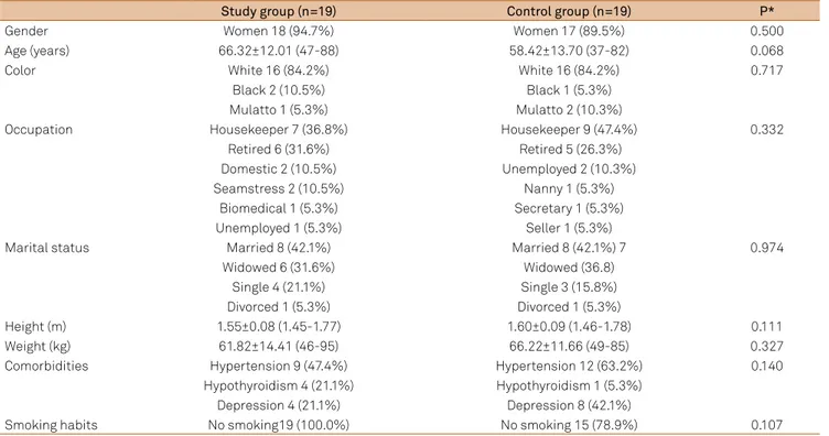
Table 1. Comparison of demographic data between groups (N=38).
Study group (n=19) Control group (n=19) P*
Gender Women 18 (94.7%) Women 17 (89.5%) 0.500
Age (years) 66.32±12.01 (47-88) 58.42±13.70 (37-82) 0.068
Color White 16 (84.2%) White 16 (84.2%) 0.717
Black 2 (10.5%) Black 1 (5.3%)
Mulatto 1 (5.3%) Mulatto 2 (10.3%)
Occupation Housekeeper 7 (36.8%) Housekeeper 9 (47.4%) 0.332
Retired 6 (31.6%) Retired 5 (26.3%)
Domestic 2 (10.5%) Unemployed 2 (10.3%)
Seamstress 2 (10.5%) Nanny 1 (5.3%)
Biomedical 1 (5.3%) Secretary 1 (5.3%)
Unemployed 1 (5.3%) Seller 1 (5.3%)
Marital status Married 8 (42.1%) Married 8 (42.1%) 7 0.974
Widowed 6 (31.6%) Widowed (36.8)
Single 4 (21.1%) Single 3 (15.8%)
Divorced 1 (5.3%) Divorced 1 (5.3%)
Height (m) 1.55±0.08 (1.45-1.77) 1.60±0.09 (1.46-1.78) 0.111
Weight (kg) 61.82±14.41 (46-95) 66.22±11.66 (49-85) 0.327
Comorbidities Hypertension 9 (47.4%) Hypertension 12 (63.2%) 0.140
Hypothyroidism 4 (21.1%) Hypothyroidism 1 (5.3%)
Depression 4 (21.1%) Depression 8 (42.1%)
Smoking habits No smoking19 (100.0%) No smoking 15 (78.9%) 0.107
smoking habits (Table 1), as well as orofacial pain chara cteristics
(Table 2). he patients were evaluated according to the pres -ence of bruxism, earache, headache, generalized pain, chew-ing quality, and other orofacial complaints. Four (21.1%) pa-tients and 3 (15.8%) controls had bruxism (Chi-square, P=0.444), 5 (26.3%) patients and 4 (21.0%) controls had earache (Chi-square, P=0.741), 13 (68.4%) patients and 11 (57.9%) controls had headache (Fisher’s exact test, P=0.288), 12 (63.2%) patients and 10 (52.6%) controls had generalized pain (Fisher’s exact test, P=0.372), 11 (57.9%) patients and 8 (42.2%) controls had good chewing quality, and 8 (42.1%) patients and 11 (57.9%) controls had poor or very poor chewing (Chi-square, P=0.495).
he evaluation of xerostomia and associated complaints identiied 14 (73.7%) patients and 14 (73.7%) controls with dry
mouth sensation (Fisher’s exact test, P=0.643), 2 (10.5%)
pa-tients and 7 (36.8%) controls with diiculty of chewing due
to xerostomia (Fisher’s exact test, P=0.062), 4 (21.1%) patients
and 5 (26.3%) controls with diiculty talking due to xerostomia
(Fisher’s exact test P=0.500), 2 (10.5%) patients and 5 (26.2%)
controls who drank luids at meals (Fisher’s exact test, P=0.500),
8 (42.1%) patients and 9 (47.4%) controls who drank liquids dur-ing the night (Fisher’s exact test, P=0.212), 7 (36.8%) patients and 5 (26.3%) controls with ha litosis (Fisher’s exact test, P=0.364), 6 (31.6%) patients and 4 (21.1%) controls with throat ache (Fisher’s
exact test, P=0.357). Other digestive complaints were: 7 (36.8%)
patients and 3 (15.8%) controls with stomach ache (Fisher’s ex-act test, P=0.135), 9 (47.4%) patients and 8 (42.1%) controls with
abnormal intestinal low (Chi-square, P=0.717), 13 (68.4%) pa -tients and 17 (89.5%) controls with normal digestion (Fisher’s exact test P=0.116), and 5 (26.4%) patients and 7 (36.9%)
con-trols with food-related digestion problems. hese patients re
-ported complaints associated with the following: sour and acid lavors, breads, milk and dairy products, su gars and fats, and
vegetables and beans (Chi-square, P=0.495).
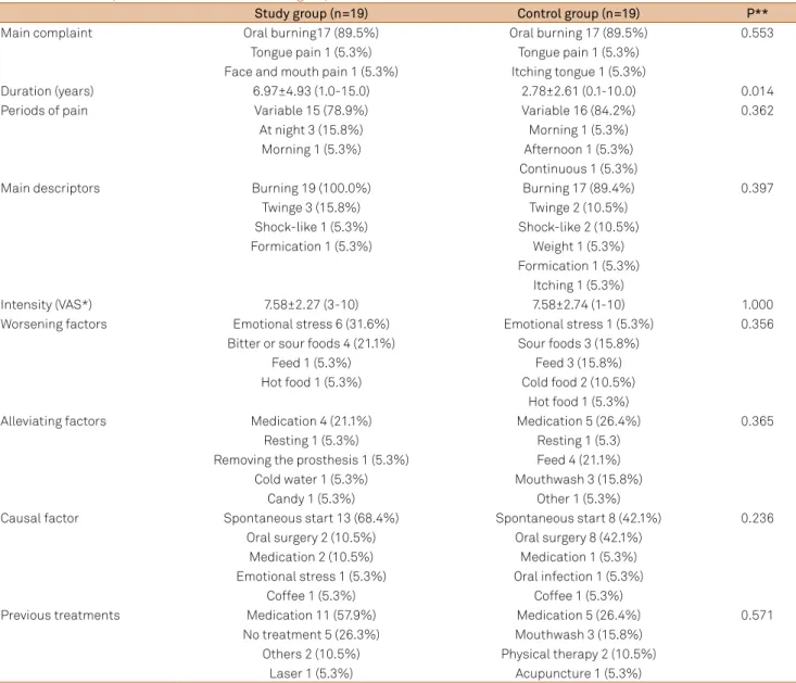
Table 2. Orofacial pain characteristics between groups (N=38).
Study group (n=19) Control group (n=19) P**
Main complaint Oral burning17 (89.5%) Oral burning 17 (89.5%) 0.553
Tongue pain 1 (5.3%) Tongue pain 1 (5.3%) Face and mouth pain 1 (5.3%) Itching tongue 1 (5.3%)
Duration (years) 6.97±4.93 (1.0-15.0) 2.78±2.61 (0.1-10.0) 0.014
Periods of pain Variable 15 (78.9%) Variable 16 (84.2%) 0.362
At night 3 (15.8%) Morning 1 (5.3%)
Morning 1 (5.3%) Afternoon 1 (5.3%)
Continuous 1 (5.3%)
Main descriptors Burning 19 (100.0%) Burning 17 (89.4%) 0.397
Twinge 3 (15.8%) Twinge 2 (10.5%)
Shock-like 1 (5.3%) Shock-like 2 (10.5%)
Formication 1 (5.3%) Weight 1 (5.3%)
Formication 1 (5.3%) Itching 1 (5.3%)
Intensity (VAS*) 7.58±2.27 (3-10) 7.58±2.74 (1-10) 1.000
Worsening factors Emotional stress 6 (31.6%) Emotional stress 1 (5.3%) 0.356
Bitter or sour foods 4 (21.1%) Sour foods 3 (15.8%)
Feed 1 (5.3%) Feed 3 (15.8%)
Hot food 1 (5.3%) Cold food 2 (10.5%)
Hot food 1 (5.3%)
Alleviating factors Medication 4 (21.1%) Medication 5 (26.4%) 0.365
Resting 1 (5.3%) Resting 1 (5.3)
Removing the prosthesis 1 (5.3%) Feed 4 (21.1%)
Cold water 1 (5.3%) Mouthwash 3 (15.8%)
Candy 1 (5.3%) Other 1 (5.3%)
Causal factor Spontaneous start 13 (68.4%) Spontaneous start 8 (42.1%) 0.236
Oral surgery 2 (10.5%) Oral surgery 8 (42.1%)
Medication 2 (10.5%) Medication 1 (5.3%)
Emotional stress 1 (5.3%) Oral infection 1 (5.3%)
Coffee 1 (5.3%) Coffee 1 (5.3%)
Previous treatments Medication 11 (57.9%) Medication 5 (26.4%) 0.571
No treatment 5 (26.3%) Mouthwash 3 (15.8%) Others 2 (10.5%) Physical therapy 2 (10.5%)
Laser 1 (5.3%) Acupuncture 1 (5.3%)
Post-treatment evaluation
Among the 38 patients that were included in this sample, 25 (65.9%) returned for the re-evaluation (12 from the study and 13 from the control group). Fifteen (60.0%) of them re-ported improvement with the treatments (7 from the study
and 8 from the control group) (ANOVA, P=0.336). Overall, there were no diferences before and after treatment with re -gard to orofacial pain, xerostomia, or digestive abnormalities.
However, signiicant improvements in mastication quality
(Mann-Whitney, P=0.041) and generalized body pain com-plaints were observed (Fisher’s exact test, P=0.014).
Pain duration was longer in the study group than in the control group (Mann-Whitney P=0.014) and there were
no diferences in pain intensity after treatment (Mann– Whitney, P=0.882). here were also no diferences in salivary low (Mann–Whitney, P=0.320) or thresholds for gustation (Mann–Whitney; sweet P=0.376, salty P=0.689, sour P=0.689, and bitter P=0.689) or olfaction (Mann–Whitney, P=0.979) (Figure 1). he groups exhibited similar corneal relex abnor
-malities (Fisher’s exact test: right P=0.202, left P>0.999).
Somatosensory thresholds
hermal detection: here were no diferences at the
trigeminal branches or distant areas in cold detection (Mann-Whitney, P>0.05), except at the maxillary branch
(Mann-Whitney, P=0.019), in which the study group showed
Figure 1. Salivary flow, olfactory, and gustative thresholds between groups (N=38).
A: Olfactory thresholds; B: Salivary flow; C: Sweet thresholds; D: Salty thresholds; E: Sour thresholds; F: Bitter thresholds. Statistical tests: Mann–Whitney U tests.
20.0
15.0
10.0
5.0
0.0
0.3
0.2
0.1
0.0
Study group Control group Study group Control group
Mean o
f salivary fl
o
w (g
/min)
Mean ol
fac
tory thr
eshol
d (
%)
Moment of avaliation
Pre-treatment Post-treatment Moment of
avaliation Pre-treatment Post-treatment
A p=0.979 B p=0.320
6.0
4.0
2.0
0.0
3.0
2.0
1.0
0.0
Study group Control group Study group Control group
Mean o
f s
w
ee
t thr
eshol
d (M)
Mean o
f salty thr
eshol
d (M)
Moment of avaliation
Pre-treatment Post-treatment
Moment of avaliation
Pre-treatment Post-treatment
D
C p=0.376 p=0.689
5.0 4.0 3.0 2.0
1.0 0.0
2.5 2.0 1.5 1.0 0.5 0.0
Study group Control group Study group Control group
Mean o
f sour thr
eshol
d (M)
Mean o
f bitt
er thr
eshol
d (M)
Moment of avaliation
Pre-treatment Post-treatment
Moment of avaliation
Pre-treatment Post-treatment
F
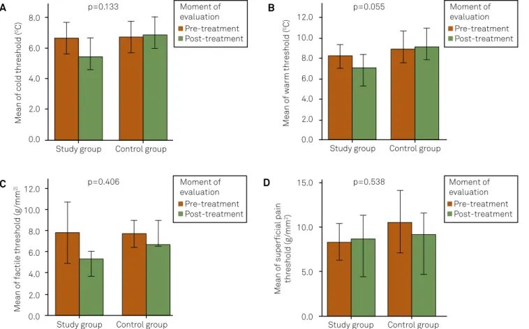
Figure 2. Somatosensory thresholds: comparison between groups (N=38). 8.0 6.0 4.0 2.0 0.0 12.0 10.0 8.0 6.0 4.0 2.0 0.0 12.0 10.0 8.0 6.0 4.0 2.0 0.0 15.0 10.0 5.0 0.0
Study group Control group
Study group Control group Study group Control group
Study group Control group
Mean o f f ac til e thr eshol d (g /mm 2) Mean o
f superficial pain
thr eshol d (g /mm 2) Mean o f w ar m thr eshol d ( 0C) Mean o f col d thr eshol d ( 0C) Moment of evaluation Pre-treatment Post-treatment Moment of evaluation Pre-treatment Post-treatment Moment of evaluation Pre-treatment Post-treatment Moment of evaluation Pre-treatment Post-treatment A B D C p=0.133 p=0.055 p=0.538 p=0.406
A: Cold thresholds; B: Warm thresholds; C: Tactile thresholds; D: Superficial pain thresholds Statistical tests: Mann–Whitney U tests.
No Litle Much Absence of pain Improvement of pain
0.3 0.2 0.1 0.0 -0.1 -0.2 -0.3 -0.4 Salivary fl o w (gr ams/ pos t-pr e) P=0.021
Figure 3. Correlation analysis between salivary flow and pain improvement (N=38).
Statistical tests: Spearman’s. lower thresholds after treatment than controls. Warm
detec-tion did not show diferences at the re-evaluadetec-tion (Figure 2). Mechanical thresholds (tactile and vibration): here were no diferences at the trigeminal branches or distant areas in
tactile detection (Mann-Whitney,P>0.05, Figure 2) except at the ophthalmic branch (Mann-Whitney,P=0.035), in which the study group showed lower thresholds after treatment
than the controls. he vibration detection results did not re
-veal diferences at the re-evaluation.
Electrical skin and teeth thresholds: here were no statis
-tical diferences in electrical thresholds between the groups
at any place investigated (Mann-Whitney,P>0.05).
Pressure and supericial pain perception: Pressure pain
detection showed higher thresholds in the study group than in controls at right temporalis (Mann-Whitney, P=0.010), bi lateral masseters (Mann-Whitney, P=0.002 and P=0.040), bilateral tibiae (Mann-Whitney, P=0.007 and 0.004) and means (Mann-Whitney,
P=0.004), after the treatment. here were no statistical diferen-ces in supericial pain thresholds between the groups at any place investigated (Mann–Whitney,P>0.05, Figure 2).
Associations and correlations: here was an associa -tion between improvement and higher salty thresholds (Spearman’s, P=0.048), higher pressure pain thresholds in the right masseter (Spearman’s, P=0.004), tibia (Spearman’s, P=0.008 and P=0.016) and maxillary branch (Spearman’s,
P=0.036) in the control group. he improvement of pain in both groups was associated with increased salivary low
(Spearman’s P=0.021) (Figure 3), increased pressure pain thresholds at the tibia (Spearman’s P=0.040), and increased
electrical teeth thresholds (Spearman’s, P=0.039). he pain
was negatively correlated with trigeminal cold thresholds (Spearman’s, P=0.046), tibia tactile threshold (Spearman’s,
P=0.044), supericial pain thresholds at the maxillary branch
(Spearman’s P=0.009), and electrical mandibular threshold
(Spearman’s P=0.008). here was also a negative correlation
between pain duration and means of pressure pain thresholds (Spearman’s P=0.020), mean tactile threshold (Spearman’s
P=0.026), maxillary supericial pain threshold (Spearman’s
P=0.043), and mandibular electrical threshold (P=0.043). Pain intensity was negatively correlated with tibia tactile threshold in the study group (Spearman’s, P=0.030) and man-dibular tactile threshold in the control group (Spearman’s, P=0.046). For both groups, there was a positive correlation bet-ween pain intensity and electric thresholds at ophthalmic and maxillary right branches (Spearman’s, P=0.022 and P=0.028, res -pectively), left mandibular branch (Spearman’s, P=0.009), right tibia (Spearman’s, P=0.036), and mean electrical skin and teeth thresholds (Spearman’s, P=0.005 and P=0.012, respectively).
DISCUSSION
his paper reports the results of a randomized clinical trial of topical medication for xerostomia. he study group received
the active substance urea, and the control group received place-bo. We observed similar improvement in both groups (60.0%), which probably means that regardless of the vehicle used, oral
cavity hydration results in an anti-xerostomic efect noticed in the association between improvement and salivation. his re
-sult conirms that protecting the oral mucosa with topic medi -cation can be useful in the control of BMS, a chronic complex condition with a poor prognosis, and that improvement was higher than that attributable to placebo (30%), showing that
the vehicle in placebo was also efective.
In general, there were no diferences in socio- demographic
characteristics between the groups, and this corresponds to the literature (elderly women are the majority of patients)26,27.
Pain characteristics (burning, moderate to severe,
conti-nuous) and abnormal salivary low were the most common
symptoms reported in previous studies2,9. here was a high
prevalence of headache and generalized pain in this sample, which had been previously described in trigeminal neuralgia28.
It is possible that these complaints and the high prevalence of temporomandibular joint dysfunction (TMD) in these patients
are relexes of pain chroniication, afecting the primary pain
cause among the reported symptoms by the patient, and sug-gested that they also need appropriate treatment for TMD28. In
this study, there was an improvement in chewing and genera-lized pain complaints, supporting the role of saliva in oral health
and the pathophysiological efects of local chronic pain28. In general, there were no statistical diferences between
the groups in sensory thresholds or xerostomia complaints
(P>0.05). QST tests were performed to verify similar neuro -pathic impairment in the BMS groups and revealed that the
study group had a longer pain duration, which could afect sensory indings in chronic pain patients, and this could
compromise the results of this study. Because of the double-blinded design, we could not match pain duration between
the groups, which could have prevented this diference. A
previous study reported a negative correlation between pain duration and sensory thresholds, which means that patients with longer histories of pain had lower thresholds than other patients, suggesting the presence of a sensory interaction29.
here were several correlations and associations between
sensory thresholds and pain improvement, as well as with
higher levels of salivary low. hese indings are interesting because they indicate a dynamic inluence of pain in neuro -logical responses and perceptions, according to its intensity30. his inluence was not only in sensory thresholds but also in eferents (salivation), and these data, which were objectively
measured with instruments, are reliable.
he limitations of this study are the loss of part of the
sample, which was small.In addition, it would be helpful to match the groups for all pain characteristics, including pain
duration, to avoid the efect of the pain history on sensory
thresholds and the results.
In conclusion, there were no diferences between the
groups; both showed an association between improvement and salivation following oral cavity hydration, regardless of the makeup of the treatment.
References
1. Ship JA, Grushka M, Lipton JA, Mott AE, Sessle BJ, Dionne RA. Burning mouth syndrome: An update. J Am Dent Assoc 1995;126:842-853.
2. Nasri C, Teixeira MJ, Siqueira JTT. Ardência bucal. Avaliação de uma amostra clínica. Rev Simbidor 2000;1:75-82
3. Siviero M, Teixeira MJ, Siqueira JT, Siqueira SR. Central mechanisms in burning mouth syndrome involving the olfactory nerve: a preliminary study. Clinics 2011;66:509-512.
4. Grushka M, Sessle BJ. Burning mouth syndrome. Dent Clin North Am 1991;35:171-184.
5. Siqueira JTT, Turbino CL, Nasri C. Dor orofacial em pacientes com disfunção temporomandibular e secura bucal: necessidade de
diagnósticos diferencial - discussão clínica. J Bras Ortodont Ortop Facial 1998;3:39-45.
6. Nasri-Heir C, Gomes J, Heir GM, et al. The role of sensory input of the chorda tympani nerve and the number of fungiform papillae in burning mouth syndrome. Oral Surg Oral Med Oral Pathol Oral Radiol Endod 2011;112:65-72.
7. Cho MA, Ko JY, Kim YK, Kho HS. Salivary flow rate and clinical characteristics of patients with xerostomia according to its aetiology. J Oral Rehab 2010;37:185-193.
8. Kho HS, Lee JS, Lee EJ, Lee JY. The effects of parafunctional
with burning mouth syndrome (BMS). Arch Gerontol Geriatr 2010;51:95-99.
9. Marino R, Torretta S, Capaccio P, Pignataro L, Spadari F. Different therapeutic strategies for burning mouth syndrome: preliminary data. J Oral Pathol Med 2010;39:611-616.
10. Patton LL, Siegel MA, Benoliel R, deLaat A, Hill C, Lauderdale F. Management of burning mouth syndrome: systematic review and management recommendations. Oral Surg Oral Med Oral Pathol Oral Radiol Endod 2007;103(Suppl 1):S39.
11. Thomson WM. Issues in the epidemiological investigation of dry mouth. Gerodontol 2005;22:65-76.
12. Siqueira SR, Nóbrega JCM, Teixeira MJ, Siqueira JT. Olfactory threshold increase in trigeminal neuralgia after balloon compression. Clin Neurol Neurosurg 2006;108:721-725.
13. Maltsman-Tseikhin A, Moricca P, Niv D. Burning mouth syndrome: will better understanding yield better management? Pain Practice 2007;7:151-162.
14. Ademola J, Frazier C, Kim SJ, Theaux C,Saudez X. Clinical evaluation of 40% urea and 12% ammonium lactate in the treatment of xerosis. Am J Clin Dermatol 2002;3:217-222.
15. Serup J. A double-blind comparison of two creams containing urea as the active ingredient. Assessment of efficacy and side-effects by non-invasive techniques and a clinical scoring scheme. Acta Dermato Venereol Suppl (Stockholm) 1992;177:34-43.
16. Merskey H, Bogduk N. Classification of chronic pain. Seattle, Washington: IASP Press, 1994.
17. Siqueira JTT, Ching LH, Nasri C, et al. Clinical study of patients with persistent orofacial pain. Arq Neuropsiquiatr 2004;62:988-996.
18. Korn GP, Pupo DB, Quedas A, Filho IB. Correlation between xerostomia degree and sialometry in patients with Sjogren Syndrome. Rev Bras Otorrinolaringol 2002;68:1-2.
19. Siviero M, Teixeira MJ, de Siqueira JT, Siqueira SR. Somesthetic, gustatory, olfactory function and salivary flow in patients with neuropathic trigeminal pain. Oral Dis 2010;16:482-487.
20. Pupo DB, Bussoloti Filho I, Liquidato BM, Korn GP. Proposta de um método prático de sialometria. Rev Bras Otorrinolaringol 2002;68:219-222.
21. Da Silva LA, Teixeira MJ, Siqueira JTT, Siqueira SRDT. Xerostomia and salivary flow in patients with orofacial pain compared with controls. Arch Oral Biol 2011;56:1142-1147.
22. Bartoshuk L. Clinical evaluation of the sense of taste. Ear Nose Throat J 1989;68 331-337.
23. Davidson TM, Murphy C. Rapid clinical evaluation of anosmia. Arch Otolaryngol Head Neck Surg 1997;123:591-594.
24. Cain WS. Testing olfaction in a clinical setting. Ear Nose Throat J 1989;68:316-328.
25. Kirkwood BR, Sterne JAC. Essential Medical Statistics. 2nd ed. Maiden, Massachusetts: Blackwell Science, 2006.
26. Mignogna MD, Pollio A, Fortuna G, et al. Unexplained somatic
comorbidities in patients with burning mouth syndrome: a controlled clinical study. J Orofacial Pain 2011;25:131-140.
27. Silvestre-Rangil J, Silvestre FJ, Tamarit-Santafé C, Bautista D. Burning mouth syndrome: correlation of treatment to clinical variables of the disease. Med Oral Patol Oral Cir Bucal 2011;16:e890-894.
28. De Siqueira SR, da Nóbrega JC, Teixeira MJ, de Siqueira JT. Masticatory problems after balloon compression for trigeminal neuralgia, a longitudinal study. J Oral Rehabil 2007;34:88-96.
29. Melzack R, Wall PD. Pain mechanisms: a new theory. Science
1965;150:971-979.