Arq
u
ivo
s
d
e
C
iê
n
ci
a
s
d
o
Ma
r
REPRODUCTIVE CYCLE OF Olivancillaria vesica
auricularia (LAMARCK, 1810) (MOLLUSCA:
GASTROPODA: OLIVIDAE) In SOUThERn BRAZIL
1
Ciclo reprodutivo de
Olivancillaria vesica auricularia
(Lamarck,
1810) (Mollusca: Gastropoda: Olividae) no Sul do Brasil
Cristina de Almeida Rocha-Barreira2
1 Pesquisa financiada pelo PICDT/CAPES.
2 Professora Adjunta e Pesquisadora do Instituto de Ciências do Mar, Universidade Federal do Ceará.
ABSTRACT
The reproductive cycle of Olivancillaria vesica auricularia on Cassino Beach, Rio Grande, RS was analyzed to determine the stages of gonad maturation according to the presence of germinate cells, correlating them to the environmental factors. Between December, 1996 and June, 1998 ten adults of each sex were collected monthly. The
gonad showed ive stages: “partially ripe”; “ripe”, “partially spawned”, “spawned” and, only in males, “restinging”. The temperature variations have signiicant inluence on the reproductive cycle of Olivancillaria vesica auricularia. Gonadal maturation was observed from September 1997 to December 1997, followed by the spawn from January 1997 to April/1997 and from December 1997 to March 1998. The resting period was recorded during fall and winter. During
this study, the reproductive cycle has suffered the inluence of environmental changes due to the severe storms occurred on January and March 1998, which suspended and transported luid mud to the beach.
Key words: reproductive cycle, histology, Olivancillaria vesica auricularia, Cassino Beach.
RESUMO
O ciclo reprodutivo de Olivancillaria vesica auricularia na praia do Cassino, Rio Grande, RS foi analisado a im
de determinar os estágios de maturação gonadal, considerando a presença das células germinativas e os fatores ambientais. Entre dezembro de 1996 e junho de 1998, dez adultos de cada sexo foram coletados mensalmente. A gônada, de ambos os sexos,
apresentou cinco estágios de desenvolvimento: “parcialmente maduro”; “maduro”, “parcialmente desovado”, “desovado” e, somente em machos, “repouso”. As variações de temperatura apresentaram inluência signiicativa sobre o ciclo de reprodução
de Olivancillaria vesica auricularia. A maturação gonadal foi observada de setembro de 1997 a dezembro de 1997, seguido por um período de desova de janeiro a abril de 1997 e dezembro de 1997 a março de 1998. A período de repouso foi observado
durante o outono e o inverno. Durante este estudo, o ciclo reprodutivo sofreu inluência de fortes tempestades que ocorreram em janeiro e março de 1998, e que suspenderam e transportaram lama luida para a praia..
INTRODUCTION
Periodically, the gonad of marine animals partially renews some of its tissue elements, and suffers several cellular transformations in order to produce gametes. Several times, this cellular dynamics is synchronous in some and between individuals of the same population (Christiansen et al., 1973).
Organisms reproduce continuously throughout the year or concentrate their reproduction over a certain period of the year (Grahame & Branch, 1985).
Gametes maturation and spawn could be inluenced by
environmental conditions (Fretter, 1984). Among the main environmental conditions that can regulate and synchronize the reproductive cycle of molluscs and most marine invertebrates, temperature and salinity are the most important. Brown (1984) suggested that seasonal variability in temperature plays a major role in determining the extent and duration of gametogenesis, whilst spawning is more dependent upon the attainment of a limited range of minimum temperatures. According to Fretter &
Graham (1962), frequently species spawn during spring and summer, related to the changes on water temperature and food availability. In temperate zones, seasonal
temperature luctuations associated to the
differences in illumination are known to be a controlling factor in gametogenesis, however, in warmer areas temperature
luctuations are not critical (Fretter, 1984). Early studies about the olivids reproduction, basically refers to mate behavior and larval development (Marcus & Marcus, 1959; Habe, 1960; Paine, 1962; Edwards, 1968; Zeigler & Porreca, 1969; Bandel, 1976), and there is none taking in to account the determination of the gonadal maturation cycle of a species of this family.
Olivancillaria vesica auricularia
is dioicous, with intern fertilization, not presenting external sexual dimorphism. Considering the little knowledge about the reproductive biology of gastropod molluscs that inhabit the surf zone of sandy beaches, in general and especially olivid species, this study aim to investigate the reproductive cycle of Olivancillaria vesica auricularia, by means of the histological analysis of the gonads; to identify the main
modiications occurring in the gonad
tissues determining different repro
ductive cycle stages and to correlate them with some environmental factors.
MATERIAL AND METHODS
Individuals of Olivancillaria vesica auricularia
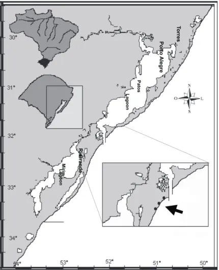
were collected in monthly samples at the Cassino Beach, Rio Grande, Rio Grande do Sul State, Brazil (Figure 1), from January 1997 to June 1998, in the surf zone, through dredging perpendicular, using rectangular dredge, with dimensions of 40 x 15 cm and nylon net with opening 10 mm among the knots. Manual collections were also made in the washing zone and 60 m inside of the surf zone.
The temperature of seawater and of the air was measured accurately through a thermometer of 0.5ºC. The salinity of the seawater in the surf zone was measured through a refractometer.
In the laboratory, the shell size was measured with a vernier caliper. The sex was determined macroscopically based on the observation of the presence or not of a copulator organ, which can be
visualized easily on the right side of the head of the animal. Young individuals were not studied in this paper. The sex ratio observed in the months studied were tested by ANOVA using Statistica software v.07.
The study of reproductive cycle was accomplished with the aid of histology techniques. 10 adult animals of each sex were randomly sampled
monthly. The animals were ixed in saline Bouin
solution for 24 hours. The histological procedure followed the routine, dehydration by alcoholic series
and cleared in xylen, until the inclusion in parafin.
Cross sections 5 to 7 mm thick were stained with Harris’hematoxilin and 1% aqueous eosin (HE). The determination of the cellular types was based on the literature (Dohmen, 1983; Rocha, 2002). In this study, were analyzed 96 males and 104 females of the subspecies Olivancillaria vesica auricularia.
All the histological photographs were taken thought a Zeiss Axiostar microscope with a digital camera.
RESULTS
Temperature, salinity and rainfall
The temperature of the seawater varied between 8°C to 24,5°C, showing a marked seasonality, with
the highest temperatures from December to March and the lowest ones in July and August (Figure 2). Seawater temperature in the surf zone was strongly
inluenced by variations of the air temperature since
the temperature variations observed were similar.
The salinity showed signiicant differences,
during the study period, with higher values from December 1996 to September 1997 (maximum 36,5) and lower among October 1997 to June 1998 (minimum 18) (Figure 2).
Sex-ratio
The sexratio was about 1:1. The variations could be attributed to the distribution not gregarious of this subspecies in the studied area. The total of 55,9% of the individuals were males and 44,1% were
females, showing no signiicant differences (p =
0.0914) between the organisms of each sex collected in every month of study (Figure 3).
Characterization of the male gonad
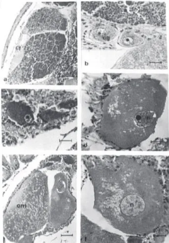
The germ cells present in the testis of
Olivancillaria vesica auricularia, it was possible to identify spermatogonia, spermatocytes I, spermatocytes II, spermatids and spermatozoa. It is important to highlight that during the spermatogenesis process,
the cells suffer divisions and transformations that can give them differentiated aspects, morphologically evident and easily recognizable (Figure 4a). The spermatogonia (Figure 4b) are located close to the wall of the testicular ducts. These cells are strongly basophilic. The cytoplasm presents a reduced volume in relation to the nucleus, which is big and it also contains several basophilic nucleolus. The spermatocytes (Figure 4c) are found more distant from the testicular wall duct. These cells are also basophilic. They are characterized by the proportional growth of the nucleus in relation to the cytoplasm. The chromatin is granular and dispersed. The cytoplasm is still more reduced than in the spermatogonia. The spermatocytes II (Figure 4d) are basophilic and has a nucleus almost occupies the whole cellular volume, with a larger condensation of the chromatin than in previous cellular stages. The spermatids (Figure 4e) are quite small, presenting little perceptible basophilic circular nucleus. The spermatozoid (Figure 4e) is relatively elongated in
the area of the head and the l agellum (tail) is long
and very thin. The spermatozoa are located in the lumen of the testicular ducts.
Characterization of the female gonad
It was possible to identify the following female germ cells: oogonia, previtellogenic oocytes, vitellogenic oocytes and ripe oocytes. These cells are inside of wrappers formed by follicular cells, called ovarian follicles (Figure 5a). The development of the germ cells occurs in the germinal epithelium, located
over a basal membrane of follicular wall. The oogonia
represent the i rst stage of the
germ lineage. These cells are small and oval, measuring approximately 36.25µ (N=60,
SD=7.5), being attached to the
follicular walls (Figure 5b). They present cytoplasm slightly basophilic and spherical nucleus quite developed, which contains a basophilic nucleolus. The previtellogenic oocytes are larger cells than the oogonia. Usually, they are slightly prolonged, measuring about 160.7µ (N=47, SD=26.1), and can be or not linked to the follicular wall (Figure 5c). The cytoplasm comes slightly
Figure 3 Sexual proportion of Olivancillaria vesica auricularia collected among January 1997 to June 1998, in the Cassino Beach, Rio Grande, Rio Grande do Sul State, Brazil.
acidophilus. The voluminous nucleus presents a big nucleolus and chromatin condensed stick inside of the nuclear membrane. The vitellogenic oocytes present a great variation in size, due to the gradual vitellus accumulation measuring approximately 217µ in diameter (N=28, SD=32.3) (Figure 5d). The cytoplasm is acidophilus. The nucleus also increases in size, reacting weakly to the HE. The ripe oocytes present larger size than the vitellogenic oocytes, measuring 270µ in diameter (N=25, SD= 24.02), they also have irregular shape (Figure 5e, 5f). They are free in the follicle lumen. The cellular membrane is thick, slightly basophilic, involving the whole oocyte.
The voluminous cytoplasm is i lled with acidophilus
spherical globules, containing nutritious substances. The nucleus becomes less evident, once the condensed chromatin is not observed, and the whole nucleus presents uniform coloration (Figure 5f).
Characterization of gonad development stages
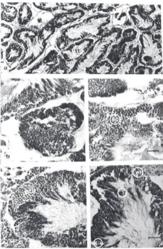
The “partially ripe” stage is characterized by intense cellular activity in both sexes. A great amount of spermatogonia is present close to the wall of the testicular ducts. The spermatocytes I and II are also quite abundant. The spermatozoids are absent and the spermatids are not very numerous in the central area of the duct (Figure 6a). The testicular ducts that are more distant to the seminal vesicle present more spermatogonia, while the closer ones contain more spermatocytes probably indicating that the process of cell maturation is not a synchronized event inside the testicles. The seminal vesicle when empty is narrow. Its wall is formed by a simple cylindrical epithelium with central nucleus (Figure 6b). In females, all the cellular types can be visualized (Figure 8a). The follicular walls are thick with to the presence of oogonia. Many previtellogenic oocytes are present and the vitellogenic oocytes are predominant. The seminal receptaculum is empty (Figure 8b).
Figure 5 Female germ cells of Olivancillaria vesica auricularia. (a) Details of the ovarian follicle; (b) Oogonia presents close to the wall of the follicle; (c) Previtellogenic oocytes; (d) Vitellogenic oocyte; (e) Ripe oocytes; (f) Ripe oocyte presenting nucleus with uniform coloration by virtue of the absence of condensed chromatin. cf = folicular cells; n = nucleus; og = oogonia; om = ripe oocyte; opv = previtellogenic oocyte; ov = viteloogenic oocyte.
In the ripe stage the spermatozoids were predominant (Figure 6c). In the ducts close to the seminal vesicle, a larger amount of spermatozoids were observed, while in the more distant ducts prevail spermatocytes. The seminal vesicle presents a central
area i lled out with great amount of spermatozoids
(Figure 6d). In the ripe ovary, there is a prevalence
of ripe oocytes that totally i ll out the interior of
the follicles, there is little or no interfolicular space (Figure 8c). The follicle wall is thin, formed by basophilic epithelial cells. In some ripe females, the seminal receptaculum was empty, similar to that found in the “partially ripe” stage. In others, it was plenty of spermatozoids “nests” (Figure 8d).
In the “partially spawned” stage there was observed that some testicular ducts are empty or they present spaces among the cells (Figure 7a). However, in some points of the testicle it is possible
to i nd ducts containing spermatocytes and many
spermatozoa. Among the testicular ducts, spaces are
also observed, in some points they are i lled out by
connective tissue. The seminal vesicle still contains spermatozoa (Figure 7b). The ovary presents some interfollicular spaces. Inside the follicles, it is observed the presence of previtellogenic and vitellogenic oocytes and the predominance of ripe oocytes (Figure 9a). Some intrafollicular space is also observed, containing phagocytes. The seminal receptaculum presents few spermatozoa inside (Figure 9b).
In the “spawned” stage a total emptiness of the testicle was observed. Some germ cells can still be found dispersed, however, most of the testicular ducts are empty. The phagocyte cells inside the ducts were also observed, mainly around of remaining germ cells (Figure 7c). The seminal vesicle does not contain spermatozoa, however a great amount of connective tissue and phagocyte cells are present. The
Figure 7 Gonadal development stages of males of Olivancillaria vesica auricularia. Part 2. (a) Testicle in the “partially spawned” stage; (b) Seminal vesicle in the “partially spawned” stage, still containing spermatozoa; (c) Testicle and seminal vesicle in the “eliminated” stage; (d) Testicle and seminal vesicle in “spent” stage. ez = spermatozoa; tt = testicle ducts; vs = seminal vesicle.
spawned females still present vitellogenic and ripe oocytes dispersed inside the follicles, however, these are in degeneration. A great amount of connective tissue occurs inside of the follicles, which present thin walls (Figure 9c). The seminal receptaculum does not contain spermatozoids (Figure 9d).
After the reproduction period, the testicle only starts its cellular recovery slowly, yet ripening process will be resumed in the next reproductive event. In the “spent” stage a great amount of connective tissue inside the testicle is still noticeable. The seminal vesicle is empty and presents connective tissue inside (Figure 7d). A “spent” stage cannot be characterized in females, because no ovary was found with follicles completely empty or only presenting oogonia, although some females have had ovaries with small amount of cells, mostly in degeneration.
Reproductive cycle
A gradual temporal displacement of the modes of gonad development stages can be observed along
the study period (Figure 10). According to this modal behavior, the reproductive cycle of Olivancillaria vesica auricularia, in the Cassino Beach, during
the study period, can be divided in three dei ned
periods: “ripening period”, “reproductive period” and “resting period”.
The ripening period was characterized by the predominance of animals in the “partially ripe” and “ripe” stages. This period occurred from September to December 1997, when approximately 50% of the individuals were in these development stages. Individuals “ripe” were observed, in minor percentage in December 1997 (Figure 11). During the
Figure 9 Gonadal development stages of females of Olivancillaria vesica auricularia. Part 2. (a) General view of the ovary partially spawned; (b) Seminal receptaculum in the “partially spawned” stage, still containing spermatozoa; (c) Ovarian follicles in the phase “eliminated”; (d) Seminal receptaculum in the “spawned” stage. cm = phagocytes; ez = spermatozoa; om = ripe oocyte.
Figure 10 Relative frequency of males and females of Olivancillaria vesica auricularia in agreement with the gonadal development stages from January 1997 to June 1998, in the Cassino Beach, Rio Grande. M = “ripe” stage; PE = “partially spawned” stage; PM = “partially ripe” stage; R = “spent” stage; E = “spawned” stage.
study, two reproductive periods were observed. The
irst one occurs from January to April 1997, and the
second from December 1997 to March 1998, when 50% of the organisms were in the “partially spawned” and “spawned” stages. Minor percentages were observed in May 1997 and in March 1998. After the reproduction period, before a new ripening process, a gonadal “resting” period was observed, which was much more characteristic in males. During the study, two resting periods were observed, namely,
the irst between April and August, 1997 and the
second, from March to June, 1998 when about 50% of the observed individuals were in “resting” stage.
DISCUSSION
Some histological differences of gonadal development of Olivancillaria vesica auricularia between males and females were observed. Females did not show completely empty follicles in the “spawned” stage, such as occurs in males. Many vitellogenic and ripe oocytes dispersed inside the follicles were still found, the majority in degeneration.
Another signiicant difference between males
and females was observed in the “spent” stage. In males, the testicular ducts were empty, a slow cellular recover begin and this stage stand until the next reproductive period. In females, a condition of complete spawning of oocytes was not observed. This fact indicates that the females do not get to use all of the produced oocytes and that the remainder are slowly reabsorbed during the period that would correspond to the “spent” stage.
The reproductive cycle can be considered, fundamentally, as the total of the gametogenic cycles of each individual of the population, which can occur in a synchronized way or not (Fretter & Graham, 1962; Christiansen et al., 1973). According to Giese (1975), the reproductive cycle of a population includes a series of successive events, as activation, growth and gametogenesis, gametes ripening and degeneration or reabsorption. Considering the frequencies of the gonadal development stages, Olivancillaria vesica auricularia presents synchronous reproductive cycle
very deined, with evident successive ripening,
reproduction and resting periods.
The reproductive cycle stages can be inluenced
by environmental factors (Fretter & Graham, 1962), being the temperature the most important one. According to Amio (1963) and Brown (1982), the spawns of the Prosobranchia are more common during the spring and summer, as an answer to the increase in the temperature of the seawater. In the studied population of Olivancillaria vesica
auricularia, a relationship was observed between the reproductive cycle stages and the temperature variations at the Cassino Beach during the study period. The seawater temperature varied between 8°C to 24.5°C, underscoring a wellmarked season, with the highest temperatures in the summer and the lowest in the winter. The ripening period begins with the arrival of the spring, when a gradual increase of temperature was observed in the seawater. The reproduction period extended from the end of the spring to summer, when the highest temperatures were registered in the area. The resting period was
veriied at begin of the fall, when a decrease in the
temperature that extends to the whole winter. The reproduction period coincided with the months in which the eggs capsules of Olivancillaria vesica auricularia were found in abundance in the
ield, from November, 1996 to March, 1997 and
from November, 1997 to February, 1998 (Rocha Barreira, 2003), a result that agrees too with the one obtained by Borzone (1991). This author, considering
the occurrence of spawning capsules in the ield,
registered that this species reproduces during the months of November to March.
Other authors reported the reproduction period of olivid species considering the presence of pairs in sexual behavior and capsules of eggs in the beach. Habe (1960) observed that the spawning period of the Japanese species Olivella fulgurata (A Adams & Reeve, 1850) occurs from May to July. Paine (1962) registered that Californian Olivella mutica (Say, 1822) spawns during the spring. Edwards (1968) observed that Olivella biplicata (Sowerby, 1825) presents a pattern of continuous mating and presence of egg capsules throughout the year. Such phenomenon is related with a neotropical origin of the genus Olivella, considering that the tropical forms present a longer reproduction season than the temperate ones.
Stormy conditions (Hs ~ 3.5 m and Ts ~ 15 s), associated with periodic cold front passages between March and May of 1998, suspended, transported and
deposited luid mud on the shoreface, surf zone and
foreshore of Cassino beach (Calliari et al., 2001). The mud and to a lesser extent, the decrease in salinity probably caused a mass mortality of infaunal and epifauna organisms inhabiting the surf zone and swash of the waves. Hundreds of young and adults of
vesica auricularia, but probably also for other benthic organisms that reproduce during the summer.
Acknowledgements The author wishes to express
her gratitude to Prof. Dr. Norton Mattos Gianuca from Oceanography Department of Fundação Universidade Federal de Rio Grande, for advising this research, and special thanks to Nilton Abreu,
for helping in the ield sampling; to Pablo Enrique
Penchaszadeh for reading the manuscript.
REFERENCES
Amio, M.A Comparative embriology of marine gastropods, with ecological considerations. Jour. Shimonoseki Coll. Fish., v.12, n. 2/3, p. 231357, 1963. Bandel, K. Spawning, development and ecology of some higher Neogastropoda from the Caribbean sea of Columbia (South America). The Veliger, v. 19, n. 2, p.176193, 1976.
Borzone, C.A. Ecologia de los moluscos del infralitoral raso de una playa arenosa del sur del Brasil. Tese de Doutorado , Universidad de Buenos Aires, Buenos Aires, Argentina, 221p, 1991.
Brown, A.C. The biology of sandy beach whelks of the genus Bullia (Nassariidae). Oceanogr. Mar. Biol. Ann. Rev., v. 20, p. 309361, 1982.
Brown, R.A. Geographical variations in the reproduction of the horse mussel, Modiolus modiolus
(Mollusca: Bivalvia). J. Mar. Biol. Assoc.iation UK, v.64, p 751770, 1984.
Calliari, L. J., Speranski, N. S., Torronteguy, M., Oliveira, M. B. The mud banks of Cassino Beach, Southern Brazil: characteristics, processes and effects. J. Coast. Res., v. 34, p. 318325, 2001.
Christiansen, H. E., Brodsky, R. S. & Cabrera, M. E. La microscopia aplicada con criterio poblacional en el estudio de las gónadas de los vertebrados y invertebrados marinos. Physis, sec. A, v. 32, n. 35, p. 467480, 1973.
Dohmen, M. R. Gametogenesis. In: K. M. Wilbur and C. M. Yonge (eds.), The Mollusca, v. 3, Development. Academic press, New York, pp.148, 1983.
Edwards, D.C. Reproduction in Olivella biplicata. The Veliger, v. 10, n. 4, p. 297304, 1968.
Fretter, V. Prosobranchs. In: K. M. Wilbur and C. M. Yonge (eds.), The Mollusca, v. 7, Reproduction. Academic Press, New York, 145, 1984.
Fretter, V. & Graham, A. British prosobranch molluscs (their functional, anatomical and ecology). Royal Society of London, London, 755p, 1962.
Galtsoff, P.S. Physiology of reproduction in molluscs.
Am. Zoologist, v. 1, p. 273289, 1961.
Giese, A. C. Reproductive cycles of marine invertebrates. An. Acad. Bras. Ciên., v. 47, p. 49 57, 1975.
Grahame, J. & Branch, G.M. Reproductive patterns of marine invertebrates. Oceanogr. Mar. Biol. Ann. Res., v. 23, p. 373398, 1985.
Habe, T. Egg masses and egg capsules of some Japanese marine prosobranchiate gastropods. Bull. Mar. Biol. Sta. Asamushi, v. 10, p. 121126, 1960. Marcus, E. & Marcus, E. On reproduction of Olivella.
Bolm. Fac. Filos. Ciênc. Univ. São Paulo., v. 22, p. 188 199, 1959.
Paine, R.T. Reproduction of Olivella mutica. Nautilus, v. 75, n. 4, p. 1391242, 1962.
RochaBarreira, C.A. Biologia de Olivancillaria vesica auricularia (Lamarck, 1810) (Mollusca, Gastropoda, Olividae) na Praia do Cassino, Rio Grande, Rio Grande do Sul, Brasil. Tese do Doutrorado, Fundação Federal Universidade de Rio Grande, 244 p., Rio Grande, 2001.
RochaBarreira, C. A. Gonad characterization and reproductive cycle of Collisella subrugosa (Orbigny, 1846) (Gastropoda; Acmaeidae) in the Northeastern Brazil. Braz. J. Biol., v. 62, n. 4B, p. 885895, 2002. RochaBarreira, C.A. Desenvolvimento embrionário e larval de Olivancillaria vesica auricularia (Lamarck, 1810) (Mollusca, Gastropoda, Olividae) em condições de laboratório. Atlântica, v. 25, n. 2, p. 107116, 2003.