THALAMUS AND LANGUAGE
Interface with attention, memory
and executive functions
Marcia Radanovic
1, Mariana Azambuja
2, Letícia Lessa Mansur
3,
Cláudia Sellitto Porto
4, Milberto Scaff
5ABSTRACT - Subcortical structures are in a strategic functional position within the cognitive networks. Their lesion can interfere with a great number of functions. We studied six patients with thalamic vascular lesions (three left sided, two right sided and one bilateral), to characterize their repercussion in the communicative abilities and the interface between language alterations and other cognitive abilities, as attention, memory and frontal executive. All patients were evaluated through a functional interview (discourse analysis), and the following batteries: Boston Diagnostic Aphasia Examination, Boston Naming Test, Token Test, Benton Visual Retention Test, Trail Making, Wisconsin Card Sorting and frontal scripts. All patients performed MRI and five underwent SPECT. Results show that these patients present impairment in several cognitive domains, especially attention and executive functions (working memory, planning and self-monitoring); those with right lesions have an additional visuospatial impairment. Such alterations interfere with language abilities, and this fact must be considered in the rehabilitation efforts.
KEY WORDS: thalamus, language, attention, memory, prefrontal cortex.
Tálamo e linguagem: interface com atenção, memória e funções executivas Tálamo e linguagem: interface com atenção, memória e funções executivasTálamo e linguagem: interface com atenção, memória e funções executivas Tálamo e linguagem: interface com atenção, memória e funções executivasTálamo e linguagem: interface com atenção, memória e funções executivas
RESUMO - As estruturas subcorticais ocupam posições funcionais estratégicas nas redes cognitivas. Sua lesão pode interferir com grande número de funções. Estudamos seis pacientes com lesão vascular talâmica (três à esqueda, dois à direita e um bilateral), para caracterizar a repercussão da lesão nas suas habilidades comunicativas e a interface entre alterações de linguagem e outras habilidades cognitivas, como atenção, memória e executivas frontais. Os pacientes foram avaliados através de entrevista funcional (análise do discurso), testes de Boston para Diagnóstico da Afasia, Nomeação Boston, Token, Múltipla Escolha de Benton, Trail Making, Wisconsin Card Sorting e scripts frontais. Todos os pacientes realizaram ressonância magnética encefálica (RM) e cinco realizaram SPECT. Os resultados mostram que os pacientes apresentam prejuízo em diversos domínios cognitivos, especialmente atenção e funções executivas (memória operacional, planejamento e automonitoração); nos lesados à direita, ocorre prejuízo visuoespacial associado. Tais alterações repercutem nas habilidades de linguagem, o que deve ser levado em conta para reabilitação.
PALAVRAS-CHAVE: tálamo, linguagem, atenção, memória, córtex pré-frontal.
Department of Neurology and Department of Physiotherapy, Speech Pathology and Occupational Therapy, University of São Paulo School of Medicine, São Paulo SP Brazil (FMUSP): 1MD, PhD, Neurologist; 2Phonoaudiologist, Neurolinguistic Specialist; 3Phonoaudiologist,
Master and PhD in Linguistics; 4Psychologist, Master in Experimental Pathophysiology; 5MD, PhD, Full Professor of Neurology.
Received 15 March 2002, received in final form 31 July 2002. Accepted 20 August 2002.
Dra. Márcia Radanovic - Rua Cristiano Viana 163 /92 - 05411-000 São Paulo SP - FAX : 55 11 3088 9708. E-mail: [email protected]
The study of the role of subcortical structures in cognitive processes, largely impelled by neuroima-ging development has brought interest in the fron-tostriatal circuits and the thalamus, and their parti-cipation in the linguistic processing. Fisher, in 19591,
was the first author to describe aphasia in left tha-lamic hemorrhagic lesions. Aphasia with decreased speech output (although fluent), anomia, parapha-sias (that could deteriorate into jargon),
comprehen-sion less affected than production, and no repetition problems (or minimum) are described following ven-trolateral (VL) and ventral anterior (VA) nuclei lesions; a fluent aphasia, with non-words, may occur espe-cially in lesions of the pulvinar and the posterior late-ral (PL) nucleus2. Left dorsomedial (DM) lesions are
related to anomia, verbal memory deficits and a fron-tal syndrome3,4. Thalamic aphasia is more frequently
(polar) and interpeduncular profunda (paramedian thalamic) arteries, and since they are small vessels, there is a low probability that some associated cor-tical dysfunction may exist5. Thalamic lesions may
provoke an association of disorders: disfluency and conditions similar to transcortical aphasia in lesions involving the left VL, pulvinar and paramedian nuclei6,7 or leading to a disconnection between the
frontal area – VA; arousal disturbances (intrusions, contaminations, non-words); comprehension altera-tions, because of interruption in thalamus - frontal area - superior longitudinal fascicle - Wernicke’s area connections; hypophonia, by interruption of pallido-nigral afferents to the thalamus8.
The attempts to explain the mechanisms by which thalamic lesions lead to language disturbances are based upon theories such as cortical activation and integration, modulation and monitoration of the formulated language9-12. Some authors hypothesized
about the integrative circuits involving the cerebral cortex, the thalamus and the brainstem, especially the ascending reticular activating system, with the participation of verbal memory13-15. Cappa and
Vignolo16 attributed the handling of words as
se-mantic units to the thalamus, excluding its partici-pation in phonemic processes, which could explain the few repetition disturbances in subjects with tha-lamic aphasia. Nadeau and Crosson17 suggested that
the mediothalamic-frontocortical system (including the frontal lobes – inferior thalamic peduncle – nu-cleus reticularis and centromedian nunu-cleus) could participate in the control of lexical selection proces-ses, its failure leading to a loss of any difference in the activation among a certain lexical item and others semantically related, thus resulting in anomia and semantic paraphasias.
The interaction between language and other cognitive domains is an important factor to be consi-dered, especially in lesions affecting structures that have extensive reciprocal connections with the cere-bral cortex, such as the thalamic nuclei. Based on a preliminary study18, we noted a great interference
of factors such as memory and attention in the lin-guistic performance of patients with thalamic lesions. Damage of short-term memory (phonological loop) can be expressed clinically by sentence compre-hension deficits19, either due to the absence of a
phonological memory and/or semantic or syntactic memory. Likewise, the short-term memory deficit may impair sentence planning, leading to verbal pro-duction and vocabulary acquisition disturbances20.
Primary attentional disorders can also disturb lin-guistic processing. Attention deficits can occur in
frontal lesions, and in these, we frequently find verbal fluency alterations, compromising speech initiative in left hemisphere lesions, and of pragmatic aspects of speech in right hemisphere lesions21. Neglect dyslexia
also was described following thalamic lesions17.
The aphasias caused by subcortical lesion usually escape the possibility of classification and the use of linguistic tests alone may not be sufficient to identify the language disturbance, which can lead to thera-peutic inefficacy. Our purpose in this study was to describe the profile of language disturbances found after right and left thalamic lesions, and to verify the interrelation between these findings and other cognitive impairments, such as attention, memory and executive functions.
METHOD
We studied six patients with vascular thalamic lesions; two patients had right-sided lesions, three had left-sided lesions and in one there were bilateral lesions. All patients were right-handed and Portuguese native speakers. The selection of cases was based on anatomical features (site of lesion in neuroimaging performed when the patient was admitted in the hospital). Patient 6, although presen-ting bilateral lesion, was included when he suffered a right thalamic stroke, and began to demonstrate the described clinical picture. Exclusion criteria were: delay in language acquisition, antecedents of psychiatric and/or neurological disease, alcoholism, hearing deficiency, associated cortical involvement (based on clinical history and neurological examination). Demographic data are shown in Table 1; neuroimaging features are shown in Table 1 and Figure 1. All the participants of this study signed an agreement form that was included in the project approved by the Commission of Ethics for Analysis of Research Projects -Hospital das Clínicas – FMUSP.
Instruments
The language evaluation was performed using a func-tional interview (narrative, argumentative and procedural discourses), the Boston Diagnostic Aphasia Examination (including the Boston Naming Test)22 and the Token Test23.
These tests evaluate the phonological, morphological, syn-tactic and semantic aspects that are present in the pro-cesses of language comprehension (word discrimination, commands and text interpretation) and language pro-duction (naming, repetition of words and phrases, text production). They take into account the effects related to the modalities of stimuli input and answer output and also those task-specific, in order to control linguistic and non-linguistic variables. The Brazilian reference values to BDAE and Token Test were based on previous studies con-ducted by the authors24,25.
An additional test of frontal scripts, translated into Portuguese from Allain’s et al. original publication26, was
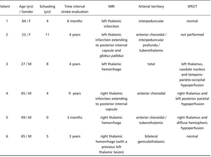
Table 1. Demographic and neuroimaging data.
Patient Age (yrs) Schooling Time interval MRI Arterial territory SPECT / Gender (yrs) stroke-evaluation
1 64 / F 4 6 months left thalamic interpeduncular normal
infarction
2 22 / F 11 4 years left thalamic anterior choroidal / not performed infarction extending interpeduncular
to posterior internal profunda / capsule and tuberothalamic globus pallidus
3 27 / M 8 4 years left thalamic total left thalamus,
hemorrhage caudate nucleus
and temporo-parieto-occipital
hypoperfusion 4 65 / M 4 9 years right thalamic anterior choroidal right thalamus and
infarction extending left posterior parietal to posterior internal hypoperfusion
capsule
5 49 / M 0 3 months right thalamic anterior choroidal / right thalamus and hemorrhage tuberothalamic diffuse hemispheric hypoperfusion
6 65 / M 5 3 years right thalamic bilateral normal
hemorrhage (with a geniculothalamic previous left
thalamic lesion)
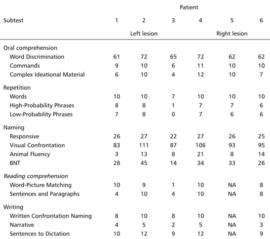
Table 2. Performance of patients in the Boston Diagnostic Aphasia Examination. Patient
Subtest 1 2 3 4 5 6
Left lesion Right lesion Oral comprehension
Word Discrimination 61 72 65 72 62 62
Commands 9 10 6 11 10 10
Complex Ideational Material 6 10 4 12 10 7
Repetition
Words 10 10 7 10 10 10
High-Probability Phrases 8 8 1 7 7 6
Low-Probability Phrases 7 8 0 7 6 6
Naming
Responsive 26 27 22 27 26 25
Visual Confrontation 83 111 87 106 93 95
Animal Fluency 3 13 8 21 8 14
BNT 28 45 14 34 33 26
Reading comprehension
Word-Picture Matching 10 9 1 10 NA 8
Sentences and Paragraphs 4 10 4 10 NA 8
Writing
Written Confrontation Naming 8 10 8 10 NA 10
Narrative 4 5 2 5 NA 3
Sentences to Dictation 10 12 9 12 NA 9
NA, not applicable.
Table 3 . Performance of patients in the Token Test. Patient
Part 1 2 3 4 5 6
Left lesion Right lesion
1 10 10 9 10 8 10
2 4 6 6 6 4 6
3 10 10 10 9 9 9
4 2 10 4 9 3 5
5 8 21 8 14 8 10
right lesion) and nine normal individuals, for comparison (normal subjects had an average of 2.6 ordination mistakes and no intrusions). This task investigates the abilities of information recovery using common action sequences and it is sensitive to frontal dysfunction. The subjects are asked to establish the hierarchical organization of six scripts (each one containing from 10 to 17 actions) obeying to the same temporal sequence in which they are usually executed. The actions of each script are presented in mixed cards that must be organized by the subject. The title of each script is presented in a separated card, remaining accessible
Attention, memory and executive functions were eva-luated through the Benton Visual Retention (BVRT), Trail Making and Wisconsin Card Sorting (WCST) tests. The BVRT consists of several series of line drawings that must be reproduced by the patient, and it was designed to assess visual perception and short-term visual memory27. The Trail
Making test measures visual skills (searching and sequen-cing) and the ability to make alternating conceptual shifts, as it requires the patient to draw a line connecting 25 circles in a certain order, as rapidly and accurately as pos-sible28. The WCST assesses abstract ability, conceptual set
shifting and the capacity of “learning”, as the subject is asked to sort a series of printed cards according to criteria that are changed without previous notice (from colors to forms, numbers, etc)29. In the neuropsychological tests,
we adopted a performance of 90% as a cut-off score for normality, because there are no standardized values for the Brazilian population, especially low-educated. All patients were able to comprehend the instructions for test accomplishment. The clinical evaluations were performed within a minimum interval of six weeks after symptom instal-lation. All patients performed SPECT and cranial MRI, after a minimum interval of two months from onset of symptoms. The patients were tested by a speech therapist and a neuropsychologist, both experienced, according to the pro-cedures recommended by Lezak30,31. Their language and
neurological evaluation were reviewed and analyzed by two authors (respectively LLM and MR).
RESULTS
Language: Five patients persistently complained of memory and word finding difficulties that inter-fered in their daily activities (exception: Patient 5).
Left lesions
Patient 1: This patient presented moderate com-prehension deficit in more complex linguistics tasks (Complex Ideational Material and Reading Sentences and Paragraphs in the BDAE and especially in the fourth and fifth blocks in the Token Test), semantic paraphasias and anomia. This patient had difficulties in changing semantic categories and sequences in formal tests (from letters to numbers, days of the week to months), showing perseverations; her per-formance in the animal fluency was reduced. In the scripts, she had two ordination mistakes and no in-trusions. During the examination, she often com-plained that she could not remember the instructions or that she forgot the original stimulus while searching for the correct answer. Besides, she was disorganized and inattentive during the search for answers in reading tasks, having difficulties to choose just one answer.
Patient 2 had a normal performance in the for-mal tests and made no mistakes in the frontal scripts. Patient 3 showed moderate to severe comprehension, repetition and naming deficits, with phonemic para-phasias. His reading and writing were also defective.
Right lesions
Patient 4 had interpretation difficulties, empha-sizing descriptive aspects in the Cookie Theft narra-tive. In the scripts he had 20 ordination mistakes and no intrusions.
Patient 5 had a slight anomia for visual presented
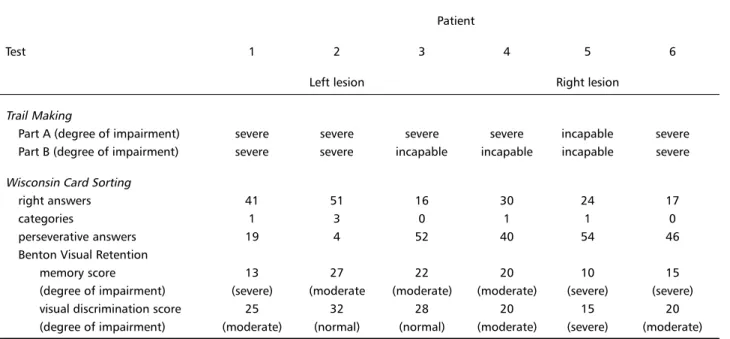
Table 4. Neuropsychological data.
Patient
Test 1 2 3 4 5 6
Left lesion Right lesion
Trail Making
Part A (degree of impairment) severe severe severe severe incapable severe Part B (degree of impairment) severe severe incapable incapable incapable severe Wisconsin Card Sorting
right answers 41 51 16 30 24 17
categories 1 3 0 1 1 0
perseverative answers 19 4 52 40 54 46
Benton Visual Retention
memory score 13 27 22 20 10 15
(degree of impairment) (severe) (moderate (moderate) (moderate) (severe) (severe)
visual discrimination score 25 32 28 20 15 20
stimuli, bad performance in all blocks of the Token test and severe difficulties in the integration of the several set elements in the Cookie Theft picture. Although these impairments are difficult to interpret (the patient is illiterate), we consider them as relevant because the patient had subjective complaints in these specific skills, and we could observe anomia in spontaneous speech.
Patient 6: This patient showed scores below nor-mal for repetition, related to sentence extension, and Visual Confrontation Naming, as well as deficits in narrative organization, with digressions and perse-verations. In the Token test, he showed bad perfor-mance in the fourth and fifth blocks. In the scripts, he had 21 ordination mistakes and 4 intrusions.
Neuropsychological examination: All six patients had performances below normal in the attention and memory tests. All of them, except case two, presen-ted performance below normal in tests of executive functions. The group with right lesions had worse performance in the visual discrimination test. The results are exposed in Table 4.
DISCUSSION
The interaction between language and other cog-nitive functions is receiving greater attention by rese-archers in Neuropsychology. Subcortical lesions can interfere with cortical networks in several ways, due to the complexity of cortico-striato-thalamic-cortical loops and also because small changes in the lesion sites can completely change the connections affected and their functional repercussion on the cerebral cor-tex (through deafferentation mechanisms)4,12,32,33.
Language alterations in subcortical lesions are often mild, although the patients persistently complain of naming difficulties or that “the thoughts are con-fused”. These deficits are rarely as disabling as those found in cortical lesions (unless the patient has some professional activity which requires a higher level of cognitive/verbal abilities: academic students, lawyers), but they have a significant impact in the subjects’ quality of life. We will try to describe the relation between language alterations and the atten-tion, memory and executive function disorders found in our patients.
The comprehension deficits in thalamic aphasias are mild to moderate, and the written compre-hension tends to be more preserved than the oral comprehension16,34. Verbal memory alterations can
contribute to the low scores in these tasks35. The
con-comitant attention and memory deficits found in
cases 1 and 3 suggest that these functions can be implied in the comprehension impairment, as we no-te a worsening of performance relano-ted to the stimu-lus extension and syntactic complexity (both proces-ses are linked to working memory capacity). Patient 1 also presented some findings that suggest an addi-tional deficit of attention, mnesic and planning/moni-toring abilities such as impairment in changing cate-gories and sequences (from days of the week to months, for example) with perseverations, reduction in animal fluency, difficulties in scanning strategies and concentration. Furthermore, her complaint that she could not remember the task or the stimuli presented also point to attention and working me-mory deficits. It is interesting to notice that Patient 2, who did not present any dysfunction in formal tests, had attention and memory alterations, but not of executive dysfunction. Although she performed well, she showed a great delay in answers. This patient complained that she could not talk and write as she did before (especially names), and that “when I read anything long, I do not remember the be-ginning”, being unable to attend her college courses. Her complaint supports the existence of a working memory alteration.
Repetition disturbances were found in Patient 3 and in Patient 6, who had bilateral lesions. It is our impression that attention and working memory dysfunction can also explain these repetition alte-rations, if we consider that there is a correlation bet-ween the decline of repetition capacity and increases in sentence length. Perseverations were found in pa-tients 1 and 6. Their existence in thalamic aphasia is considered as related to attention and vigilance me-chanisms11,15, and may be present in VA lesions8.
Paraphasias were found in Patients 1 and 3. Pa-tient 1 had semantic paraphasias, especially in na-ming tests, where she benefited from phonemic cues. Patient 3 showed phonemic paraphasias. These patients also presented anomia. These findings sus-tain the most consistent theoretical model about the thalamus role as a “floodgate”, responsible for the best lexical selection12,17, with the participation of
attention and working memory36. A category-specific
anomia37 and proper name anomia38 were already
impair-ment in visuospatial, discursive and script tasks. In Patient 5, the visuospatial alteration led to low scores in Visual Confrontation Naming and in the Token Test associated to an inability to adequately distinguish the elements in the Cookie Theft picture. These im-pairments are difficult to interpret because this pa-tient is illiterate, and there are not reference values for Brazilian illiterates in most neuropsychological tests. In patient 6, the Visual Confrontation Naming task was impaired too. These patients with right thalamic damage were those who had the worst per-formances in the Benton visual discrimination test.
The ability to elaborate a discourse macrostruc-ture and to integrate information in a context are impaired in patients with right hemisphere lesions, who present difficulties in extracting sentence meaning, discarding irrelevant information and integrating the apprehended meaning within a cohe-rent whole, in order to generate an organized narra-tive. Contrarily, they have a tendency to fix on indi-vidual, isolated and/or less important elements, wi-thout performing a connection between them to form a whole39. The coherent discourse
comprehen-sion, in MRI studies, provokes larger activation in the right frontal lobe than the reading of non-related sentences40.
The impact of the executive system dysfunction in language is a complex issue. Alexander et al., in 198921, tried to correlate language deficits and
fron-tal lesions. They considered four levels in their theo-retic model: motor, linguistic-cognitive, activation and formulation, the latter being the most important in our discussion. Thus, in left frontal lesions (espe-cially prefrontal and those in the posterior lateral convexity), the formulation impairment reflects an inability in language structuration and organization (when the subject tries to deal with elements that are more complex or abstract) and of conceptual association. In the right frontal lesions (also pre-frontal or of its projections to the posterior con-vexity), formulation difficulties are more contextual, as goal selection and self-monitoring, leading to tan-genciality, inadequate topic changes, improper dis-course and confabulation. Frontal symptoms like con-crete thinking, deficiency in behavior planning and monitoring, perseveration, apathy and impairment in conceptualization can certainly compromise the discursive ability, generating problems with its macrostructure, organization, cohesion, and infe-rences as those found in our cases. The attention and working memory deficits can also be inserted in the context of frontal alterations.
An interesting finding was the difference in per-formance between individuals with left and right le-sions in the frontal scripts. According to the cognitive theory, scripts are a class of organized structures that are stored as knowledge nets representing events, stories and frequent activities41. Thus, the frontal
dysexecutive syndrome also includes impairment in the temporal and sequential processing of this pre-existing mental plan42, although the exact nature of
this storage process is still unknown. Studies with functional MRI showed bilateral prefrontal dorso-lateral activation in tasks involving frontal scripts, thus evidencing the participation of these areas in the analysis of action sequences43.
Most errors in our patients occurred in the actions ordination, and not in the choice of which actions belonged to the scripts. This fact puts into evidence a specific inability in performing the temporal-sequential ordination discussed above. An intriguing fact is the preponderance of this dysfunction in pa-tients with right lesion, since most studies point to the participation of bilateral prefrontal cortices in this task. We can speculate that the great demand for spatial movement in most scripts (going to the supermarket, taking the subway) generated an additional disadvantage for these patients. Patient 2, that did not present any mistakes, also did not have executive dysfunction in WCST. Disorders of thought that are independent of primary language disturbances were described in dominant thalamic lesions, and seem to reflect a primary disorganization that can rely on frontal executive mechanisms44.
Many authors have proposed that the pathophy-siological mechanisms underlying subcortical aphasia (and other cognitive symptoms) are associated with cerebral blood flow and metabolism alterations45-48.
These abnormalities reflect the remote consequences of focal damage (diaschisis of the ipsilesional cortex) and it may explain why these changes are sometimes transient and their improvement follows the pattern of the cortical reperfusion49,50. Considering that the
anterior and medial thalamus project to the frontal cortex and the lateral thalamus projects to posterior cortex47, most thalamic lesions will lead to some
cor-tical involvement in associative areas, and thus pro-voke cognitive symptoms.
perfu-sion studies (Patients 1 and 6) were symptomatic. Patient 2, whose results in tests were normal, refused to undergo the procedure. In case 3, there was a relative overlapping between the hypoperfusion pattern (left posterior) and his clinical picture; this patient presented the largest lesion, that affected all the thalamus and extended to the adjacent pos-terior periventricular white matter, suggesting a more severe deafferentation mechanism. In Patient 5, there was diffuse hypoperfusion in the right hemisphere, making any detailed analysis impossible. In Patient 4, there was an apparent contralateral functional reper-cussion (left posterior parietal), but of little clinical repercussion. We believe that the small number of cases is the main reason for these inconclusive results. The study of subcortical-induced language altera-tions provides very complex and diverse types of clini-cal syndromes, which helps us to understand how other cognitive processes interact with language and vice-versa. Based on our results, we agree with the interpretation that the causes for a loss of efficiency in the language use caused by thalamic lesions are multifactorial and transcend the boundaries of lin-guistic processing itself7,11,13-15. Lesions in both
he-mispheres can generate dysfunctions that, although different in nature, become disabling. From an ana-tomical view, the amnesic syndrome (resembling that caused by the temporal medial lesion) appears more specifically after lesions of the mammillo-thalamic tract (MTT); different memory deficit patterns can occur after lesions of other thalamic structures, even when the MTT is preserved. The dysexecutive syndro-me can be found after lesions in DM, midline, intra-laminar (IL) or any combination of these nuclei51. As
our patients had widespread lesions, frequently in more than one arterial territory, and taking into account the complexity and diversity of the thalamic vascularization, is not possible for us to make preci-se anatomical correlations. We can speculate that MTT, IL and the ventral portion of DM are damaged in Patients 1 and 2, in the latter possibly existing a VA lesion; in Patients 4, 5 and 6, the posterior lesions predominate, probably in pulvinar, PL, posterior por-tion of DM and lateral geniculate. Patient 3 had a total thalamic lesion.
In many cases, the alterations objectively found in language batteries can be slight and dispro-portionate to the patient’s complaints. Executive, attention, mnesic and visuospatial dysfunctions, cau-sed by the thalamus insertion in the respective cogni-tive networks, are elements that contribute to lan-guage deficits, and must be adequately evaluated
and measured to improve the efficiency of thera-peutic approaches.
REFERENCES
1. Fisher CM. The pathologic and clinical aspects of thalamic hemorrhage. Trans Am Neurol Assoc 1959;84:56-59.
2. Graff-Radford NR. Syndromes due to acquired thalamic damage. In Feinberg TE, Farah MJ (ed.)Behavioral neurology and neuropsy-chology. New York: McGraw-Hill, 1997:433-443.
3. Sandson TA, Daffner KR, Carvalho PA, Mesulam M-M. Frontal lobe dysfuntion following infarction of the left-sided medial thalamus. Arch Neurol 1991;48:1300-1303.
4. Mesulam M-M. Behavioral neuroanatomy. In Mesulam M-M (ed). Prin-ciples of behavioral and cognitive neurology 2.Ed. New York: Oxford Univ Press, 2000:1-120.
5. Crosson B, Nadeau SE. The role of subcortical structures in linguistic pro-cesses. recent developments. In Stemmer B, Whitaker HA (eds.) Handbook of neurolinguistics. San Diego: Academic Press, 1998:431-445.
6. Riklan M, Cooper IS. Psychometric studies of verbal functions following thalamic lesions in humans. Brain Lang 1975;2:45-64.
7. Lazzarino LG, Nicolai A, Valassi F Biasizzo E. Language disturbances from mesencephalo-thalamic infarcts: identification of thalamic nuclei by CT-reconstruction. Neuroradiology 1991;33:300-304.
8. Gorelick PB, Hier DB, Benevento L, Levitt S, Tan W. Aphasia after left thalamic infarction. Arch Neurol 1984;41:1296-1298.
9. Samra K, Riklan M, Levita E, et al. Language and speech correlates of anatomically verified lesions in thalamic surgery for parkinsonism. J Speech Hear Res 1969;12:510-540.
10. Darley FL, Brown JR, Swenson WM. Language changes after neurosurgery for parkinsonism. Brain Lang 1975;2:65-69.
11. Luria AR On quasi-aphasic speech disturbances in lesions of the deep structures of the brain. Brain Lang 1977; 4: 432-459.
12. Wallesch C-W, Papagno C. Subcortical aphasia. In Rose FC, Whurr R, Wyke MA (ed) Aphasia. London: Whurr 1988: 256-287.
13. Reynolds AF, Turner PT, Harris AB, Ojemann GA, Davis LE. Left thalamic hemorrhage with dysphasia: a report of five cases. Brain Lang 1979;7:62-73.
14. Metter EJ, Riege WH, Hanson WR, et al. Comparison of metabolic rates, language, and memory in subcortical aphasias. Brain Lang 1983;19: 33-47. 15. Ojemann G. Brain organization for language from the perspective of
electrical stimulation mapping. Behav Brain Sci 1983;6:189-230. 16. Cappa SF, Vignolo LA. “Transcortical “ features of aphasia following
left thalamic hemorrhage. Cortex 1979;15:121-130.
17. Nadeau SE, Crosson B. Subcortical aphasia. Brain Lang 1997;58: 355-402.
18. Radanovic M, Scaff M. Speech and language disturbances due to subcortical lesions. Brain Lang, in press.
19. Vallar G, Baddeley AD. Fractionation of working memory: neuropsy-chological evidence for a phonological short-term store. J Verbal Learn Verbal Behav 1984;23:151-161.
20. Baddeley A, Gathercole S, Papagno C. The phonological loop as a language learning device. Psychol Rev 1998;105:158-173.
21. Alexander MP, Benson DF, Stuss DT. Frontal lobes and language. Brain Lang 1989;37:656-691.
22. Goodglass H, Kaplan E. The assessment of aphasia and related disorders. 2.Ed. Philadelphia: Lea & Febiger, 1983.
23. DeRenzi E, Vignolo LA. The Token Test: a sensitive test to detect receptive disturbances in aphasics. Brain 1962;85:665-678.
24. Radanovic M, Mansur LL. Performance of a Brazilian population sample in the Boston Diagnostic Aphasia Examination: a pilot study. Braz J Med Biol Res 2002;35:305-317.
25. Radanovic M. Disturbances of speech and language in subcortical lesions.Thesis. São Paulo University School of Medicine. São Paulo, 1999. 26. Allain P, Le Gall D, Etcharry-Bouyx F, Aubin G, Emile J. Mental
representation of knowledge following frontal-lobe lesion: dissociations on tasks using scripts. J Clin Exper Neuropsychol 1999;21:643-665. 27. Benton AL. he revised visual retention test. 4.Ed. New York:
Psycho-logical Corporation, 1974.
28. Reitan RM. Validity of the Trail-Making Test as an indicator of organic brain damage. Percept Mot Skills 1958;8:271-276.
29. Grant DA, Berg E. The Wisconsin Card Sort Test Random Layout: directions for administration and scoring. Madison, Wisconsin: Wells Printing, 1980.
31. Lezak MD. The neuropsychological examination: interpretation. In Lezak MD. Neuropsychological assesment. 3.Ed. Oxford: Oxford Univ Press, 1995:144-169.
32. Naeser MA, Palumbo CL, Helm-Estabrooks N, Stiassny-Eder D, Albert ML. Severe nonfluency in aphasia. Brain 1989;112:1-38.
33. Alexander MP. Speech and language deficits after subcortical lesions of the left hemisphere: a clinical, CT, and PET study. In Vallar G (ed). Neuropsychological disorders associated with subcortical lesions. New York: Oxford Univ Press, 1992:455-477.
34. Graff-Radford NR, Eslinger PJ, Damasio AR, Yamada T. Nonhemor-rhagic infarction of the thalamus: behavioral, anatomic, and physiologic correlates. Neurology 1984;34:14-23.
35. Puel M, Cardebat D, Demonet JF, et al. Le role du thalamus dans les aphasies sous-corticales. Rev Neurol (Paris) 1986;142:431-440. 36. Crosson B. Subcortical mechanisms in language: lexical-semantic
mechanisms and the thalamus. Brain Cogn 1999;40:414-438. 37. Crosson B, Moberg PJ, Boone JR, Rothi LJG, Raymer A.
Category-specific naming deficit for medical terms after dominant thalamic/ capsular hemorrhage. Brain and Lang 1997;60:407-442.
38. Lucchelli F, DeRenzi E. Proper name anomia. Cortex 1992; 28: 221-230. 39. Joanette Y, Goulet P, Hannequin D. Alterações da comunicação verbal nos destros com lesões cerebrais no hemisfério direito. In Mansur LL, Rodrigues N (eds.), Temas em neurolingüística. São Paulo: Tec Art, 1993:23-30.
40. Kircher TT, Brammer M, Tous Andreu N, Williams SC, McGuire PK. Engagement of right temporal cortex during processing of linguistic context. Neuropsychologia 2001;39:798-809.
41. Schank R, Abelson P. Scripts, plans, goals and understanding. Hillsdale: Erlbaum, 1977.
42. Shallice T, Burgess PW. Deficits in strategy application following fron-tal lobe damage in man. Brain 1991;114:727-741.
43. Crozier S, Sirigu A, Lehéricy S, van der Moortele P-F, Pillon B, Grafman J et al. Distinct prefrontal activations in processing sequence at the sentence and script level: an fMRI study. Neuropsychologia 1999;37: 1469-1476. 44. Chatterjee A, Yapundich R, Mennemeier M, et al. Thalamic thought
disorder: on being “ a bit addled”. Cortex 1997;33:419-440.
45. Perani D, Vallar G, Cappa S, Messa C, Fazio F. Aphasia and neglect after subcortical stroke. Brain 1987;110:1211-1229.
46. Démonet JF, Puel M, Celsis P, Cardebat D. “Subcortical” aphasia: some proposed pathophysiological mechanisms and their rCBF correlates revealed by SPECT. J Neuroling 1991;6:319-344.
47. Metter EJ. Role of subcortical structures in aphasia: evidence from studies of resting cerebral glucose metabolism. In Vallar G. Neuropsychological disorders associated with subcortical lesions. New York: Oxford Univ Press, 1992:478-500.
48. Okuda B, Tanaka H, Tachibana H, Kawabata K, Sugita M. Cerebral blood flow in subcortical global aphasia: perisylvian cortical hypoperfusion as a crucial role. Stroke 1994;25:1495-1499.
49. Olsen TS, Bruhn P, Öberg RGE. Cortical hypoperfusion as a possible cause of “subcortical aphasia”. Brain 1986;109:393-410.
50. Vallar G, Perani D, Cappa SF, Messa C, Lenzi GL, Fazio F. Recovery from aphasia and neglect after subcortical stroke: neuropsychological and cere-bral perfusion study. J Neurol Neurosurg Psychiatry 1988; 51:1269-1276. 51. Van der Werf YD, Witter MP, Uylings HBM, Jolles J. Neuropsychology of