288
Brazilian Journal of Cardiovascular Surgery
Relationship between High Red Cell Distribution
Width and Systemic Inflammatory Response
Syndrome after Extracorporeal Circulation
Harsh Sateesh Seth
1, M.S; Prashant Mishra
1, M.Ch; Jayant V. Khandekar
1, M.Ch; Chaitanya Raut
1, M.Ch; Chandan
Kumar Ray Mohapatra
1, M.S; Ganesh Kumar K. Ammannaya
1, M.S; Jaskaran Singh Saini
1, M.S; Vaibhav Shah
1, M.S
Abstract
Objective: Cardiac surgical operations involving extracorporeal circulation may develop severe inflammatory response. This severe inflammatory response syndrome (SIRS) is usually associated with poor outcome with no predictive marker. Red cell distribution width (RDW) is a routine hematological marker with a role in inflammation. We aim to determine the relationship between RDW and SIRS through our study.
Methods: A total of 1250 patients who underwent cardiac surgery with extracorporeal circulation were retrospectively analyzed out of which 26 fell into the SIRS criteria and 26 consecutive control patients were taken. RDW, preoperative clinical data, operative time and postoperative data were compared between SIRS and control groups.
Results: The demographic profile of the patients was similar. RDW was significantly higher in the SIRS versus control group
(15.5±2.0 vs. 13.03±1.90), respectively with P value <0.0001. There was significant mortality in the SIRS group, 20 (76.92%) as compared to 2 (7.6%) in control group with a P value of <0.005. Multiple logistic regression analysis revealed that there was significant association with high RDW and development of SIRS after extracorporeal circulation (OR for RDW levels exceeding 13.5%; 95% CI 1.0-1.2; P<0.05).
Conclusion: Increased RDW was significantly associated with increased risk of SIRS after extracorporeal circulation. Thus, RDW can act as a useful tool to predict SIRS in patients undergoing cardiac surgery with extracorporeal circulation. Hence, more aggressive measures can be taken in patients with high RDW to prevent postoperative morbidity and mortality.
Keywords: Erythrocyte Indices. Extracorporeal Circulation. Systemic Inflammatory Response Syndrome.
DOI: 10.21470/1678-9741-2017-0023
1Department of Cardiovascular and Thoracic Surgery Lokmanya Tilak Municipal
Medical College and General Hospital
This study was carried out at
Lokmanya Tilak Municipal Medical College and General Hospital, Mumbai, India
No financial support. No conflict of interest.
Correspondence Address: Harsh Sateesh Seth
Department of Cardiovascular and Thoracic Surgery
Lokmanya Tilak Municipal Medical College and General Hospital, Sion West, Mumbai, Maharashtra, India - Zip code: 400022
Email: [email protected]
Article received on February 2nd, 2017.
Article accepted on March 21st, 2017.
Abbreviations, acronyms & symbols
ACT AMI CABG CBC CPB ECC ET ESR EuroSCORE HCT hs-CRP ICU
= Activated clotting time = Acute myocardial infarction = Coronary artery bypass grafting = Complete blood count
= Cardiopulmonary bypass = Extracorporeal circulation = Endothelin
= Erythrocyte sedimentation rate
= European system for cardiac operative risk evaluation = Hematocrit
= High-sensitive C-reactive protein = Intensive care unit
IL-6 MCHC MCV MOD PaCO2 RBC RDW ROC SD SIRS TNF-alpha WBC
= Interleukin-6
= Mean cell hemoglobin concentration = Mean corpuscular volume
= Multi Organ Dysfunction
= Partial arterial carbon dioxide pressure = Red blood cell
= Red cell distribution width = Receiver operator curve = Standard deviation
= Severe inflammatory response syndrome = Tumor necrosis factor-alpha
INTRODUCTION
Cardiac surgery using cardiopulmonary bypass (CPB) provokes a systemic inflammatory response. This is mainly triggered by contact activation of blood by artificial surfaces of the extracorporeal circuit. Although often remaining subclinical and resolving promptly at the end of CPB, in its most extreme form this inflammatory response may be associated with the development of the systemic inflammatory response syndrome (SIRS) that can often lead to multi organ dysfunction (MOD) and death[1].
Red cell distribution width (RDW) is a quantitative measure of anisocytosis, the variability in size of the circulating erythrocytes. It is routinely measured by automated haematology analysers and is reported as a component of the complete blood count (CBC)[2].
RDW is a recently described novel biomarker that has been shown to be predictive of adverse outcomes in multiple cardiovascular disease settings, including stable coronary artery disease, chronic heart failure and acute myocardial infarction
(AMI)[3-5]. Although the plausible pathobiological mechanisms
explaining the relationship of RDW with adverse cardiovascular outcomes are yet to be elucidated, both inflammation and oxidative stress are believed to play a role[6].
The molecular basis of the above mentioned association has been mainly attributed to the ability of RDW’s capability to reliably reflect an increase in the levels of circulating cytokines, such as Interleukin-6 (IL-6), Tumor Necrosis Factor-alpha (TNF-alpha) and hepcidin[7].
SIRS is a dreaded complication of any surgery. It is known to have a poor prognosis and in the current scenario there are no useful laboratory and clinical parameters to predict it. There has not been much work done elucidating the possible association between high RDW and development of SIRS after extracorporeal circulation (ECC).
METHODS
After approval of the study by ethics committee of our institution (IEC/67/16), we retrospectively evaluated 1250 patients who underwent elective cardiac surgery with ECC from August 2012 to August 2016. Two groups were formed (SIRS and control groups), according to the following criteria.
SIRS Group
According to this criterion, we identified 26 patients with SIRS, which presented with two or more of the following features:
· Temperature ≥38°C or ≤36°C; Heart rate >90 beats/min.
· Respiratory rate >20 breaths/min or partial arterial carbon
dioxide pressure (PaCO2) <32 mmHg.
· White blood cell (WBC) count ≥12,000/µl or ≤ 4,000/µl[8].
Exclusion criteria: Mechanical ventilation more than 48 hours before surgery, preoperative infection, death during surgical intervention or in the first 48 hours after surgery, proved postoperative infection within the first 5 days, record with incomplete data.
Control Group
Twenty-six consecutive patients with similar demographic parameters who underwent cardiac surgery with ECC were included in the study and who met the inclusion criteria (elective operation, no preoperative infection, no coagulopathy, ejection fraction >35%). These patients were operated in the similar timeframe as that of the corresponding SIRS patient. Patients who developed SIRS were excluded from the control group.
Data Collection
Demographic parameters were recorded as the patients were included in the study: age, gender, weight, left ventricular ejection fraction, European system for cardiac operative risk evaluation (EuroSCORE), hematological and biochemical parameters. Perioperative data were taken as: type of surgery, cross-clamp time, ECC duration and oxygenator type. Postoperative collected data were also taken into consideration and recorded as: need for inotropic support, postoperative complications such as SIRS, acute kidney injury (increased serum creatininemia ≥1.5 or
urine output <0.6 ml/kg/h during six consecutive hours)[9] and
mortality. Clinical signs such as heart rate, body temperature and respiration rate were also recorded hourly in the intensive care unit (ICU). Serial arterial blood gas analysis was done in the ICU.
Hematologic and Biochemical Measurements
The complete blood count (CBC) and biochemistry panel of our patients were measured routinely after 12 hours from fasting at the time of admission. Baseline RDW values were measured with the use of the Sysmex XT-2000i Automated Hematology Analyzer (Roche Diagnostics, Mannheim, Germany) in our hospital’s laboratory and were reported as a coefficient of variation (percentage) of red blood cell (RBC) volume. The normal reference range for RDW in our laboratory is between 11.6% and 14.8%.
Operative details
290
Brazilian Journal of Cardiovascular Surgery
Statistical Analysis
Statistical analysis was performed using MedCalc statistical software version 10.3.0.0 for Windows. Data are presented as the mean ± standard deviation (SD) for continuous variables and percentage for categorical variables. Student’s t-test was used to compare continuous variables and the chi-square test was used for categorical variables. A P-value <0.05 indicates statistical significance. Multiple logistic regression analysis was used to evaluate the independent predictors of SIRS occurrence.
RESULTS
After the retrospective analysis of patients as per the inclusion and exclusion criteria, 26 patients came under SIRS group and 26 consecutive patients were assigned in the control group.
The distribution of inclusion criteria was as follows: temperature (>38°C) in 5 (19.2%) patients, heart rate >90/min in
18 (69.2%) patients, respiratory rate abnormalities in 22 (84.6%) patients and WBC criteria in 10 (38.4%) patients.
Baseline clinical and demographic parameters are summarized in Table 1.
Both groups were homogeneous in terms of age, gender, smoking habits, presence of diabetes mellitus and predisposition to allergy, presence of hypercholesterolemia, preoperative ejection fraction and the presence of chronic obstructive lung disease, hypertension and EuroSCORE. Operative data of the patients are summarized in Table 2. We found significant differences between the two groups regarding total operation time, number of units of blood transfused and need for inotropic support. Significant postoperative mortality occurred among the SIRS group patients of whom 20 (76.92%) were lost, whereas
this turned out to be 2 (7.6%) in the control group (P<0.001).
The hematological parameters are presented in Table 3. WBC, RBC, hematocrit (HCT), mean corpuscular volume (MCV), mean
Table 2. Operative data of both groups.
Parameter SIRS group (n=26) Control group (n=26) Pvalue
Combined surgery (coronary/valve) 8 6 0.531
Isolated valve 8 10 0.559
Isolated CABG 10 10 1.000
Operation time (min) 182.7±44.2 132.6±38.8 0.001*
Cardiopulmonary bypass time (min) 136±40.3 121±30.5 0.136
Cross-clamp time (min) 98.3±40.6 88.2±20.8 0.264
Need for inotropic support 23 8 0.0002*
Number of blood transfusion (unit) 2±1.2 1.2±0.8 0.006*
Intra-aortic balloon pump 3 1 0.297
Major complications __ __ 0
CABG=coronary artery bypass grafting; SIRS=systemic inflammatory response syndrome * Statistically significant
Table 1. Baseline clinical and demographic parameters.
Parameter SIRS group (n=26) Control group (n=26) P value
Age 61.66±9.2 56.8±19.2 0.250
Gender (F/M) 10 -16 12 -14 0.574
Diabetes mellitus 8 3 0.085
Hypercholesterolemia 5 3 0.447
Ejection fraction 48.5±9.5 50.5±9.5 0.566
Active smoker 11 10 0.777
Hypertension 8 7 0.759
Chronic obstructive lung disease 9 6 0.358
EuroSCORE 4.52±2.38 4.10±2.20 0.503
291
Brazilian Journal of Cardiovascular Surgery
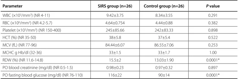
Table 3. Laboratory parameters.
Parameter SIRS group (n=26) Control group (n=26) P value
WBC (×103/mm3) (NR 4-11) 9.42±3.75 8.34±3.55 0.291
RBC (×106/mm3) (NR 4.2-5.7) 4.64±0.754 4.44±0.88 0.382
Platelet (×103/mm3) (NR 150-400) 245±85.66 242±83.33 0.898
HCT (%) (NR 35-50) 38±5.8 37±5.4 0.522
MCV (fL) (NR 77-96) 84.44±6.07 86.55±7.06 0.253
MCHC g Hb/dl (32-36) 33±1.5 33±1.7 1.00
RDW (%) (NR 11.6-14.8) 15.5±2 13.03±1.90 0.0001*
PO blood creatinine (mg/dl) (NR 0.5-1.5) 0.98±0.23 0.97±0.32 0.897
PO fasting blood glucose (mg/dl) (NR 76-110) 116±22 90±14 0.0001*
Hb=hemoglobin; HCT=hematocrit; NR=normal range; RBC=red blood cell; RDW=red cell distribution width; SIRS=systemic inflammatory response syndrome; WBC=white blood cell; MCV=mean corpuscular volume; MCHC=mean cell hemoglobin concentration; PO=postoperative. * Statistically significant
0
20
40
60
80
100
120
140
160
180
200
SI
RS
g
ro
up
Co
nt
ro
l g
ro
up
Preoperative glucose levels
(mg/dl)
Operation time (mins)
cell hemoglobin concentration (MCHC), platelet, and creatinine levels were similar in both groups. Of the total 1250 patients, 33 of them had high preoperative RDW. Out of the 26 patients who developed SIRS, 22 of them had high preoperative RDW values.
However, RDW was significantly higher in the SIRS group versus
the control group (15.5±2.0 vs. 13.03±1.90), respectively, P<0.0001. In addition, preoperative blood glucose levels were also found to
be significantly higher in the SIRS group versus the control group
(116±22 vs. 90±14), respectively, P<0.0001 (Figure 1). Multiple
logistic regression analyses showed an association between high RDW levels and SIRS development (OR for RDW levels exceeding
Fig. 2 - Comparison of levels of red cell distribution width in SIRS and control groups.
SIRS GROUP CONTROL GROUP
0 20 40 60 80 100 120 140 160 180 200
SI
RS
g
ro
up
nt
ro
l g
ro
up
Preoperative glucose levels (mg/dl)
Operation time (mins)
Preoperative glucose (mg/dl) Operative time (mins)
0 20 40 60 80 100 120 140 160 180 200
SI
RS
g
ro
up
Co
nt
ro
l g
ro
up
Preoperative glucose levels (mg/dl)
Operation time (mins)
Fig. 1 - Graphical representation of preoperative glucose levels and operation time between SIRS and control groups.
13.5%; 95% CI 1-1.2; P<0.05) (Figure 2). The receiver operator
curve (ROC) analysis suggested that the optimum cut-off level of RDW for SIRS was 12.9% (sensitivity: 93.74%; specificity: 76%; area under the curve: 0.851, P<0.05).
DISCUSSION
This study shows us the importance of preoperative RDW as a predictive marker for development of SIRS after ECC. RDW is a routine parameter available in complete blood count.
This syndrome occurs in about 0.5-1.7% of patients after ECC
17
16
15
14
13
12
RD
W%
SIRS GROUP CONTROL GROUP
292
Brazilian Journal of Cardiovascular Surgery
and can be associated with multiple organ failure which has
a mortality of 40-60%, which can also be higher[10]. Although
perioperative SIRS occurs in about 2% of all ECC procedures, the
mortality is high and comparable to that of severe sepsis[11].
In our study, the incidence of SIRS was 2.08% of the patients with a mortality of 76.92%. The patients who expired in the SIRS group ultimately had multiorgan failure. ECC is crucial part of many cardiac surgical operations. Multiple factors associated with the use of CPB contribute toward the generation of perioperative SIRS. These include the generation of shear forces from roller pumps driving blood through the bypass circuit, hypothermia as blood is passed through the extracorporeal circuit, and contact activation of plasma protein systems as circulating blood is exposed to artificial surfaces in the bypass circuit. The generation and release of endogenous inflammatory
mediators leading to the development of SIRS follow this[1]. The
main underlying molecular mechanisms of such inflammation are activation of the complement system, increasing production of cytokines, oxygen radicals, release of endothelin (ET) and the expression of adhesion molecules on leukocytes and the endothelium[12,13].
In 2007, Felker et al.[13] firstly discovered that increasing RDW
is an independent predictor for the prognosis of heart failure patients, and researchers gradually discovered that RDW is closely associated with the prognosis of cardiovascular diseases. Recent studies showed that increasing RDW is not only a predictor for poor prognosis of heart failure. But it also exhibits a predictive value towards the prognosis of stable coronary artery disease patients who have underwent percutaneous coronary intervention therapy.
Red cell differentiation is also related to oxidative stress and to the release of cytokines in response to inflammation induced by
cardiac surgery[14]. Oxidative stress directly damages erythrocytes
and leads to shortened erythrocyte survival, resulting in elevated
RDW[15]. Lippi et al.[16] showed a correlation between RDW
and indices of inflammation, such as elevated erythrocyte sedimentation rate (ESR) and high-sensitive C-reactive protein (hs-CRP), identifying a strong and graded increase in both ESR and hs-CRP across various RDW values. In a study by Semba et
al.[17] it was found that antioxidant status might influence RDW
and play a role in the relationship between increased RDW and worsened clinical prognosis. These cytokines attenuate the activity of erythropoietin and cause the production of ineffective
red blood cells, leading to elevated RDW[18].
Perlstein et al.[19] showed that RDW strongly predicted
all-cause and cardiovascular mortality. Lappé et al.[20] demonstrated
that RDW was associated with mortality in patients with stable coronary disease and in normal coronary subjects. In addition, RDW is also an independent prognostic factor for patients with peripheral arterial disease. In one study, a 10% increased risk of
mortality was observed with a 1% increase in RDW[21]. In our
study, patients with a high RDW value over 13.5% had increased incidence of SIRS and the relation became even stronger if the
RDW value was more than 15%. In a study by Kumar et al.[22],
patients with higher RDW had a longer ICU stay (155.6±71.3
vs. 122.4±61.3 hours, P=0.02). It was consisent with our study
showing significant morbidity in the SIRS group as compared to
control group in terms of length of ICU stay (158.2±72.3 hours vs.
100.2±44.2 hours, P<0.001). Cemin et al.[23] also found that RDW
was a significant predictor of AMI, exhibiting an area under the curve of 0.61 (95% CI, 0.54-0.68).
The sensitivity and specificity of RDW at the 13.7% cut-off value were 0.75 and 0.52, respectively. In our study, ROC analysis suggested that the optimum cut-off level of RDW for SIRS was 12.9% (sensitivity: 93.74%; specificity: 76%; area under the curve:
0.851, P<0.05), and the mean operation period was significantly
longer in the SIRS group than the control group.
In accordance with total operation time, ECC time was found longer in the SIRS group, but it did not reach to statistical
significance. Kirklin et al.[24] emphasized that an increase in ECC
time from 60 to 120 minutes would also increase postoperative morbidity in all age groups. There was a difference in the operating times, but the clamp time and ECC times were similar in the two groups. There was not one particular cause for the same but some of them are as follows: there was increased time taken to harvest the left internal mammary artery in few patients of CABG. In some patients, there were cardiotomy-bleeding points, which needed reinforcement sutures. In some there was bleeding from the aortic line, which needed reinforcement sutures. In some patients after coming off CPB, there was an oozy field, which needed to be addressed hence took time before chest closure. The fact that clamp time and ECC times were similar in our study suggests that the ECC time could not be considered as a confounding factor. The operating time, which was higher in the SIRS group, was the time either before onset or after termination of ECC.
Preoperative high blood glucose levels were also found to be significant in the SIRS group in comparison with the control group. This condition can hypothetically be explained with cardiac and pulmonary stress induced by catecholamine release,
which results in increased preoperative glucose levels[25]. In
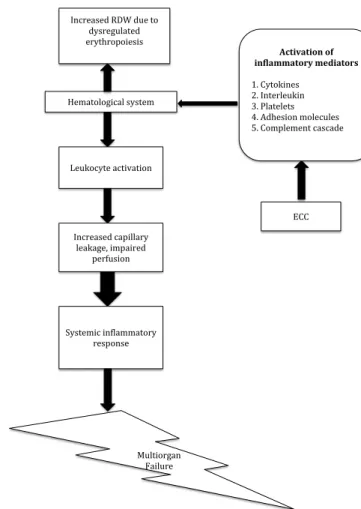
our study, there was a significant correlation between number of units of blood transfused and development of SIRS. These results suggest that high preoperative RDW can be used as an effective predictive marker for SIRS in patients undergoing cardiac surgery with ECC. As postulated, patients with high RDW have dysregulated erythropoiesis. These patients may also have qualitative defects in their platelets, which may lead to increase bleeding after ECC. This may be the reason that our patients with high RDW required more transfusion. However, our study was not designed to establish objective evidence of qualitative platelet dysfunction and to determine the possible causes of SIRS, but we can only speculate on the possible causes (Figure 3).
The reason for high RDW is that under pulmonary or cardiovascular stress such as hypoxia or low cardiac output, there is increased cytokine level, which attenuates the activity of erythropoietin. This results in production of ineffective red blood
cells leading to an elevated RDW[23].
operative time in a few patients of the SIRS group. Nevertheless, high RDW had a significant association in the development of SIRS after ECC in our study. Therefore, we also recommend that a similar study with a higher sample size, prospective design and randomized control should be done to validate these findings even further. This was not possible in our setup.
CONCLUSION
In conclusion, the main finding to be noted is that there is a significant association between elevated RDW and development of SIRS after ECC. This finding can provide us with valuable information for predicting SIRS in patients undergoing open-heart surgery without any additional costs, as RDW is a part of routine complete blood count. This valuable piece of information can also be made to use such that we find various alternatives to achieve the best result for our patient: (1) avoiding CPB altogether (off pump surgery); (2) removing activated neutrophils (leukodepletion filters); (3) using hemofiltration in appropriate patients; (4) we as clinicians should be watchful in patients with elevated RDW and take appropriate aggressive measures.
Multiorgan Failure
ECC
Activation of inflammatory mediators
1. Cytokines 2. Interleukin 3. Platelets 4. Adhesion molecules 5. Complement cascade Hematological system
Increased RDW due to dysregulated erythropoiesis
Leukocyte activation
Increased capillary leakage, impaired
perfusion
Systemic inflammatory response
Fig. 3 - Proposed hypothesis for correlation between high red cell distribution width and SIRS.
REFERENCES
1. Day JR, Taylor KM. The systemic inflammatory response syndrome and cardiopulmonary bypass. Int J Surg. 2005;3(2):129-40.
2. van Kimmenade RR, Mohammed AA, Uthamalingam S, van der Meer P, Felker GM, Januzzi JL Jr. Red blood cell distribution width and 1-year mortality in acute heart failure. Eur J Heart Fail. 2010;12(2):129-36. 3. Ozkalemkas F, Ali R, Ozkocaman V, Ozcelik T, Ozan U, Ozturk H, et al.
The bone marrow aspirate and biopsy in the diagnosis of unsuspected nonhematologic malignancy: a clinical study of 19 cases. BMC Cancer. 2005;5:144.
4. Dabbah S, Hammerman H, Markiewicz W, Aronson D. Relation between red cell distribution width and clinical outcomes after acute myocardial infarction. Am J Cardiol. 2010;105(3):312-7.
5. Patel KV, Ferrucci L, Ershler WB, Longo DL, Guralnik JM. Red blood cell distribution width and the risk of death in middle-aged and older adults. Arch Intern Med. 2009;169(5):515-23.
6. Tonelli M, Sacks F, Arnold M, Moye L, Davis B, Pfeffer M; for the Cholesterol and Recurrent Events (CARE) Trial Investigators. Relation between red blood cell distribution width and cardiovascular event rate in people with coronary disease. Circulation. 2008;117(2):163-8.
7. Gonzalo-Calvo D, Luxán-Delgado B, Rodríguez- González S, García-Macia M, Suárez FM, Solano JJ, et al. Interleukin 6, soluble tumor necrosis factor receptor I and red blood cell distribution width as biological markers of functional dependence in an elderly population: a translational approach. Cytokine. 2012;58(2):193-8.
8. Laffey JG, Boylan JF, Cheng DC. The systemic inflammatory response to cardiac surgery: implications for the anesthesiologist. Anesthesiology. 2002;97(1):215-52.
9. Nashef SA, Roques F, Hammill BG, Peterson ED, Michel P, Grover FL, et al; EuroSCORE Project Group. Validation of European System for Cardiac Operative Risk Evaluation (EuroSCORE) in North American cardiac surgery. Eur J Cardiothorac Surg. 2002;22(1):101-5.
10. Bellomo R, Ronco C, Kellum JA, Mehta RL, Palevsky P; Acute Dialysis Quality Initiative workgroup. Acute renal failure: definition, outcome measures, animal models, fluid therapy and information technology needs: the Second International Consensus Conference of the Acute Dialysis Quality Initiative (ADQI) Group. Crit Care. 2004;8(4):R204-12.
Authors’ roles & responsibilities
HSS
PM
JVK
CR
CKRM
GKKA
JSS VS
Substantial contributions to the conception or design of the work; or the acquisition, analysis, or interpretation of data for the work; final approval of the version to be published
Substantial contributions to the conception or design of the work; or the acquisition, analysis; final approval of the version to be published
Substantial contributions to the conception or design of the work; or the acquisition; final approval of the version to be published
Substantial contributions to the conception or design of the work; or the acquisition; final approval of the version to be published
Substantial contributions to the analysis; final approval of the version to be published
Interpretation of data for the work; final approval of the version to be published
294
Brazilian Journal of Cardiovascular Surgery
11. Asimakopoulos G, Smith PL, Ratnatunga CP, Taylor KM. Lung injury and acute respiratory distress syndrome after cardiopulmonary bypass. Ann Thorac Surg. 1999;68(3):1107-15.
12. Engel C, Brunkhorst FM, Bone HG, Brunkhorst R, Gerlach H, Grond S, et al. Epidemiology of sepsis in Germany: results from a national prospective multicenter study. Intensive Care Med. 2007;33(4):606-18. 13. Sessler CN, Shepherd W. New concepts in sepsis. Curr Opin Crit Care.
2002;8(5):465-72.
14. Felker GM, Allen LA, Pocock SJ, Shaw LK, McMurray JJ, Pfeffer MA, et al; CHARM Investigators. Red cell distribution width as a novel prognostic marker in heart failure: data from the CHARM Program and the Duke Databank. J Am Coll Cardiol. 2007;50(1):40-7.
15. Friedman JS, Lopez MF, Fleming MD, Rivera A, Martin FM, Welsh ML, et al. SOD2-deficiency anemia: protein oxidation and altered protein expression reveal targets of damage, stress response, and antioxidant responsiveness. Blood. 2004;104(8):2565-73.
16. Lippi G, Targher G, Montagnana M, Salvagno GL, Zoppini G, Guidi GC. Relation between red blood cell distribution width and inflammatory biomarkers in a large cohort of unselected outpatients. Arch Pathol Lab Med. 2009;133(4):628-32.
17. Semba RD, Patel KV, Ferrucci L, Sun K, Roy CN, Guralnik JM, et al. Serum antioxidants and inflammation predict red cell distribution width in older women: the Women’s Health and Aging Study I. Clin Nutr. 2010;29(5):600-4.
18. Ferrucci L, Guralnik JM, Woodman RC, Bandinelli S, Lauretani F, Corsi AM,
et al. Proinflammatory state and circulating erythropoietin in persons with and without anemia. Am J Med. 2005;118(11):1288.
19. Perlstein TS, Weuve J, Pfeffer MA, Beckman JA. Red blood cell distribution width and mortality risk in a community-based prospective cohort. Arch Intern Med. 2009;169(6):588-94.
20. Lappé JM, Horne BD, Shah SH, May HT, Muhlestein JB, Lappé DL, et al. Red cell distribution width, C-reactive protein, the complete blood count, and mortality in patients with coronary disease and a normal comparison population. Clin Chim Acta. 2011;412(23-24):2094-9. 21. Ye Z, Smith C, Kullo IJ. Usefulness of red cell distribution width to predict
mortality in patients with peripheral artery disease. Am J Cardiol. 2011;107(8):1241-5.
22. Kumar S, Sudhakar A, Mohan M, Balachandran R, Raj B, Sumangala SG, et al. Elevated red cell distribution width is associated with delayed postoperative recovery after correction of Tetralogy of Fallot. Ann Pediatr Cardiol. 2013;6(2):121-5.
23. Cemin R, Donazzan L, Lippi G, Clari F, Daves M. Blood cells characteristics as determinants of acute myocardial infarction. Clin Chem Lab Med. 2011;49(7):1231-6.
24. Kirklin JK, Westaby S, Blackstone EH, Kirklin JW, Chenoweth DE, Pacifico AD. Complement and the damaging effects of cardiopulmonary bypass. J Thorac Cardiovasc Surg. 1983;86(6):845-57.