The aim of this study was to conduct a retrospective evaluation of the survival and success rates of dental implants with acid-etched surfaces after 8-10 years of function. Forty-four patients who received 183 implants 8-10 years ago were evaluated. Clinical examinations were performed around the implants and natural teeth. The following parameters were measured: visible plaque index (VPI), marginal bleeding index (MBI), probing depth (PD), bleeding on probing (BOP) and clinical attachment level (CAL). To considerer an implant as a success case, the following criteria were considered: absence of peri-implant infection and suppuration, absence of implant mobility, absence of persistent pain and dysesthesia and absence of radiolucency around the implant. Overall, 178 implants were categorized as surviving (97.3%), 155 were categorized as successful (84.7%), 5 implants (2.7%) were lost (1 in the maxilla and 4 in the mandible), and 3 implants were not under functional load (2.0 %). 20 implants were diagnosed with peri-implantitis. Thus, the survival rate was 97% and the success rate was 85%. In conclusion, implants with acid-etched surfaces showed high survival and success rates after a period of 8 to 10 years of function.
S u r v i v a l / S u c c e s s o f D e n t a l
I m p l a n t s w i t h A c i d - E t c h e d
S u r f a c e s : A R e t r o s p e c t i v e
Evaluation After 8 to 10 Years
Lélis Gustavo Nicoli1, Guilherme José Pimentel Lopes de Oliveira1,2, Beatriz Maria Valério Lopes1, Cláudio Marcantonio2, Daniela Leal Zandim-Barcelos1, Elcio Marcantonio Jr1
1Department of Diagnosis and
Surgery, Araraquara Dental School, UNESP - Universidade Estadual Paulista, Araraquara, SP, Brazil
2Department of Health Sciences,
Dental School, UNIARA - Universidade de Araraquara -, Araraquara, SP, Brazil
Correspondence: Dr. Elcio Marcantonio Junior, Rua Humaitá, 1680, 14801-903, Araraquara, SP, Brasil. Tel: +55-16-3301-6376. e-mail: [email protected]
Key Words: osseointegration, prostheses and implants, survival rate.
Introduction
Despite the information that the treatment of edentulism with dental implants presents predictable results (1), the long-term evaluation of implants has shown that some factors such as smoking, radiotherapy history, presence of periodontitis, uncontrolled diabetes, lack of keratinized gingiva and presence of biofilm appear to be risk factors that may have negative impact on implant success and survival rates (2).
To minimize these failure rates, various changes to the macro- and microstructure of implants have been proposed. Modifications of the implant surface through subtractive and additive methods that provide increased surface roughness promote better initial response of the osseointegration process (3,4). Among these methods of implant surface modification, acid-etched surfaces have been shown to accelerate the process of osseointegration (5) and produced high rates of implant survival (97.6% - 98.4%) after 3-5 years (6,7).
Despite the great advantages offered by the increased contact area of bone and acid-etched implant surface in conditions of peri-implant bone loss, the presence of a rough surface appears to favor biofilm accumulation and thereby promote the development of peri-implantitis, which is the main reason for the dental implants failure (8-10). Because peri-implantitis is a chronic pathological
process, the evaluation of acid-etched implants after long periods of use is needed in order to determine the impact of this type of surface on the survival and success rates of these implants. Therefore, the present study aimed to evaluate the success and survival rates of implants with surfaces treated with double acid attack and used for a period of 8-10 years.
Material and Methods
331
Success of acid-etched surfaces implants
Clinical and Radiographic Analysis
Prior to the clinical examination, a careful anamnesis was performed in order to update the information about the systemic/behavior condition of each patient. A periodontal/ peri-implant clinical examination was carried out by a single trained and calibrated examiner (Wilcoxon test p>0.05; Spearman correlation r=0.81) (LN). The following clinical parameters were measured: visible plaque index (VPI), gingival bleeding index (GBI), probing depth (PD), bleeding on probing (BOP) and clinical attachment level (CAL). These parameters were evaluated at six sites per tooth using a North Carolina millimeter periodontal probe (Hu-Friedy, Chicago, IL, USA). Then, the following parameters were evaluated around each implant: VPI, GBI, PD, BOP, presence of suppuration, presence and amount of keratinized tissue. These parameters were also measured at six sites per implant using a millimeter plastic periodontal probe (Hu-Friedy,). The distance between the implant platform and the bone-implant contact was measured in periapical radiography. The radiographs were obtained using the long-cone paralleling technique and X-ray positioners. A diagnosis of peri-implantitis was established when the PD was ≥5 mm, with the presence of BOP and/or suppuration, and radiographic confirmation of bone loss. Mucositis was defined as the presence of BOP and PD <5 mm or BOP and PD ≥5 mm without radiographic confirmation of bone loss (11). The case definition of periodontitis was determined as the presence of four or more teeth with at least one site with PD ≥4 mm, CAL ≥3 mm and BOP (12).
Criteria for Implant Success Evaluation
To determine the success of the implants, the following criteria were considered: absence of peri-implant infection, lack of mobility, absence of persistent pain or dysesthesia and absence of continuous radiolucency around the implant (13). Thus, the implants were classified as failed, surviving or successful.
Statistical Analysis
The results were analyzed using descriptive statistical analysis of the obtained data. The data were allocated in relation to the sampling units as individuals and implants. The analysis was performed using measures of the central tendency, including the means and medians, and the standard deviation was used as a dispersion parameter measurement. The GraphPad Prism software 6 (San Diego, CA, USA) was used for the descriptive statistical analysis.
Results
Seventy-four patients underwent oral rehabilitation with acid-etched dental implant surfaces at the Specialization Course in Implantology of the FOAr -
UNESP from January 2003 to December 2005. Among these patients, only 44 showed up to perform the clinical and radiographic examination and were included in this study. Among these 44 patients, 18 were male (40.9%) and 26 were female (59.1%), their average age at the time of implant installation was 50.07±11.61 years, and 34.1% of the patients were older than 65 years. At the time of re-evaluation, the average age of the patients was 59.57±11.61 years. Among all the patients, 6.8% were smokers, 22.7% were former smokers and 70.4% were never smokers. According to the anamnesis, 15.9% of the patients reported having diabetes, 9.1% hypothyroidism, 9.1% cardiovascular diseases, 9.1% had undergone radiotherapy and/or chemotherapy, 15.9% had osteoporosis and 6.8% were bisphosphonate users. Regarding the periodontal and peri-implant condition, 45% of the patients had chronic periodontitis and 25% had peri-implantitis (Table 1).
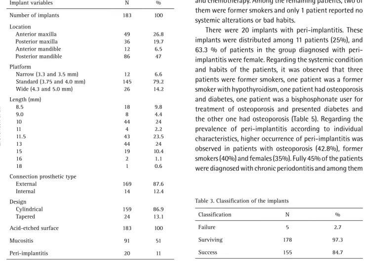
All the implants had acid-etched surfaces (Porous®, Conexão Sistemas de Próteses, Arujá, SP, Brazil) obtained by treating their surface with hydrochloric and sulfuric acids. The number of placed implants was 183, most of them in the posterior region of the mandible (47%). The platforms of the implants were classified as narrow (implants 3.3 to 3.5 mm diameter), regular (implants 3.75 to 4.0 mm diameter) and large (4.3 to 5.0 mm diameter). One hundred and forty-five of the placed implants presented a prevalence of standard platform (79.2%). The length of the implants ranged between 8.5 mm and 18 mm, most
Table 1. Distribution of different variables in relation to patients
Patient variables N %
Number of patients 44 100
Male Female
18 26
40.9 59.1
Age at implant placement > 65 years Age at implant placement < 65 years
15 29
34.1 65.9
Smoker Non-smoker Former smoker
3 31 10
6.8 70.4 22.7
Diabetes 7 15.9
Bisphosphonate user 3 6.8
Radiotherapy/Chemotherapy history 4 9.1
Hypothyroidism 4 9.1
Cardiovascular disease 4 9.1
Osteoporosis 7 15.9
Mucositis Peri-implantitis
32 11
72.7 25
Chronic periodontitis Periodontally healthy
20 24
L.G. Nicoli et al.
of them between 10 and 13 mm. Most of the implants presented an external hexagon type prosthetic connection (169 implants, 87.6%), in multiple and unitary prostheses. The other 14 implants (12.4%) had an internal hexagon type connection and were used for different types of prosthesis. The implants were divided into tapered and cylindrical shapes and most of the installed implants had a cylindrical shape (159, 86.9%) (Table 2).
After the re-evaluation, five implants were lost and 178
had survived; survival was associated with clinical success in 155 implants. Thus, the survival rate observed was 97.3 %, and the success rate was 84.7 % (Table 3). Among the surviving implants, 64 presented inflammation (36%), three were not functioning (2%), 91 had mucositis (51%) and 20 were diagnosed with peri-implantitis (11%) (Table 2).
The characteristics of the lost implants and of those diagnosed with peri-implantitis are presented in Tables 4 and 5, respectively. Four patients lost implants (approximately 10% of the patients), one of them lost 2 implants and the others 3 lost one implant each (Table 4). The patient who lost 2 implants was a female that had undergone radiotherapy and chemotherapy. Among the remaining patients, two of them were former smokers and only 1 patient reported no systemic alterations or bad habits.
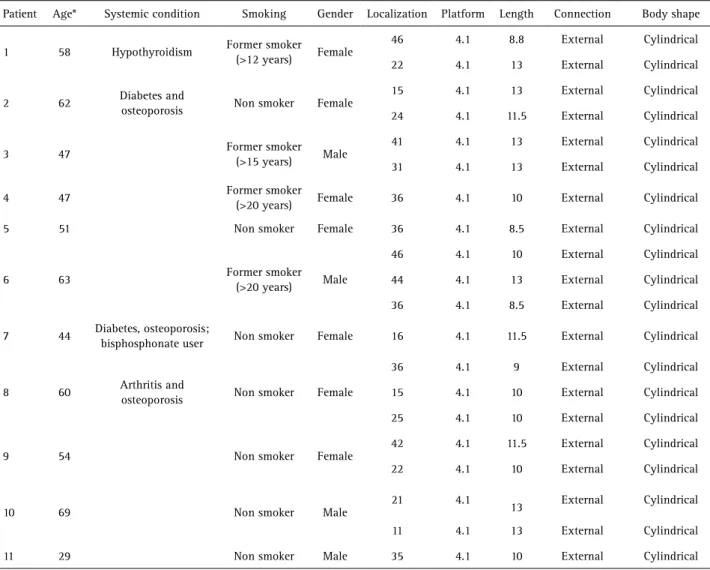
There were 20 implants with peri-implantitis. These implants were distributed among 11 patients (25%), and 63.3 % of patients in the group diagnosed with peri-implantitis were female. Regarding the systemic condition and habits of the patients, it was observed that three patients were former smokers, one patient was a former smoker with hypothyroidism, one patient had osteoporosis and diabetes, one patient was a bisphosphonate user for treatment of osteoporosis and presented diabetes and the other one had osteoporosis (Table 5). Regarding the prevalence of peri-implantitis according to individual characteristics, higher occurrence of peri-implantitis was observed in patients with osteoporosis (42.8%), former smokers (40%) and females (35%). Fully 45% of the patients were diagnosed with chronic periodontitis and among them
Table 3. Classification of the implants
Classification N %
Failure 5 2.7
Surviving 178 97.3
Success 155 84.7
Table 2. Distribution of the different variables in relation to the implants
Implant variables N %
Number of implants 183 100
Location
Anterior maxilla Posterior maxilla Anterior mandible Posterior mandible
49 36 12 86
26.8 19.7 6.5 47
Platform
Narrow (3.3 and 3.5 mm) Standard (3.75 and 4.0 mm) Wide (4.3 and 5.0 mm)
12 145
26
6.6 79.2 14.2
Length (mm) 8.5 9.0 10 11 11.5 13 15 16 18
18 8 44
4 43 44 19 2 1
9.8 4.4 24 2.2 23.5
24 10.4
1.1 0.6
Connection prosthetic type External
Internal
169 14
87.6 12.4
Design Cylindrical Tapered
159 24
86.9 13.1
Acid-etched surface 183 100
Mucositis 91 51
Peri-implantitis 20 11
Table 4. Characteristics of the lost implants
Patient Age* Medical history Smoking Sex Localization Platform Length Connection Shape
1 56
Breast cancer (radiotherapy and
chemotherapy)
Non-smoker Female
15 4.1 10 External Cylindrical
36 4.1 10 External Cylindrical
2 47 Healthy Former smoker
(stopped >20 years) Female 35 4.1 10 External Cylindrical
3 51 Healthy Non-smoker Female 34 4.1 8.5 External Cylindrical
4 76 Cardiovascular
disease
Former smoker
(stopped >20 years) Male 45 4.1 15 External Cylindrical
333
Success of acid-etched surfaces implants
15 % presented peri-implantitis (Table 6).
The peri-implant condition was also analyzed regarding the type of prosthesis, abutments and adaptation of the prosthesis. The partial (15.3%) and full arch screwed (13.3%) prosthesis presented higher prevalence of peri-implantitis than the single (10%) and partial cemented prostheses (9.4%) and the single screwed prosthesis (6.9%). The esthetic and UCLA abutments (screwed) (14.3 and 14.6%), and the custom-angled abutment (cemented) (20%) presented higher prevalence of peri-implantitis. The disconnection of the prosthesis produced a subtle increase in the peri-implantitis prevalence compared with the prosthesis with a good adaptation (14.8 vs. 10.8%) (Table 7).
Discussion
This retrospective study aimed to evaluate the long-term survival and success rates of implants with acid-etched surfaces. After 8 to 10 years of follow-up, the survival rate was 97.3 % and the success rate was 84.7 %. A similar
survival rate (97.6 %) was observed in a previous study that evaluated implants with acid-etched surfaces after a median follow-up of 37 months (6). Comparable survival rates were reported in a study that evaluated the outcomes of short and standard implants with acid-etched surfaces. The implants with acid-etched surfaces presented high survival rates regardless of the length (short 97.7 % vs. 98.4 % standard) after 5 years of follow-up (7). The high survival rates of implants with acid-etched surfaces can be explained by the greater contact area between bone and implant provided by this surface and its roughness, which seems to favor strongly the induction and maintenance of osseointegration for long-term periods.
The failure rate in the present study was 2.7 %, relative to only 5 implants distributed among 4 patients (10%). Interestingly, 2 implants failed in 1 patient who had undergone radiotherapy, 2 implants failed in 2 ex-smokers and 1 implant failed in 1 patient who had no systemic alterations. The radiotherapy performed in patients with
Table 5. Characteristics of the implants with peri-implantitis
Patient Age* Systemic condition Smoking Gender Localization Platform Length Connection Body shape
1 58 Hypothyroidism Former smoker
(>12 years) Female
46 4.1 8.8 External Cylindrical
22 4.1 13 External Cylindrical
2 62 Diabetes and
osteoporosis Non smoker Female
15 4.1 13 External Cylindrical
24 4.1 11.5 External Cylindrical
3 47 Former smoker
(>15 years) Male
41 4.1 13 External Cylindrical
31 4.1 13 External Cylindrical
4 47 Former smoker
(>20 years) Female 36 4.1 10 External Cylindrical
5 51 Non smoker Female 36 4.1 8.5 External Cylindrical
6 63 Former smoker
(>20 years) Male
46 4.1 10 External Cylindrical
44 4.1 13 External Cylindrical
36 4.1 8.5 External Cylindrical
7 44 Diabetes, osteoporosis;
bisphosphonate user Non smoker Female 16 4.1 11.5 External Cylindrical
8 60 Arthritis and
osteoporosis Non smoker Female
36 4.1 9 External Cylindrical
15 4.1 10 External Cylindrical
25 4.1 10 External Cylindrical
9 54 Non smoker Female
42 4.1 11.5 External Cylindrical
22 4.1 10 External Cylindrical
10 69 Non smoker Male
21 4.1
13 External Cylindrical
11 4.1 13 External Cylindrical
11 29 Non smoker Male 35 4.1 10 External Cylindrical
L.G. Nicoli et al.
cancer may cause progressive fibrosis of blood vessels and soft tissues, leading to a decrease in vascularization and healing capacity. This decrease in vascularization may be related to the lower survival rate of implants placed in patients irradiated in head and neck (14,15). Likewise, the vasoconstriction caused by smoking seems to have a strong relationship with peri-implant bone resorption in smokers (16). Even knowing the limitations regarding the limited sample size of this research, the result suggests the hypothesis that radiotherapy and smoking habit history may increase the failure rate of dental implants.
Despite the methodological variations observed in the literature with respect to the clinical parameters used for the diagnosis of peri-implantitis, most definitions used currently are equivalent in the sense that all of them assume that peri-implantitis is always associated with inflammation of the soft tissue and collapse of the bone structure around the implant (11,17,18). The parameters used for the determination of the peri-implantitis cases used in this study are consistent with those already reported in other studies (11,13). The implants with peri-implantitis (11%) were classified as survivors and corresponded to 20 implants distributed among 11 patients. Retrospective studies using similar parameters to determine peri-implantitis cases as in this study, demonstrated a similar prevalence of this pathology in comparison to the present study (8%) (11,19).
Among the 11 patients with peri-implantitis, four were former smokers, seven were women and three were women with osteoporosis. Although the limited sample size does not allow a statistically significant relationship between these individual characteristics of the patients and the prevalence of peri-implantitis, these data may suggest that
certain habits, hormonal changes or undesirable systemic conditions may influence the occurrence of this disease.
It has been shown that changes in estrogen levels may promote variations in the concentration of proinflammatory cytokines and consequently are directly related to immune function. The decrease of the level of estrogen in postmenopausal women can be a determining factor in the occurrence of osteoporosis, a metabolic disease that promotes imbalance in the bone remodeling process with a constant decrease in the quality and volume of bone tissue (20). Low bone density can be regarded as a risk factor for peri-implantitis and low density bone regions have been related to osteoporotic conditions (21). Although osteoporosis has not been considered a risk factor for peri-implantitis, the low bone density resulting from this disease deserves attention due to a possible relationship between osteoporosis and decreased implant success (21), as observed in this study. The present results showed considerable incidence rates of peri-implantitis in women with and without osteoporosis. Osteoporosis cases were only considered in patients who reported being diagnosed by a prior medical consultation. Despite the deficiency of this study in the determination of the diagnosis of osteoporosis, all the women who had peri-implantitis had matched age of premenopausal period,
Table 7. Characteristics of prostheses. abutments and adaptation of the prosthesis to the abutments or to the implants connections in relation to the incidence peri-implantitis
Type of prosthesis n Peri-implantitis (%) Healthy (%)
Single cemented 40 04 (10) 36 (90)
Single screwed 29 02 (6.9) 27 (93.1)
Partial cemented 32 03 (9.4) 29 (90.6)
Partial screwed 59 09 (15.3) 50 (84.7)
Full arch screwed 15 02 (13.3) 13 (86.7)
Type of abutment n Peri-implantitis (%) Healthy (%)
Micro unit screwed 55 06 (10.9) 49 (89.1)
UCLA screwed 41 06 (14.6) 35 (85.4)
Estheticone screwed 7 01 (14.3) 06 (85.7)
Custom-straight abutments cemented
66 06 (9.1) 60 (90.9)
Custom-angled abutments cemented
5 01 (20) 04 (80)
Adaptation n Peri-implantitis (%) Healthy (%)
Adapted 148 16 (10.8) 132 (89.2)
Non-adapted 27 04 (14.8) 23 (85.2)
Table 6. Characteristics of individuals in relation to the incidence of peri-implantitis
Characteristics n Peri-implantitis (%)
Healthy (%)
Number of patients 44 11 (25) 33 (75)
Female 26 07 (35) 19(65)
Male 18 04 (13.3) 14 (86.7)
Smoker 03 00 (00) 03 (100)
Former smoker 10 04 (40) 06 (60)
Non smoker 31 07 (22.6) 24 (77.4)
Diabetes 07 02 (28.6) 05 (71.4)
Osteoporosis 07 03 (42.8) 04 (57.2)
Cardiovascular disease 04 01 (25) 03 (75)
Hypothyroidism 04 01 (25) 03 (75)
335
Success of acid-etched surfaces implants
menopause or postmenopausal period (22). Thus, in this sample, women with peri-implantitis and who did not self-report as osteoporotic could also have changes in estrogen levels, which may relate to the higher incidence of peri-implantitis in females. Despite the small sample size of the study, 42.8% of the patients with osteoporosis presented implants with peri-implantitis. Additionally, it was found that 75% of the patients who had osteoporosis also had diabetes, and diabetes is related to the reduction in bone density (23). More studies are needed to evaluate a possible relationship between diabetes and consequent low bone density caused by osteoporosis as a risk factor for peri-implant diseases.
The effect of tobacco consumption in smokers can create a more vulnerable environment for peri-implant bone loss (16). According to the obtained results, former smokers who stopped smoking more than five years ago and who had smoking habit for at least 10 years, had a higher susceptibility to present peri-implantitis. This suggests that local and systemic changes induced by smoking can continue even after the habit is stopped. This result is in accordance with previously published data where greater peri-implant bone loss occurred in former smokers when compared with nonsmokers (16). On the other hand, the smokers in this sample showed no peri-implantitis. The most likely hypothesis for this contradictory result is related to the low prevalence of smokers in the sample.
A history of periodontitis has been considered a risk factor for peri-implantitis (24,25). The increased susceptibility of these patients occurs due to the inability of their host immune response to face the microbial challenge adequately; thus, patients affected previously by periodontitis may have a higher probability of peri-implantitis development. This association between periodontitis and the prevalence of peri-implantitis could be observed in previous reports (18,25). However, despite the high prevalence of patients who presented chronic periodontitis (45%), only 15% of these patients presented peri-implantitis and this information is not in accordance with a significant number of studies (2,17,24). It is likely that the lack of well-established criteria for the diagnosis of periodontitis and peri-implantitis has led to a large variation in disease indices in the literature.
In relation to the prosthetic data, a higher prevalence of peri-implantitis was associated with screwed prostheses and abutments and with maladaptation of the prosthesis abutment to the implants platform. On the other hand, the literature demonstrates that cemented prostheses have been associated with a higher risk for peri-implantitis probably due to the occurrence of residual cement in the peri-implant sulcus (26). The greater possibility of misfit or fractures of the screwed prostheses may have
increased the gap size and contributed to the presence of peri-implantitis (27). However, the information about the prosthetic complications was not provided by this study. Furthermore, the sample size limitations already mentioned do not allow the establishment of prosthetic risk factors peri-implantitis.
This study has several limitations. The retrospective study design prevented a certain determination of the real impact of the different risk factors that influenced the implant success rate, a fact complicated by a low patient acceptance rate to be evaluated in this study, which resulted in a reduced sample size. However, it is important to note that long-term follow-up studies are scarce and that this study provided valuable information about the predictability of rehabilitation treatment with acid-etched implant surfaces, which is one of the types of surfaces most commonly used in daily practice. The implementation of long-term prospective studies will be important to confirm the hypotheses raised by this study.
Implants with acid-etched surfaces showed high survival and success rates after a period of 8 to 10 years of function. This result demonstrates that implants with acid-etched surfaces were effective and predictable for obtaining good clinical outcomes maintained for a long period of observation.
Resumo
O objetivo deste estudo foi avaliar retrospectivamente a taxa de sobrevivência e sucesso de implantes com superfície tratada por ataque ácido após 8-10 anos de função. Um total de 44 pacientes que receberam 183 implantes há 8-10 anos foram avaliados. Foi realizado exame clínico de todos os dentes e implantes presentes na cavidade bucal. Os seguintes parâmetros foram avaliados: índice de placa visível (IPV), índice de sangramento gengival (ISG), profundidade de sondagem (PS), sangramento à sondagem (SS), nível clínico de inserção (NCI). Para a classificação de sucesso dos implantes foram considerados os seguintes critérios: ausência de infecção peri-implantar com supuração, ausência de mobilidade, ausência de dor persistente ou disestesia e ausência de radiolucência contínua ao redor do implante. Após avaliação, 178 (97.3%) implantes foram classificados como sobreviventes, 155 (84.7%) aderiram aos critérios de sucesso, 5 implantes (2.7%) foram perdidos (1 na maxila e 4 na mandíbula) e 3 implantes (2.0%) não estavam em função. 20 (11%) implantes foram diagnosticados com peri-implantite. Dessa forma, a taxa de sobrevivência foi de 97% e a taxa de sucesso de 85%. Pode-se concluir que os implantes com superfície tratada por ataque ácido apresentaram altas taxas de sobrevivência e sucesso após 8-10 em função.
References
1. van Velzen FJ, Ofec R, Schulten EA, Ten Bruggenkate CM. 10-year survival rate and the incidence of peri-implant disease of 374 titanium dental implants with a SLA surface: a prospective cohort study in 177 fully and partially edentulous patients. Clin Oral Implants Res 2015;26:1121-1128.
2. Renvert S, Polyzois I. Risk indicators for peri-implant mucositis: a systematic literature review. J Clin Periodontol 2015;42 Supp 16:S172-186.
L.G. Nicoli et al.
and osseotite implant surfaces. Int J Periodontics Restorative Dent 2002;22:535-545.
4. Lang NP, Jepsen S, Working G. Implant surfaces and design (Working Group 4). Clin Oral Implants Res 2009;20 Suppl4:228-231.
5. Santiago AS, Santos EA, Sader MS, Santiago MF, Soares GA. Response of osteoblastic cells to titanium submitted to three different surface treatments. Braz Oral Res 2005;19:203-208.
6. Sesma N, Pannuti C, Cardaropoli G. Retrospective clinical study of 988 dual acid-etched implants placed in grafted and native bone for single-tooth replacement. Int J Oral Maxillofac Implants 2012;27:1243-1248. 7. Feldman S, Boitel N, Weng D, Kohles SS, Stach RM. Five-year survival
distributions of short-length (10 mm or less) machined-surfaced and Osseotite implants. Clin Implant Dent Relat Res 2004;6:16-23. 8. Teughels W, Van Assche N, Sliepen I, Quirynen M. Effect of material
characteristics and/or surface topography on biofilm development. Clin Oral Implants Res 2006;17 Suppl 2:68-81.
9. Albouy JP, Abrahamsson I, Berglundh T. Spontaneous progression of experimental peri-implantitis at implants with different surface characteristics: an experimental study in dogs. J Clin Periodontol 2012;39:182-187.
10. Burgers R, Gerlach T, Hahnel S, Schwarz F, Handel G, Gosau M. In vivo
and in vitro biofilm formation on two different titanium implant
surfaces. Clin Oral Implants Res 2010;21:156-164.
11. Ferreira SD, Silva GL, Cortelli JR, Costa JE, Costa FO. Prevalence and risk variables for peri-implant disease in Brazilian subjects. J Clin Periodontol 2006;33:929-935.
12. Lopez NJ, Smith PC, Gutierrez J. Periodontal therapy may reduce the risk of preterm low birth weight in women with periodontal disease: a randomized controlled trial. J Periodontol 2002;73:911-924. 13. Buser D, Janner SF, Wittneben JG, Bragger U, Ramseier CA, Salvi
GE. 10-year survival and success rates of 511 titanium implants with a sandblasted and acid-etched surface: a retrospective study in 303 partially edentulous patients. Clin Implant Dent Relat Res 2012;14:839-851.
14. Pompa G, Saccucci M, Di Carlo G, Brauner E, Valentini V, Di Carlo S, et al.. Survival of dental implants in patients with oral cancer treated by surgery and radiotherapy: a retrospective study. BMC Oral Health 2015;15:5.
15. Mancha de la Plata M, Gias LN, Diez PM, Munoz-Guerra M, Gonzalez-Garcia R, Lee GY, et al.. Osseointegrated implant rehabilitation of irradiated oral cancer patients. J Oral Maxillofac Surg 2012;70:1052-1063.
16. Levin L, Hertzberg R, Har-Nes S, Schwartz-Arad D. Long-term marginal
bone loss around single dental implants affected by current and past smoking habits. Implant Dent 2008;17:422-429.
17. Atieh MA, Alsabeeha NH, Faggion CM Jr., Duncan WJ. The frequency of peri-implant diseases: a systematic review and meta-analysis. J Periodontol 2013;84:1586-1598.
18. Simonis P, Dufour T, Tenenbaum H. Long-term implant survival and success: a 10-16-year follow-up of non-submerged dental implants. Clin Oral Implants Res 2010;21:772-777.
19. Fransson C, Lekholm U, Jemt T, Berglundh T. Prevalence of subjects with progressive bone loss at implants. Clin Oral Implants Res 2005;16:440-446.
20. Dvorak G, Arnhart C, Heuberer S, Huber CD, Watzek G, Gruber R. Peri-implantitis and late implant failures in postmenopausal women: a cross-sectional study. J Clin Periodontol 2011;38:950-955.
21. Liddelow G, Klineberg I. Patient-related risk factors for implant therapy. A critique of pertinent literature. Aust Dent J 2011;56:417-426; quiz 441.
22. Cheng Q, Tang W, Sheu TJ, Du Y, Gan J, Li H, et al.. Circulating TGF-beta1 levels are negatively correlated with sclerostin levels in early postmenopausal women. Clin Chim Acta 2016.
23. Abbassy MA, Watari I, Soma K. The effect of diabetes mellitus on rat mandibular bone formation and microarchitecture. Eur J Oral Sci 2010;118:364-369.
24. Monje A, Aranda L, Diaz KT, Alarcon MA, Bagramian RA, Wang HL, et al.. Impact of maintenance therapy for the prevention of peri-implant diseases: a systematic review and meta-analysis. J Dent Res 2016;95:372-379.
25. Renvert S, Lindahl C, Rutger Persson G. The incidence of peri-implantitis for two different implant systems over a period of thirteen years. J Clin Periodontol 2012;39:1191-1197.
26. Dalago HR, Schuldt Filho G, Rodrigues MA, Renvert S, Bianchini MA. Risk indicators for peri-implantitis. A cross-sectional study with 916 implants. Clin Oral Implants Res 2016.
27. Anchieta RB, Machado LS, Hirata R, Bonfante EA, Coelho PG. Platform-switching for cemented versus screwed fixed dental prostheses: reliability and failure modes: an in vitro study. Clin Implant Dent Relat Res 2016;18:830-839.
Received December 24, 2016