RBCCV 44205-1562 DOI 10.5935/1678-9741.20140043
Evaluation of peripheral muscle strength of
patients undergoing elective cardiac surgery:
a longitudinal study
Avaliação da força muscular periférica de pacientes submetidos à cirurgia cardíaca eletiva: estudo longitudinal
Kelli Maria Souza Santos
1; Manoel Luiz de Cerqueira Neto, PhD
1; Vitor Oliveira Carvalho
1, PhD;
Valter Joviniano de Santana Filho
1, MD; Walderi Monteiro da Silva Junior
1, MD; Amaro Afrânio
Araújo Filho
2, Ms; Telma Cristina Fontes Cerqueira
2, MsC; Lucas de Assis Pereira Cacau
1, MsC
1Universidade Federal de Sergipe (UFS), Aracaju, SE, Brazil 2Universidade Tiradentes, (Unit), Aracaju, SE, Brazil.
This study was carried out at Fundação Beneicência Hospital Cirurgia and Universidade Federal de Sergipe (UFS), Aracaju, SE, Brazil.
No inancial support.
Correspondence Address: Lucas de Assis Pereira Cacau
Rua Cláudio Batista, s/n - Santo Antônio – Aracaju, SE Brazil - Zip code: 49060-100
E-mail: [email protected]
Article received on September 4th, 2013
Article accepted on January 14th, 2014
Abstract
Introduction: Peripheral muscle strength has been little ex-plored in the literature in the context of cardiac rehabilitation. Objective: To evaluate the peripheral muscle strength of patients undergoing elective cardiac surgery.
Methods: This was a longitudinal observational study. The peripheral muscle strength was measured using isometric
dy-namometry lower limb (knee extensors and lexors) at three different times: preoperatively (M1), the day of discharge (M2) and hospital discharge (M3). Participants received physiotherapy
pre and postoperatively during the days of hospitalization during the morning and afternoon.
Results: Twenty-two patients were evaluated. The values of peripheral muscle strength of knee extensors preoperative found were about 50% lower than those predicted for the healthy
population. When comparing muscle strength prior (M1), with the remaining evaluation, found himself in a fall of 29% for the movement of knee extension and 25% for knee lexion in M2 and
a decrease of 10% movement for knee extension and 13% for
knee lexion in M3 when comparing with M1.
Conclusion: The values of peripheral muscle strength prior of the study patients were lower than predicted for the healthy population of the same age. After the surgical event this reduction
is even more remarkable, being reestablished until the time of discharge, to values close to baseline.
Descriptors: Muscle Strength. Muscle Strength Dynamometer. Heart Diseases.
Resumo
Introdução: A força muscular periférica tem sido pouco explorada na literatura atual no contexto da reabilitação car-diovascular.
Objetivo: Avaliar a força muscular periférica de pacientes submetidos à cirurgia cardíaca eletiva.
Métodos: Trata-se de um estudo observacional e longitudinal. A força muscular periférica foi mensurada por meio de
deep vein thrombosis, deambulation, walking up and down stairs, active-assisted and active kinesiotherapy of upper and lower limbs, and muscle stretching. The rehabilitation pro-tocol followed the guidelines of the Brazilian Consensus on Cardiovascular Rehabilitation[6].
Study Population
Patients were recruited from a tertiary hospital of Cardiology between September and November 2012. Patients undergoing elective cardiac surgery (both genders and aged between 18 and 80 years) were eligible for this study. Inclusion criteria were:
ab-sence of neurocognitive decline or dementia that could inluence
the process of communication, absence of musculoskeletal dis-order and previous pulmonary diseases (chronic obstructive pul-monary disease), presence of hemodynamic instability (Mean
Arterial Pressure >60 mmHg), dyspnea with oxygen saturation
less than 90%. The patient, who refused to sign the Written In-formed Consent and those who had the time of stay in the ICU
more than 7 days were excluded from the study.
This research protocol was approved by the Re-search Ethics Committee of our institution (CAAE nº 01521012.2.0000.0058). All patients signed a written in-formed consent prior to participation in this research.
Data collected from medical records
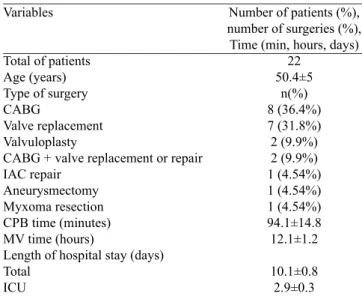
Clinical history, diagnosis, vital signs, type of surgery, total days ofhospital and ICU stay, time of cardiopulmonary bypass and mechanical ventilation time were collected, as well as the patient’s characteristics involved in the study (Table 1).
Assessment of peripheral muscle strength
The PMS was assessed at three different time points: pre-operatively (day before surgery) (M1), on the 1st day after ICU discharge (M2) and the day of discharge (M3), in the
form of isometrics for extension and lexion of the right and
left knees. The instrument used in this assessment was the portable digital dynamometer, IMPAC®
, IP-90DI model with INTRODUCTION
Cardiovascular diseases are responsible for high rates of morbidity and mortality in Brazil and in the world[1]. For treatment of many of these cardiac
con-ditions, surgery is the main resource[2]. Although very
safe, heart surgery is a procedure considered large and is accompanied by the need for general anesthesia, cardiopul-monary bypass, mechanical ventilation and relative restric-tion to bed rest[3].
The heart disease patients showed loss of functional
ca-pacity resulting from decreased oxidative caca-pacity of skeletal
muscle and reduced muscle perfusion, such loss being aggra-vated in case of hospitalization for bed rest, impacting loss of peripheral muscle function[4]. Peripheral muscle strength
(PMS) can be considered as a predictor of overall muscular strength, and is associated with functional and nutritional pre-sentation[4]. The PMS is of great interest to cardiac rehabilita-tion, but it has been little explored in the current literature[5].
The aim of this study was to assess peripheral muscle strength in patients undergoing elective heart surgery.
METHODS
Study design
This is a pilot, observational, longitudinal study performed
at the Fundação de Beneicência Hospital de Cirurgia (FBHC)
in the city of Aracaju - SE. The patients’ assessments were performed at three time points: preoperatively (a day before surgery) (M1), on the 1st day after ICU discharge (M2) and the day of discharge (M3), in the form of lower limb isometrics.
All study participants received physiotherapy (physical and respiratory therapy), in the pre- and postoperative peri-ods, twice a day for every day of hospitalization in the morn-ing and afternoon. The treatment protocol included:
breath-ing exercises (ventilatory), lung re-expansion maneuvers, bronchial hygiene, postural drainage, exercises to prevent
Abbreviations, acronyms & symbols
CABG Coronary Artery Bypass Grafting
FBHC Fundação de Beneicência Hospital de Cirurgia PMS Peripheral muscle force
pós-operatório durante os dias do internamento, nos períodos ma -tutino e vespertino.
Resultados: Foram avaliados 22 pacientes. Os valores de força
muscular periférica de extensores de joelho pré-operatórios en -contrados foram cerca de 50% menores do que os preditos para a população saudável. Ao comparar a força muscular prévia
(M1), com os demais momentos de avaliação, encontrou-se em M2 queda de 29% para o movimento de extensão do joelho e 25% para o movimento de lexão de joelho e queda de 10% para o movimento de extensão do joelho e 13% para o movimento de lexão de joelho em M3 ao comparar com M1.
Conclusão: Os valores de força muscular periférica prévia dos pacientes do estudo foram menores do que o predito para a população saudável com a mesma faixa etária. Após o evento
cirúrgico, essa redução é ainda mais notável, sendo reestabelecida
até o momento da alta hospitalar a valores próximos ao basal.
scale in kgf, associated with an anatomical adapter for better coupling of the lower end segment[7].
In the assessment of knee isometric strength was observed
muscle performance in lexion and extension, bilaterally. The
procedure followed in the following standardization: the
patient remained seated, hips at 90° of lexion and abduc -tion at shoulder width. Patients performed the movements
required under verbal command of the evaluator, exerting
a continuous isometric force for 5 seconds. Three indepen-dent measurements and with one-minute interval between them for each movement and collection of averaging of such measurements were performed. The digital dynamometer
re-mained leaning against a lat surface in order to maintain the stability of the instrument under the pressure exerted by the
subject at the time of isometrics. Patients were instructed not to perform the Valsalva maneuver. The analgesic therapy in the study patients was standardized.
Statistical analysis
Data arepresented as mean and standard deviation. The data presented normal behavior (Shapiro Wilk test) and for the analysis of PMS at different times, we used one-way
ANOVA test and Bonferroni post-test with 95% conidence
interval. We consider the level of less than 5% (P<0.05)
sig-niicance. For statistical analysis we used SPSS (Chicago IL,
USA) version 13.0. RESULTS
In this study, 31 patients were listed for surgery of which
22 met the inclusion criteria. Nine patients were excluded and
22 were assessed until the end of the study (Figure 1, Table 1). Concerning body composition of the sample, BMI (Body
Mass Index) had values classiied as normal weight (37.5%
men, 64.29% women), overweight (50% men, 14.29% women) and obesity grade 1 (12.5% men, 21.42% women). All subjects were considered physically inactive with respect to the level of
physical activity after submission to the speciic questionnaire.
The preoperative values of peripheral muscle strength
for knee extension found in study patients (3.5±8.94) were
nearly 50% lower than predicted for the healthy population
with the same mean age (18.2±2.3)[8].
There was a 29% reduction in the PMS for the
move-ment of knee lexion in the interval between assessmove-ments of M1 (7.06±2.8), CI=(3.30, 14.50) and M2 (5.29±1.9) CI
(2.55, 10.67) (P=0.056). Comparing M1 and M3 (6.35±2.4), CI=(3.05, 12.82) there is still a deicit of PMS of 10% for movement of knee lexion (P=0.99).
For movement of knee extension, there was also a 25% reduction of M1 (8.94±3.5), CI=(5.10, 20.45) for M2 (6.34 ± 2.24), CI=(2.34, 11.92) (P=0.016) and further reduction of 13% when comparing M1 with M3 (7.74±3), CI=(3.91,
17.19) (P=0.057) (Figure 2).
Table 1. Characteristics of the study patients.
CABG=coronary artery bypass grafting, IAC=interatrial septal defect, CPB= cardiopulmonary bypass, MV=mechanical ventilation, ICU=intensive care unit
Variables
Total of patients Age (years) Type of surgery CABG
Valve replacement Valvuloplasty
CABG + valve replacement or repair IAC repair
Aneurysmectomy
Myxoma resection
CPB time (minutes) MV time (hours)
Length of hospital stay (days) Total
ICU
Number of patients (%), number of surgeries (%), Time (min, hours, days)
22
50.4±5
n(%) 8 (36.4%) 7 (31.8%) 2 (9.9%) 2 (9.9%) 1 (4.54%) 1 (4.54%) 1 (4.54%)
94.1±14.8 12.1±1.2
10.1±0.8 2.9±0.3
Fig. 2 - Peripheral muscle strength. Peripheral muscle strength in patients undergoing cardiac surgery. M1 (preoperative assessment), M2 (assessment on the day of discharge from ICU), M3 (assessment on the day of hospital discharge). The data were assessed using the one-way ANOVA test and Bonferroni post-test.
DISCUSSION
The main inding of our study was the reduction in mus -cle strength of the lower limbs of patients undergoing elec-tive cardiac surgery procedure.
Some studies have reported that postoperative complica-tions of cardiac surgery may contribute to the increased stay-ing time of intensive care unit[3], especially respiratory and
metabolic complications[9]. Nevertheless, decrease in
postop-erative functionality of these patients has been reported[10].
Among the postoperative respiratory complications, one aggravating factor is the loss of respiratory muscle strength[11],
but so far, it is unknown whether such loss is also reproduced in peripheral muscle strength.
Peripheral muscle force (PMS) can be regarded as a pre-dictor of overall muscular strength, and is associated with functional and nutritional presentation of the patient[12,13]. Nevertheless, the PMS has been little explored in the litera
-ture, especially in the context of complications from surgery,
the reason why the study was proposed.
Diffuse neuromuscular abnormalities have been reported in 50% of patients admitted to the ICU after staying on me-chanical ventilation, with the primary clinical sign the physi-cal deconditioning and muscle weakness[14].
Immobility in bed following surgical procedures, the use of cardiopulmonary bypass (CPB) and mechanical ventila-tion, the pain caused by sternotomy and saphenectomy, the presence of drains and venous access, peripheral edema, among other factors, are conditions that limit mobility in bed and consequently the functionality[6].
It is known that the functionality of cardiac patients can be severely limited by the reduction of muscle mass and
strength, decreased oxidative capacity and a signiicant dei
-cit in blood low affecting the energy intake to the muscle[6].
In the present study, after ten days, on average, CABG patients were discharged from hospital (M3) and currently has detected an increase in the PMS values compared to the values listed in M2, demonstrating loss of only 10% when compared with M1.
Even with the limitation of the study, taking into con-sideration the design and duration of patient follow-up, it is not possible to identify the causes of incomplete resto-ration of PMS in patients postoperatively, but it is note-worthy that the patients had a good recovery of strength
muscle, which was demonstrated by the absence of signii -cant differences when comparing the previous values with the day of discharge.
However, it is clear that the moment of greatest re -striction on the bed (due to the drains, access and pain)
is closely linked to the largest deicit in muscle strength
(M2), emphasizing the need for early inclusion of this pa-tient in cardiac rehabilitation protocols with supervised
physical exercise.
Authors’ roles & responsibilities
KMSS Data collection MLCN Editing
VOC Guidance, supervision
VJSF scientiic writing and supervision WMSJ training and supervision of staff AAAF Data Collection
TCFC Evaluation of patients LAPC Coordination and guidance
REFERENCES
1. Azambuja MIR, Foppa M, Maranhão MFC, Achutti AC. Impacto econômico dos casos de doença cardiovascular grave no Brasil: uma estimativa baseada em dados secundários. Arq Bras Cardiol. 2008;91(3):163-71.
2. Braile DM, Godoy MF. History of heart surgery in the world. 1996.
Rev Bras Cir Cardiovasc. 2012;27(1):125-36.
3. Laizo A, Delgado FEF, Rocha GM. Complications that increase the time of hospitalization at ICU of patients submitted to cardiac surgery. Rev Bras Cir Cardiovasc. 2010;25(2):166-71.
4. Savage PA, Shaw AO, Miller MS, VanBuren P, LeWinter MM, Ades PA, et al. Effect of resistance training on physical disability
in chronic heart failure. Med Sci Sports Exerc. 2011; 43(8):
1379-86.
5. Cacau LAP, Oliveira GU, Maynard LG, Araujo-Filho AA, Silva Júnior WM, Cerqueira Neto ML, et al. The use of the virtual reality as intervention tool in the postoperative of cardiac surgery.
Rev Bras Cir Cardiovasc. 2013;28(2):281-9.
6. Sociedade Brasileira de Cardiologia. Diretriz de Reabilitação Cardíaca. Arq Bras Cardiol. 2005;84(5):431-40.
Limitations of the study
The fact that this was a pilot study limits the interpreta-tion of some data. Even in the absence of a gold standard for assessment of peripheral muscle strength, dynamometry is an effective, practical and reproducible method has demonstrat-ed in the literature[15].
CONCLUSION
Patients undergoing cardiac surgical procedures may present reduction in peripheral muscle strength in the imme-diate postoperative period, with a tendency to gain strength in subsequent days until the day of discharge.
Potential conlict of interest
359
7. Nadler SF, DePrince ML, Hauessien N, Malanga GA, Stitik TP,Price E. Portable dynamometer anchoring station for measuring
strength of the hip extensors and abductors. Arch Phys Med
Rehabil. 2000;81(8):1072-6.
8. Baun K, Hildebrandt U, Edel K, Bertram R, Hahmann H, Bremer FJ et
al. Comparison of skeletal muscle strength between cardiac patients and age-matched healthy controls. Int J Med Sci. 2009;6(4):184-91.
9. Desai SV, Law TJ, Needham DM. Long-term complications of critical care. Crit Care Med. 2011;39(2):371-9.
10. Morais DB, Lopes ACR, Sá VM, Silva Júnior WM, Cerqueira Neto ML. Evaluation of functional performance in patients undergoig cardiac surgery. Rev Bras Cardiol. 2010;23(5):263-9.
11. Matheus GB, Dragosavac D, Trevisan P, Costa CE, Lopes MM, Ribeiro GCA. Postoperative muscle training improves tidal volume and vital capacity in the postoperative period of CABG surgery. Rev Bras Cir Cardiovasc. 2012;27(3):362-9.
12. Dallan LAO, Jatene FB. Myocardial revascularization in the XXI century. Rev Bras Cir Cardiovasc. 2013;28(1):137-44.
13. Baptista VC, Palhares LC, Oliveira PPM, Silveira Filho LM, Vilarinho KAS, Severino ESBO, Lavagnoli CFR, Petrucci O.
Six-minute walk test as a tool for assessing the quality of life in
patients undergoig coronary artery bypass grafting surgery. Rev Bras Cir Cardiovasc. 2012;27(2):231-9.
14. De Jonghe B, Bastuji-Garin S, Durand MC, Malissin I, Rodrigues
P, Cerf C, Outin H, Sharshar T. Groupe de rélexion et d’etude des neuromyopathies en réanimation. Respiratory weakness is
associated with limb weakness and delayed weaning in critical illness. Crit Care Med. 2007;35(9):2007-15.
15. Poulsen E, Christensen HW, Penny JP, Overgaard S, Vach W, Hartvigsen J. Reproducibility of range of motion