RBCCV 44205-1610 DOI 10.5935/1678-9741.20140103
Risk factors for transient dysfunction of gas
exchange after cardiac surgery
Fatores de risco para disfunção transitória da troca gasosa após a cirurgia cardíaca
Cristiane Delgado Alves Rodrigues
1, MsC; Marcos Mello Moreira
1, MsC, PhD; Núbia Maria Freire
Vieira Lima
1, MsC, PhD; Luciana Castilho de Figueirêdo
1, MsC, PhD; Antônio Luis Eiras Falcão
1,
MD, MsC, PhD; Orlando Petrucci Junior
1, MD, MsC, PhD; Desanka Dragosavac
1, MD, PhD
1Universidade Estadual de Campinas (UNICAMP), Campinas, SP, Brazil. This study was carried out at Universidade Estadual de Campinas (UNICAMP), Campinas, SP, Brazil.
No inancial support.
Correspondence address: Cristiane Delgado Alves Rodrigues
Universidade Estadual de Campinas (UNICAMP) Cidade Universitária “Zeferino Vaz”
Distrito de Barão Geraldo, Campinas, SP, Brazil – Zip code: 13083-970 E-mail: [email protected]
Article received on December 28th, 2013 Article accepted on August 4th, 2014
Abstract
Objective: A retrospective cohort study was preformed
aiming to verify the presence of transient dysfunction of gas exchange in the postoperative period of cardiac surgery and determine if this disorder is linked to cardiorespiratory events.
Methods: We included 942 consecutive patients undergoing
cardiac surgery and cardiac procedures who were referred to the Intensive Care Unit between June 2007 and November 2011.
Results: Fifteen patients had acute respiratory distress syn-drome (2%), 199 (27.75%) had mild transient dysfunction of gas exchange, 402 (56.1%) had moderate transient dysfunction of gas exchange, and 39 (5.4%) had severe transient dysfunction of gas exchange. Hypertension and cardiogenic shock were as-sociated with the emergence of moderate transient dysfunction of gas exchange postoperatively (P=0.02 and P=0.019, respec-tively) and were risk factors for this dysfunction (P=0.0023 and
P=0.0017, respectively). Diabetes mellitus was also a risk factor for transient dysfunction of gas exchange (P=0.03). Pneumonia was present in 8.9% of cases and correlated with the presence of moderate transient dysfunction of gas exchange (P=0.001). Severe transient dysfunction of gas exchange was associated with patients who had renal replacement therapy (P=0.0005), hemotherapy (P=0.0001), enteral nutrition (P=0.0012), or car-diac arrhythmia (P=0.0451).
Conclusion: Preoperative hypertension and
cardiogen-ic shock were associated with the occurrence of postoperative transient dysfunction of gas exchange. The preoperative risk factors included hypertension, cardiogenic shock, and diabetes. Postoperatively, pneumonia, ventilator-associated pneumonia, renal replacement therapy, hemotherapy, and cardiac arrhyth-mia were associated with the appearance of some degree of transient dysfunction of gas exchange, which was a risk factor for reintubation, pneumonia, ventilator-associated pneumonia, and renal replacement therapy in the postoperative period of cardiac surgery and cardiac procedures.
Descriptors: Thoracic Surgery. Postoperative
Complica-tions. Risk Factors. Intensive Care Units.
Resumo
Objetivo: Estudo de coorte retrospectivo com objetivo de
veriicar a presença de disfunção transitória da troca gasosa no pós-operatório de cirurgia cardíaca e determinar se esse transtorno está relacionado a eventos cardiorrespiratórios.
Métodos: Foram incluídos 942 pacientes consecutivos
INTRODUCTION
Cardiac surgery has a direct inluence on the respiratory system in patients with heart disease, affecting the morbidity and mortality of patients postoperatively. Respiratory com-plications after cardiac surgery have been described in the literature since 1965[1]. Respiratory system dysfunction can
occur due to general anesthesia, median sternotomy (which leads to instability of the upper chest), cardiopulmonary by-pass (CPB), prolonged myocardial ischemia, manipulation during surgery, and number of chest tubes[1-4]. Changes in
pulmonary function occurring in the postoperative period of cardiac surgery with CPB are secondary to the reaction of heparin with protamine complex, edema, congestion, and lung damage, in addition to microatelectasis. In most cases, there is an absence of mechanical ventilation during CPB, which, coupled with the inlammatory response due to sur -gical trauma, leads to changes in respiratory mechanics[2,5].
During CPB, blood contact occurs with non-endothelial surfaces, leading to blood clotting; this clotting occurs along with the activation of the inlammatory cascades and contrib -utes to the increased weight of the pulmonary parenchyma and the additional breakdown of cellular units, further impairing gas exchange in these patients. Thus, the presence of postoper-ative hypoxemia is secondary to all these changes that impair the ventilation/perfusion ratio[6]. Due to the occurrence of
tran-sient dysfunction of gas exchange (called TDGE in this study) after surgery, the terms acute respiratory distress syndrome
Resultados: A síndrome do desconforto respiratório
agu-do foi observada em 15 (2%) pacientes, 199 (27,75%) pacientes apresentaram disfunção transitória da troca gasosa leve, disfun -ção transitória da troca gasosa moderada foi observada em 402 (56,1%) pacientes e disfunção transitória da troca gasosa grave em 39 (5,4%). A presença de hipertensão arterial sistêmica e choque cardiogênico foi associada ao surgimento de disfunção
Abbreviations, acronyms & symbols
APACHE II Acute Physiology and Chronic Health Evaluation II ALI Acute Lung Injury
ARDS Acute Respiratory Distress Syndrome CH Clinics Hospital
CPB Cardiopulmonary Bypass FiO2 Fraction of inspired oxygen ICU Intensive Care Unit PaO2 Partial pressure of oxygen PCWP Pulmonary capillary wedge pressure PEEP Positive end-expiratory pressure SD Standard deviation
SIRS Systemic inlammatory response syndrome
TDGE Transient Dysfunction of Gas Exchange UNICAMP Universidade Estadual de Campinas VAP Ventilator-Associated Pneumonia
(ARDS) and acute lung injury (ALI) have been widely used in patients after cardiac surgery with CPB in recent decades. According to the criteria of the irst consensus deinitions of ARDS and ALI (1994), several studies reported the incidence of these disorders after such a procedure[4].
Recently, the criteria for the diagnosis of ARDS have been changed. The current deinition is based on the degree of hy -poxemia, represented by the partial pressure of oxygen (PaO2)/ fraction of inspired oxygen (FiO2), namely, mild ARDS (PaO2/ FiO2 between 200 mmHg and 300 mmHg), moderate ARDS (PaO2/FiO2 between 100 mmHg and 200 mmHg), and severe ARDS (PaO2/FiO2<100 mmHg). In addition, four factors were included for the diagnosis of severe ARDS (refractory hypox-emia, radiographic severity, respiratory system compliance, and positive end-expiratory pressure)[7].
The criteria for deining DTGE this study were the same ones used in the latest rankings of ARDS, described above. Therefore, the primary objective of the present study was to evaluate the presence of TDGE after cardiac surgery and to de-termine if there is an association between post-cardiac surgery TDGE and cardiorespiratory events. As a secondary objective, the presence of risk factors for the development of cardiorespi-ratory complications in the postoperative period was evaluated.
METHODS
Study design and ethical considerations
The research project was approved by the ethics com-transitória da troca gasosa moderada no período pós-operatório (P=0,02 e P=0,019, respectivamente) e foram considerados fato-res de risco para essa disfunção (P=0,0023 e P=0,0017, respecti-vamente). A presença de diabetes mellitus também foi considera-da um fator de risco para disfunção transitória considera-da troca gasosa (P=0,03). Houve correlação entre a presença de pneumonia e a
presença de disfunção transitória da troca gasosa moderada em 8,9% dos casos (P=0,001). A presença de disfunção transitória da
troca gasosa grave foi associada a pacientes que necessitaram de hemodiálise (P=0,0005), hemoterapia (P=0,0001), nutrição
ente-ral (P=0,0012), ou arritmia cardíaca (P=0,0451).
Conclusão: A presença de hipertensão arterial sistêmica pré --operatória e choque cardiogênico foi associada à ocorrência de disfunção transitória da troca gasosa pós-operatória. Os fatores de risco pré-operatórios foram hipertensão arterial sistêmica, choque cardiogênico e diabetes. No pós-operatório, pneumonia, pneumonia associada à ventilação, hemodiálise, hemoterapia e arritmia cardíaca foram associadas com certo grau de disfun -ção transitória da troca gasosa, que foi fator de risco para rein-tubação, pneumonia, pneumonia associada à ventilação e hemo-diálise no pós-operatório de cirurgia cardíaca.
Descritores: Cirurgia Torácica. Complicações Pós-Operató
mittee of the institution, under the assigned number 409,460/2013. During the research, the medical information and the privacy of the patients were kept conidential. The data from this study were obtained from the database and charts of patients from the Clinics Hospital (CH) of the State University of Campinas (UNICAMP).
Population
Consecutive patients undergoing cardiac surgery and car-diac procedures referred to the ICU in between June 2007 and November 2011 were included in this study. The patients included males and females and over the age of 14 years. The initial observed sample included 942 patients, whose data were collected prospectively and consecutively and stored in the database of the ICU of Clinics Hospital at UNICAMP. Data collection was performed daily, at the bedside by spe-cially trained professionals (1998/3432 Ordinance). The data from patients who had been readmitted and the data from pa-tients with incomplete medical records were excluded from the statistical analysis.
The demographic and clinical variables assessed in this study were as follows:
1) type of surgery or cardiac procedure performed; 2) occurrence of TDGE after surgery and cardiac proce-dures. TDGE represents changes in PaO2/FiO2 present up to 48 hours after surgery;
3) preoperative cardiorespiratory system background and its association with postoperative TDGE type;
4) presence of complications, infections, and post-oper-ative interventions and their associations with postoperpost-oper-ative TDGE type;
5) length of stay in the ICU, occurrence of death in the ICU, Acute Physiology and Chronic Health Evaluation II (APACHE II) score, mortality provided by APACHE II, and their association with TDGE;
6) risk factors for developing cardiorespiratory complica-tions in the postoperative period.
Measuring instruments
The APACHE II scores were calculated for the patient’s irst 24 hours in the ICU; the patients’ mortality igures were provided by that score[8].
Calculation of the ratio PaO2/FiO2
Tracks of the current classiication for deining ARDS were employed to determine four patient groups with study TDGE: 1) absence of TDGE (PaO2/FiO2>300 mmHg), 2) mild TDGE (PaO2/FiO2 between 200 mmHg and 300 mmHg), 3) moderate TDGE (PaO2/FiO2 between 100 mmHg and 200 mmHg), and 4) severe TDGE (PaO2/ FiO2<100 mmHg). The values of PaO2/FiO2 were from the arterial blood gas analysis on the irst postoperative day, i.e., up to the 48th hour in the ICU.
ICU protocols and procedures in the surgical center The heart surgeries and procedures were carried out by the same team for all patients. Surgical techniques and procedures complied with the standards described by the work-processes manuals of CH/UNICAMP. At the end of the surgery or termination of the procedure, patients presenting favorable clinical conditions were extubated in the surgical room. If the patient’s clinical conditions were not favorable, they were referred to the postoperative ICU, intubated, and mechanically ventilated. Postoperative ICU patients were ad-mitted by the multidisciplinary team and were then processed using the admission protocol for surgical patients in the ICU. The patients were then under the care of the ICU staff, who attended to the patients according to the speciic protocols of the unit.
Phases of the retrospective study
This study consisted of a data collection period followed by the assessment and review of the database and the medical records of patients. The tabulation of the data was revised, and inally, the statistical analysis was performed. For the statistical analysis, the program SAS System for Windows, version 9.2 was used. Continuous variables are presented as the mean ± SD. Categorical variables are expressed as abso-lute values and percentages. To verify an association, the chi-square test or Fisher’s exact test was used when necessary. To test the relationship between the disease preoperatively and TDGE, as well as the relationship between complica-tions and intervencomplica-tions in the ICU and TDGE, the logistic regression model with proportional odds was used. ANOVA by ranks with transformation followed by the Tukey test was used to compare TDGE groups (no, mild, moderate, severe) with respect to the length of hospitalization in intensive care, APACHE II and predicted mortality. To verify a linear trend in proportions, the Cochran-Armitage test was applied. We used Cox regression analysis to identify factors associated with univariate complications and death. P<0.05 was consid-ered signiicant for all tests.
RESULTS
Of the initial sample, 717 patients fulilled the criteria of the study. Patients who were readmitted or had data miss-ing from their charts were excluded from the study. There were 442 (61.6%) male and 275 (38.4%) female patients. The average age of the patients was 56.1 years (SD=13.7). The types of surgery and cardiac procedures performed are described in Table 1.
In Table 2, the frequencies for TDGE are presented. In Table 3, the background and preoperative comorbidities and their correlations with postoperative TDGE are stated.
Table 1. Data of heart procedures.
Procedures performed Myocardial revascularization Valve exchange
Correction of aortic aneurysm Atrial septal defect or interventricular communication
Coronary artery bypass grafting combined with valve replacement
Resection of cardiac tumor Pacemaker implant
Reconstruction of left ventricle Pericardial drainage Cardiac transplantation Other* n 354 212 68 35 17 4 6 6 4 3 8 717 % 49.4% 29.5% 9.5% 4.9% 2.4% 0.6% 0.8% 0.8% 0.6% 0.4% 1.1% 100%
*Percentage of total group in parentheses
Table 2. The impact of postoperative TDGE.
TDGE levels Absence Mild Moderate Severe n 77 199 402 39 717 % 10.7% 27.8% 56.1% 5.4% 100%
TDGE=Transient Dysfunction of Gas Exchange. Absence of TDGE=PaO2/FiO2>300 mmHg; Mild TDGE=PaO2/FiO2 between 200 mmHg and 300 mmHg; Moderate TDGE=PaO2/FiO2 between 100 mmHg and 200 mmHg; Severe TDGE=PaO2/FiO2<100 mmHg
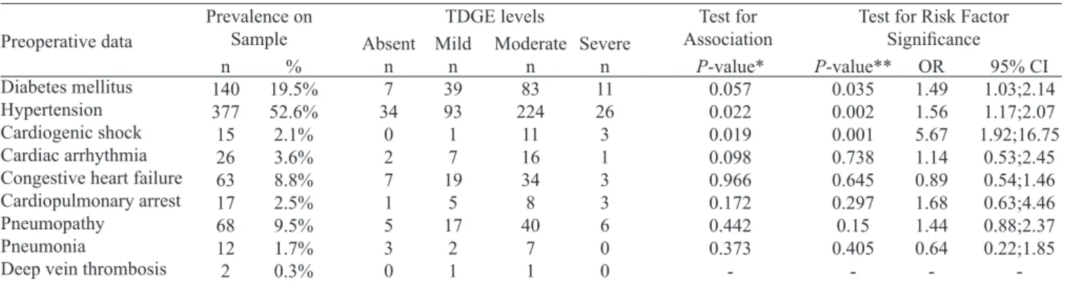
Table 3. Association of history and preoperative comorbidities with postoperative TDGE.
Preoperative data
Diabetes mellitus Hypertension Cardiogenic shock Cardiac arrhythmia Congestive heart failure Cardiopulmonary arrest Pneumopathy
Pneumonia
Deep vein thrombosis n 140 377 15 26 63 17 68 12 2 % 19.5% 52.6% 2.1% 3.6% 8.8% 2.5% 9.5% 1.7% 0.3%
TDGE= transient dysfunction of gas exchange; TDGE absent=PaO2/FiO2>300 mmHg; TDGE mild=PaO2/FiO2 between 200 mmHg and 300 mmHg; TDGE moderate=PaO2/FiO2 between 100 mmHg and 200 mmHg; severe TDGE=PaO2/FiO2<100 mmHg; Parentheses=percentage of the
total group OR=odds ratio; CI=conidence interval; P-value* (chi-square test); P-value** (logistic regression); Deep vein thrombosis: There
were not enough patients in the deep vein thrombosis group for statistical calculatios
Prevalence on Sample TDGE levels Absent n 7 34 0 2 7 1 5 3 0 Mild n 39 93 1 7 19 5 17 2 1 Moderate n 83 224 11 16 34 8 40 7 1 Severe n 11 26 3 1 3 3 6 0 0 P-value* 0.057 0.022 0.019 0.098 0.966 0.172 0.442 0.373
-Test for Risk Factor
Signiicance P-value** 0.035 0.002 0.001 0.738 0.645 0.297 0.15 0.405 -OR 1.49 1.56 5.67 1.14 0.89 1.68 1.44 0.64 -95% CI 1.03;2.14 1.17;2.07 1.92;16.75 0.53;2.45 0.54;1.46 0.63;4.46 0.88;2.37 0.22;1.85 -Test for Association
Table 4. Association of age with TDGE (n=717).
Age Classes ≤44 45-54 55-64 65-74 ≥75 n 124 162 229 153 49 717 % 17.3% 22.6% 31.9% 21.4% 6.8% 100%
TDGE=transient dysfunction of gas exchange; TDGE absent=PaO2/FiO2 > 300 mmHg; TDGE mild= PaO2/FiO2 between 200 mmHg and 300 mmHg; TDGE moderate= PaO2/FiO2 between 100 mmHg and 200 mmHg; severe TDGE=PaO2/FiO2<100 mmHg; parentheses=percentage of total
group; OR=odds ratio; CI=conidence interval; * P-value (chi-square test); ** P-value (logistic regression) of lower risk compared to the ≤ 44-year group
TDGE levels Absent n 26 16 21 8 6 Mild n 42 55 52 45 5 Moderate n 49 86 139 91 37 Severe n 7 5 17 9 1 P-value* 0.0001 0.0001 0.0001 0.0001 0.0001
Test for Risk Factor Signiicance
P-value** 0.0001 0,0001 0.0001 0.0001 OR
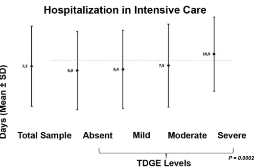
Interventions and complications in the ICU and their asso-ciation with the appearance of TDGE are described in Table 5. The occurrence of death within 48 hours and after 48 hours are described in Table 6. The he average length of stay in the ICU and the mortality predicted by APACHE II are described in Figure 1 and Figure 2.
Death within the irst 48 hours after surgery and death in the ICU were associated with the APACHE II score (P=0.0105 and P<0.0001, respectively). Death within the irst 48 hours after surgery and death in the ICU were both as -sociated with predicted mortality (P=0.0001, and P<0.0114, respectively).
DISCUSSION
Demographic variables, background, and preopera-tive comorbidities
The majority of the study population was male (61.6%), with an average age of 56.1 years. This inding is consistent with reports that the male population has a higher surgical incidence (70.1%) than females in the under-60 population[9].
In this study, myocardial revascularization surgery was the most common (49.4%), corroborating the indings of Laizo
et al.[10]. The preoperative comorbidities diabetes mellitus
(19.5%) and hypertension (52.6%) were prevalent. A pre-vious study observed diabetes mellitus in 29.6% of cases[8].
Compared with other developed countries, Brazil has a high-er incidence of hyphigh-ertension (90.7% vs. 60%) and diabetes mellitus (37.2% vs. 29%)[9]. Diseases such as hypertension,
autoimmune diseases, peripheral vascular disorders, and metabolic syndrome must be controlled and require great care in the immediate postoperative period[10].
Other studies report preoperative comorbidities of dys-lipidemia in 48% of cases and a family background of coro-nary artery disease in 38% of cases but report hypertension in 75%[11] to 79%[2] of patients. In developed countries,
car-diovascular diseases are the leading causes of death and are increasing in occurrence in developing countries[12]. Oliveira
et al.[11] measured morbidity by measuring the occurrence
of the postoperative complications described above and the mortality as the number of deaths. In our study, 0.84% of individuals died within 48 hours, and 9.21% died after 48 hours of intensive care.
In Brazil, the mortality rate for myocardial revasculariza-tion is 6.2%. Several postoperative management protocols of cardiac surgery have been studied with the purpose of
pre-Table 6. The occurrence of death within 48 hours and after 48 hours after surgery.
Postoperative data
Death within 48 hours Death after 48 hours
n 6 66 % 0.8% 9,2%
TDGE=transient dysfunction of gas exchange; TDGE absent=PaO2/FiO2>300 mmHg; TDGE mild=PaO2/ FiO2 between 200 mmHg and 300 mmHg; TDGE moderate=PaO2/FiO2 between 100 mmHg and 200 mmHg; severe TDGE=PaO2/FiO2<100 mmHg; * P-value=Tukey test for comparison of measures between TDGE groups Prevalence on Sample TDGE levels Absent n 0 5 Mild n 1 13 Moderate n 4 40 Severe n 1 8 P-value* 0.5420 0.0350 Test for Association Table 5. Complications and interventions in the ICU and their associations with TDGE.
Postoperative data Pneumonia VAP Non-invasive mechanical ventilation Reintubation Tracheostomy
Renal replacement therapy Hemotherapy Cardiac arrhythmia n 64 4 15 69 26 62 247 26 % 8.9% 0.6% 2.1% 9.6% 3.6% 8.6% 34.4% 3.6%
ICU=intensive care unit; TDGE=transient dysfunction of gas exchange; TDGE absent=PaO2/FiO2>300 mmHg; TDGE mild=PaO2/FiO2 between 200 mmHg and 300 mmHg; TDGE moderate=PaO2/FiO2 between 100 mmHg and 200 mmHg; severe TDGE=PaO2/FiO2<100 mmHg;
Parentheses= percentage of total group; VAP=mechanical ventilation-associated pneumonia; P-value* (chi-square test); P-value** (logistic regression); CI= conidence interval
Prevalence on Sample TDGE levels Absent n 4 0 0 4 3 6 21 2 Mild n 12 0 4 15 6 10 60 7 Moderate n 37 2 10 45 15 36 139 12 Severe n 11 2 1 5 2 10 27 5 P-value* 0.0001 0.0420 0.5400 0.2250 0.8460 0.0005 0.0001 0.0450
Test for Risk Factor
Fig. 1- The average length of stay in the Intensive Care Unit.
TDGE=transient dysfunction of gas exchange; TDGE absent=PaO2/FiO2>300 mmHg; TDGE mild=PaO2/ FiO2 between 200 mmHg and 300 mmHg; TDGE moderate=PaO2/FiO2 between 100 mmHg and 200 mmHg; severe TDGE=PaO2/FiO2<100 mmHg; * P-value=* ANOVA followed by transformation with posts by
Tukey’s test. TDGE Record signiicantly proved superior to other levels of TDGE regarding Apache II score.
Fig. 2 - Mortality predicted by APACHE II.
TDGE=transient dysfunction of gas exchange; TDGE absent=PaO2/FiO2>300 mmHg; TDGE mild=PaO2/ FiO2 between 200 mmHg and 300 mmHg; TDGE moderate=PaO2/FiO2 between 100 mmHg and 200 mmHg; severe TDGE=PaO2/FiO2 <100 mmHg; * P-value: ANOVA followed by transformation with posts by
dicting mortality[10]. In the study of Ribeiro et al.[13],
espe-cially in the postoperative period of cardiac surgery, 115,021 pulmonary complications contributed to the overall mortality rate of 8%. In Brazil, the mortality rate after cardiovascular surgery is approximately 8% in the Uniied Health System[14].
APACHE II is an index that classiies critical patients accord -ing to the severity of their condition[15]. Our patients had high
APACHE II scores, similar to the study by Feijó et al.[16]. In a
prospective trial of 520 patients lasting 13 months in the ICU of Hospital Sao Paulo, in which the APACHE II prognostic index was applied, scores > 25 were associated with a higher risk of death[15]. In our study, the APACHE II score (12.8 ±
4.2 points) and predicted mortality (17.9±9.5 points) were correlated with the occurrence of severe TDGE (P=0.0001 for both), demonstrating that APACHE II score was effec-tive to determine the severity of the patients in this study. Al-though mortality was high (10.5%), it was less than expected.
Occurrence of TDGE after surgery and cardiac procedures
Respiratory failure after heart surgery is an important fac-tor of postoperative morbidity and mortality[4]. In this study,
15 (2%) patients had ARDS, 27.7% mild TDGE, 56.1% mod-erate TDGE, and 5.4% severe TDGE. In a study by Szeles et al.[5], the PaO
2/FiO2 ratio was used to assess the impact of the
seriousness of the hypoxemia in the immediate postoperative period. The patients were divided into three groups: PaO2/ FiO2>200 (45.8%); PaO2/FiO2 between 150 and 200 (26.9%) and PaO2/FiO2<150 (27.3%). That study reported that tran-sient hypoxemia was not affected by an increase of mechan-ical ventilation time and that the inlammatory response to surgical trauma and CBP caused lung damage, explaining the transitional hypoxemia[5].
ARDS was present in 0.4% to 1.32% of the patients[4].
ARDS was irst described by Ashbaugh & Petty, in 1967, with these common characteristics: tachypnea, hypoxemia, persistent opaciication on chest X-ray, decreased compla -cency, and high mortality. In 1994, the irst consensus for ARDS was published, which deined four aspects: acute pre -sentation, presence of hypoxemia (PaO2/FiO2<200 mmHg), iniltrated bilateral on chest X-ray, and pulmonary capil -lary wedge pressure (PCWP) less than 18 cmH2O (to rule out heart failure). Also deined in this consensus, similar to ARDS, was ALI, deined as being present when the PaO2/
FiO2 ratio is between 200 and 300 mmHg[17].
Over the years, the criteria for ARDS did not correspond to the manifestations of the syndrome. In 2012, the criteria were revised using the following classiication: the term acute was deined as the occurrence of events of manifesta -tion in one week or less; the term ALI was abandoned; and the measurement of the PaO2/FiO2 relationship was amended to require a minimal amount of positive end-expiratory pres-sure (PEEP)[7]. In addition, three categories of ARDS were
proposed: mild, moderate, and severe, based on the PaO2/ FiO2 ratio; chest X-ray criteria were clariied to improve reliability between examiners; and the PCWP criterion was removed and clarity was added to improve the ability to rule out cardiac causes of bilateral iniltrates[17]. The last deini
-tion includes the measurement of PEEP, limiting the possibil-ities of diagnosis for patients without mechanical ventilation, but the diagnosis of ARDS does not exclude these patients.
To evaluate our patients under the new classiication of severity of the ARDS PaO2/FiO2 ratio, we found that 89.26% it this criterion for diagnosis, suggesting a high incidence of ARDS in this population. However, using only the PaO2/FiO2 ratio is not suficient to deine the presence of ARDS. Oth -er factors are included in the dysfunctional gas exchange in the postoperative period of cardiac surgery, such as systemic inlammatory response syndrome (SIRS) with pulmonary re -percussion, hyperdynamic frame, interstitial pulmonary ede-ma, microatelectasis, and reduced surfactant. In most cases, the pulmonary radiological image is normal, undercommit-ted lung compliance, and patients recover from this dysfunc-tion in a few hours, including extubadysfunc-tion after a few hours in intensive care.
In the present study, of 89.26% of patients who presented with TDGE, only 2% evolved with ARDS, according to the criteria of the last consensus. The classiication of ARDS as mild, moderate, and severe was used to determine the degree of TDGE. Based on these indings, the term ARDS should only be used for patients who meet all the diagnostic criteria. Therefore, we suggest that this acute and transient hypox-ia that occurs in the postoperative period of cardhypox-iac surgery (within 48 hours) be called transient dysfunction of gas ex-change, as we have in this work.
Association of TDGE with preoperative and postop -erative factors
In our sample, increasing age was associated with increas-ing severity of TDGE, especially in patients older than 75 years, whose risk is three times higher than patients younger than 44 years. In the study of Oliveira et al.[9], age>70 years was
sig-niicantly associated with greater mortality (P<0.002). In this study, the average length of stay in the ICU was 7.3±11.9 days, similar to the average of 4.16±3.76 days in the study by Laizo et al.[10]. There was a tendency for the prevalence of severe
TDGE to increase from 2007 to 2011(P=0.01).
chang-Authors’ roles & responsibilities
CDAR Analysis and/or interpretation of data, inal approval of the
manuscript, study design, manuscript writing or critical review of its content
MMM Final approval of the manuscript
NMFL Analysis and/or interpretation of data, statistical analysis,
inal approval of the manuscript, study design, manuscript
writing or critical review of its content LCF Final approval of the manuscript ALEF Final approval of the manuscript OPJ Final approval of the manuscript
DD Analysis and/or interpretation of data, inal approval of the
manuscript, study design, manuscript writing or critical review of its content
es in gas exchange.
The postoperative complications inluence the length of hospitalization of the patient, generating increased costs and hospital mortality[18]. In this study, pneumonia was present
in 8.9% of cases and was associated with moderate TDGE (P=0.001), which was a risk factor (P=0.0005) for the oc-currence of postoperative pneumonia. Ventilator-associated pneumonia (VAP) was present in 0.6% of the cases and was associated with severe TDGE (P=0.042), which in turn was a risk factor (P=0.003) for the development of VAP.
The low incidence of VAP in the study may be explained by the implementation of prophylactic measures and the awareness of the professionals of the ICU at the beginning of 2007. In a previous study, lung infection was more common among the infectious complications (15.3%)[19]. In another
study, 7332 patients undergoing cardiac surgery, infectious outbreaks were identiied in 29 patients, 55% of them pri -mary infections[14]. However, there is no study reporting the
occurrence of postoperative pulmonary complications[20].
Oliveira et al.[11] indicated the following as predictors of
postoperative infections: body mass index ≥ 40 kg/m2,
pre-operative hemodialysis, cardiogenic shock, prepre-operative age ≥ 85 years, preoperative immunosuppressive treatment, dia -betes mellitus, CPB ≥ 200 minutes, and the revascularization of three or more vessels.
Among the complications encountered in the period after surgery, those that occur in the respiratory system contrib-ute signiicantly to morbidity and mortality related to cardiac surgery, as 3.5% to 10% of these morbidities and mortali-ties are caused by respiratory complications[21]. In large part,
these complications are explained by the use of CPB, which causes an increase of inlammatory mediators leading to de -creased ventricular contractility, which consequently increas-es vascular permeability and rincreas-esistance of the organs.
Speciically in the pulmonary circulation, inlammatory luid accumulates interstitially, leading to the formation of microatelectasis, hypoxemia, and hypoxic vasoconstriction. These conditions decrease the local production of pulmo-nary surfactant, which leads to worsening pressures, lung collapse, and pulmonary dysfunction, generating losses in respiratory mechanics and increased respiratory work[12,22].
This situation results in pneumonic complications in respi-ratory mechanics[23]. Morsch et al.[2] used pulmonary
radio-logical changes as diagnostic criteria for unventilated areas and/or consolidation, pleural effusion, and collapsed lung. Diagnostic criteria of postoperative lung infections are not standard in the scientiic literature and are associated or not with radiological changes, leukocytosis, body hyperther-mia, and isolation of pathogens in culture or in microscopic analysis of[21].
Hypoxemia is among the leading pulmonary complica-tions in the postoperative period of cardiac surgery; however, other complications, such as pleural effusion, pneumonia,
pneumothorax, reintubation, and ventilatory insuficiency, are also observed[24]. We used non-invasive mechanical
venti-lation therapeutically in 2.1% of cases, reintubation in 9.6%, and tracheostomy in 3.6%. Reintubation was a risk factor (P=0.042) for only postoperative TDGE.
Severe TDGE was associated with patients who had renal replacement therapy (P=0.0005), hemotherapy (P=0.0001), and cardiac arrhythmia (P=0.0450), with respective risk fac-tors 2.34, 1.72, and 1.79. A large amount of information in this study was collected from patient records, so the loss of information is inexcusable. We sorted the information by cat-egory to extract the largest amount of available information.
CONCLUSION
We concluded that TDGE, in varying degrees, was present at the time of surgery and during postoperative cardiac proce-dures. Preoperative hypertension and cardiogenic shock were associated with the occurrence of postoperative TDGE. The preoperative risk factors included hypertension, cardiogenic shock, and diabetes. Postoperatively, pneumonia, VAP, renal replacement therapy, hemotherapy, and cardiac arrhythmia were associated with the appearance of a certain degree of TDGE, which was a risk factor for reintubation, pneumonia, VAP, and renal replacement therapy during the postoperative period from cardiac surgery and during cardiac procedures.
ACKNOWLEDGMENTS
The authors acknowledge the work done by the Ofice of Biostatistics, School of Medical Sciences, State University of Campinas. In particular, we would like to thank Cleide Apa-recida Moreira Silva for his dedication to this work.
REFERENCES
1. Guizilini S, Gomes WJ, Faresin SM, Carvalho ACC, Jaramillo JI, Alves FA, et al. Effects of the pleural drain site on the pulmonary function after coronary artery bypass grafting. Rev Bras Cir Cardiovasc. 2004;19(1):47-54.
2. Morsch KT, Leguisamo CP, Camargo MD, Coronel CC, Mattos
W, Ortiz LDN, et al. Peril ventilatório dos pacientes submetidos
a cirurgia de revascularização do miocárdio. Rev Bras Cir Cardiovasc. 2009;24(2):180-7.
3. Arcêncio L, Souza MD, Bortolin BS, Fernandes ACM, Rodrigues AJ, Évora PRB. Cuidados pré e pós-operatórios em cirurgia
cardiotorácica: uma abordagem isioterapêutica. Rev Bras Cir
Cardiovasc. 2008;23(3):400-10.
4. Rodrigues CDA, Oliveira RARA, Soares SMTP, Figueiredo LC, Araújo S, Dragosavac D. Lesão pulmonar e ventilação mecânica em cirurgia cardíaca: revisão. Rev Bras Ter Intensiva. 2010;22(4):375-83.
5. Szeles TF, Yoshinaga EM, Alencar W, Brudniewski M, Ferreira FS, Auler Jr JOC, et al. Hipoxemia após revascularização miocárdica: análise dos fatores de risco. Rev Bras Anestesiol. 2008;58(2):124-36.
6. Padovani C, Cavenaghi OM. Recrutamento alveolar em pacientes no pós-operatório imediato de cirurgia cardíaca. Rev Bras Cir Cardiovasc. 2011;26(1):116-21.
7. ARDS Definition Task Force, Ranieri VM, Rubenfeld GD, Thompson BT, Ferguson ND, Caldwell E, Fan E, et al. Acute
respiratory distress syndrome: the Berlin Deinition. JAMA.
2012;307(23):2526-33.
8. Ledur P, Almeida L, Pellanda LC, Schaan BD. Preditores de infecção no pós-operatório de cirurgia de revascularização miocárdica. Rev Bras Cir Cardiovasc. 2011;26(2):190-6.
9. Oliveira EL, Westphal GA, Mastroeni MF. Características
clínico-demográicas de pacientes submetidos à cirurgia de
revascularização do miocárdio e sua relação com a mortalidade. Rev Bras Cir Cardiovasc. 2012;27(1):52-60.
10. Laizo A, Delgado FEF, Rocha GM. Complicações que aumentam o tempo de permanência na unidade de terapia intensiva na cirurgia cardíaca. Rev Bras Cir Cardiovasc. 2010;25(2):166-71.
11. Oliveira DC, Oliveira Filho JB, Silva RF, Moura SS, Silva DJ, Egito EST, et al. Sepse no pós-operatório de cirurgia cardíaca: descrição do problema. Arq Bras Cardiol. 2010;94(3):332-6.
12. Renault JA, Costa-Val R, Rossetti MB. Fisioterapia respiratória na disfunção pulmonar pós-cirurgia cardíaca. Rev Bras Cir Cardiovasc. 2008;23(4):562-9.
13. Ribeiro AL, Gagliardi SP, Nogueira JL, Silveira LM, Colosimo EA, Lopes do Nascimento CA. Mortality related to cardiac surgery in Brazil, 2000-2003. J Thorac Cardiovasc Surg. 2006;131(4):907-9.
14. Gomes WJ, Mendonça JT, Braile DM. Resultados em cirurgia cardiovascular: oportunidade para rediscutir o atendimento médico e cardiológico no sistema público de saúde do país. Rev Bras Cir Cardiovasc. 2007;22(4):III-VI.
15. Costa JI, Gomes do Amaral JL, Munechika M, Juliano Y, Bezerra-Filho JG. Severity and prognosis in intensive care: prospective application of the APACHE II index. Sao Paulo Med J. 1999;117(5):205-14.
16. Feijó CAR, Leite Júnior FO, Martins ACS, Furtado Júnior AH, Cruz LLS, Meneses FA. Gravidade dos pacientes admitidos
à unidade de terapia intensiva de um hospital universitário
brasileiro. Rev Bras Ter Intensiva. 2006;18(1):18-21.
17. Angus DC. The acute respiratory distress syndrome: what’s in a name? JAMA. 2012;307(23):2542-4.
18. Piotto RF, Ferreira FB, Colósimo FC, Silva GS, Sousa AG, Braile DM. Fatores preditores independentes de ventilação
mecânica prolongada em pacientes submetidos à cirurgia
de revascularização miocárdica. Rev Bras Cir Cardiovasc. 2012;27(4):520-8.
19. Strabelli TMV, Stolf NAG, Uip DE. Uso prático de um índice de risco de complicações após cirurgia cardíaca. Arq Bras Cardiol. 2008;91(5):342-7.
20. Westerdahl E, Lindmark B, Almgren SO, Tenling A. Chest physiotherapy after coronary artery bypass graft surgery: a comparison of three different deep breathing techniques. J Rehabil Med. 2001;33(2):79-84.
21. Brasher PA, McClelland KH, Denehy L, Story I. Does removal of deep breathing exercises from a physiotherapy program including pre-operative education and early mobilisation after cardiac surgery alter patient outcomes? Aust J Physiother. 2003;49(3):165-73.
22. Barbosa RAG, Carmona MJC. Avaliação da função pulmonar
em pacientes submetidos à cirurgia cardíaca com circulação
extracorpórea. Rev Bras Anestesiol. 2002;52(6):689-99.
23. Taylor GJ, Mikell FL, Moses HW, Dove JT, Katholi RE, Malik SA, et al. Determinants of hospital charges for coronary artery bypass surgery: the economic consequences of postoperative complications. Am J Cardiol. 1990;65(5):309-13.