Protein supplementation may be an alternative to reduce the erosive potential of acidic drinks. The aim of this in vitro study was to evaluate the erosive potential of an orange juice modified by dietary proteins. A commercially available orange juice was added 0.2 g/L casein, 2.0 g/L ovalbumin and their combination. The juice with no additives and a commercially available calcium-modified juice were used as negative and positive controls, respectively. Human enamel and dentin specimens (n=11) were tested in an erosion-remineralization cycling model. Enamel was analyzed by surface microhardness and profilometry, whilst dentin by profilometry only. Statistical analyses were performed using one-way ANOVA followed by Tukey’s test (p<0.05). Calcium-modified juice showed the lowest erosive potential for both analyses (p<0.05). For enamel, the protein-added groups did not differ from each other (p>0.05) and showed significantly lower enamel loss compared to negative control (p<0.05). Regarding surface microhardness, casein showed the highest values compared to negative control (p<0.05). For dentin, none of the protein-added groups showed lower values of surface loss compared to negative control (p>0.05). In conclusion, for enamel the protein-modified orange juices presented reduced erosion of enamel, with casein showing a trend for better protection. For dentin, no reduction in the erosive potential was observed for the tested protein-modified orange juices.
Supplementation of an Orange
J u i c e w i t h D i e t a r y P r o t e i n s t o
Prevent Enamel and Dentin Erosion
Stella S. Ferreira1, Tais Scaramucci1, Anderson T. Hara2, Idalina V. Aoki3, Maria Angela Pita Sobral1
1Department of Restorative
Dentistry, School of Dentistry, USP - Universidade de São Paulo, São Paulo, SP, Brazil
2Oral Health Research Institute,
Department of Preventive and Community Dentistry, Indiana University School of Dentistry, Indianapolis, IN, USA
3Department of Chemical
Engineering, Polytechnic School, USP - Universidade de São Paulo, São Paulo, SP, Brazil
Correspondence: Maria Angela Pita Sobral, Avenida Prof. Lineu Prestes 2227, 05508-900 São Paulo, SP, Brasil. Tel: +55-11-3091-7843. e-mail: [email protected]
Key Words: dental enamel, dentin, dietary proteins, ovalbumin, tooth erosion.
Introduction
As the population ages, the teeth remain longer in the mouth, and in consequence the incidence of non-carious cervical lesions has increased (1). It is known that a change in the population lifestyle has led to an increase in the consumption of acidic beverages, and reports have associated this fact with a higher prevalence of dental erosion (2). In this sense, the major concern is related to frequent dentin exposure, due to surface loss, because if some strong acids can remove crystals from enamel the same is expected to occur in the exposed dentin (3).
Considering the irreversible nature of erosive tooth wear, prevention is highly important. A commonly suggested preventive measure is the addition of calcium and/or phosphate salts (4) to acidic drinks, which increases its saturation in relation to hydroxyapatite (5) and decreases dental dissolution by the common-ion effect (6). Although in previous investigations (5,7) some of these calcium-modified drinks have demonstrated positive results in reducing erosion, the large amounts of calcium usually present in these beverages may change their taste, making it unacceptable to some consumers (8). In an attempt to improve this negative effect of calcium supplementation, potential alternative supplements for acidic drink modification have been considered, like some
proteins with larger molecules, bringing positive results (9). Among these proteins, casein and ovalbumin are approved for use as food additives, inexpensive and soluble in acidic aqueous solutions. Caseins account for nearly 80% of the total protein in bovine milk (10), and ovalbumin is the major protein found in the whites of hen’s eggs (11). Earlier investigations have reported that casein and ovalbumin may be adsorbed on an enamel surface, modifying the dissolution process (12,13) and reducing hydroxyapatite dissolution in dental caries models (14-16). Since these investigations, some studies (6,11) also demonstrated that these proteins can efficiently reduce the erosive potential of acidic solutions and commercially available soft drinks (17); however, to the best of the authors’ knowledge, these studies used as substrates only hydroxyapatite discs and dental enamel, not assessing the role of these proteins in preventing dentin erosion.
264
S.S. Ferreira et al.
Material and Methods
Experimental DesignIn this in vitro study, each of two dietary proteins and a combination of them were added to a commercially available orange juice. This study was conducted at five levels: C-, orange juice with no additives (negative control); CAS, orange juice with sodium caseinate; OVA, orange juice with ovalbumin; CAS + OVA, orange juice with sodium caseinate and ovalbumin; and C+, orange juice with calcium (positive control). Then, the erosive potentials of these experimental juices and of the positive and negative controls were tested on human enamel and root dentin substrates, using an erosion-remineralization model (n=11). The response variables were surface loss (SL), expressed in μm measured by optical profilometry for both substrates, and surface microhardness (SMH) determined by the Knoop hardness number for enamel. This study was carried out after approval of the local Ethics Board (Protocol 155/09).
Modified Juice Preparation
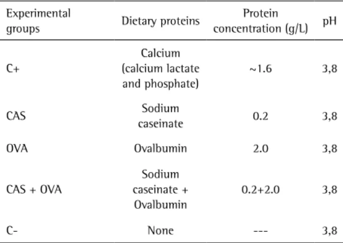
Two food-approved proteins were added alone or in combination to a commercially available orange juice (Minute Maid Original®; The Coca-Cola Company, Atlanta, GA, USA), creating the experimental groups described in Table 1. The proteins selected as additives were sodium caseinate (BDH, Poole, UK) and ovalbumin (MP Biochemicals, Solon, OH, USA). The used amounts were based on previous publications (6,11). The original juice without additives was the negative control (C-), and a commercially available calcium-modified juice (Minute Maid Calcium®; The Coca-Cola Company) with approximately 1.6 g/L of calcium (20) was the positive control (21) (C+). After the addition of proteins, no change in the pH values of the experimental juices was observed. The pH values were determined in triplicate, using a calibrated pH electrode (Accumet 13-620-530, Fisher Scientific, Pittsburgh, PA, USA). Table 1 describes the experimental groups, the dietary proteins, the protein concentration and the pH values of the juices.
Specimens Preparation
Fifty-five enamel and 55 root dentin specimens (4 x 4 x 2 mm) were obtained from included human third molars, selected for not being in contact with the oral cavity. The crowns and the roots were sectioned in the microtome, and the specimens obtained were embedded in acrylic resin (Varidur; Buehler, Lake Bluff, IL, USA), ground flat with water-cooled abrasive discs 500-, 1200-, 2400- and 4000-grit carbide papers, (MD-Fuga; Struers Inc., Cleveland, OH, USA), and polished in polishing pads with diamond-particle dispersion (1 µm, Struers Inc.). The polished surfaces were covered with an adhesive tape (control area), leaving a central test area of 4 x 1 mm
exposed to the erosive challenge.
Erosive Challenge
The enamel and dentin specimens obtained were randomly allocated into the five experimental groups (n=11). Then, they were submitted to erosion-remineralization cycles (21). A complete cycle consisted of a 5-min immersion in 10 mL/specimen of the test juice, with no agitation and at room temperature; followed by a 60-min immersion in 10 mL/specimen of artificial saliva (0.213 g/L of CaCl2.2H2O, 0.738 g/L of KH2PO4, 1.114 g/L of KCl, 0.381 g/L of NaCl, 12 g/L of tris-buffer, and 2.2 g/L of porcine gastric mucin, pH 7.0) at 150 rpm and at room temperature. This cycle was repeated 6 times a day for 5 days. After the demineralization and remineralization episodes, the specimens were rinsed with distilled water and gently dried with paper towel. The specimens were stored overnight in artificial saliva (150 rpm at room temperature).
Surface Analysis
After the cycling phase, the tapes were removed, and the surface loss (enamel and dentin) and surface microhardness (enamel) of the specimens were analyzed.
The SL was assessed by profilometric analysis, performed on the enamel and dentin specimens using an optical profilometer (Proscan 2000; Scantron, Taunton, Somerset, UK). An area 2 mm long (x-axis) and 1 mm wide (y-axis) was scanned. The step size was set at 0.01 mm (x-axis) and 0.1 mm (y-axis), and the number of steps was set at 200 (x-axis) and 20 (y-axis). The scanned area covered both the central testing area and the two lateral control areas. The surface loss was determined by calculating the difference in height (µm) of the lesion area in relation to the reference surfaces using a dedicated software (Proscan 2000, Scantron).
The SMH was assessed on enamel specimens only, using
Table 1. Experimental groups, dietary proteins, protein concentration and the pH values of the juices
Experimental
groups Dietary proteins
Protein
concentration (g/L) pH
C+
Calcium (calcium lactate and phosphate)
~1.6 3,8
CAS Sodium
caseinate 0.2 3,8 OVA Ovalbumin 2.0 3,8
CAS + OVA
Sodium caseinate + Ovalbumin
0.2+2.0 3,8
Protein-modified juice to reduce erosion
a Knoop diamond indenter (Tukon 2100; Wilson-Instron, Norwood, MA, USA), with a 50 g load applied for 15 s. Three indentations were made 100 µm apart, in both the reference and experimental areas. The mean value (in Knoop numbers) of these indentations was considered the response variable.
Data Analysis
Homoscedasticity and normal distribution of the data were checked by the Levene and Shapiro-Wilk tests, respectively. Once these assumptions were satisfied, one-way ANOVA and Tukey test were carried out to detect and identify differences among the experimental groups. All analyses were performed considering a significance level of 5% using the SPSS for Windows statistical software (version 17.0, SPSS Inc., Chicago, IL, USA).
Results
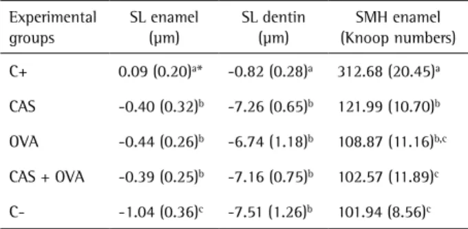
The results of surface loss for enamel and dentin are shown in Table 2. C+ showed the lowest erosive potential, with lower surface loss for both tested dental substrates (p<0.05). For enamel, the protein-added groups (CAS, CAS+OVA, and OVA) did not differ from each other (p>0.05) and presented significantly less surface loss compared to C- (p<0.05). For dentin, the surface loss values of the CAS, OVA and CAS+OVA groups were not significantly different from C- (p>0.05).
Regarding SMH, the results are also shown in Table 2. C+ presented the highest values of SMH, followed by CAS. The SMH values of the groups OVA and CAS+OVA were not significantly different from C-.
Discussion
According to the obtained results, the commercially available calcium-modified juice (positive control) showed the best protective effect against erosion for both enamel and dentin. It significantly reduced the loss of both tested
substrates and also led to higher values of enamel SMH than did the juice without additives. This positive result was expected due to the high amount of calcium in this juice, which is why it was chosen as positive control. This result agrees with those of Scaramucci et al. (21), who used the same calcium-modified orange juice as their control group.
Regarding the dietary proteins tested, both proteins alone and their combination prevented enamel surface loss. On the other hand, concerning the surface microhardness, only casein was able to provide protection against enamel surface softening. In an attempt to explain these differences, it was hypothesized that the characteristics related to the nature and molecular weight of the tested proteins may influence their adsorption to enamel.
Casein presents a relatively hydrophobic nature (10), with a weaker interaction with water. Its high molecular weight (375 kDa) provides it a long chain of amino acids, enabling more interactions with the dental surfaces. These interactions are made by their hydrophobic groups (apolar) through van der Waals bonds, which, despite being weak, can be efficient if a large number of molecules is present very close to each other. As result, a more uniform and persistent protective layer can be formed. Another important aspect is that casein is a phosphoprotein made up by phosphoserine and phosphothreonine (22), which contains chemically bounded phosphoric acid and calcium ions. This composition favors the interactions between the phosphate groups and calcium ions from one molecule to another (casein-casein), as well as between the casein molecules and ions present in the hydroxyapatite crystals of the dental substrate (casein-enamel).
As suggested by Barbour et al. (6), the protective layer formed by casein restricts the access of H+ ions, preventing release of calcium and phosphate ions. This protein acts differently from CPP-ACP, which is a complex formed by casein phosphopeptide (CPP), another milk-derived protein, and an amorphous calcium phosphate (ACP). The CPP-ACP is described as a transport for Ca+2 and PO
4-3 ions to the tooth surface (23), releasing them during the erosive attack and supersaturating the medium. Although this complex has shown positive results when added to sports drinks (24), its use in commercial tooth care products to prevent erosion is very controversial (25). It should be stressed that the results of the present study show the protective effect against dental erosion derived from sodium caseinate without the addition of calcium phosphate.
In contrast to casein, ovalbumin has a stronger hydrophilic nature (26); the polar groups in its molecules allow for a better interaction between this protein and water, making it more soluble. On the other hand, its low molecular weight (45 kDa) means its molecules are relatively small in size, which could be a positive factor,
Table 2. Means (SD) of surface loss (SL) for enamel and dentin, and surface microhardness (SMH) for enamel
Experimental groups
SL enamel (µm)
SL dentin (µm)
SMH enamel (Knoop numbers) C+ 0.09 (0.20)a* -0.82 (0.28)a 312.68 (20.45)a
CAS -0.40 (0.32)b -7.26 (0.65)b 121.99 (10.70)b
OVA -0.44 (0.26)b -6.74 (1.18)b 108.87 (11.16)b,c
CAS + OVA -0.39 (0.25)b -7.16 (0.75)b 102.57 (11.89)c
C- -1.04 (0.36)c -7.51 (1.26)b 101.94 (8.56)c
266
S.S. Ferreira et al.
assuming that these molecules would easier reach all active sites of adsorption present in the hydroxyapatite crystals. Due to its greater solubility, ovalbumin was used at a concentration 10x higher than casein, and for this reason, it was expected that these proteins yielded a better result. Although both tested proteins showed similar results for enamel surface loss, OVA showed no protection regarding enamel softening. It may be assumed that despite the high availability and ease of adsorption on the hydroxyapatite, the hydrophilic nature of ovalbumin molecules with many carboxylic acid groups (aspartic and glutamic acid) allowed interactions and permeation of H+ ions through the adsorbed layer, facilitating enamel demineralization. H+ ions can interact with amino acids, particularly with those that have additional amino groups, like leucine, isoleucine, and valine, which explains the easy permeation through the adsorbed ovalbumin layer.
There are few studies investigating the use of casein and ovalbumin as potential anti-erosive additives to drinks. To the best of the authors’ knowledge, the only study that investigated the effects of casein and ovalbumin added as anti-erosive agents in different commercially available beverages, including a natural orange juice and using the human enamel as substrate, was the study by Hemingway (17). These authors added the proteins (casein and ovalbumin) separately to the beverages, for a 0.2% concentration and concluded that ovalbumin reduced erosion in only one drink, while casein reduced erosion in all drinks except orange juice. Considering that the enamel specimens were analyzed regarding surface loss by optical profilometer, these findings do not agree with the results of the present. This may be explained by differences in relevant factors between the two investigations, including the time of exposure to the beverages, the used concentration of the proteins and the orange juice brand.
When the proteins were tested in combination (CAS+OVA), the results were similar to the ovalbumin-alone group, with no prevention of enamel softening but with significant reduction in surface loss. Therefore, one of the initially proposed study hypotheses was rejected. Although casein has demonstrated a better adsorption to hydroxyapatite than ovalbumin (12), it suggests that a competition between these proteins may have occurred, with predominance of ovalbumin. This was probably strongly affected by the difference in the proteins’ concentration (0.2 g/L of casein vs. 2.0 g/L of ovalbumin), where the presence of only a few chains of casein was not enough to form an effective barrier against H+ ions, similar to when casein was used alone.
For dentin, none of the tested proteins showed a positive effect in reducing the erosive potential of the orange juice. It was assumed that the lack of protection may be due to
the different structure and composition of this substrate in comparison to enamel. Dentin has a lower mineral content and a more irregular structure (with tubules), which may have contributed to a lower adsorption of proteins. In addition, the crystals in dentin are smaller than those in enamel, so the surface area per gram of dentin is much higher, providing more available surface for the acid to affect (27) and for the proteins to react with it. Hence, the protein concentration (speaking mainly about casein) used in this study may not have been enough to react with the entire available dentin surface. Comparison with other studies is difficult since, to the authors’ knowledge, this is the first study evaluating drink supplementation with proteins using human dentin as a substrate.
The assessment of the erosion in this study was performed by both surface microhardness, which is a suitable method to evaluate tooth-tissue softening (28), and optical profilometry, which is more suitable to evaluate the loss of tooth tissue in more advanced stages (28). In this sense, both methods were chosen due to the use of substrates with different susceptibility to erosion and solutions with different erosive potentials (20,21). In agreement with other studies, the enamel erosion was assessed by surface microhardness and optical profilometry (17,20,21) and dentin erosion by optical profilometry (20,21,29).
Although, a reduction of erosion by the protein-added juices has been observed in the current study, none of them was able to completely inhibit the harmful effects of the orange juice or do so as effectively as the positive control. The use of these proteins in association with calcium could possibly allow the use of smaller amounts of this ion, reducing the drawbacks related to drink taste and stability changes commonly found in juices modified with large amounts of calcium (4).
Within the limitations of this in vitro study, it may be concluded that, regarding the tested proteins, casein yielded better results for enamel in terms of the reduction of erosion. The protein combination did not lead to an additional erosion reduction. For dentin, neither of the tested proteins prevented erosion. The commercially available calcium-modified juice yielded reduced erosion of both enamel and dentin.
Resumo
Protein-modified juice to reduce erosion por microdureza de superfície e perfilometria, enquanto que a dentina,
apenas por perfilometria. As análises estatísticas foram realizadas utilizando ANOVA um fator seguido pelo Teste de Tukey (p<0.05). O suco suplementado com cálcio mostrou o menor potencial erosivo para ambas as análises (p<0,05). Em relação ao esmalte, os grupos com adição de proteína não diferiram entre si (p>0,05) e mostraram significativamente uma menor perda de esmalte em relação ao grupo controle negativo (p<0,05). Para a microdureza, a caseína apresentou os maiores valores em relação ao controle negativo (p<0,05). Para a dentina, nenhum dos grupos com adição de proteína apresentou valores de perda de superfície menores quando comparados ao grupo controle negativo (p>0,05). Conclui-se que, para o esmalte os sucos de laranja modificados por proteínas apresentaram uma redução da erosão, com a caseína mostrando uma tendência para melhor proteção. Para a dentina, nenhuma redução da erosão foi observada para os sucos de laranja modificados por proteínas testados neste estudo.
Acknowledgements
The authors report no conflict of interest and would like to thank CNPq (National Council of Technological and Scientific Development) for the first author’s scholarship (#134944/2009-7).
References
1. Wood I, Jawad Z, Paisley C, Brunton P. Non-carious cervical tooth surface loss: A literature review. J Dent 2008;36:759-766.
2. Dugmore CR, Rock WP. A multifactorial analysis of factors associated with dental erosion. Brit Dent J 2004 Mar;196:283-286.
3. White I, McIntyre J, Logan R. Studies on dental erosion: An in vitro model of root surface erosion. Aust Dent J 2001;46:203-207. 4. Grenby TH. Lessening dental erosive potential by product modification.
Eur J Oral Sci 1996;104:221-228.
5. Larsen MJ, Nyvad B. Enamel erosion by some soft drinks and orange juices relative to their pH, buffering effect and contents of calcium phosphate. Caries Res 1999;33:81-87.
6. Barbour ME, Shellis RP, Parker DM, Allen GC, Addy M. Inhibition of hydroxyapatite dissolution by whole casein: the effects of pH, protein concentration, calcium, and ionic strength. Eur J Oral Sci 2008;116:473-478.
7. Davis RE, Marshall TA, Qian F, Warren JJ, Wefel JS. In vitro protection against dental erosion afforded by commercially available, calcium-fortified 100 percent juices. J Am Dent Assoc 2007;138:1593-1598. 8. Barbour ME, Lussi A. Erosion in relation to nutrition and the
environment. Monogr Oral Sci 2014;25:143-154.
9. Ganss C. Is erosive tooth wear an oral disease? Monogr Oral Sci 2014;25:16-21.
10. Aimutis WR. Bioactive properties of milk proteins with particular focus on anticariogenesis. J Nutr 2004;134:989S-995S.
11. Hemingway CA, Shellis RP, Parker DM, Addy M, Barbour ME. Inhibition of hydroxyapatite dissolution by ovalbumin as a function of pH, calcium concentration, protein concentration and acid type. Caries Res
2008;42:348-353.
12. Pearce EIF, Bibby BG. Protein adsorption on bovine enamel. Arch Oral Bio 1966;11:329-336.
13. Weiss ME, Bibby BG. Effects of milk on enamel solubility. Arch Oral Bio 1966;11:49-57.
14. Arends J, Schuthof J, Christoffersen J. Inhibition of enamel demineralization by albumin in vitro. Caries Res 1986;20:337-340. 15. Reynolds EC, Black CL. Reduction of chocolate cariogenicity by
supplementation with sodium caseinate. Caries Res 1987;21:445-451. 16. Reynolds EC, Black CL. Cariogenicity of a confection supplemented with
sodium caseinate at a palatable level. Caries Res 1989;23:368-370. 17. Hemingway CA, White AJ, Shellis RP, Addy M, Parker DM, Barbour ME.
Enamel erosion in dietary acids: inhibition by food proteins in vitro. Caries Res 2010;44:525-530.
18. West NX, Maxwell A, Hughes JA, Parker DM, Newcombe RG, Addy M. A method to measure clinical erosion: the effect of orange juice consumption on erosion of enamel. J Dent 1998;26:329-335. 19. Murrell S, Marshall TA, Moynihan PJ, Qian F, Wefel JS. Comparison of
in vitro erosion potentials between beverages available in the United Kingdom and the United States. J Dent 2010;38:284-289.
20. Hara AT, Zero DT. Analysis of the erosive potential of calcium-containing acidic beverages. Eur J Oral Sci 2008;116:60-65. 21. Scaramucci T, Hara AT, Zero DT, Ferreira SS, Aoki IV, Sobral MAP. In vitro
evaluation of the erosive potential of orange juice modified by food additives in enamel and dentin. J Dent 2011;39:841-848.
22. Wind M, Feldmann I, Jakubowski N, Lehmann WD. Spotting and quantification of phosphoproteins purified by gel electrophoresis and laser ablation-element mass spectrometry with phosphorus-31 detection. Electrophoresis 2003;24:1276-1280.
23. Reynolds EC. Casein phosphopeptide-amorphous calcium phosphate: the scientific evidence. Adv Dent Res 2009;21:25-29.
24. Ramalingam L, Messer LB, Reynolds EC. Adding casein phosphopeptideamorphous calcium phosphate to sports drinks to eliminate in vitro erosion. Paediatr Dent 2005;27:61-67.
25. Wiegand A, Attin T. Randomised in situ trial on the effect of milk and CPP-ACP on dental erosion. J Dent 2014;42:1210-1215.
26. Christoffersen MR, Christoffersen J, Ibsen P, Ipsen H. Adsorption of ovalbumin on calcium hydroxyapatite crystals. Colloids Surf 1986;18:1-8.
27. Featherstone JDB, Lussi A. Understanding the chemistry of dental erosion. Monogr Oral Sci 2006;20:66-76.
28. Stefanski T, Postek-Stefanska L. Possible ways of reducing dental erosive potential of acidic beverages. Aust Dent J 2014;59:280-288. 29. Caneppele TM, Jeronymo RD, Di Nicol. R, Ara.jo MA, Soares LE. In vitro
assessment of dentin erosion after immersion in acidic beverages: surface profile analysis and energy-dispersive X-ray fluorescence spectrometry study. Braz Dent J 2012;23:373-378.