1 6 Maria Helena R odrigues Macedo The role of L -arginine in t
he modulation of oral biofilms in periodont
al disease
Escola de Engenharia
Maria Helena Rodrigues Macedo
The role of L-arginine in the modulation
of oral biofilms in periodontal disease
Dissertation for the MSc in Biomedical Engineering
Area of Clinical Engineering
Supervisor
Professor Mariana Henriques – Universidade do Minho
Co-supervisor
Professor Wim Teughels – KU Leuven
Maria Helena Rodrigues Macedo
The role of L-arginine in the modulation
of oral biofilms in periodontal disease
A
CKNOWLEDGEMENTS
I sincerely would like to thank everyone that, one way or another, contributed to the realization of this project.
I would like to thank professor Mariana Henriques for the opportunity of doing Erasmus and develop this project, for her constant interest and dedication and for always helping me either in Portugal or in Belgium, which was very important in order to accomplish this work.
I would also like to thank professor Wim Teughels for welcoming me in Belgium, giving me the opportunity to join his research group during the Erasmus project and for his availability to always help.
A special thank you to Vera Slomka, the person who helped me the most during my time in Belgium and who continued helping me throughout the writing process. Her guidance and support were extremely important for the conclusion of this project and, without her, this work would not be possible.
Also, I want to thank all the people that worked in the laboratory for their friendship and for making the lab such a fun and nice place to work, it was very important. I will never forget them. To Margarida Araújo, a great friend who shared with me the experience of Erasmus and made it an even better experience.
To all my friends, for their support and for being a source of energy and fun and always being present in my life, providing so many good moments and memories that I will never forget. A special thanks to my friends who visited me during my stay abroad, it meant a lot to me.
Finally, I want to thank my whole family, especially my parents, my brother and my boyfriend, the most important people in my life, for their unconditional support and for making me want to be better every day.
A
BSTRACTPeriodontitis is an infectious oral disease and remains one of the most prevalent chronic diseases worldwide. Although the exact microbial aetiology is unknown, oral biofilms, also known as dental plaque, seem to play an important role in the development of this disease. Studies already shown that bacterial composition of dental biofilms differs between healthy and diseased sites. To date, the occurrence of this disease is associated with three main factors: (1) the presence of pathogenic bacteria, (2) the absence of beneficial bacteria and (3) the susceptibility of the host.
This research project focussed on evaluating the interaction with the outgrowth of beneficial bacteria, where selective nutritional stimulation, here referred to as prebiotics, aimed to modulate oral biofilms by stimulating the growth of beneficial bacteria and, thereby, supress the outgrowth of pathogens, shifting a complex microbiota towards a more healthy-associated composition.
The effect of the amino acid L-arginine on selective stimulation of beneficial oral bacteria was tested in single-, dual- and multi-species assays. Further, it was investigated if L-arginine may prevent pathogen incorporation into established biofilms.
It was demonstrated that L-arginine treatment could selectively trigger the outgrowth of beneficial bacteria throughout the experiments, leading to a shift in multi-species biofilms in vitro. Although a direct preventive effect of L-arginine could not be observed, continuous treatment showed a reduction in the pathogenic proportion in the complex microbial biofilms. Also, it was demonstrated that predictions about multi-species assays cannot be made based on the results obtained with single and dual-species assays.
In conclusion, this study showed that L-arginine has a promising potential to be used as a prebiotic compound in oral health, especially to treat periodontitis, as it is able to modulate oral biofilms towards a more beneficial state. However, in vivo studies are necessary to confirm the observed in vitro prebiotic effect.
R
ESUMOA periodontite é uma doença oral infeciosa e é uma das doenças crónicas mais prevalentes a nível mundial. Apesar da etiologia exata da doença ser desconhecida, os biofilmes orais, também conhecidos como placa bacteriana, parecem desempenhar um papel importante no seu desenvolvimento. Alguns estudos já demonstraram que a composição dos biofilmes orais de pacientes saudáveis são diferentes dos de pacientes doentes. Até hoje a ocorrência desta doença está associada com três fatores principais: (1) a presença de bactérias patogénicas, (2) a ausência de bactérias benéficas e (3) a suscetibilidade do hospedeiro.
Este projeto de investigação focou-se em avaliar a interação com o crescimento de bactérias benéficas, onde uma estimulação nutricional seletiva, aqui referido como prebióticos, visou modular os biofilmes orais, estimulando o crescimento das bactérias benéficas e, desse modo, suprimindo o crescimento dos patogénicos, levando a uma mudança do microbioma oral em direção a um estado mais saudável.
O efeito da estimulação seletiva das bactérias benéficas foi testado usando o aminoácido L-arginina em ensaios de mono-, dual- e multi-espécies. Foi ainda investigado se a L-arginina é capaz evitar a incorporação de bactérias patogénicas em biofilmes já estabelecidos na cavidade oral.
O tratamento com L-arginina revelou que esta consegue estimular o crescimento das bactérias benéficas em detrimento dos patogénicos, levando a uma mudança nos biofilmes multi-espécie in vitro. Apesar de não se ter observado um efeito preventivo direto com o uso da L-arginina, o tratamento contínuo com este aminoácido demonstrou uma redução na proporção de espécies patogénicas no biofilme. Percebeu-se ainda que os resultados obtidos com ensaios mono-, dual- e multi-espécies diferem entre simono-, o que significa que previsões sobre o comportamento das espécies em biofilmes complexos não podem ser feitas através de ensaios onde são apenas testadas uma ou duas espécies.
Concluindo, este estudo demonstrou que a L-arginina tem um enorme potencial para ser usada como um prebiótico na saúde oral, com um grande potencial para tratar a periodontite, visto que é capaz de modular os biofilmes orais. No entanto, estudos in vivo são necessários para confirmar os efeitos observados in vitro.
I
NDEXACKNOWLEDGEMENTS ... III ABSTRACT ... V RESUMO ... VII INDEX OF FIGURES ... XI INDEX OF TABLES ... XV CHAPTER 1 – MOTIVATION AND OBJECTIVES ... 1 1.1. MOTIVATION ... 3 1.2. OBJECTIVES ... 4 CHAPTER 2 – LITERATURE REVIEW ... 5
2.1. THE ORAL CAVITY AND ITS MICROBIOTA ... 7
2.2. ORAL DISEASES ... 9 2.3. PERIODONTITIS ... 11 2.3.1. Why does Periodontitis occur? ... 11 2.3.3. Pathogens Involved in Periodontitis ... 14 2.3.3. Current Approaches to Treat Periodontitis ... 17 2.4. PROBIOTICS ... 18 2.5. PREBIOTICS ... 21 CHAPTER 3 – MATERIALS AND METHODS ... 23
3.1. STRAINS AND CULTURE CONDITIONS ... 25
3.2. GROWTH AND BIOFILM FORMATION IN THE PRESENCE OF L‐ARGININE ... 25
3.3. DUAL‐SPECIES COMPETITION ASSAYS ... 26
3.4. MULTI‐SPECIES ASSAYS ... 29
3.4.1. Set‐up Bioreactor ... 29 3.4.2. Biofilm Treatment with L‐arginine ... 29 3.4.3. Preventive Effect of L‐arginine ... 30 3.4.4. Preventive Effect of L‐arginine with Continuous Treatment ... 31 3.5. DATA ANALYSIS ... 31 CHAPTER 4 – RESULTS ... 33
4.1. GROWTH AND BIOFILM FORMATION IN THE PRESENCE OF L‐ARGININE ... 35
4.2. DUAL‐SPECIES COMPETITION ASSAYS ... 36
4.3. MULTI‐SPECIES ASSAYS ... 37
4.3.1. Biofilm Treatment with L‐arginine ... 37 4.3.2. Preventive Effect of L‐arginine ... 38 4.3.3. Preventive Effect of L‐arginine with Continuous Treatment ... 39 CHAPTER 5 – DISCUSSION ... 41 5. DISCUSSION ... 43 CHAPTER 6 – CONCLUSIONS ... 49 6.1. CONCLUSIONS ... 51 6.2. FUTURE PERSPECTIVES ... 51
CHAPTER 7 – BIBLIOGRAPHY ... 53 ANNEXES ... 65
I
NDEX OFF
IGURES
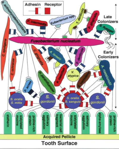
Figure 1 ‐ Model of bacterial colonization showing early and late colonizers and F. nucleatum acting as a bridge between them [12]. ... 8 Figure 2 ‐ Difference between periodontal health and periodontitis. A – Schematic representation of the difference between periodontal health and periodontal disease [101]. B – Difference in the appearance of the mouth of a patient with
healthy teeth and a patient with periodontitis
(http://www.esthetixdentalspa.com/uncategorized/5‐things‐you‐should‐know‐ about‐gum‐disease‐periodontitis/). ... 11 Figure 3 ‐ Combination of factors that lead to periodontitis (adapted from [35]). ... 12 Figure 4 ‐ Schematic representation of the "Ecological Plaque Hypothesis" in relation to gingivitis and periodontitis [18]. ... 13 Figure 5 ‐ Statistically significant values regarding the growth stimulation assays (A) and the biofilm formation assays (B) and the respective color key scale (p<0,05), the reference control was set to a value of 1. Aa: Aggregatibacter actinomycetemcomitans, Fn: Fusobacterium nucleatum, Pg: Porphyromonas gingivalis, Pi: Prevotella intermedia, Smut: Streptococcus mutans, Ssob: Streptococcus sobrinus, Avisc: Actinomyces viscosus, Ssal: Streptococcus salivarius, Smitis: Streptococcus mitis, Ssang: Streptococcus sanguinis, Sgord: Streptococcus gordonii, Soralis: Streptococcus oralis. ... 36 Figure 6 ‐ Decrease/increase of pathogenic species in dual species biofilms in comparison to the no substrate control [%] (p<0.05). Grey area shows non‐ significant data. Aa: Aggregatibacter actinomycetemcomitans, Fn: Fusobacterium nucleatum, Pg: Porphyromonas gingivalis, Pi: Prevotella intermedia, Smut: Streptococcus mutans, Ssob: Streptococcus sobrinus, Avisc: Actinomyces viscosus, Ssal: Streptococcus salivarius, Smitis: Streptococcus mitis, Ssang: Streptococcus sanguinis, Sgord: Streptococcus gordonii, Soralis: Streptococcus oralis. ... 37 Figure 7 – Growth curves for A. actinomycetemcomitans (A), F. nucleatum (B) and P. gingivalis (C) in the presence of different concentrations of L‐arginine. ... 67 Figure 8 ‐ Growth curves for P. intermedia (A), S. mutans (B) and S. sobrinus (C) in the presence of different concentrations of L‐arginine. ... 68 Figure 9 ‐ Growth curves for A. naeslundii (A), S. gordonii (B) and C. sputigena (C) in the presence of different concentrations of L‐arginine. ... 69
Figure 10 ‐ Growth curves for S. salivarius (A), A. viscosus (B) and S. sanguinis (C) in the presence of different concentrations of L‐arginine. ... 70 Figure 11 ‐ Growth curves for S. oralis (A), S. mitis (B) and V. parvulla (C) in the presence of different concentrations of L‐arginine. ... 71 Figure 12 – Biofilm formation for A. actinomycetemcomitans (A), F. nucleatum (B) and P. gingivalis (C) in the presence of different concentrations of L‐arginine. Light grey – 24h incubation; Dark grey – 48h incubation. ... 72 Figure 13 ‐ Biofilm formation for P. intermedia (A), S. mutans (B) and S. sobrinus (C) in the presence of different concentrations of L‐arginine. Light grey – 24h incubation; Dark grey – 48h incubation. ... 73 Figure 14 ‐ Biofilm formation for A. naesludii (A), S. gordonii (B) and C. sputigena (C) in the presence of different concentrations of L‐arginine. Light grey – 24h incubation; Dark grey – 48h incubation. ... 74 Figure 15 – Biofilm formation for S. salivarius (A), A. viscosus (B) and S. sanguinis (C) in the presence of different concentrations of L‐arginine. Light grey – 24h incubation; Dark grey – 48h incubation. ... 75 Figure 16 ‐ Biofilm formation for S. oralis (A), S. mitis (B) and V. parvula (C) in the presence of different concentrations of L‐arginine. Light grey – 24h incubation; Dark grey – 48h incubation. ... 76 Figure 17 – Biofilm percentage when the beneficial species Sgord: Streptococcus gordonii is incubated with the pathogenic species Aa: Aggregatibacter actinomycetemcomitans, Fn: Fusobacterium nucleatum, Pg: Porphyromonas gingivalis, Pi: Prevotella intermedia, Smut: Streptococcus mutans and Ssob: Streptococcus sobrinus. ... 77 Figure 18 ‐ Biofilm percentage when the beneficial species Avisc: Actinomyces viscosus
is incubated with the pathogenic species Aa: Aggregatibacter
actinomycetemcomitans, Fn: Fusobacterium nucleatum, Pg: Porphyromonas gingivalis, Pi: Prevotella intermedia, Smut: Streptococcus mutans and Ssob: Streptococcus sobrinus. ... 78 Figure 19 ‐ Biofilm percentage when the beneficial species Ssal: Streptococcus salivarius
is incubated with the pathogenic species Aa: Aggregatibacter
actinomycetemcomitans, Fn: Fusobacterium nucleatum, Pg: Porphyromonas gingivalis, Pi: Prevotella intermedia, Smut: Streptococcus mutans and Ssob: Streptococcus sobrinus. ... 79
Figure 20 ‐ Biofilm percentage when the beneficial species Ssang: Streptococcus sanguinis is incubated with the pathogenic species Aa: Aggregatibacter actinomycetemcomitans, Fn: Fusobacterium nucleatum, Pg: Porphyromonas gingivalis, Pi: Prevotella intermedia, Smut: Streptococcus mutans and Ssob: Streptococcus sobrinus. ... 80 Figure 21 ‐ Biofilm percentage when the beneficial species Smitis: Streptococcus mitis is
incubated with the pathogenic species Aa: Aggregatibacter
actinomycetemcomitans, Fn: Fusobacterium nucleatum, Pg: Porphyromonas gingivalis, Pi: Prevotella intermedia, Smut: Streptococcus mutans and Ssob: Streptococcus sobrinus. ... 81 Figure 22 ‐ Biofilm percentage when the beneficial species Soralis: Streptococcus oralis
is incubated with the pathogenic species Aa: Aggregatibacter
actinomycetemcomitans, Fn: Fusobacterium nucleatum, Pg: Porphyromonas gingivalis, Pi: Prevotella intermedia, Smut: Streptococcus mutans and Ssob: Streptococcus sobrinus. ... 82
I
NDEX OFT
ABLESTable 1 ‐ Primers/probes used in qPCR ... 28 Table 2 ‐ Bacterial numbers shown as log(CFU/ml) comparing treatment of biofilms with L‐arginine and the control (PBS). Reduction of pathogenic species and increase of beneficial species in comparison to the negative control are highlighted with green background. Bold numbers indicate a ‐1log reduction in comparison to the negative control. Significant reduction of pathogenic species are highlighted by black rectangles (p<0.05). Aa: Aggregatibacter actinomycetemcomitans, Fn: Fusobacterium nucleatum, Pg: Porphyromonas gingivalis, Pi: Prevotella intermedia, Smut: Streptococcus mutans, Ssob: Streptococcus sobrinus, Avisc: Actinomyces viscosus, Ssal: Streptococcus salivarius, Smitis: Streptococcus mitis, Ssang: Streptococcus sanguinis, Sgord: Streptococcus gordonii, Soralis: Streptococcus oralis. ... 38 Table 3 ‐ Bacterial numbers shown as log(CFU/ml) comparing preventive treatment of biofilms with L‐arginine and the control (PBS). Biofilms were grown on the bottom of 24‐well plates (polyethylene) or on hydroxyapatite discs. Reduction of pathogenic species and increase of beneficial species in comparison to the control are highlighted with green background. Significant reduction of pathogenic species or increase of beneficial species are highlighted by black rectangles (p<0.05). Aa: Aggregatibacter actinomycetemcomitans, Fn: Fusobacterium nucleatum, Pg: Porphyromonas gingivalis, Pi: Prevotella intermedia, Smut: Streptococcus mutans, Ssob: Streptococcus sobrinus, Avisc: Actinomyces viscosus, Ssal: Streptococcus salivarius, Smitis: Streptococcus mitis, Ssang: Streptococcus sanguinis, Sgord: Streptococcus gordonii, Soralis: Streptococcus oralis. ... 39 Table 4 ‐ Bacterial numbers shown as log(CFU/ml) comparing preventive, albeit continuous treatment of biofilms with L‐arginine and the control (PBS). Biofilms were grown on the bottom of 24‐well plates (polyethylene) or on hydroxyapatite discs. Reduction of pathogenic species and increase of beneficial species in comparison to the negative control are highlighted with green background. Bold numbers indicate a ‐1log reduction in comparison to the negative control. Significant reduction of pathogenic species are highlighted by black rectangles (p<0.05). Aa: Aggregatibacter actinomycetemcomitans, Fn: Fusobacterium nucleatum, Pg: Porphyromonas gingivalis, Pi: Prevotella intermedia, Smut: Streptococcus mutans, Ssob: Streptococcus sobrinus, Avisc: Actinomyces viscosus, Ssal: Streptococcus salivarius, Smitis: Streptococcus mitis, Ssang: Streptococcus sanguinis, Sgord: Streptococcus gordonii, Soralis: Streptococcus oralis. ... 40
1.1. M
OTIVATION
Periodontitis is an infectious disease that affect millions of people worldwide and, today, oral healthcare professionals are still lacking and effective and durable treatment to fight this disease.
Studies have been showing that the development of this periodontal disease is probably related to oral biofilms and its composition. The presence of pathogenic bacteria, the absence of the so-called beneficial bacteria and the susceptibility of the host seem to be the three main factors that lead to the development of the disease.
As so, modulating oral biofilms towards a more healthy-associated microbiota might be a promising way to treat or even prevent the development of the disease.
Probiotics, also known as “beneficial bacteria”, have been studied in the oral field and have shown promising results in maintaining oral health. Besides probiotics, prebiotics have also been studied, as they are substances that are able to selectively stimulate the growth of beneficial bacteria, and thus, improve host health. However, their effect on the oral cavity has not yet been clarified since studies are still scarce.
Prebiotics might be a promising mechanism to modulate the oral microbiota by selectively stimulating the growth of beneficial bacteria. Moreover, promoting the outgrowth of beneficial bacteria, and as so, preventing the growth of pathogens in the biofilm might be important to prevent the establishment of periodontal diseases.
Therefore, this work was conducted in order to test if the amino-acid L-arginine could act as a prebiotic and trigger the growth of beneficial bacteria in the oral biofilm, thus reducing the proportion of pathogenic bacteria. If L-arginine is able to modulate the oral biofilm, it may be seen as a promising substrate to be used in oral healthcare.
1.2. O
BJECTIVES
The main goal of this project was to selectively trigger the outgrowth of beneficial oral bacteria in complex biofilms by means of nutritional stimulation using the amino-acid L-arginine. To achieve this goal, the project was divided into following parts:
- Evaluating which specific bacteria are stimulated by L-arginine (growth and biofilm formation)
- Evaluating the selective stimulation of beneficial species by L-arginine and subsequent outgrowth over a pathogenic species in dual-species competition assays
- Inducing a shift towards a beneficial-dominated microbial composition in multispecies biofilms by L-arginine treatment
- Evaluating if L-arginine treatment causes a preventive effect in the incorporation of pathogenic species into established beneficial biofilms
C
HAPTER
2
–
L
ITERATURE
R
EVIEW
2.1.
T
HEO
RALC
AVITY AND ITSM
ICROBIOTA
The oral cavity is of major importance in the human body. Although some physicians tend to believe it is resumed to a confined compartment within the human organism, from an anatomic point of view, the oral cavity is connected to the nasopharynx, the larynx, the tonsils, the middle ear and the gastrointestinal tract. As it has such a strong connection with different parts of the human body, it influences general health and it is also influenced by the state of the body [1]. It is known, for example, that inflammations of the mouth seem to weaken the ability of the body to control blood sugar, further complicating diabetes since the inflammation impairs the body’s ability to use insulin. On the other hand, patients with diabetes tend to have a greater amount of pathogenic bacteria, which can lead to periodontal diseases [2]. Besides, epidemiological studies focusing on periodontitis have shown that this disease can increase the risk of systemic infections, coronary heart diseases, myocardial infarction, osteoporosis, among others [3].
The oral microbiota is a biological system of extremely high complexity [4]. More than 700 species have been detected in the human mouth being that over 400 have been identified in the periodontal pocket [5]. Thus, the oral microbiota is at least as complex as the gastrointestinal or vaginal microbiota.
In nature, as well as in the human body, the vast majority of microorganisms are not found in a planktonic state, but attached to surfaces where they form biofilms. Biofilms can be defined as matrix-enclosed bacterial populations that adhere to each other and/or to surfaces or interfaces [6]. The biofilm matrix is produced by the bacteria and is generally composed of water and microbial macromolecules [7]. Different bacterial species specifically attach to different surfaces or co-aggregate with specific partners in the mouth [8,9]. Often one species can co-aggregate with more than one partner and the partners, in their turn, can co-aggregate with other bacteria to form a dense biofilm composed of different species [9]. This strategy allows bacteria to survive and grow in hostiles environments because they are able to protect each other and try to create the perfect conditions for each species to thrive [9]. Bacteria in biofilms exhibit ‘emergent properties’ such as metabolic co-dependence and cooperation, facilitated horizontal gene transfer, and increased resistance to antimicrobial agents which differ from bacteria in the planktonic state [8,10]. These emergent properties may lead to alterations in their gene expression, which results in bacteria having a different phenotype [8]. These changes can make biofilms at least 500 times more resistant to antibacterial agents [6].
In the oral cavity biofilm formation is referred to as dental plaque [11]. Kolenbrander et al. [12] developed a model of bacterial adhesion to tooth surface and co-aggregation among bacterial species. Based on this model, bacteria can be considered as either early or late colonizers (Figure 1). Early colonizers, usually streptococci, bind to salivary receptors. Their ability to bind to other early colonizers and host molecules may confer an advantage in establishing early dental plaque [12]. Fusobacterium nucleatum, however, acts as a bridging organism between early and late colonizers, playing a special role in the development of dental plaque. F. nucleatum is the most numerous gram-negative species in healthy sites, with increasing abundance in oral diseased sites. It has also been noticed that this species is always present whenever Treponema denticola and Porphyromonas gingivalis are also present, which suggests that its presence may be required for the colonization of these two species [13]. Interestingly, F. nucleatum coaggregates with several late colonizers, although no coaggregation among the late colonizers have been observed [12,14,15]. As such, F. nucleatum may play a key role in the establishment of dental plaque and, consequently, the development of dental diseases.
Figure 1 ‐ Model of bacterial colonization showing early and late colonizers and F.
Taken the complexity and important interspecies interactions into account, oral biofilms display difficult therapeutic targets [16]. Further, bacterial diversity is rather subject specific, and prone to changes during the lifetime of each individual [5]. As such, treatment of oral diseases requires a microbial community-based approach instead of targeting one specific oral pathogen, as this oral pathogen may be causing disease in some individuals but not in others [11,17]. Hence, the exact microbial aetiology of oral diseases remains unknown, although a different bacterial community composition was found between health and diseased sites.
In a healthy patient, the resident microflora acts as an important component of the host defences forming a barrier against exogenous and potentially pathogenic populations – preventing their establishment in the mouth – and by regulating the inflammatory host response to oral commensal bacteria [18]. Further, oral commensal bacteria have been shown to maintain healthy oral tissues by influencing the expression of mediators such as intracellular adhesion molecule 1 (ICAM-1), E-selectin and Interleukin 8 (IL-8), and modulating immune responses and enhancing cellular homeostatic mechanisms [19–22].
The resident microbiota also contributes to the normal development of the physiology, nutrition and defence systems of the organism, protecting the host from disease. Thus, beneficial organisms that colonize the oral cavity live in a stable symbiotic balance with the host [23]. Only when this balance is disturbed, an outbreak of disease may occur [18,24]. According to Devine and Marsh [25], a disturbance of the balance occurs “if the host becomes immunocompromised, if the resident microbial populations are suppressed or if microorganisms reach sites they normally do not have access to (e.g. through trauma)”.
2.2. O
RALD
ISEASES
There are several types of diseases that can affect the human cavity, like caries, halitosis, gingivitis, periodontitis, herpes, and oral candidiasis.
Dental caries is one of the most common disease in the oral cavity and one of the most prevalent infectious disease affecting humans worldwide [26]. This disease appears because of disturbances that occur in the bacterial biofilm on the tooth surface, disturbing the ecological balance and leading to loss of tooth mineral [27]. Some bacteria present in the mouth are classified as cariogenic, such as Streptococcus mutans and Streptococcus sobrinus, meaning that they can
induce the formation of caries [28]. These microorganisms are present in the dental plaque and are known to produce acids, due to the periodic ingestion of dietary carbohydrates, that dissolve the mineral matrix of the teeth [26,29]. Bacterial glycolysis leads to the production of acid, which induce repeated cycles of demineralization of the tooth enamel [29]. The demineralization phases are followed by periods of alkalinisation, which restore the integrity of enamel. Caries occur when the acidification phases outweigh the alkalinisation phases, translating in the establishment of a more acidogenic microbiota, which results in lower plaque pH values leading to enhanced enamel demineralization [29]. The production of acids, and consequently demineralization of the enamel is influenced by three factors, the host, the presence of bacteria and nutrients. Only the interplay of all three factors lead to the occurrence of caries [30]. Nowadays, the preventive strategies applied to dental caries focus on targeting the host and dietary factors and removing the dental plaque [31].
Although any disorder of the periodontium – tissue surrounding and supporting the teeth – can be defined as a periodontal disease, this term is usually used when referring to gingivitis and periodontitis, which are inflammatory disorders caused by a pathogenic microflora [32]. Basically, periodontal diseases develop when dental plaque accumulates at the gingival margin triggering an inflammatory response of the host [33]. The increase of plaque mass is followed by an increase of obligatory anaerobic and proteolytic bacteria, many of which are Gram-negative pathogens [25].
Gingivitis is the mildest form of periodontal disease and is reversible when the correct oral hygiene habits are adapted [32]. At this stage, the infection is limited to the gums. The chronic form of the disease results in mild bleeding from the gums during tooth brushing which is not considered a major inconvenience unless the patient suffers from a bleeding disorder [32].
Periodontitis appears when gingivitis is not treated and the disease progresses to a more severe state (Figure 2). The inflammatory process extends deep in to the tissues and leads to the loss of supporting connective tissue and alveolar bone [32]. Soft tissue pockets or deepened crevices are formed between the gingiva and the root of the tooth and can cause pain and discomfort to the patient. The mastication process may also be impaired and eventually the tooth can be lost. Further, severe periodontitis can be associated with periodontal abscesses and halitosis. Moreover, if the biofilm is not thoroughly removed the flow of the gingival crevicular fluid is increased which also introduces molecules such as haemoglobin, haptoglobin and transferrin which select for the proteolytic bacteria that can degrade host molecules that regulates inflammation, resulting in an exaggerated and inappropriate inflammatory response [23]. Loss of
connective tissue attachment and loss of alveolar bone are the two main diagnostic criteria used for the detection of periodontitis [34]. Periodontitis is highly prevalent, affecting billions of people around the world [34].
2.3. P
ERIODONTITIS2.3.1. W
HY DOESP
ERIODONTITIS OCCUR?
To date it is assumed that the occurrence of periodontitis is linked to several factors (Figure 3). There are three main factors that, when combined, lead to disease [35]:
• The presence of pathogenic bacteria • The absence of beneficial bacteria • The susceptibility of the host
A B
Figure 2 ‐ Difference between periodontal health and periodontitis. A – Schematic representation of the difference between periodontal health and periodontal disease [101]. B – Difference in the appearance of the mouth of a patient with healthy teeth and a patient with periodontitis (http://www.esthetixdentalspa.com/uncategorized/5‐things‐you‐should‐know‐about‐gum‐ disease‐periodontitis/).
This is in accordance with the “Ecological Plaque Hypothesis” introduced by Marsh [36], trying to describe and explain the dynamic relationship between the oral microflora and the host in health and disease. In the past, the occurrence of periodontitis was explained by varied hypothesis. First, the “Specific Plaque Hypothesis” was proposed. This hypothesis claimed that some specific pathogenic bacteria are solely responsible for the formation and aggravation of oral diseases [37]. However, the fact that disease sometimes occurred even in the absence of these pathogenic species lead to the rejection of this theory. So, the “Non-specific Plaque Hypothesis” arose. This theory claimed that bacteria present in the oral biofilm produce specific factors and substances and the accumulation of these products are responsible for inflammation and damage to the gingival tissues and lead to oral disease [38]. Here, the quantity of plaque, rather than its composition was considered to be the rationale for oral diseases. However, with the aid of technological advances, it was possible to observe that, in fact, there are some bacteria that are more associated with the occurrence of disease than others. As so, the “Ecological Plaque Hypothesis” arose as a combination of the key points of the previously two mentioned hypotheses. It links the local environment changes with the activity and composition of the biofilm, where any change in the environment induces a response in the microflora (Figure 4) [18].
Figure 4 ‐ Schematic representation of the "Ecological Plaque Hypothesis" in relation to gingivitis and periodontitis (Eh = redox potential) [18]
This hypothesis combines environmental factors of the host with the biofilm itself. The accumulation of plaque might trigger an inflammatory response by the host, which in turn leads to changes in the local environment and induces the production of new proteins which might favour the growth of mostly anaerobic Gram-negative bacteria – the pathogens [18].
The shift that occurs from positive bacteria dominated biofilms to mostly Gram-negative anaerobic biofilms might trigger the occurrence of periodontal diseases [39]. The presence of these periodontopathogens is clearly linked to periodontitis, as microbiota from diseased sites was shown to be markedly different from the one seen in healthy patients [40].
Although a specific microbial aetiology is not known, there are several strains that could be related to the disease and play an important role in its development. When these bacteria are present in the oral cavity of a susceptible host, and abundant in sufficient amounts to cause a shift in the oral microbiota, disease might occur. Each species in the pathogenic biofilm has a different role and some are more involved in the disease and are more virulent than others. For example, it is considered that by the time P. gingivalis or Aggregatibacter actinomycetemcomitans are prevalent, the disease is well established and the site is already affected [25].
The same way some species are defined as pathogens, others are considered to be commensal or beneficial and they are considered to play a positive role in the community. Beneficial/commensal bacteria may have an effect in protecting the host from pathogens. Therefore, some authors have started to focus on the fact that the reduction or absence of these bacteria is another factor for plaque-related periodontal inflammation [12]. Thus, the presence of
these species in the oral cavity might prevent the shift form a health towards a disease associated biofilm composition [1]. Kilian et al [41] listed Streptococcus mitis, Streptococcus oralis, Actinomyces naeslundii, F. nucleatum and some species of Prevotella as being oral commensal/beneficial bacteria.
2.3.3.
P
ATHOGENSI
NVOLVED INP
ERIODONTITIS• Aggregatibacter actinomycetemcomitans
A. actinomycetemcomitans (formerly known as Actinobacillus actinomycetemcomitans) is an exogenous Gram-negative microorganism. It has been considered, for many years, the most likely aetiological agent in aggressive periodontitis (AgP) and has also been implicated in chronic periodontitis and other severe non-oral infections [34,42–44]. The term aggressive periodontitis was suggested in order to differentiate certain rapidly progression forms of periodontitis from the more common form of chronic periodontitis [45]. AgP was formerly known as juvenile periodontitis as it generally affects systemically healthy individuals aged less than 30 years old [46]. Two forms of AgP have been distinguished, based on the number of teeth affected and the distribution of lesions: the localized form (LAgP) and the generalized form (GAgP) [34]. Some scientists believe that the main organism implicated in localized AgP is A. actinomycetemcomitans, colonizing the subgingival plaque biofilm, whereas generalized AgP usually affects more teeth and is caused by P. gingivalis [46]. A. actinomycetemcomitans is present in 90% of localized AgP and in 30 to 50% of severe adult periodontitis [43].
A. actinomycetemcomitans is considered to be an opportunist, with varied virulence factors and mechanisms. Its ability to produce virulence factors leads to serious infections, transmissible among exposed individuals [43]. This bacterium moves from the initial colonization site to the gingival crevices where it competes with other bacteria [34]. The production of leukotoxin is considered a major virulence factor of A. actinomycetemcomitans, since it enables the bacterium to evade the host response by killing leukocytes [47].
A study in West African individuals showed that there was a significantly increased risk of attachment loss among young individuals when A. actinomycetemcomitans was present in the oral cavity compared with the ones who did not harbour this bacterium [48]. Also, several studies on
various adolescent’s populations have shown that the presence of this species in the subgingival plaque increases the risk of attachment loss [34].
Although A. actinomycetemcomitans has been considered by many as the major pathogen in AgP, this is a controversial concept. Different studies showed different results and there is one study that shows no significant association between this bacterium and periodontal diseases, whereas the prevalence and levels of P. gingivalis, T. denticola and Prevotella intermedia were significantly associated with the disease, in descending order [42,49]. This shows that more evidence is needed to fully understand the role of bacteria and their interactions in dental biofilms.
• Porphyromonas gingivalis
P. gingivalis is a black pigmented, non-motile, Gram-negative bacterium that requires anaerobic conditions and that has been associated with periodontitis for a long time [50]. It is considered a natural member of the human microbiota, that under perturbations to the host or microflora can cause pathology [51]. It was found in samples of epithelial cells obtained from periodontal pockets, gingival crevices, and buccal mucosa specimens collected from patients with periodontitis but also from subjects with healthy gingivae [52]. Its metabolic energy is obtained through fermentation of amino acids, which is decisive for survival in deep periodontal pockets, where sugars are extremely scarce [53]. It is the species most associated with the chronic form of periodontitis, leading to chronic inflammation and destruction of the soft and hard tissues that support the teeth and can be detected in up to 85% of diseased sites [54–57]. It is considered a late colonizer of the periodontal biofilm as can be observed in Figure 1.
According to Sakanaka et al, P. gingivalis can elevate the virulence of the periodontal biofilm, even in low numbers, by subverting host responses, altering biofilm community structures and facilitating an increase in overall bacteria load leading to its designation as a keystone pathogen [54]. Inter-bacterial interactions between P. gingivalis and other species of the bacterial community are considered to play a critical role in promoting the pathogenicity of this bacterium in the biofilm [54]. P. gingivalis possesses a great number of virulence factors such as cysteine proteinases, lipopolysaccharide (LPS), capsule and fimbriae, which enable the bacterium to colonise and invade periodontal pockets [57,58].
P. gingivalis invades cells and tissues in the host as a strategy to survive, avoiding, this way, immune surveillance. It can invade gingival epithelial cells, maintaining their viability and replication [53,59,60].
• Fusobacterium nucleatum
F. nucleatum can be associated with a great number of diseases that affect the human body, but on the other hand, can also be isolated from healthy subjects, being the most numerous Gram-negative species present in healthy sites [61]. Thus F. nucleatum can be considered either as an oral commensal or as a periodontal pathogen that is associated with a variety of human diseases. It is one of the first Gram-negative species to become established in plaque biofilms acting as a bridge between early and late colonizers [12]. F. nucleatum is a central species in oral biofilms since it enables the aggregation between Gram-positive and Gram-negative species [12,62].
This bacterium is ubiquitous in the oral cavity and is dominant within the periodontium and dental plaque biofilms and hence, plays an important role in biofilm ecology and infectious diseases in humans. It has been demonstrated that it can be invasive and pro-inflammatory in human oral epithelial cells, eliciting the secretion of IL-8. [39,61–63].
In a study performed with several species to evaluate the adherence and invasion of different bacteria to epithelial cells (using P. gingivalis as a positive control since it is considered a periodontal pathogen), results showed that F. nucleatum was different from the rest of the group of bacterial species in the invasion assays. This organism showed a high capacity to invade cells, with its activity being comparable to P. gingivalis [39].
The presence of this species is affected by environmental factors such as smoking. Smoking increases the number of these bacteria in the oral cavity in both healthy and diseased patients [64].
• Prevotella intermedia
P. intermedia is an obligated anaerobic gram-negative, black-pigmented, rod-shape bacterium that was previously classified in the genus Bacteroides. P. intermedia has long been known to be associated with periodontal disease [65]. It has been implicated as a putative periodontal pathogen due to its isolation from lesions of subjects with early and advanced periodontitis [66]. P. intermedia has also been shown to invade human coronary artery endothelial and smooth muscle cells in vitro [66]. It is the most commonly isolated species of the genus Prevotella from dental plaque [67].
Although P. intermedia is considered less virulent than P. gingivalis, it has several surface properties which may be regarded as potential virulence factors [67].
Information about this pathogen and its role in disease is still limited and more studies need to be conducted in order to fully understand its role in diseases like periodontitis.
• Streptococcus mutans and Streptococcus sobrinus
Oral streptococci are early colonizers of the oral cavity and are abundantly found in dental plaque, considering the oral cavity as their natural habitat [68].
S. mutans and S. sobrinus are the two most important bacteria of the group mutans streptococci, the major pathogenic bacteria in caries process [69]. These bacteria are Gram-positive, catalase-negative, non-motile, aerobic members of the genus Streptococcus. These two species are closely related and they are both pathogenic within humans, enhancing the formation of dental caries, as they produce organic acids that demineralize hard tissues [70,71]. They are considered the two most significant human pathogens regarding dental caries [72].
S. mutans is a major matrix producer and when dietary sucrose and starch are present, it can rapidly modulate the formation of cariogenic biofilms [11,73]. S. mutans thrives in cariogenic plaque because of its ability to tolerate, to grow and to metabolize carbohydrates in an environment with a low pH [74–76]. It is considered a key aetiological agent in dental caries, being predominant in carious lesions microflora [68].
Studies performed on subjects with caries do not always show the same results. While some studies showed that S. sobrinus is more acidogenic and more cariogenic than S. mutans, others did not found this difference [72]. However, it is possible to understand that both species have a great influence in the development of caries and, as such, are considered pathogenic.
2.3.3.
C
URRENTA
PPROACHES TOT
REATP
ERIODONTITIS
In the treatment of periodontitis, initial therapy focuses on reducing dental plaque [77]. It is based on a mechanical approach, namely scaling and root planning (SRP) and eventually can include surgery in order to reduce the depth of the periodontal pocket, with instructions for an improved correct oral hygiene by tooth brushing and interdental cleaning [33,78,79]. Although SPR can reduce the number of pathogens, they rapidly re-colonize the treated sites [80]. The establishment of a more aggressive microbiota occurs within weeks or months [81]. Treatment can be combined with the use of antimicrobials, however the shift of the oral microbiota is only
temporary, since the use of antibiotics have not been proven to improve the long term effect of periodontal therapy [78]. This may be due to the failure of the antimicrobial agent in penetrating the full depth of the biofilm [9].
Like periodontitis, most of the infectious diseases that affect the human population have been treated with antibiotics. However, infectious diseases remain one of the most important health problems affecting the world population nowadays. In fact, hospital infection rates are not declining and multi-drug-resistant bacteria continue to emerge [1]. This widespread use of antibiotics also reflects on the level of resistance in the subgingival microbiota of adults with periodontitis [82]. Also, antibiotics can also lead to undesired side effects such as antibiotic-associated diarrhoea [83].
Taking this into account, there is a great necessity to develop novel non-antibiotic treatment therapies. New emerging concepts for treatment or prevention of periodontal diseases might be the use of probiotics and prebiotics in dental health care [25].
2.4. P
ROBIOTICS
Over the last decades, an increasing interest in the use of probiotics, also known as beneficial bacteria, and their use to modulate the microbiota and maintain health has been reported [1].
Probiotics are defined by the World Health Organization (WHO) as living microorganism that confer a health benefit to the host when administered in sufficient amounts (http://www.who.int/foodsafety/fs_management/en/probiotic_guidelines.pdf). They are referred to as “health-promoting” bacteria and might be used for therapeutic purposes in humans [81].
In order for periodontitis to develop a combination of three factors is needed: a susceptible host, the presence of pathogens and the reduction or absence of beneficial species [35]. Since it is difficult to modulate the host immune response, researchers are focusing on the third aetiological factor, namely restoring the number of beneficial bacteria [1]. Probiotics are seen as a promising strategy to treat periodontitis since they might be able to increase the number of beneficial bacteria in dental plaque, and thus, leading to a decrease of pathogenic bacteria.
Healthcare professionals are focusing more and more on probiotics as their oral intake, to promote general health, has already shown promising effects [81]. For example, probiotics have been shown to reduce mutans streptococci counts in saliva and plaque, which is promising since
they are key aetiological agents in caries [84]. Moreover, studies using Lactobacillus species show significant decreases in gingivitis when compared to the control [83]. Researchers have also observed that a strain of Lactobacillus salivarius named L. salivarius TI 2711 starts to kill P. gingivalis, P. intermedia and P. nigrescens after 6-12h in co-culture in vitro [85]. The same researchers also tested L. salivarius in vivo, letting tablets containing this bacterium to dissolve in subject’s mouth 5 times a day for a period of 8 weeks. Results showed significant reductions of salivary black pigmented anaerobic species for subjects treated with probiotics compared to the control [85]. A study by Krasse et al. [86] using Lactobacillus reuteri in patients with gingivitis showed a significant reduction of gingivitis and plaque compared to the control. Also, a study performed in a beagle dog model showed that beneficial oral bacteria such as Streptococcus sanguinis, Streptococcus salivarius and Streptococcus mitis delayed the recolonization of the oral cavity by periodontal pathogens, reduced inflammation and improved density and bone levels [87]. In addition, positive effects have been shown on the gastrointestinal tract for diarrhoea, inflammatory bowel disease and irritable bowel syndrome, being that probiotics also showed promising effects for atopic diseases and cancer [88].
For now, it is known that probiotics have at least the following modes of action [1,89]: • Modulation of the host defences (innate and acquired immune system) – probiotics can
modulate the immune system towards an anti-inflammatory action, acting on different cells. Host cells such as immune cells and epithelial cells are able to recognize bacteria and their products. Also they can regulate the expression of phagocytosis receptors in the neutrophils and enhance natural killer cell activity [89–92]. They are capable of reducing the production of pro-inflammatory cytokines and increase the production of anti-inflammatory cytokines such as Interleukin 10 (IL-10) and host defence peptides such as beta-defensin 2 [25];
• Production of antimicrobial substances – probiotics can exert a direct effect against pathogens by producing several compounds such as lactic acid, hydrogen peroxide (H2O2), bacteriocins and bacteriocin-like inhibitory substances that act as microbial agents against periodontopathogens and that are capable of inhibiting their growth or kill them [25,83,89];
• Competitive exclusion mechanisms – probiotic can also exert an indirect effect against pathogenic bacteria as they can compete against pathogens for the same nutrients or hamper their adhesion. If beneficial strains are better adapted to the respective niche, the
adhesion of pathogens to the surfaces may be prevented by them, impairing pathogens growth and vitality [13,83]. This indirect effect is very important as the adhesion of pathogens to host surfaces is the first step of infection [33].
However, it is likely that one single probiotic strain does not exhibit all three mechanisms, so probiotic strains are used in combination with each other to increase their beneficial effects [1,93]. It is also likely that these mechanisms vary according to the combinations of probiotics used, the condition that is being treated and its stage and also the presence of prebiotics [94].
Studies concerning probiotics have already shown that certain streptococci, such as S. salivarius, S. mitis and S. sanguinis can attenuate the IL-8 response on epithelial cells induced by pathogens such as F. nucleatum and A. actinomycetemcomitans and that these streptococci are also capable of producing hydrogen peroxide and inhibiting the outgrowth of periodontopathogens [22,95,96].
Teughels et al. [80] showed that the application of probiotics in a beagle dog model after SRP delayed and reduced the recolonization of the site by periodonpathogens, when compared to the control treatment.
Probiotics can be used as a method to prevent oral diseases, supplementing the oral cavity with beneficial bacteria, creating a healthy oral biofilm, preventing pathogens and consequently disease to become established in the mouth [97]. When disease is already established at periodontal sites, probiotics might be used as a method of restoring beneficial bacteria at those sites and shifting the microflora to a healthy state and preventing the growth of pathogens, treating the disease [97].
It is also important to understand that probiotics are more likely to have a durable effect on young individuals since their resident microbiota seems to be less stable and more subject to flux than the microbiota of adults, so perhaps it is in childhood that long-term changes on the oral microbiota can be achieved. In adults, shifting the oral microbiota through probiotics may be difficult as a definitive shift may be hard to accomplish [1,25]. It has been shown that the probiotic effect might disappear if the patient discontinues its use [1].
Besides age of the subject, a careful selection of the probiotic must be performed according to the disease, also the mode and time of administration can be crucial, as well as the general health state of the patient. All of these are determinant factors for the success of probiotics [25].
2.5. P
REBIOTICS
Similar to probiotics, the objective of prebiotic use can be defined as to improve host health through modulation of the microbiota [81].
The definition of prebiotics is adapted to the gastrointestinal tract, since studies of prebiotics have been mainly focused on the gastrointestinal microbiota [25]. Prebiotics are defined as “non digestible food ingredients that beneficially affect the host by selectively stimulating the growth and/or activity of one or a limited number of bacterial species already established in the colon and thus in effect improve host health” [98] or more recently as “selectively fermented ingredients that allows specific changes, both in the composition and/or activity in the gastrointestinal microflora that confers benefits upon host well-being and health” [99]. However, beneficial effects of prebiotics might be extended to other parts of the body, in this case to the oral cavity.
To put simply, prebiotics might be seen as selective triggers to stimulate beneficial bacteria, as they aim to enhance their growth and proliferation [25]. The combination of probiotics and prebiotics is defined as a symbioses, beneficially affecting the host [81].
The main mechanism of action of prebiotics is thought to be indirect since they facilitate the proliferation of beneficial bacteria of the microflora, exerting probiotic effects. However, they may also have direct effects on the host, which include the stimulation of expression of the anti-inflammatory cytokine IL-10 and interferon gamma, the enhancement of secretion of IgA and the modulation of inflammatory responses [25].
Prebiotics might be a promising mechanism to modulate the human resident microbiota without disturbing the health associated homeostasis. However, to date, very few studies on prebiotics in oral health have been conducted.
3.1.
S
TRAINS ANDC
ULTUREC
ONDITIONS
In total 15 bacterial species known from the oral cavity were used as model organisms. Aggregatibacter actinomycetemcomitans ATCC 43718, Fusobacterium nucleatum ATCC 10953, Porphyromonas gingivalis ATCC 33277, Prevotella intermedia ATCC 25611, Streptococcus mutans ATCC 25175 and Streptococcus sobrinus ATCC 33478 were considered as pathogenic species. Commensal, albeit potentially beneficial species included Actinomyces naeslundii ATCC 51655, Capnocytophaga sputigena ATCC 33612, Streptococcus gordonii ATCC 49818, Actinomyces viscosus ATCC 15987, Streptococcus mitis ATCC 49456, Streptococcus oralis clinical isolate, Streptococcus salivarius TOVE-R, Streptococcus sanguinis LMG 14657 and Veillonella parvula DSM 2008.
All bacterial strains were cultured on blood agar plates (Blood Agar Base No 2, Oxoid Ltd, Basingstoke, UK) supplemented with 5 μg/ml hemin (Sigma-Aldrich Co, St. Louis, MO, USA), 1 μg/ml menadione (Sigma-Aldrich Co, St. Louis, MO, USA) and 5% sterile horse blood (Defibrinated Horse Blood – E&O Laboratories Ltd, Burnhouse, Bonnybridge, Scotland). A. actinomycetemcomitans, S. mutans, S. sobrinus, S. gordonii, S. mitis, S. oralis, S. salivarius and S. sanguinis were incubated at 37ºC in a 5% CO2 environment. F. nucleatum, P. gingivalis, P. intermedia, A. naeslundii, C. sputigena, A. viscosus and V. parvula were incubated at 37°C under anaerobic conditions (10% CO2, 10% H2, 80% N2).
3.2.
G
ROWTH ANDB
IOFILMF
ORMATION IN THEP
RESENCE OFL‐
ARGININE
In order to observe if L-arginine promotes the growth and biofilm formation of the different oral bacteria, single-species growth curves and biofilm formation assays were established. Bacteria were collected from the blood agar plates and transferred to 10 ml of brain hearth infusion broth (BHI) (Difco Laboratories, Detroit, MI, USA). The cultures were incubated overnight at 37ºC either under anaerobic conditions or in a 5% CO2 environment. The bacterial concentration of the overnight cultures was adjusted with fresh BHI to 1x10⁷ Colony Forming Units (CFU)/ml by measuring the optical density (OD) at 600 nm.
Stock solutions of L-arginine (Sigma-Aldrich Co, St. Louis, MO, USA) were prepared in demineralized water with concentrations of 250, 200, 100 and 50 μmol/ml and filter sterilized through a 0.2 μm filter (Pall Corporation, Ann Arbor, MI, USA).
In 96-well plates (Greiner Bio-One, Wemmel, Belgium), 200 μl of bacteria were added to either 20 μl of 0.03% chlorhexidine (CHX) (Sigma-Aldrich Co, St. Louis, MO, USA) (negative control), or 20 μl of L-arginine. Also, in each assay there was one well with just 200 μl BHI, without bacteria or substrate (white), and one well with just 200 μl of bacteria (positive control). Final concentrations of L-arginine were 22.73, 18.18, 9.09 and 4.55 μmol/ml. Four technical replicates for each condition were used. The plates were incubated at 37ºC under anaerobic conditions or in a 5% CO2 environment. Experiments were repeated three times at different days.
The growth was monitored by measuring the OD at 630 nm. For aerobic species the measurements were made every hour from 0h to 9h, and at 24h and 48h. For anaerobic species measurements were made at 0h, 24h and 48h.
At 24h and 48h, biofilm formation was monitored. The supernatant was removed and the wells were washed 2 times with 100 μl of sterilized 1 x phosphate buffered saline (PBS). Afterwards, 100 μl of ethanol (EtOH) were added to each well to fix the biofilms for 20 min. EtOH was removed and the plates were dried. Biofilms were stained by adding 100 μl/well of a crystal violet solution (Sigma-Aldrich Co, St. Louis, MO, USA). After 15 min, the plates were rinsed with demineralized water to remove the remaining crystal violet and subsequently left to dry. The bound dye was solubilized by adding 75 μl/well of 5% acetic acid for about 30-45 min. Finally, OD was measured at 630 nm.
3.3.
D
UAL‐S
PECIESC
OMPETITIONA
SSAYS
Overnight cultures of the pathogenic species A. actinomycetemcomitans, F. nucleatum, P. gingivalis, P. intermedia, S. mutans, S. sobrinus and the commensal species S. gordonii, A. viscosus, S. mitis, S. oralis, S. salivarius and S. sanguinis were prepared. Both C. sputigena and V. parvula were not used in this assay since previous experiences performed by the research team shown that these species were not able to grow. Since it is not known if Actinomyces are truly beneficial bacteria or just commensals, only A. viscosus was used in the assay since it showed significant results in the first assay, unlike A. naeslundii. Bacteria were collected from the blood
agar plates and transferred to 10 ml of BHI. The cultures were incubated overnight at 37ºC either under anaerobic conditions or in a 5% CO2 environment and concentration was adjusted to 1x10⁷ CFU/ml.
A mixture of 1 ml of the beneficial culture and 1 ml of the pathogenic culture were added to 24-well plates (Greiner Bio-One, Wemmel, Belgium). 200 μl of L-arginine (250 μmol/ml) or BHI as a negative control were added to the cultures and the plates were incubated for 24 h under anaerobic conditions at 37ºC.
Biofilms at the bottom of the plates were gently washed with 1xPBS to detach the non-adhered cells and were disrupted by trypsinization (Trypsin-EDTA (0.05%), Life Technologies, Paisley, UK) for 15 min under anaerobic conditions at 37ºC. Bacterial cells were harvested by centrifugation (5 min, 6,000 x g) and were resuspended in 400 μl of 1xPBS. Aliquots of biofilm samples were immediately incubated with propidium monoazide (PMA) (Biotium, Hayward, CA) at a final concentration of 100 μg/ml as described by Loozen et al [100]. Briefly, this process consisted of adding 10 μl of PMA to 90 μl of each sample. The samples were incubated for 5 min in the dark, and subsequently exposed for 10 min to a 400 W halogen light source placed 20 cm above the samples to induce the cross-linking of PMA. During this step samples were kept on ice to avoid excess heating. Samples were centrifuged at 20,000 x g for 10 min and DNA extraction was performed using the QIAamp DNA Mini kit (Qiagen, Hilden, Germany) in accordance to the manufacturer’s instructions.
A quantitative PCR (qPCR) assay was performed using a CFX96 Real-Time System (Biorad, Hercules, CA, USA). The Taqman 5’ nuclease assay PCR method was used for detection and quantification of bacterial DNA. Specific primers and Taqman probes for each species are shown in Table 1. Taqman reaction contained 12.5 μl Mastermix (Eurogentec, Seraing, Belgium), 4.5 μl sterile distilled water, 1 μl of each primer and probe and 5 μl template DNA. Assay conditions for all primer/probe sets consisted of an initial 2 min at 50º, followed by a denaturation step for 10 min at 95ºC, followed by a denaturation step for 10 min at 95ºC, followed by 45 cycles of 95ºC for 15 sec and 60ºC for 60 sec. Quantification was based on a plasmid standard curve.
Table 1 ‐ Primers/probes used in qPCR
STRAIN Primer/Probe (5’‐3’) Fragment
length Aggregatibacter actinomycetemcomitans Forward Reverse Probe GAA CCT TAC CTA CTC TTG ACA TCC GAA TGC AGC ACC TGT CTC AAA GC AGA ACT CAG AGA TGG GTT TGT GCC TTA GGG 80 bp Fusobacterium nucleatum Forward Reverse Probe GGA TTT ATT GGG CGT AAA GC GGC ATT CCT ACA AAT ATC TAC GAA CTC TAC ACT TGT AGT TCC G 162 bp Porphyromonas gingivalis Forward Reverse Probe GCG CTC AAC GTT CAG CC CAC GAA TTC CGC CTG C CAC TGA ACT CAA GCC CGG CAG TTT CAA 68 bp Prevotella intermedia Forward Reverse Probe CGG TCT GTT AAG CGT GTT GTG CAC CAT GAA TTC CGC ATA CG TGG CGG ACT TGA GTG CAC GC 99 bp Streptococcus mutans Forward Reverse Probe GCC TAC AGC TCA GAG ATG CTA TTC T GCC ATA CAC CAC TCA TGA ATT GA TGG AAA TGA CGG TCG CCG TTA TGA A 114 bp Streptococcus sobrinus Forward Reverse Probe TTC AAA GCC AAG ACC AAG CTA GT CCA GCC TGA GAT TCA GCT TGT CCT GCT CCA GCG ACA AAG GCA GC 88 bp Actinomyces naeslundii Forward Reverse Probe TCG AAA CTC AGC AAG TAG CCG AGA GGA GGG CCA CAA AAG AAA GGG TAC TCT AGT CCA AAC TGG CGG ATA GCG 96 bp Streptococcus gordonii Forward Reverse Probe CGG ATG ATG CTA ATC AAG TGA CC GTT AGC TGT TGG ATT GGT TGC C AGA ACA GTC CGC TGT TCA GAG CAA 177 bp Actinomyces viscosus Forward Reverse Probe GTG AAG GAG CCA GCT TGC TGG TTC TG CGG AAC AAA CCT TTC CCA GGC ATG AGT GGC GAA CGG GTG AGT AAC 155 bp Streptococcus salivarius Forward Reverse Probe AAC GTT GAC CTT ACG CTA GC ACC GTA ACG TGG GAA AAC TG GTA GCG TCA GAG TGG TTG AC 192 bp Streptococcus oralis Forward Reverse Probe ACC AGC AGA TAC GAA AGA AGC AT AGG TTC GGG CAA GCG ATC TTT CT AAG GCT GCT GTT GCT GAA GAA GT 229 bp Streptococcus mitis Forward Reverse Probe GGC TCG TAG TCT GGA GAT GG TAG GTC GTC GTC CCA AGG AA CGA AGA GCA CCA ATA GCA CCT CCC 133 bp Streptococcus sanguinis Forward Reverse Probe CAA AAT TGT TGC AAA TCC AAA GG GCT ATC GCT CCC TGT CTT TGA AAA GAA AGA TCG CTT GCC AGA ACC GG 75 bp Veillonella parvula Forward Reverse Probe GAC GAA AGT CTG ACG GAG CA TGC CAC CTA CGT ATT ACC GC AGC TCT GTT AAT CGG GAC GAA AGG C 171 bp
3.4.
M
ULTI‐S
PECIESA
SSAYS
3.4.1. S
ET‐UP BIOREACTOR
To establish multi-species communities, a Biostat B Twin 1L bioreactor (Sartorius Stedim Biotech Gmbh, Goettingen, Germany) was used. Each of the culture vessels contained 750 ml BHI (37g/L) (Difco Laboratories, Detroit, MI, USA) supplemented with 2.5 g/L mucin from porcine stomach type III (Sigma-Aldrich Co, St. Louis, MO, USA), 1.0 g/L yeast extract (Oxoid Ltd, Basingstoke, Hampshire, England), 0.1 g/L L-cysteine (Calbiochem, San Diego, CA, USA), 2.0 g/L sodium bicarbonate, 5 mg/L hemin, 1 mg/L menadione and 0.25% (w/v) glutamic acid (all Sigma-Aldrich Co, St. Louis, MO, USA) (from now on referred as BHI 2). Growth conditions were set to 37°C, 0% O2, 5% CO2, pH 6.7 ± 0.1, stirring at 300 rpm.
The chemostat culture was prepared, containing 14 bacterial species including beneficial as well as pathogenic species. Overnight cultures of the pathogenic species A. actinomycetemcomitans, F. nucleatum, P. gingivalis, P. intermedia, S. mutans, S. sobrinus and the commensal/beneficial species A. naeslundii, S. gordonii, A. viscosus, S. mitis, S. oralis, S. salivarius, S. sanguinis and V. parvula were prepared. C. sputigena was not used for the reason described under 3.3. First, S. mitis was inoculated into the chemostat and grown until late exponential phase. The OD600nm of each of the remaining species was adjusted to 1.4 and a volume of 750 μl of each species was inoculated to the chemostat culture. To stabilize the composition of the community the culture was kept undiluted for 48h and subsequently kept in continuous culture with an exchange of medium of 200 ml within 24h.
3.4.2. B
IOFILMT
REATMENT WITHL‐
ARGININE
A sample of the chemostat culture was taken and diluted 1:10 in fresh BHI 2 medium. Respectively, 2 ml of the culture were added to two wells of a 24-well plate. The plates were incubated for 24h under anaerobic conditions at 37ºC.
![Figure 2 ‐ Difference between periodontal health and periodontitis. A – Schematic representation of the difference between periodontal health and periodontal disease [101]. B – Difference in the appearance of the mouth of a patient with healthy teet](https://thumb-eu.123doks.com/thumbv2/123dok_br/17570130.817991/28.892.181.761.262.525/difference-periodontal-periodontitis-schematic-representation-periodontal-periodontal-difference.webp)
![Figure 3 ‐ Combination of factors that lead to periodontitis (adapted from [35]).](https://thumb-eu.123doks.com/thumbv2/123dok_br/17570130.817991/29.892.161.734.114.354/figure-combination-factors-that-lead-periodontitis-adapted-from.webp)
![Figure 4 ‐ Schematic representation of the "Ecological Plaque Hypothesis" in relation to gingivitis and periodontitis (Eh = redox potential) [18]](https://thumb-eu.123doks.com/thumbv2/123dok_br/17570130.817991/30.892.152.749.116.404/schematic-representation-ecological-hypothesis-relation-gingivitis-periodontitis-potential.webp)


![Figure 6 ‐ Decrease/increase of pathogenic species in dual species biofilms in comparison to the no substrate control [%] (p<0.05). Grey area shows non‐significant data. Aa: Aggregatibacter actinomycetemcomitans, Fn:](https://thumb-eu.123doks.com/thumbv2/123dok_br/17570130.817991/54.892.198.706.110.492/decrease-increase-pathogenic-comparison-substrate-significant-aggregatibacter-actinomycetemcomitans.webp)


