DEPARTAMENTO DE QU´IMICA E BIOQU´IMICA
Application of QM and MM methodologies
to cytochrome c
3: Charge parametrization of the heme
group for classic force fields
Jo˜ao Manuel Almeida Henriques
Mestrado em Bioqu´ımica
DEPARTAMENTO DE QU´IMICA E BIOQU´IMICA
Application of QM and MM methodologies
to cytochrome c
3: Charge parametrization of the heme
group for classic force fields
Jo˜ao Manuel Almeida Henriques
Tese orientada por Doutor Miguel Machuqueiro
Mestrado em Bioqu´ımica
In 2009, Machuqueiro and Baptista used Desulfovibrio vulgaris Hildenborough type I cy-tochrome c3 in the validation of their new implementation of the stochastic titration method,
the constant-(pH,E) MD method. Dv H-TpI-c3 is a small globular and monomeric tetraheme
protein present in the periplasm of the sulfate reducing bacteria Dv H. It is constituted by 107 residues plus four hemes covalently bound to cysteines in the polypeptide chain together with bis-histidinyl axial ligation. Therefore, it constitutes a tight densely packed structure where the heme reduction potentials are very close, making it a very demanding test case in terms of prediction. When tested, this method was shown to yield better results when a high dielectric constant, ε, was assigned to the protein region. This dependence on ε was never found in previous constant-pH MD simulations and poor heme charge parametrization may have been responsible for this effect.
Hence, the main goal of this work was to refine the QM parametrization of the redox centers, i.e. obtain new and more accurate charge sets, incorporate them in the new 53A6 GROMOS force field, and finally run constant-(pH,E) MD simulations with a ε of 2. Single and multi-conformational approaches were tested using the RESP scheme. Merz-Kollman and CHELPG methods were also used.
All tested charge sets and respective charge parametrization methods were found to be valid, and there is not enough margin to unequivocally select one method as the absolute best. Still, the RESP fitting procedure is much more versatile than CHELPG and Merz-Kollman, making it a better suited tool for a generalized derivation of partial atomic charges. When applied to the constant-(pH,E) MD method, these new and more accurate charge sets, enabled us to improve Machuqueiro and Baptista’s results. We were, however, unsuccessful in fully repro-ducing all experimental data. Theoretical and experimental results systematically disagreed when predicting the Ehalf values for hemes III and IV. The reason behind this incongruity is something we plan to investigate in future work.
Keywords: cytochrome c3, heme group, charge parametrization, redox titration,
constant-(pH,E) MD
O citocromo c3 tipo I de Desulfovibrio vulgaris Hildenborough (Dv H-TpI-c3) ´e uma pequena
prote´ına globular e monom´erica, que se encontra presente no periplasma das bact´erias reduc-toras de sulfato Desulfovibrio vulgaris Hildenborough (Dv H). ´E constitu´ıdo por 107 red´ıduos de amino´acido e quatro grupos hemo, ligados covalentemente a ciste´ınas da cadeia polipept´ıdica. Para al´em disto, os hemos encontram-se ainda coordenados axialmente a dois res´ıduos de histidina.
Factores como o tamanho reduzido, a estabilidade e a elevada solubilidade desta prote´ına, fizeram com que ela pudesse ser caracterizada extensivamente por uma larga gama de t´ecnicas. Entre estas t´ecnicas contam-se a espectroscopia de RMN, que contribuiu largamente para o seu conhecimento estrutural e termodinˆamico; e a espectroscopia de raios-X, que permitiu obter estruturas cristalogr´aficas bastante detalhadas da prote´ına em quest˜ao, tornando-a na prote´ına multi-h´emica mais bem caracterizada de todas.
Todos os estudos anteriormente mencionados, mostram que os quatro hemos do Dv H-TpI-c3 se
encontram muito pr´oximos uns dos outros, exibindo, por isso, um forte acoplamento entre si e com grupos ´acido/base na vizinhan¸ca. Como tal, n˜ao ´e surpreendente verificar que o potencial de redu¸c˜ao de cada hemo ´e dependente do estado de oxida¸c˜ao dos outros trˆes hemos (potencial de interac¸c˜ao redox ou cooperatividade homotr´opica), e para al´em disto, depende ainda do pH (efeito redox-Bohr ou cooperatividade heterotr´opica). Estes fen´omenos tˆem vindo a ser extensivamente estudados atrav´es de m´etodos experimentais e te´oricos, tendo consitu´ıdo uma ´
area de investiga¸c˜ao priorit´aria no Instituto de Tecnologia Qu´ımica e Biol´ogica (ITQB), sob a al¸cada do Professor Ant´onio V. Xavier.
O efeito redox-Bohr pode ser descrito como uma forte dependˆencia dos potenciais de redu¸c˜ao dos grupos hemo com o pH, o que em termos termodinˆamicos, corresponde a uma dependˆencia dos potenciais de redu¸c˜ao de centros redox com o estado de inoniza¸c˜ao de centros proton´aveis, atrav´es de interac¸c˜oes electroest´aticas. Isto ´e tido como sendo de especial importˆancia biol´ogica em citocromos c3, uma vez que ocorre a valores fisiol´ogicos de pH. Deste modo, pensa-se que este
efeito seja fulcral `a fun¸c˜ao deste tipo de prote´ınas, e em especial ao Dv H-TpI-c3, uma vez que
a aquisi¸c˜ao concertada de electr˜oes e prot˜oes na mesma mol´ecula, constitui um meio fisiol´ogico relevante de transferˆencia dos electr˜oes e prot˜oes gerados pela hidrogenase, ap´os cat´alise da oxida¸c˜ao revers´ıvel do hidrog´enio molecular. De um modo termodinˆamico, a aquisi¸c˜ao de electr˜oes, gerados pela hidrogenase, por parte do Dv H-TpI-c3, leva a uma mudan¸ca do estado
redox dos grupos hemo, e consequentemente `a altera¸c˜ao da sua electrost´atica. Esta mudan¸ca tem efeito em grupos ´acido/base nas redondezas, que por sua vez sofrem altera¸c˜oes no seu pKa, tornando-se mais receptivos a eventos de (des)protona¸c˜ao.
Em 2009, Machuqueiro e Baptista utilizaram esta prote´ına como meio de valida¸c˜ao da sua nova implementa¸c˜ao do m´etodo estoc´astico de titula¸c˜ao, o m´etodo de dinˆamica molecular a pH e potencial de redu¸c˜ao, E, constantes. Como o pr´oprio nome indica, este m´etodo possibilita a realiza¸c˜ao de simula¸c˜oes de dinˆamica molecular a pH e E constantes, e com isto permite a amostragem conjunta de conforma¸c˜ao proteica, protona¸c˜ao, e estados redox. Isto torna o m´etodo em quest˜ao ´unico, uma vez que n˜ao se conhecem outras abordagens com caracter´ısticas semelhantes. Tendo em considera¸c˜ao as particularidades da prote´ına em quest˜ao, a escolha desta para testar a performance deste novo m´etodo torna-se bastante intuitiva. Se a isto se acrescentar o facto que os potenciais de redu¸c˜ao de cada hemo s˜ao bastante pr´oximos, i.e. dentro de um intervalo de 80 mV, ou 1.3 unidades de pH; e que o erro associado `a determina¸c˜ao de pKa do m´etodo de titula¸c˜ao estoc´astico ´e da ordem das 0.8 unidades de pH, tem-se um
teste bastante exigente `as capacidades do m´etodo de dinˆamica molecular a pH e E constantes. Embora tenham obtido resultados encorajadores, os autores do m´etodo em quest˜ao n˜ao pu-deram deixar de reparar num resultado em particular. Foi verificado que a qualidade dos resul-tados obtidos com esta metodologia se encontrava dependente do valor da constante diel´ectrica, ε, atribuida `a prote´ına. Isto ´e algo que nunca tinha sido verificado anteriormente, em qualquer um dos estudos efectuados com o m´etodo de dinˆamica molecular a pH constante. Na realidade, este tipo de resultado ´e algo que seria esperado caso se estivesse na presen¸ca de um m´etodo de estrutura r´ıgida de Poisson-Boltzmann/Monte Carlo, onde toda a reorganiza¸c˜ao estrutural ´e assegurada pela constante diel´ectrica. Como tal, pensa-se que na origem do problema anteri-ormente mencionado, estejam eventos n˜ao estruturais, como por exemplo, a redistribui¸c˜ao das cargas ap´os oxida¸c˜ao/redu¸c˜ao dos grupos hemo.
Posto isto, o principal objectivo deste trabalho prende-se com a resolu¸c˜ao do problema an-teriormente formulado, atrav´es da reformula¸c˜ao/refinamento da parametriza¸c˜ao quˆantica dos centros redox (hemos), de modo a obter novos grupos de cargas mais precisos.
Primeiramente, decidiu-se efectuar toda uma revis˜ao ao composto modelo representativo dos grupos hemo de Dv H-TpI-c3. Isto deve-se ao facto de se pretender introduzir mais detalhe
ao modelo original. Para tal testou-se a inclus˜ao de propionatos desprotonados, uma vez que o pKa dos propionatos ´e inferior ao pH fisiol´ogico a ser simulado. No entanto, estudos
quˆanticos preliminares do novo modelo, revelaram que este n˜ao possui validade qu´ımica a um n´ıvel electr´onico, como se provou atrav´es da an´alise das orbitais moleculares de valˆencia e da densidade de spin electr´onico, realizada com a prote´ına em v´acuo. Na sequˆencia destes resultados, proposeram-se modelos e metodologias alternativas, cujo teste n˜ao foi poss´ıvel, dado o car´acter temporal limitado do trabalho aqui apresentado. Assim sendo, foi decido manter o mesmo modelo utilizado em estudos anteriores. No entanto, antes de proceder a qualquer outro tipo estudo, provou-se pela primeira vez a validade deste modelo a um n´ıvel quˆantico. Uma vez definido o modelo, procedeu-se ao design racional de toda a metodologia a empregar, de modo a obter novos conjuntos de cargas para o modelo de hemo de Dv H-TpI-c3. Embora
os quatro hemos da prote´ına em quest˜ao sejam todos do mesmo tipo, i.e. estruturalmente idˆenticos, estes apresentam uma variabilidade conformacional significativa entre si, o que ´e mais que suficiente para originar cargas at´omicas parciais diferentes para cada um dos grupos hemo. Como tal, foi dada como prioridade, a obten¸c˜ao de cargas atrav´es de m´etodos multi-conformacionais, como ´e o caso do RESP. No entanto, porque as cargas determinadas por Oliveira et al. em 2005, e utilizadas por Machuqueiro e Baptista em 2009, s˜ao obtidas tendo em conta uma ´unica conforma¸c˜ao, a do hemo I, decidiu-se explorar igualmente m´etodos uni-conformacionais como o CHELPG e o Merz-Kollman. O m´etodo de obten¸c˜ao de cargas RESP foi igualmente utilizado numa abordagem uni-conformacional, `a semelhan¸ca do que foi feito anteriormente. Decidiu-se manter o hemo I como sendo a estrutura a utilizar nas abordagens uni-conformacionais, dado o facto de ser o hemo cujo RMSD cruzado ´e menor, quando se compara a sua estrutura contra as outras trˆes.
De um modo geral, foram testadas seis tipos de abordagens. RESP multi-conformacional das estruturas de raios-X dos quatro hemos presentes na estrutura cristalogr´afica; RESP multi-conformacional das estruturas dos quatro hemos ap´os optimiza¸c˜ao de geometria livre; RESP multi-conformacional das estruturas dos quatro hemos ap´os optimiza¸c˜ao de geometria fixa, em que quatro ´atomos chave de cada hemo foram restringidos `a sua posi¸c˜ao original da estrutura de raios-X; CHELPG, Merz-Kollman e RESP atendendo apenas `a estrutura cristalogr´afica do hemo I.
A an´alise comparativa dos seis conjuntos de cargas obtidos, revelou que o m´etodo CHELPG originou as cargas mais diferentes de todas, o que n˜ao ´e de estranhar face `as diferen¸cas si-gnificativas no algoritmo de amostragem utilizado neste m´etodo, em rela¸c˜ao ao Merz-Kollman, e em ´ultima an´alise, ao RESP, que ´e uma extens˜ao do Merz-Kollman. Para al´em disto, as abordagens multi-conformacionais originam resultados diferentes dos obtidos com abordagens uni-conformacionais. Isto ´e, mais uma vez, algo que seria esperado uma vez que conforma¸c˜oes diferentes, tˆem, em princ´ıpio, potˆenciais electrost´aticos diferentes, e consequentemente dife-rentes cargas at´omicas parciais. Em suma, e num compto geral, os resultados obtidos em cada um dos conjuntos de cargas, revelaram ser bastante concordantes com previs˜oes baseadas na
intui¸c˜ao qu´ımica.
Outra descoberta interessante, prende-se com o facto de os ´atomos mais perto do centro de coordena¸c˜ao apresentarem as maiores varia¸c˜oes de cargas entre os seis conjuntos testados. Este resultado ´e encarado como sendo relevante, na medida em que estes ´atomos est˜ao directamente envolvidos nos eventos de oxida¸c˜ao/redu¸c˜ao que ocorrem em cada ´atomo de ferro pertencente ao centro de coordena¸c˜ao. A sensibilidade electrost´atica destes ´atomos corrobora a delicadeza do sistema aqui estudado. Para al´em disso, real¸ca a importˆancia de se realizarem diversas parametriza¸c˜oes de cargas deste modelo.
Tendo determinado seis conjuntos de cargas adequados ao modelo em quest˜ao, procedeu-se ao teste dos mesmos com o m´etodo de dinˆamica molecular a pH e E constantes. Foram realizados triplicados de cada conjunto de cargas a oito potenciais de redu¸c˜ao diferentes. Utilizou-se, ainda, um novo campo de for¸cas, o GROMOS 53A6. Em suma, foram realizadas 144 si-mula¸c˜oes, com 10 ns cada, obtendo-se um total de 1.44 µs de simula¸c˜ao de dinˆamica molecular a pH e E constantes.
Cada uma das simula¸c˜oes previamente mencionadas, foi analisada atrav´es do seu RMSD ao longo do tempo e da frac¸c˜ao de redu¸c˜ao para cada um dos quatro grupos hemo. De um modo geral, todas as simula¸c˜oes, independentemente do conjunto de cargas utilizado, mostraram encontrar-se equilibradas `a passagem do quinto nanosegundo de simula¸c˜ao. Para al´em disto, ficou demonstrado que todos os hemos se encontram efectivamente a titular durante toda a simula¸c˜ao.
Quanto `a avalia¸c˜ao da estabilidade proteica, efectuaram-se analises de raio de gira¸c˜ao, per´ımetro h´emico, fluctua¸c˜oes (RMSF), e de estrutura secund´aria por meio de DSSP. Destas an´alises, conclui-se que a grande maioria das simula¸c˜oes ´e, de facto, est´avel. Foram, no entanto, encon-trados casos pontuais em que a prote´ına demonstra um certo grau de instabilidade. Na origem desta instabilidade est˜ao os elevados valores de RMSF de res´ıduos de amino´acido envolvidos em motivos de estrutura secund´aria como “hairpins”, “turns” e h´elices. A variabilidade asso-ciada a “hairpins” e “turns” ´e tida como sendo uma consequˆencia da adapta¸c˜ao da prote´ına ao meio aquoso, sem grande relevˆancia na instabilidade proteica. A instabilidade dos motivos em h´elice, por sua vez, pode ser atribu´ıda ao pr´oprio campo de for¸cas usado, que foi indiciado como sendo respons´avel pela desestabiliza¸c˜ao de h´elices em p´eptidos e prote´ınas. Ainda assim, estes casos pontuais de instabilidade foram considerados insignificantes quando comparados com a maioria das simula¸c˜oes, levando a concluir que n˜ao afectam, de modo algum, a vali-dade das simula¸c˜oes. Para al´em disto, nas simula¸c˜oes em que se verificou haver algum ind´ıcio de instabilidade proteica, ficou provado que os hemos se encontram a titular normalmente, atendendo aos padr˜oes estabelecidos.
uma enorme consistˆencia entre si, considerando quer a sua elonga¸c˜ao, quer o seu declive. Mais ainda, as curvas revelam que as titula¸c˜oes est˜ao compreendidas num intervalo de potencial de redu¸c˜ao mais plaus´ıvel que o obtido previamente por Machuqueiro e Baptista, em 2009. Ap´os ajuste de cada curva ao modelo experimental, adaptado de Turner et al. (1996), obtiveram-se resultados bastante satisfact´orios, tendo em conta alguma falta de amostragem.
Os valores de Emod, Ehalf e RMSD, determinados para cada conjunto de cargas, revelaram, uma vez mais, grande consistˆencia entre si. Esta consistˆencia entre conjuntos de cargas, leva-nos a concluir que o m´etodo de titula¸c˜ao estoc´astica, de um modo geral, mostra ser bastante robusto a pequenas diferen¸cas nos parˆametros de carga at´omica. Num plano individual, os va-lores de Emodaqui obtidos mostram ser bastante discordantes em rela¸c˜ao ao valor apresentado anteriormente, no trabalho realizado por Machuqueiro e Baptista. A not´oria desloca¸c˜ao dos Emod para valores mais negativos que o Ehalf (-220 mV) apresentado por Harbury et al., em 1965, para um derivado octapept´ıdico de coordena¸c˜ao bis-histidinil, ´e algo que seria esperado `
a partida, e constitui uma melhoria relativamente ao trabalho anterior. Tendo em conta os Ehalf obtidos neste trabalho, torna-se not´orio que os hemos I e II se encontram a titular a potenciais de redu¸c˜ao bastante pr´oximos. No entanto, os hemos III e IV mostraram um enorme divergˆencia quanto aos seus valores de Ehalf. O facto de se encontrarem a titular a potenciais de redu¸c˜ao t˜ao distantes reflecte-se no intervalo m´aximo de Ehalf.
Quanto `as ordens de redu¸c˜ao dos hemos, obtidos atrav´es da organiza¸c˜ao dos Ehalf de modo crescente, nota-se que, `a excep¸c˜ao dos conjuntos de cargas obtidos pelos m´etodos RESP MC Fixo e CHELPG, foi poss´ıvel prever a ordem de redu¸c˜ao correcta dos hemos I e II, entre si. Os hemos III e IV encontram-se sempre desordenados, algo que n˜ao ´e novidade atendendo ao estudo anterior. No entanto, a natureza sistem´atica destes resultados ´e algo que n˜ao pode ser ignorada. Face a estes resultados, decidiu efectuar-se um pequeno teste, que consistiu em trocar os Ehalf obtidos experimentalmente para os hemos III e IV, e posteriormente recalcular
todos os RMSD para cada conjunto de cargas em rela¸c˜ao ao dados experimentais alterados. Os resultados deste simples teste foram surpreendentes, na medida em que, se obtiveram descidas concertadas dos RMSD para valores baix´ıssimos, reflectindo uma precis˜ao incr´ıvel. Para al´em disto, se se considerar que a ordem experimental dos hemos III e IV est´a trocada, ´e poss´ıvel prever a ordem completa de redu¸c˜ao dos quatro hemos para a maioria dos conjuntos de cargas. Este resultado, absolutamente in´edito, ´e algo que deve ser investigado com maior detalhe em estudos posteriores.
O acoplamento entre grupos ´acido/base e centros redox foi estudado atrav´es do c´alculo das suas correla¸c˜oes. Num compto geral, os resultados obtidos mostraram ser parcialmente concor-dantes com estudos anteriores de estrutura r´ıgida de Poisson-Boltzmann/Monte Carlo e com os resultados apresentados ulteriormente, em 2009. No entanto, foi poss´ıvel capturar uma maior quantidade de interac¸c˜oes directas e, especialmente, indirectas, entre hemos, mediadas pelo
res´ıduo de histidina 67. Este tipo de observa¸c˜ao corrobora a precis˜ao do modelo electrost´atico utilizado. Contudo, n˜ao foi poss´ıvel determinar qualquer tipo de cooperatividade positiva entre os hemos I e II, `a semelhan¸ca do que fora reportado experimentalmente. No entanto, e por mais que uma vez, tem sido argumentado que esta cooperatividade possa ser um artefacto do procedimento de ajuste dos dados experimentais.
Tamb´em ficou demonstrado que o Dv H-TpI-c3, quando parcialmente reduzido, pode acomodar
um n´umero de electr˜oes titul´aveis acima ou abaixo do seu valor m´edio, o que se pensa ser fulcral `
a fun¸c˜ao desta prote´ına em condi¸c˜oes de n˜ao equil´ıbrio.
Em suma, neste trabalho, todos os conjuntos de cargas utilizados e respectivos m´etodos de parametriza¸c˜ao s˜ao v´alidos. Tendo em conta os resultados obtidos de um modo geral, n˜ao ´e poss´ıvel afirmar com certeza absoluta qual dos m´etodos ´e o melhor. Contudo, o m´etodo de ajuste restrito ao potencial electrost´atico (RESP), pela sua versatilidade, parece ser a ferra-menta mais indicada para a deriva¸c˜ao generalizada de cargas at´omicas parciais. Para al´em disto, ficou claro, que as defini¸c˜oes e parˆametros utilizadas nos estudos de mecˆanica quˆantica, efectuados neste trabalho, s˜ao bastante apropriados para o modelo em quest˜ao, visto terem apresentado um bom compromisso entre precis˜ao, velocidade e transferibilidade. A aplica¸c˜ao de novos, e mais detalhados, conjuntos de cargas ao m´etodo de dinˆamica molecular a pH e E constantes, permitiu obter melhorias significativas aos resultados previamente publicados por Machuqueiro e Baptista. Mesmo assim, n˜ao foi poss´ıvel reproduzir todos os resultados experimentais, verificando-se uma discordˆancia sistem´atica aquando da previs˜ao dos valores de Ehalf para os hemos III e IV. Esta incongruˆencia ´e algo para a qual est´a planeada uma investiga¸c˜ao em detalhe num futuro pr´oximo. Uma primeira abordagem passar´a, certamente, pela revis˜ao exaustiva dos trabalhos pioneiros de Turner et al. (1992-96).
Palavras-chave: citocromo c3, hemo, parametriza¸c˜ao de cargas, titula¸c˜ao redox, dinˆamica
First of all I would like to stress that the acknowledgments made here come in protocolar order. No secondary interpretations should be attempted.
My first acknowledgments go to Dr. Miguel Machuqueiro, Prof. Maria Jos´e Calhorda, Dr. Paulo Costa and Diogo Vila Vi¸cosa. The reason why I put them all together is very simple: for the past year I have spent most of my time surrounded by these people. I even dare to say that I have certainly spent more than twice as much time with them than with my family. Therefore, they were like a second family to me, an academic family. Each one played a very special and unique role in this family.
Hence, I’m deeply thankful to Miguel and Prof. Calhorda for accepting me as a collaborator of the Theoretical and Inorganic Chemistry Group at FCUL. On an individual note, I would like to thank Miguel for taking me under his wing and always being there whenever I needed him. I could have not asked for a better supervisor, either at an academic, professional or personal level. With all do respect, Miguel is what we usually call “a good guy”, or in Portuguese “um tipo impec´avel”.
Another person which had a profound impact in my formation during this year is Paulo. His comprehensive knowledge of quantum mechanics and computational chemistry was precious to me, and I am 100% sure I would not have gone this far without his contribution. If I were religious, I would be tempted to say that Paulo was sent by God as a complement to Miguel’s knowledge and teachings. Since I am not a religious person, I would just like to say I was lucky enough to have him around whenever I needed extra help. Besides this, I appreciate people with a sense of humor, and Paulo was a constant good mood generator.
To Prof. Calhorda my heartfelt gratitude for letting me use her CPU cluster, which took quite a beating in my hands. It was also constantly occupied by me most of the time, leaving no space for her research. Thus, I’m deeply thankful for her patience and comprehension. Last but not the least, I would like to thank Diogo, someone who started as a complete stranger to me and now I am proud to call him a good friend. Our symbiotic relationship during our
journey through the world of molecular simulation was crucial, as we both learned quite a lot from each other. Still, I am very upset he didn’t thank me on his thesis’ acknowledgments. I’m “yoking” (personal joke).
Someone who also deserves a special mention is Lu´ıs Carlos Filipe, a long time friend who introduced me to Miguel Machuqueiro and to molecular simulation. Dr. Ant´onio Baptista also deserves a special mention for different reasons. He was and still is the mastermind behind the theoretical foundations of the constant-(pH,E) MD method. Also, his thesis and scripts were invaluable to me during this work.
On an affective plane I want to thank my biological family. To my parents and sister a special mention for always supporting me and my work, even though they never fully understood what it is exactly. Their support and dedication is something I will never be able to repay.
Finally, I would like to dedicate this work to someone if I may. I will also be doing it in Portuguese, as that person never got to learn English.
“Poderia aqui citar grandes autores e at´e frases que ficaram gravadas nos anais da Hu-manidade, mas n˜ao quero entrar pelos caminhos sinuosos da escrita art´ıstica, at´e porque, infelizmente, nunca tive o dom da palavra. Como tal, quero apenas dedicar esta tese de Mestrado ao meu avˆo Jos´e Afonso Serr˜ao Henriques, por quem eu tenho um enorme apre¸co e a mais convicta das certezas, de que se lhe tivessem sido dadas as mesmas oportunidades que me foram dadas a mim, teria com certeza dado outros voos. Embora estejamos se-parados por 350 km de distˆancia, a sua presen¸ca ´e uma constante, e a sua preocupa¸c˜ao e dedica¸c˜ao para comigo ´e algo que n˜ao poderia passar em claro.”
1 Introduction 1
1.1 Sulfate-reducing bacteria and hemeproteins . . . 1
1.2 Heme group characterization . . . 2
1.2.1 Structural details - porphyrin and axial ligands . . . 2
1.2.2 Redox characteristics . . . 3
1.3 Desulfovibrio vulgaris Hildenborough type I cytochrome c3 (Dv H-TpI-c3) . . . 4
1.4 Cooperativity events in Dv H-TpI-c3 . . . 5
1.5 Energy transduction mechanism . . . 6
1.6 Scope and structure of this work . . . 8
1.7 Methodology . . . 9
2 Theory and methods 13 2.1 Quantum mechanics . . . 13
2.1.1 The Schr¨odinger equation . . . 14
2.1.2 Wavefunctions of polyelectronic systems . . . 16
2.1.3 Hartree-Fock method . . . 17
2.1.4 Density functional theory . . . 18
2.1.4.1 Hybrid HF/DFT methods . . . 21
2.1.5 Basis sets . . . 22
2.1.5.1 Minimal basis sets . . . 22
2.1.5.2 Split valence basis sets . . . 23
2.1.5.3 Polarized basis sets . . . 23
2.1.5.4 Diffuse functions . . . 24
2.1.5.5 Effective core potentials . . . 25
2.1.6 Geometry optimizations . . . 25
2.1.7 Atomic charge analysis . . . 27
2.1.7.1 Mulliken population analysis . . . 28
2.1.7.2 CHELPG - Charges from electrostatic potentials using a grid based method . . . 29
2.1.7.3 Merz-Kollman method . . . 29
2.1.7.4 RESP - Restrained electrostatic potential fitting method . . . 30
2.2 Molecular mechanics . . . 31
2.2.1 Potential energy function . . . 32
2.2.2 Force fields . . . 35
2.2.2.1 United-atom force fields . . . 37
2.2.3 Molecular dynamics . . . 37
2.2.3.1 Statistical mechanics . . . 37
2.2.3.2 Equations of motion . . . 39
2.2.3.3 Finite difference methods . . . 40
2.2.3.4 Energy minimization . . . 42
2.2.3.5 Periodic boundary conditions . . . 44
2.2.3.6 Potential truncation and the minimum image convention . . . 45
2.2.3.7 Long range non-bonded interactions . . . 46
2.2.3.8 Constraints and restraints . . . 47
2.3 Continuum electrostatics . . . 51
2.3.1 The Poisson-Boltzmann equation . . . 52
2.3.1.1 Dielectric media . . . 54
2.3.2 Protonation and oxidoreduction free energy . . . 55
2.3.3 Proton isomers or tautomers . . . 57
2.4 Monte Carlo . . . 58
2.5 Constant-(pH,E ) MD . . . 59
2.6 Setup and parameters . . . 61
2.6.1 Dv H-TpI-c3 heme model . . . 61
2.6.2 Quantum mechanical calculations . . . 62
2.6.2.1 Overall settings . . . 62
2.6.2.2 Geometry optimization settings . . . 64
2.6.2.3 Atomic charge analysis settings . . . 65
2.6.3 Constant-(pH,E ) MD . . . 67
2.6.3.1 MM/MD minimization and initiation . . . 67
2.6.3.2 MM/MD settings . . . 68
2.6.3.3 PB/MC settings . . . 69
2.7 General analysis . . . 69
2.7.1 Root mean square deviation . . . 69
2.7.2 Root mean square fluctuation . . . 70
2.7.3 Radius of gyration . . . 70
2.7.4 Define secondary structure of proteins - DSSP . . . 70
3 Results and discussion 73 3.1 QM calculations for charge set determination . . . 73
3.1.2 Geometry optimization analysis . . . 81
3.1.3 Charge analysis . . . 84
3.2 Dv H-TpI-c3 titration using constant-(pH,E ) MD simulations . . . 89
3.2.1 Simulation equilibration . . . 89
3.2.2 Protein stability . . . 91
3.2.3 Redox titration curves . . . 98
4 Concluding remarks 113
A RMSD crossing tables: heme group model structures 119
B Simulation equilibration supplementary figures: temporal evolution of the
RMSD and shifting average of the reduction fraction 123
C Protein stability supplementary figures: radius of gyration, heme perime-ter, residue RMSF and secondary structure analysis (DSSP). 127
1.1 Most common heme groups found in proteins from anaerobes. . . 3
1.2 Reduction potential range covered by heme proteins. . . 4
1.3 Dv H-TpI-c3 and respective heme structure. . . 5
1.4 Oxidation states for Dv H-TpI-c3 at pH 6.5. . . 7
1.5 Energy transduction mechanisms of Dv H-TpI-c3. . . 8
2.1 Electron correlation energy . . . 19
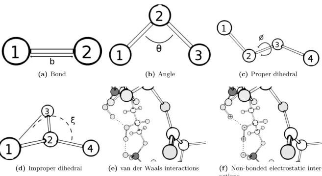
2.2 Schematic representation of the interactions present in the potential energy equation 36 2.3 Schematic representation of the leap-frog algorithm. . . 42
2.4 Schematic representation of the steepest descent algorithm. . . 44
2.5 2D representation of a periodic system with periodic boundary conditions. . . 46
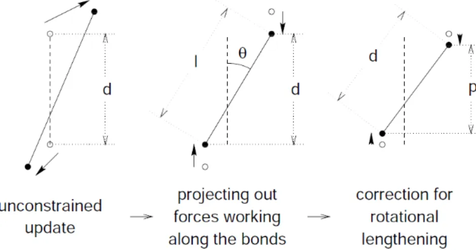
2.6 Representation of the LINCS algorithm. . . 49
2.7 General continuum electrostatics model. . . 53
2.8 Thermodynamic cycle involving protein and model compounds . . . 55
2.9 Schematic representation of the constant-(pH,E ) MD algorithm used in this thesis. 60 2.10 Heme I X-ray structure with no propionates. . . 61
2.11 Heme models with no propionates . . . 63
2.11 Heme models with no propionates . . . 64
3.1 Heme I X-ray model with deprotonated propionates. . . 74
3.2 HOMO and spin density analysis of both heme models (heme I X-ray with and
without propionates, at both redox states). . . 76
3.2 HOMO and spin density analysis of both heme models (heme I X-ray with and without propionates, at both redox states). . . 77
3.2 HOMO and spin density analysis of both heme models (heme I X-ray with and without propionates, at both redox states). . . 78
3.2 HOMO and spin density analysis of both heme models (heme I X-ray with and without propionates, at both redox states). . . 79
3.3 HOMO and spin density analysis of heme I X-ray with protonated propionates (2H), at both redox states. . . 80
3.3 HOMO and spin density analysis of heme I X-ray with protonated propionates (2H), at both redox states. . . 81
3.4 Average variation of each partial atomic charge between charge sets. . . 88
3.5 Chromatic representation of the average partial atomic charge variation. . . 88
3.6 RMSD and reduced fraction plots of a chosen replicate of the RESP charge set at the reduction potential of -260 mV. . . 90
3.7 RMSD and reduced fraction plots of a chosen replicate of the RESP MC X-ray charge set at the reduction potential of -260 mV. . . 91
3.8 Protein stability analysis relative of a chosen replicate of the RESP charge set at the reduction potential of -260 mV. . . 93
3.9 Protein stability analysis relative of a chosen replicate of the RESP MC X-ray charge set at the reduction potential of -260 mV. . . 94
3.10 Representation of Dv H-TpI-c3 2CTH PDB entry colored by secondary structure motif. . . 96
3.11 Structure alignment of Dv H-TpI-c3 2CTH PDB entry and a structurally affected RESP MC Free Opt. replicate, simulated at -240 mV. . . 97
3.12 DSSP maps of two different simulations. SS stability comparison. . . 99
3.13 Redox titration curves . . . 101
3.14 Total titration curves . . . 102
3.16 Histograms of the electron populations at different average reduction (< ne>) . . 111
3.16 Histograms of the electron populations at different average reduction (< ne>) . . 112
B.1 RMSD and reduced fraction plots of a chosen replicate of the RESP MC X-ray charge set at the reduction potential of -240 mV. . . 123 B.2 RMSD and reduced fraction plots of a chosen replicate of the RESP MC Fixed Opt.
charge set at the reduction potential of -240 mV. . . 124 B.3 RMSD and reduced fraction plots of a chosen replicate of the RESP MC Free Opt.
charge set at the reduction potential of -240 mV. . . 124 B.4 RMSD and reduced fraction plots of a chosen replicate of the CHELPG charge set
at the reduction potential of -300 mV. . . 125 B.5 RMSD and reduced fraction plots of a chosen replicate of the Merz-Kollman (MK)
charge set at the reduction potential of -240 mV. . . 125 B.6 RMSD and reduced fraction plots of a chosen replicate of the RESP MC Fixed Opt.
charge set at the reduction potential of -260 mV. . . 126 B.7 RMSD and reduced fraction plots of a chosen replicate of the RESP MC Free Opt.
charge set at the reduction potential of -240 mV. . . 126
C.1 Protein stability analysis relative of a chosen replicate of the RESP MC X-ray charge set at the reduction potential of -240 mV. . . 128 C.2 Protein stability analysis relative of a chosen replicate of the RESP MC Fixed Opt.
charge set at the reduction potential of -240 mV. . . 129 C.3 Protein stability analysis relative of a chosen replicate of the RESP MC Free Opt.
charge set at the reduction potential of -240 mV. . . 130 C.4 Protein stability analysis relative of a chosen replicate of the CHELPG charge set
at the reduction potential of -300 mV. . . 131 C.5 Protein stability analysis relative of a chosen replicate of the Merz-Kollman (MK)
charge set at the reduction potential of -240 mV. . . 132 C.6 Protein stability analysis relative of a chosen replicate of the RESP MC Fixed Opt.
charge set at the reduction potential of -260 mV. . . 133 C.7 Protein stability analysis relative of a chosen replicate of the RESP MC Free Opt.
3.1 Structure RMSD crossing table. Heme X-ray against heme X-ray . . . 83 3.2 Structure RMSD crossing table. Optimized heme structures against heme X-ray . 83 3.3 Atomic partial charge compilation. . . 86 3.4 Charge set RMSD crossing table. Charge set versus charge set, at the reduced
redox state. . . 87 3.5 Charge set RMSD crossing table. Charge set versus charge set, at the oxidized
redox state. . . 87 3.6 Charge set RMSD crossing table. Reduced versus oxidized charges of the same
charge set. . . 87 3.7 RMSD values and corresponding Emod and Ehalf . . . 104 3.8 Order of reduction of the heme groups . . . 105 3.9 Alternative RMSD values . . . 106 3.10 Alternative order of reduction of the heme groups . . . 106
A.1 Structure RMSD crossing table. All four hemes versus one another. Fixed opti-mization at the reduced redox state . . . 120 A.2 Structure RMSD crossing table. All four hemes versus one another. Free
optimiza-tion at the reduced redox state . . . 120 A.3 Structure RMSD crossing table. All four hemes versus one another. Fixed
opti-mization at the oxidized redox state . . . 120 A.4 Structure RMSD crossing table. All four hemes versus one another. Free
optimiza-tion at the oxidized redox state . . . 120
A.5 Structure RMSD crossing table. Reduced versus oxidized redox states, fixed opti-mization . . . 121 A.6 Structure RMSD crossing table. Reduced versus oxidized redox states, free
opti-mization . . . 121 A.7 Structure RMSD crossing table. Fixed versus free optimization, at the reduced
redox state . . . 121 A.8 Structure RMSD crossing table. Fixed versus free optimization, at the oxidized
Introduction
Before going into further detail regarding the main introductory details of the present work, a brief explanation of why each section was created is necessary. Since this thesis main subject revolves around quantum mechanical (QM) atomic charge parametrization of Desulfovibrio vulgaris Hildenborough type I cytochrome c3 (Dv H-TpI-c3) heme group(s) model(s), and
sub-sequent molecular dynamics (MD) simulations of the mentioned system with (hopefully) im-proved force field charge parameters, a different approach to the whole biological/biochemical background of the model protein felt imperative. Most emphasis in this chapter will be given to the mechano-chemical (thermodynamic and structural) knowledge of the system, due to its inherent importance to the understanding of the work reported in this thesis. Little, but sufficient, insight about the biological background of hemeproteins and their role in anaerobic bacteria will be given, in order to assure that the reader can become familiar with the addressed subject.
Quantum and molecular mechanics (MM), continuum electrostatics (CE) and constant-(pH,E ) molecular dynamics will be addressed in a simple way, to enable the reader to blend and make the logic connections between addressed physico-chemical fields and the studies presented here. Detailed considerations regarding these themes will be addressed in Chapter 2.
1.1
Sulfate-reducing bacteria and hemeproteins
Sulfate-reducing bacteria (SRB) are a group of microorganisms, comprising both bacteria and archaea, which derive energy from the anaerobic respiration of sulfate. They are capable of using lactate, pyruvate, formate and other organic compounds as electron donors, and can even grow chemolithotrophically using hydrogen as the electron donor. The hydrogen metabolism
plays a central role in the energy-generating mechanisms of these microorganisms, i.e. the reduction of sulfate, which is a true respiratory process, leading to oxidative phosphorylation through a still poorly understood electron-transfer pathway [1, 2].
Extensive studies have been conducted on the metabolism of these bacteria, as well as on the characterization of their protein components. A great number of periplasmic electron carriers exhibiting low redox potentials and some interesting and unique redox enzymes have been characterized in these bacteria. However, and as stressed before, the sulfate respiratory process is still poorly understood. In particular, and contrary to other types of organisms, the terminal reductases involved in sulfate reduction are not membrane-bound, but cytoplasmic, and therefore cannot be directly involved in proton translocation across the membrane [3]. Hemeproteins are widespread in all groups of living organisms, from archaea to eukaryotes, and from aerobes to anaerobes. In particular, the Desulfovibrio genus of the SRB is known for having an unusually high number of multiheme cytochromes c [4]. These cytochromes appear to be involved in the electron transfer linked to the hydrogen oxidation [5]. Among all characterized cytochromes, type I cytochrome c3(TpI-c3) is the most abundant and is present
in all Desulfovibrio species [6]. Regardless the low sequence homology between TpI-c3 of
different species, their three-dimensional structure shows a common topology organized around the four-heme cluster, suggesting that this spatial organization is essential for the function of the protein. Likewise, the multiple heme structural motif seems to be a common feature for other members of the multiheme cytochromes c. Still, despite the structural similarities, TpI-c3 possesses a distinct in-vivo role from other cytochromes c, e.g. the type II cytochrome c3,
and the 16 and 9-heme cytochrome. The former three cytochromes display affinity for the membrane [7] while TpI-c3 is a soluble protein, being their common electron donor, acting as
a mediator for electron transfer from periplasmic hydrogenases [8].
1.2
Heme group characterization
1.2.1 Structural details - porphyrin and axial ligands
The heme moiety is a porphyrin, which, through its four pyrrol nitrogens binds to one iron in an almost square planar geometry. Anaerobes usually display three common types of porphyrins (see Figure 1.1). Only the c-type hemes are covalently bound to the polypeptide chain, through thioether linkages between the porphyrin vinyl side-chains and cysteinyl residues, usually forming a common aminoacid sequence motif -Cys-X-X-Cys-His- [9]. In hemeproteins, the iron is usually, either, five- or six-coordinated, and the axial ligands are provided by aminoacid residues of the protein backbone. Only a very limited type of residues have been so far identified for this role in naturally occurring proteins. In most cases, the fifth ligand is a
Figure 1.1: Most common heme groups found in proteins from anaerobes. Taken from [4].
histidine, which, in the particular case of c-type cytochromes, is a histidine residue immediately following the two cysteines in the aforementioned sequence motif. In six-coordinated hemes, the other axial ligand is generally either a methionine or a histidine [4, 9]. Six coordinated hemes are generally low-spin in both oxidation states. The ligand field at the heme iron is very close to the crossover point for the high- and low-spin states. Variations of the strength of one of the axial ligands may change the spin state. Interference with the axial ligand nature is also known to yield a large change in the oxidation-reduction potential. Mus-Veteau et al. (1992) reported a large increase of at least 200 mV of one of the four oxidation-reduction potentials in Desulfovibrio vulgaris Hildenborough type I cytochrome c3 upon site-directed mutagenesis.
This replacement of the sixth coordination axial ligand, histidine 70, by a methionine residue occurred without significant alteration of the structure [10].
1.2.2 Redox characteristics
Heme-iron is usually limited to two stable redox states: • Fe(II), i.e., ferrous iron.
• Fe(III), i.e., ferric iron.
Even though the amplitude of stable redox states available to heme-iron is low, hemeproteins are able to cover a wide range of reduction potentials, with values ranging from ∼-500 mV to ∼+400 mV. This is probably why hemeproteins are found in most electron transfer chains, from anaerobic to aerobic systems [4].
The reduction potential of a heme group is dependent on multiple, interplaying factors, like:
Figure 1.2: Reduction potential range covered by heme proteins. Taken from [4].
• The heme environment;
• The redox-linked chemical equilibria [9–11].
In general, the reduction potential decreases with increasing σ donor capability of the axial ligand. In most cases, methionine bound hemes present the highest reduction potentials, while histidine bound ones have the lowest, as can be seen in Figure 1.2. Once again, site-directed mutagenesis experiments confirm this statement [10]. Nevertheless, heme-bound axial ligands cannot be accounted as the sole variable which controls their reduction potential. At most, axial ligands are but one of the factors behind the evident differences in reduction potential between the different naturally occurring cytochromes. Therefore, the actual reduction potential of a heme is a result of several interplaying electrostatic factors, such as electric charges on neighbor or even distant residues, dipolar and hydrogen-bonding interactions, solvent exposure, and heme-heme interactions (henceforth designated as homotropic interactions). Redox-linked equilibria also contributes to the fine control of the reduction potential. In short, the redox behavior of these proteins is extremely complex, due to multiple homotropic (heme-heme redox potential interactions) and heterotropic (heme redox potential to pKa of ionizable groups
interactions) cooperativities [12].
1.3
Desulfovibrio vulgaris Hildenborough type I cytochrome
c
3(Dv H-TpI-c
3)
Desulfovibrio vulgaris Hildenborough type I cytochrome c3(Dv H-TpI-c3) is a small (Mr13,000)
sul-(a) Dv H-TpI-c3. (b) Heme I from Dv H-TpI-c3.
Figure 1.3: Dv H-TpI-c3 and respective heme structure. (a) Dv H-TpI-c3 secondary
structure with highlighted heme numbers. (b) Atomic representation of the heme I structure.
fate reducing bacteria Desulfovibrio vulgaris Hildenborough (Dv H). It is constituted by 107 residues plus four hemes covalently bound to cysteines in the polypeptide chain together with bis-histidinyl axial ligation, as can be seen in Figure 1.3b [6]. The small size, stability and high solubility of this protein enabled its characterization by a wide variety of techniques, including NMR spectroscopy, which has been successfully applied in structural and thermo-dynamic studies [13, 14]. A detailed crystallographic structure has also been obtained by X-ray spectroscopy, making it the best characterized multiheme protein [15, 16]. All these studies showed that the Dv H-TpI-c3 hemes are covalently held together in close proximity,
exhibiting strong coupling between themselves and nearby acid/base groups. Therefore, and as stated in Section 1.2.2, it comes with no surprise that the heme reduction potential is dependent on the oxidation state of the other three hemes (redox interaction potential) and on the pH (redox-Bohr effect). These events have been extensively studied by experimental and theoretical approaches [12, 14, 17–26].
1.4
Cooperativity events in Dv H-TpI-c
3As briefly discussed in Section 1.2.2 and Section 1.3, Dv H-TpI-c3 is known to exhibit
cooperativity between the four hemes among themselves and the hemes and acid/base groups, which means each heme redox potential is dependent on the oxidation state of the other three hemes (redox interaction potentials or homotropic cooperativity) and the heme redox potentials are pH dependent (redox-Bohr effect or heterotropic cooperativity), respectively [12].
and was one major research field at the Instituto de Tecnologia Qu´ımica e Biol´ogica (ITQB), through a series of experimental and theoretical (essentially thermodynamic) studies in tetra-heme cytochromes c3, with special mention to Professor Ant´onio V. Xavier [12, 14, 17–26].
In short, this effect can be described as a strong pH dependence of heme reduction poten-tials, corresponding to a thermodynamic dependence between the reduction of redox sites and the ionization of protonable sites, due to their electrostatic interaction. This effect is thought to be of potential physiological significance in cytochromes c3, since it occurs even
at physiological values of pH. Therefore, the redox-Bohr effect is thought to allow (thermo-dynamically) correlated electron and proton capture in the same molecule, which constitutes a physiologically relevant mechanism of transferring the electrons and protons generated by hydrogenase from molecular hydrogen oxidation. While hemes receive the generated electrons, protonatable sites in the protein, by consequent changes in their pKavalues, are more prone
to (de)protonation events. The order of oxidation of the hemes has been established for quite some time, and the occurrence of positive cooperativity (higher affinity for electrons than it would be expected for independent groups) between heme I and II of Dv H-TpI-c3, is
consen-sual [12, 14, 17, 25, 26]. Although the relative positions of the hemes are known from the X-ray crystal structure [15, 16], the nature and position of the acid-base groups responsible for the redox-Bohr effect has yet to be determined unequivocally. However, most theoretical studies point towards propionate D from heme I being the major responsible for the redox-Bohr effect [18, 22, 24]. Its effect on the redox potential of individual hemes, as calculated by electrostatic calculations, correlates very well with the experimental order of influence, making it a likely candidate. Moreover, the comparison between the protein structure in the fully oxidized state, obtained by X-ray [15, 16], and a structure in the fully reduced state, obtained by multidimensional NMR [13], shows the existence of small but significant differences, mostly localized near a loop close to heme I. At last, heme I is always dominant in the redox-Bohr effect, since its redox potential is the most sensitive to changes in pH, which also suggests that the protonable site responsible for the redox-Bohr effect is indeed in close proximity.
1.5
Energy transduction mechanism
Even though the molecular basis for the energy transduction mechanism of Desulfovibrio vul-garis Hildenborough type I cytochrome c3, as well as its implication in the energy generation
mechanism, has not been unequivocally demonstrated, it is possible to propose a mechanism for its functional activity, based on the information gathered throughout the years of experimental and theoretical research.
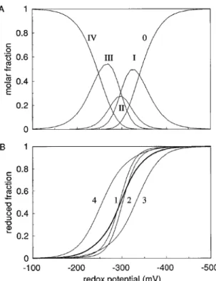
Analysis of the populations for the oxidation states of the different hemes calculated [12] at pH 6.6, the optimum pH for growth of these bacteria (Dv H) [28], suggests a concerted
Figure 1.4: Oxidation states for Dv H-TpI-c3 at pH 6.5. (a) Populations of the different
oxidation stages versus solution potential (Roman numerals number of hemes oxidized). (b) Reduced fraction of each heme (sequence numbering) versus solution potential. The bold curve represents the overall reduced fraction of the protein. Taken from [17].
proton-assisted 2e− step. Furthermore, the rather steep slope of the population curves [17] (see Figure 1.4) for the two hemes with intermediate redox potentials (heme I and II), confirm the former statement. This is, also, reflected by the relatively low population attained by stage II, which includes the molecules with two hemes oxidized.
Combined, these results clearly show that in the pH range between the pKa for stage I and
that for stage III, Dv H-TpI-c3 will perform a 2e−/2H+ concerted step. Therefore it is
par-ticularly well designed to work as a coupling protein to the enzyme hydrogenase. The energy transduction performed by this protein is called “proton thrusting” to distinguish it from the “proton pumping“ activity of transmembrane protein complexes. This coupled transfer of two electrons associated with proton transfer in the physiological pH range is fundamental for the ability of cytochrome c3 to accelerate the oxidation of hydrogen by hydrogenase, with
subse-quent transfer of electrons to the membrane-associated multiheme cytochromes, which vary depending on the organism. In conclusion, Figure 1.5 depicts the overall mechanism of charge separation promoted by Dv H-TpI-c3 [5, 17].
(a) Thermodynamic and mechanistic bases for
en-ergy transduction by Dv H-TpI-c3.
(b) Schematic illustration of the central role of
Dv H-TpI-c3 in the bioenergetic system of
sulfate-reducing bacteria.
Figure 1.5: Energy transduction mechanisms of Dv H-TpI-c3. (a) The cubane diagram
for the charge separation mechanism achieved by cycling between oxidation stages I and III is shown at the bottom. The top diagram gives the macroscopic thermodynamic parameters. Taken from [17]. (b) Functional energy transduction cycle performed by Dv H-TpI-c3. Taken
from [5].
1.6
Scope and structure of this work
As mentioned before (see Section 1.3), Desulfovibrio vulgaris Hildenborough type I cy-tochrome c3 (Dv H-TpI-c3), which is the subject of this work, has been extensively studied
by, both, experimental and theoretical means. Recently, Machuqueiro and Baptista [29] used this proteic model as a means of validation and proof of concept for their new implementation of the stochastic titration method [30], the constant-(pH,E ) MD method. This method makes it possible to perform molecular dynamics simulations at constant pH and reduction potential. It was applied to the redox titration of Dv H-TpI-c3, and enabled to obtain a major finding:
• The method showed a better performance when a high dielectric constant, ε, was assigned to the protein regions [29].
This dependence on the value of protein dielectric constant was never found in previous constant-pH MD simulations [30–33]. Several explanations were formulated in order to un-derstand this phenomenon. The most reasonable is related with the, eventually, excessively high heme-heme interactions at the low dielectric constants. Dv H-TpI-c3 constitutes a tight
densely packed structure where the heme reduction potentials are very close (within a range of 80 mV, or 1.3 pH units), making it a very demanding test case in terms of prediction, since 1.3 pH units is not so far from the constant-pH MD method pKa prediction error (∼0.8 pH units)
observed in the Dv H-TpI-c3, enhances the previously mentioned homotropic and heterotropic
cooperativities observed in this protein (see Section 1.4).
The authors also noticed that the order of reduction of the hemes (III < II ≤ I < IV, with increasing value of reduction potential) was only partially well predicted even at higher values of ε (heme III is always out of order). This need for higher ε seems to indicate a lack of reorganization effects in the simulations. The observed increase of heme interactions with the decrease of ε is what one would expect in a rigid-structure Poisson-Boltzmann/Monte Carlo (PB/MC) study, where reorganization must be entirely captured by ε. The constant-(pH,E ) MD method should not be affected by this, since structural reorganization should in principle occur explicitly via MM/MD, as discussed in [33]. However, and taking into account that the heme groups are structurally limited to planar tilting and to the small fluctuations allowed by the anchoring cysteines, it is only natural to think that in order for the hemes to achieve significant structural reorganization, its charges, in both the oxidized and reduced state, need to be accurately determined. New charge sets obtained with more accurate quantum theory, basis sets and methodology can eventually result in a more extensive conformation-reduction coupling, and might end up affecting structural reorganization because of trapping or hysteresis effects.
With the former statements in mind, the main goal of this work is to refine the parametrization of the redox centers, i.e. obtain new and more accurate charge sets; incorporate them in the new 53A6 GROMOS force field [34]; and finally run MD simulations of the new systems at constant pH (6.6, the assumed value for the physiological pH in the periplasm of Desulfovibrio vulgaris Hildenborough bacteria) and reduction potential values (within a range which comprises the mean reduction potentials for each of the four hemes).
1.7
Methodology
As stated before (see Section 1.6), even though Machuqueiro and Baptista were successful in proving the concept behind the constant-(pH,E ) MD method [29], they have met with several problems regarding the validation of their results. The main suspicion behind the poor results obtained when simulating Dv H-TpI-c3 at low ε, lies on the system’s electrostatics,
moreover, the parametrized partial atomic charge values for the heme group model, determined in [35]. These charges were calculated upon a single heme group model derived from the Dv H-TpI-c3 heme I X-ray structure. No extra care was given to the fact that each of the four
hemes present in Dv H-TpI-c3displays significant conformational differences among each other.
Moreover, positive redox cooperativity between hemes implies a redox-linked conformational change [14], i.e., whenever a heme changes its redox state within Dv H-TpI-c3, its conformation
also changes. Thus, in order to accurately parametrize the charges of the heme groups present in the aforementioned cytochrome, one need to account for conformational diversity (between hemes, and within each heme at different redox states). Charges obtained for a single heme do not apply to other hemes present in the same protein1, and do not take into account the aforementioned redox-linked conformational change. Forcing the other three hemes to share the same charge parameters obtained specifically for heme I, is expected to result in undesired electrostatic strain.
There are less than a handful of ways to obtain partial atomic charges for empirical force field parametrization. The most obvious way is perhaps a “semi-empirical approach”2, i.e. a rational partial atomic charge attribution with successive testing, which (with luck and a lot of labor) can lead to acceptable charge sets for systems with less electrostatic sensitivity. However, more sensitive systems, as Dv H-TpI-c3, require much more accurate charge sets, which can only be
obtained by specific charge derivation schemes based on highly accurate QM and QM/MM (hybrid quantum mechanical and molecular mechanical) calculations3. Enhancements to the charge parametrization of the heme groups from Dv H-TpI-c3, published in [35], can only
be made by increasing the electronic detail of the system, from which the atomic charges of the heme group models are determined. This can be achieved through the use of better and more appropriate basis functions, while improving the self-consistent field (SCF) convergence criteria as well. Full and partial geometry optimizations of each heme structure at both redox states, are also important, since crystal and solution protein structures differ in conformation. Furthermore, crystallographic X-ray structures are obtained at specific redox states, and as mentioned before, hemes undergo a redox-linked configuration change. Finally, considerable attention should also be given to the testing of several different charge derivation methods, specially to the ones which enable a multi-configurational analysis.
In sum, charge parametrization involves strenuous planning, exploration and patience. No charge derivation method is, a priori, predictably better than another, and an “intelligent” amount of possibilities must always be explored and subsequently tested. At the end of the day, the best charge set is the one which yields the best results for a given empirical force field based method; which in the case presented here, is the constant-(pH,E ) MD method. Many, many factors exert direct influence upon this observation. Moreover, one must be prepared to accept the fact that not always what seems to be more logical ends up producing better results. Sometimes, empirical force field based simulation methods work better with charge sets obtained from, often considered, “less elegant” charge derivation methods, by the simple,
1As long as their conformation and surrounding protein environment is not equal, the subjacent electrostatics
will most likely be different.
2This term appears in quotation marks for the simple fact that all charge derivation methods, even the ones
based on quantum mechanical properties, are semi-empirical at heart. See next chapter’s Section 2.1.7 for a more detailed explanation.
3
yet powerful fact that, they were developed and parametrized together. Still, a rational choice of model structure and charge derivation method is considered indispensable.
In the next chapter, the reader will be given a comprehensive presentation of the theory and methods behind the work presented here.
Theory and methods
2.1
Quantum mechanics
This chapter deals with a subject that is often considered to be difficult. Difficult or not, quantum mechanics happen to be the only way to quantify and qualify several microscopic phenomena, e.g. black body radiation, the photoelectric effect, atomic stability and spec-troscopy, among others. This stands because classical physics, whose overwhelming success until the end of the nineteenth century made people believe that the ultimate description of nature had been achieved, fails miserably when trying to explain them. For example, Einstein’s 1905 theory of relativity showed that the validity of Newtonian mechanics ceases at very high speeds, i.e. at speeds comparable to that of light. In addition to this, many other discoveries and theories by Planck, Bohr, Compton and de Broglie, led the way to what became quan-tum mechanics. Specially, Compton’s 1923 discovery gave the conclusive confirmation about the corpuscular nature of light, showing that, at this scale, classical physics fails not only quantitatively but even qualitatively and conceptually.
Even though this subject is extremely interesting, this thesis does not intend to review the main physical ideas and experimental facts that defied classical physics and led to the birth of quantum mechanics. Nor does it intend to inform the reader about the most general physical and mathematical aspects of quantum mechanics. These subjects are found in most general physical chemistry textbooks1. Furthermore, being a biochemist and not a physicist, the author of this dissertation is in no position to offer the reader a detailed description of such an extensive and complex subject as quantum mechanics. Therefore, the main aim of this chapter is to remind the basic elements of quantum mechanical methods that are most widely used in molecular modelling, specially model geometry optimization and subsequent charge derivation
1
See [36] for a useful review of quantum mechanics concepts and applications.
methodologies, which are essential to force field parametrization (see Section 2.2.2), one of the main subjects of this thesis.
2.1.1 The Schr¨odinger equation
“Although Bohr’s model for the atom produced results that agree well with experimental spectroscopy, it was criticized for lacking the ingredients of a theory. Like the “quantization” scheme introduced by Planck in 1900, the postulates and assumptions adopted by Bohr in 1913 were quite arbitrary and do not follow from the first principles of a theory. It was the dissatisfaction with the arbitrary nature of Planck’s idea and Bohr’s postulates as well as the need to fit them within the context of a consistent theory that had prompted Heisenberg and Schr¨odinger to search for the theoretical foundation underlying these new ideas. By 1925 their efforts paid off: they skillfully welded the various experimental findings as well as Bohr’s postulates into a refined theory: quantum mechanics.”
in Quantum Mechanics, Concepts and Applications (2009) [36]
Even though the starting point for any discussion of quantum mechanics is the Schr¨odinger’s equation, it is relevant to mention that Dirac showed Heisenberg’s matrix mechanics, to be equivalent to Schr¨odinger’s wave mechanics formulation. However, Schr¨odinger’s formulation is more intuitive than the matrix mechanics, describing the dynamics of microscopic matter by means of a wavefunction, ψ, which is obtained from a differential equation that is a function of space, spin and time. It returns the probability of the position of each particle in a given system over all space. Mathematically, it is a function of an infinite space that maps all the possible states of the system [37].
Considering situations where the external potential, V , is independent of time (which happens in stationary systems), the Schr¨odinger equation is written as follows:
Hψ = Eψ , (2.1)
where H is the Hamiltonian operator,
H = ¯h 2 2m∇ 2+ V , (2.2) and ∇2 = δ 2 δx2 + δ2 δy2 + δ2 δz2. (2.3)
The Schr¨odinger equation (Equation 2.1) refers to a single particle, usually an electron2, of mass m moving through space under the influence of an external field, V . E stands for the energy of the particle and ¯h is the reduced Planck constant or Dirac’s constant (¯h = 2πh). ∇2
is often mentioned as del-squared, which is the square of the vector operator del. When del operates upon a scalar function, it produces a vector which is the gradient of that function. The del and del-squared operators are very important in quantum mechanics. For instance, del shows up in the definition of the linear momentum operator (not shown here), while del-squared appears in the definition of the Hamiltonian operator (as seen in Equation 2.2).
E is often regarded as the expectation value of H for a N -body particle system. Solving the Schr¨odinger equation involves finding the values of E and functions ψ that, when the wavefunction is operated upon the Hamiltonian, it returns the wavefunction multiplied by the energy, as in Equation 2.1 [38]. Solutions to Schr¨odinger’s equation describe not only molecular, atomic and subatomic systems, but also macroscopic systems, possibly even the whole universe!
However, Schr¨odinger’s equation cannot be solved exactly for more than two interacting parti-cles at a time. Only single electron systems (where a single electron interacts with the nucleus, a two-body system), like the H, He+1, Li+2 atoms/ions, can be treated with complete detail3. Any N -body systems with N ≥ 3 cannot be calculated exactly, and need to be treated in an approximate manner.
The aforementioned mathematical problem has haunted quantum mechanics since its birth, leading one of its most prominent figures to state the following:
“It would indeed be remarkable if Nature fortified herself against further advances in knowl-edge behind the analytical difficulties of the many-body problem.”
Max Born (1960)
Until today, Max Born’s wise words remain only partially valid. The impossibility of solving exactly Schr¨odinger’s equation for the great majority of atoms and molecules led to the emer-gence of methods and, with the development of computer calculus/simulations, algorithms, that enabled the attainment of very accurate approximate solutions to this problem. Thus,
2Quantum mechanics can be applied to any particle, since in 1923, de Broglie suggested that the
wave-particle duality is not restricted to radiation. All material wave-particles should also display a dual wave-wave-particle behavior. This idea was confirmed in 1927 by Davidson and Germer, and later by Thomson, who obtained interference patterns with electrons. Even macroscopic particles display wave features. See [36] for further insight.
3
Even the simplest molecular species, like H+2 can be solved exactly, through the Born-Oppenheimer
ap-proximation [39], which states that the motion of the electrons can be decoupled from the motion of the nuclei, whose mass is much bigger. This way electrons can adjust almost instantaneously to any changes in the position of the nuclei.
solving exactly the Schr¨odinger equation is no longer the main issue nowadays. The problem lies in getting quantum mechanics to treat large systems in an acceptable time scale. But this is a subject out of the scope of this work.
As a consequence of this deadlock the wavefunction may adopt more than one form, or in other words, no form is necessarily more correct than another. In simple terms, a guessed wavefunction that yields the lowest energy distribution for all interacting particles in a system is the always the most accurate one, according to the variation method (see Section 2.1.3 for a description of the variation method).
2.1.2 Wavefunctions of polyelectronic systems
Without further considerations one of the simplest ways to approximate the wavefunction of a many-body system is to take the product of properly chosen wavefunctions of the individ-ual particles. This is, unfortunately, not correct, since it does not take into consideration the fermionic4 properties of electrons, and therefore is not valid for electron bearing systems (which compose pretty much 100% of the systems studied in molecular modelling by quantum mechanics). Multi-fermionic systems, require that the principle of antisymmetry is met. In other words, the wavefunction must respect the Pauli exclusion principle, which states the following:
“(...) No two electrons can have the same four quantum numbers, that is, if n, l, and mlare
the same, ms must be different such that the electrons have opposite spins. More generally,
no two identical fermions may occupy the same quantum state simultaneously.”
Wolfgang Pauli (1925)
In 1929, the physicist John Slater, proposed an expression which can be generalized to any number of fermions by writing it as a determinant. Thus the name Slater determinant. Slater determinants gave the means of ensuring the antisymmetry of a many-body wavefunction through the use of matrices [40]. In general terms, for a system of N electrons in spin orbitals χ1, χ2, ..., χN (where each χN is a the product of a spatial and a spin function), the Slater
determinant is defined as:
ψ(1, 2, ..., N ) = √1 N ! χ1(1) χ2(1) ... χN(1) χ1(2) χ2(2) ... χN(2) .. . ... ... χ1(N ) χ2(N ) ... χN(N ) (2.4) 4
The factor 1/√N ! is used in order to normalize the wavefunction. The former equation (E-quation 2.4), represents the simplest form of an orbital wavefunction that satisfies the anti-symmetry principle. Manipulation of any two rows of a determinant changes the sign of the determinant, leading to the antisymmetry property. Furthermore, the determinant vanishes when any two rows of a determinant are identical, which is the same as saying that no two electrons can occupy the same spin orbital. This is of significant importance, since it shows that the Slater determinant obeys Pauli’s exclusion principle, i.e. each spatial orbital can only accommodate two electrons of opposite spin.
Thus, the resulting wavefunction accounts for the exchange interaction, that is, the wavefunc-tion describing two indistinguishable particles must be inverted in sign (antisymmetric) if the labels of the two particles are changed.
Several theories which give good approximations to the real solution of the Schr¨odinger equa-tion incorporate Slater’s determinant. In the following secequa-tion (Secequa-tion 2.1.3), one of the most common ab initio5 methods will be presented.
2.1.3 Hartree-Fock method
The Hartree-Fock method (HF), often called self-consistent field method (SCF), is an approxi-mate method for the determination of the ground-state wavefunction and ground-state energy of a quantum many-body system. It was one of the first methods used in quantum computa-tions, but its inherent weaknesses and the development of computers made it quite obsolete nowadays. Its inclusion in this work is mainly due to the fact that it is at the base of most post-Hartree-Fock methods, making it a cornerstone in quantum mechanics.
It is mainly used to generate initial guesses of the system’s wavefunction, since it requires fewer computational resources, when compared with more advanced methods, usually yielding results comparable to the ones obtained by most molecular mechanics methods. In order to supplant the need of very accurate approximations to the exact Schr¨odinger’s equation solution, one should rely on more advanced quantum mechanical methods, such as the so-called post-Hartree-Fock methods and/or density functional theory.
In the HF method, a single Slater determinant is used as an approximation to the electronic wavefunction. The single-determinant approximation does not take into account Coulomb cor-relation, which accounts for the instantaneous repulsion between electrons. Instead, it uses a mean field potential. This leads to a total electronic energy different from the exact solu-tion of the non-relativistic Schr¨odinger equation within the Born-Oppenheimer approximation. Therefore the Hartree–Fock limit is always above the exact energy. This difference is often
5
![Figure 1.1: Most common heme groups found in proteins from anaerobes. Taken from [4].](https://thumb-eu.123doks.com/thumbv2/123dok_br/18218042.877220/26.892.149.711.169.350/figure-common-heme-groups-proteins-anaerobes-taken.webp)
![Figure 1.2: Reduction potential range covered by heme proteins. Taken from [4].](https://thumb-eu.123doks.com/thumbv2/123dok_br/18218042.877220/27.892.307.630.173.433/figure-reduction-potential-range-covered-heme-proteins-taken.webp)