UNIVERSIDADE DE LISBOA
FACULDADE DE CIÊNCIAS
D
EPARTAMENTO DEB
IOLOGIAV
EGETAL
L
OOKING AT
T
RICELLULIN
E
XPRESSION IN THE
B
RAIN
Cibelle Neiva Cavalcanti Mariano
Mestrado em Biologia Molecular e Genética
UNIVERSIDADE DE LISBOA
FACULDADE DE CIÊNCIAS
D
EPARTAMENTO DEB
IOLOGIAV
EGETAL
L
OOKING AT
T
RICELLULIN
E
XPRESSION IN THE
B
RAIN
Dissertação orientada por:
Maria Alexandra Brito, PhD, Faculdade de Farmácia, abrito@ff.ul.pt Maria Filomena Caeiro, PhD, Faculdade de Ciências, mfcaeiro@fc.ul.pt
Cibelle Neiva Cavalcanti Mariano
Dissertação de candidatura ao grau de Mestre em Biologia Molecular e Genética apresentada à Faculdade de Ciências da Universidade de Lisboa.
Projecto de Investigação desenvolvido no grupo de investigação Neuron Glia Biology in Health and Disease do Centro de Patogénese Molecular da Faculdade de Farmácia da Universidade de Lisboa, integrado no iMed-Research Institute for Medicines and Pharmaceutical Sciences, sob orientação de Maria Alexandra Brito, PhD, e orientação interna de Maria Filomena Caeiro, PhD, na Faculdade de Ciências.
N
ota de Abertura
O trabalho aqui desenvolvido deu origem a um resumo, submetido a apresentação oral no XVII Congresso Nacional de Bioquímica da Sociedade Portuguesa de Bioquímica:
Mariano C, Pereira P, Cardoso F, PalmelaI, Vaz AR, Fernandes A, SilvaSL, Falcão AS,
TorradoE, CamposAR, FerreiraAG, KimKS, BritesD, BritoMA. Scrutinizing the brain for the
tight junction protein tricellulin. The XVII National Congress of Biochemistry, Portugal Biochemical Society, Porto, Portugal, December, 15 – 17, 2010.
Encontra-se em fase de preparação uma revisão de conjunto sobre a tricelulina e um artigo científico para submissão a revistas internacionais.
...à minha mãe, companheira e amiga.
A
GRADECIMENTOSAs minhas primeiras palavras de agradecimento vão para a Profª. Doutora Dora Brites. Gostaria de começar por lhe agradecer o facto de me ter permitido realizar o projecto de estágio no grupo Neuron Glia Biology in Health and Disease. Agradeço a forma como me acolheu e a oportunidade de me iniciar no mundo da investigação. A sua dedicação à ciência é inspiradora! Foi um privilégio fazer parte do seu grupo de investigação!
À Profª. Doutora Alexandra Brito, orientadora deste projecto, um obrigada muito especial! Agradeço a dedicação, o empenho, a sua inteira disponibilidade e constante orientação neste trabalho. As suas preciosas sugestões, o seu rigor científico, persistência e profissionalismo merecem a minha admiração e foram fundamentais para que eu conseguisse realizar este trabalho. Gostaria também de expressar a minha gratidão pelas suas palavras de apoio e de confiança nos momentos mais difíceis deste percurso. Foi muito enriquecedor trabalhar consigo!
À Profª. Doutora Maria Filomena Caeiro, minha orientadora na Faculdade de Ciências, gostaria de agradecer o seu constante interesse pela evolução deste trabalho e o facto de estar sempre disponível para colaborar neste projecto. Obrigada por ter contribuído para o sucesso deste trabalho!
À coordenadora do Mestrado em Biologia Molecular e Genética, Profª. Doutora Rita Zilhão, agradeço a sua preocupação constante com todos os alunos e o seu empenho em organizar o mestrado da melhor forma possível. Não poderia deixar de agradecer o seu apoio nos momentos mais difíceis, a forma como me incentivou, as suas palavras de confiança. Muito obrigada!
Agradeço também ao Prof. Doutor Rui Silva a forma como me acolheu no grupo. Agradeço por todos os conhecimentos científicos que foram transmitidos e o interesse que sempre demonstrou pela evolução do meu trabalho.
Inesita, a minha “mestrinha” Inês e uma das futuras Doutoras do grupo, eu nem tenho palavras para dizer o quão importante tu foste para este trabalho… Tu, que me acompanhaste desde o início, que te interessaste pelo meu trabalho e que me ensinaste tantas coisas, um obrigada daqueles! Com muito carinho! A nossa relação foi mais do que profissional, tornou-se uma amizade… Fomo-nos conhecendo e aprendendo a gostar uma da outra. Nos momentos mais complicados, procurei muitas vezes em ti motivação para continuar, porque vejo-te com admiração. Tu és inteligente, és trabalhadora, és empenhada!
AGRADECIMENTOS
Confio plenamente no teu sucesso profissional! Sei que nem sempre é fácil, mas tu és capaz! Agradeço por tudo o que passamos juntas ao longo deste ano (sabes do que estou a falar: das margueritas, claro! Risos…), por todos os sorrisos, risos e gargalhadas… por todas
as lágrimas. A ti, agradeço por teres aturado tantas vezes as minhas “rabugices” e o meu
mau humor matinal (faz parte de qualquer relação, até da amizade. Risos…). Não há palavras suficientes para te agradecer… Obrigada por seres a “minha” Inesita!
Ema, Eminha, tu foste uma grande e agradável surpresa! Contagiaste-me com o teu jeitinho meigo de ser (às vezes, se é que me compreendes… Risos…). Admiro a tua dedicação e capacidade de trabalho. Sei que vais longe! A ti, agradeço as neuroesferas (Risos…). Agradeço todo o apoio que me deste, quer a nível profissional, quer a nível pessoal. Foste, com certeza, indispensável neste percurso que agora se finda. Tu és uma pessoa especial, que quero que permaneça na minha vida… Quero sentar na primeira fila quando for assistir à tua prova de Doutoramento. Obrigada por seres minha amiga! Obrigada por poder partilhar contigo os belos dias de sol de Verão!
Um obrigada muito especial, mesmo muito especial, para todas as minhas companheiras desta longa caminhada… Sim, porque sem vocês este trabalho não teria sido possível! Ritinha e Sandrinha, agradeço por todo o apoio que me deram neste trabalho. O vosso conhecimento, a vossa experiência, os vossos conselhos, e, principalmente, a vossa amizade… Foram fundamentais! Futuras Doutoras (está mesmo quase!), desejo-vos muito boa sorte nesta etapa que já se finda. Tenho a certeza de que a vão concluir com sucesso! Andreia, a nossa Dédé, obrigada por “colorires” os meus dias de trabalho. Foram muitas “galhofas” juntas! Com muito carinho, obrigada pelo apoio que me deste, não só a nível profissional, como também pessoal. Tu és uma pessoa super divertida e alegraste os meus dias. Tenho mesmo imensa pena por não estares presente neste momento. Quem sabe se eu não te vou visitar à Edinburgh?! Filipa, agradeço a tua disponibilidade incansável em ajudar-me. Obrigada por teres contribuído neste trabalho. Os teus conhecimentos foram indispensáveis! Boa sorte para o teu Doutoramento que ainda agora começou. Adelaide, tu és uma pessoa admirável… Não só pela tua vasta sabedoria e capacidade científica, como pela tua maneira doce de ser. Um obrigada muito especial pelos conhecimentos que me transmitiste e por te mostrares sempre disponível a ajudar-me na execução deste trabalho. Sofia, obrigada por seres assim, sempre tão serena e cativante! Foi um prazer ter a tua companhia nesta “caminhada”… Agradeço por todos os esclarecimentos científicos ao longo deste ano. Obrigada a todas as meninas pela forma como me receberam e fizeram com que
AGRADECIMENTOS
os meus dias fossem mais calorosos e divertidos! Obrigada pelo incansável apoio que me deram quando eu mais precisei! Tornaram tudo muito mais fácil!
Aos colegas com quem partilhei a sala “escura”, agradeço a vossa boa disposição que
tornou, sem dúvida, os meus dias mais “iluminados”. Obrigada pela forma como me acolheram… Foi um prazer dividir o mesmo espaço convosco!
À Inês Milagre e à Maria, agradeço a vossa inteira disponibilidade em ajudar-me. Obrigada pelos vossos conhecimentos científicos e pela vossa paciência comigo! A vossa contribuição foi fundamental na realização deste trabalho.
Pedro, tu foste mais do que indispensável para este trabalho… Agradeço por toda a ajuda na execução técnica deste estudo. Mas, mais do que isso, agradeço por seres meu amigo… Eu sei que não gostas muito de agradecimentos, mas é só desta… Prometo! Tu és uma pessoa excepcional! Obrigada por teres feito parte desta “dura” etapa da minha vida… Sim, porque não foi fácil… Mas tu estiveste lá a apoiar-me. Obrigada, com muito carinho!
Eu não poderia deixar de agradecer também ao Prof. José Ferreira, Técnico de Anatomia Patológica no Hospital São José, pelas amostras cedidas para o início deste estudo, bem como pelos conhecimentos adquiridos quando ainda era meu professor.
À minha mãe, a quem eu dedico este trabalho, a minha gratidão é eterna… Sem ti, eu não seria quem sou. Tu és a força da natureza, uma mulher fantástica! Não existem palavras suficientes para demonstrar a minha gratidão… e mais do que isso, não existem palavras que expressem o quanto significas para mim. Ambas sabemos que não foi um ano fácil, mas tivemos uma a outra, como sempre, e isso foi o mais importante. Obrigada, mãe, por todo o apoio que me deste, por teres acreditado em mim, por todos os sacrifícios que fizeste por mim ao longo destes anos... Obrigada, mãe, por me teres “aturado”! Obrigada, por seres a minha mãe! Obrigada, com todo o meu amor!
Pedro, o meu Pedro, agradeço a paciência, o apoio, o carinho e o amor que me tens mostrado… Sei que estes últimos tempos não foram muito fáceis e que nem sempre eu soube separar as coisas… Obrigada por fazeres parte da minha vida! Mesmo estando longe, estás sempre presente, iluminando a minha vida! Adoro-te!
Às minhas grandes amigas, Marta, Jo, Habib e MJ, agradeço a vossa compreensão quando, na maior parte das vezes, eu não pude estar convosco. Mas, a amizade é isso mesmo, é
AGRADECIMENTOS
saber compreender…Obrigada pelo vosso apoio, e, sobretudo, por aturarem tantas vezes as minhas “neuras” e inseguranças. A vossa amizade tornou tudo mais simples… Obrigada por serem minhas amigas! É com enorme prazer e satisfação que partilho isto convosco!
T
ABLE OFC
ONTENTS Abbreviations ... xiii Abstract ... xv Resumo ... xviC
HAPTER 1 – Introduction ... 1 1. Neurovascular unit ... 2 1. 1. Neurons ... 2 1. 2. Microglia ... 2 1. 3. Astrocytes ... 2 1. 4. Pericytes ... 3 1. 5. Endothelial cells ... 3 2. Intercellular junctions ... 4 2. 1. Tight junctions... 42. 1. 1. Tight junction proteins ... 5
2. 1. 1. 1. Occludin ... 5
2. 1. 1. 2. Claudins ... 5
2. 1. 1. 3. JAM ... 6
2. 1. 1. 4. MarvelD3 ... 6
2. 1. 1. 5. Zonula Occludens ... 7
2. 1. 2. Types of tight junctions ... 8
3. Tricellulin ... 8
3. 1. Discovery and known localization ... 8
3. 2. Tricellulin regulation ... 9
3. 3. Tricellulin function ... 11
3. 3. 1. Mutations in tricellulin gene ... 13
3. 4. Expression of tricellulin in non-epithelial cells ... 14
4. Aim ... 15
TABLE OF CONTENTS
1. Human and rat samples ... 16
2. Tissues samples preparation ... 16
2. 1. Human ... 16
2. 2. Rat ... 16
3. Cells cultures ... 16
3. 1. Primary culture of human brain microvascular endothelial cells ... 16
3. 2. Culture of human brain microvascular endothelial cell line ... 17
3. 3. Primary cultures of rat microglia and neurons ... 17
4. Immunohistochemistry ... 18
5. Immunocytochemistry ... 18
6. Western blot analysis ... 19
7. RNA isolation, reverse transcriptase and real-time RT-PCR ... 19
C
HAPTER 3 – Results ... 211. Analysis of tricellulin localization in human stomach in situ ... 21
2. Characterization of tricellulin distribution and expression in human brain microvascular endothelial cells ... 21
2. 1. Localization of tricellulin along intercellular junctions in a cell line of human brain microvascular endothelial cells ... 21
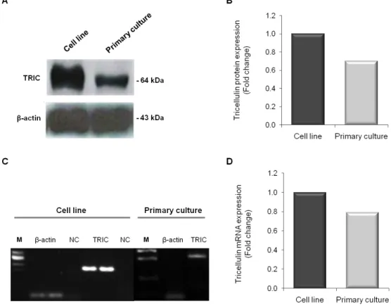
2. 2. Protein and mRNA expression levels of tricellulin in a cell line and in primary cultures of human brain microvascular endothelial cells ... 22
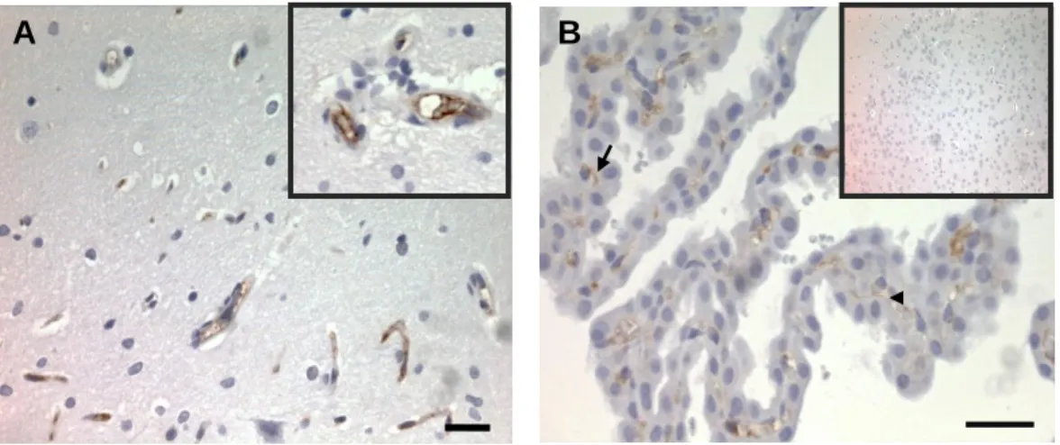
3. Assessment of tricellulin localization in rat and human brain in situ ... 23
4. Characterization of tricellulin in primary cultures of rat microglia and neurons ... 25
4. 1. Examination of tricellulin mRNA expression in rat microglia and neurons ... 25
4. 2. Inspection of tricellulin localization in rat microglia ... 26
C
HAPTER 4 – Discussion ... 27C
HAPTER 5 – References ... 31A
NNEXES... 331. Annex A ... 33
A
BBREVIATIONSAB/AM antibiotic-antimycotic
BBB blood-brain barrier
bTJ bicellular tight junction(s)
CD68 cluster of Differentiation 68
CNS central nervous system
cDNA complementary deoxyribonucleic
DAB 3,3-diaminobenzidine tetrahydrochloride
DAPI 4'-6'-diamidino-fenilindol
DIV days in vitro
DMEM Dulbecco‟s modified Eagle‟s medium
DMSO dimethyl sulphoxide
DPX di-N-butyle phthalate in xylene
EC endothelial cell(s)
ECGS endothelial cell growth supplement
EDTA ethylenediamine tetraacetic acid
FBS fetal bovine serum
Fig figure
HEPES (4-(2-hydroxyethyl)-1-piperazineethanesulfonic acid
HBMEC human brain microvascular endothelial cell(s)
HC hydrocortisone
HRP horseradish peroxidase
H2O hydrogen peroxide
IgG immunoglobulin G
JAM junctional adhesion molecule(s)
MARVEL MAL and related proteins for vesicle trafficking and membrane link
M molar ml mililitre ng nanogram nM nanomolar PBS phosphate-buffered saline PC primary culture(s)
PCR polymerase chain reaction
ABBREVIATIONS
PKC protein kinase C
PMSF phenylmenthylsulfonil fluoride
PPARγ proliferator activated receptor
RNA ribonucleic acid
RPMI Roswell Park Memorial Institute
RT room temperature
RT-PCR real-time PCR
SDS-PAGE sodium dodecyl sulphate-polyacrylamide electrophoresis gel
TEMED tetramethylethylendiamin
TER transendothelial electrical resistence
tTJ tricellular tight junction(s)
TJ tight junction(s)
TNF tumor necrosis factor
TRIC tricellulin
T-TBS tween20-Tris buffered saline
vs versus ZO zonula occludens m micrometer µl microlitre % percentage °C degree Celsius
A
BSTRACTIt was recently reported the existence of a protein, called tricellulin (TRIC), concentrated at tricellular tight junctions (TJ) but also present at bicellular contacts. Most of the studies have fallen upon epithelial tissues and, despite the similar ectodermic origin of nervous tissue, TRIC presence in brain cells has never been reported. Interestingly, TRIC mRNA expression was found in mature dendritic cells, raising the question of whether TRIC is present in the brain resident immunocompetent cells, microglia.
Immunohistochemical, western blot and real-time PCR methodologies were used to investigate the expression of TRIC in the brain. TRIC expression was examined both in paraffin sections of human brain (hippocampus) and in human brain microvascular endothelial cells (HBMEC), either in primary culture (PC) or in a cell line (CL). We further investigated the TRIC expression in rat brain sections, in addition to PC of rat neurons and microglia.
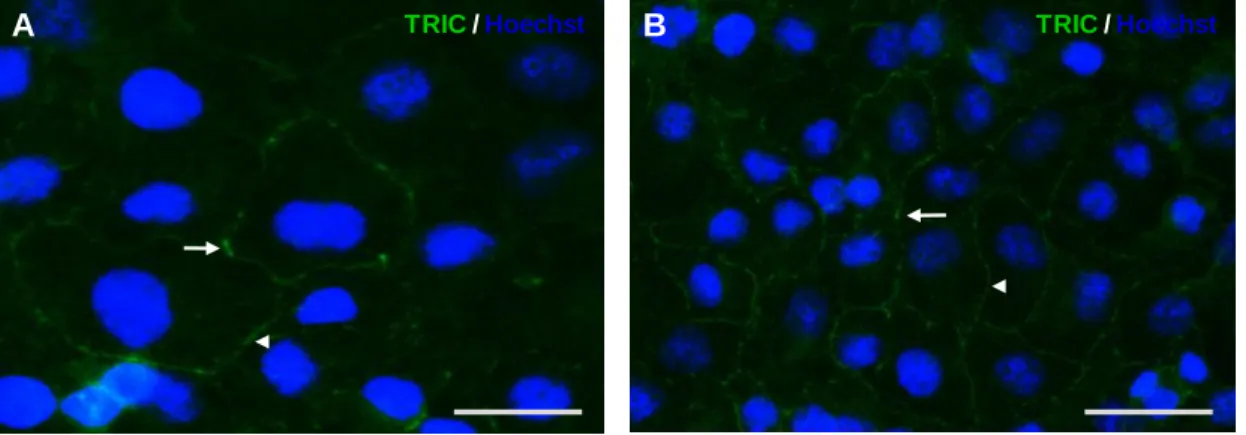
Immunofluorescence analysis of the CL revealed the presence of the protein in HBMEC and highlighted that it is concentrated at tricellular contacts, though distributed along bicellular contacts as well. Western-blot analysis of HBMEC showed a 64-kDa band corresponding to a protein of the predicted size for TRIC, and the presence of mRNA for TRIC was confirmed by real-time PCR. The presence of TRIC in brain endothelium was corroborated by immunohistochemical analysis of paraffin sections of human and rat brain. TRIC was also investigated in PC of rat neurons and microglia. TRIC mRNA was detected in neurons and microglia, although the protein was not detected by western blot. Most interesting is the clear immunofluorescence staining for TRIC in microglia cytoplasm.
By revealing the presence of TRIC in HBMEC, as well as the protein occurrence in other than epithelial cells, this study shall have an important impact in the field of cerebral vascular biology and opens new avenues for future research, namely on the role of TRIC in nerve cells.
Keywords: Neurovascular Unit; Neural cells; Brain microvascular endothelial cells; tight
R
ESUMO
A barreira hemato-encefálica (BHE) é uma estrutura complexa e dinâmica que separa o sangue do encéfalo, e que possui um papel fundamental na manutenção da homeostase do sistema nervoso central (SNC). A BHE é constituída por uma monocamada de células endoteliais justapostas, que, juntamente com a membrana basal e as restantes células que compõem a unidade neurovascular (UNV), nomeadamente os pericitos, os astrócitos, os neurónios e a micróglia, conferem as propriedades bioquímicas, bem como a sua estrutura especializada. Nas células endoteliais encontram-se estruturas elaboradas, denominadas junções intercelulares de oclusão, que possuem um papel fundamental no controlo do efluxo e influxo de substâncias entre o SNC e a circulação sistémica. Estas junções intercelulares são compostas por proteínas de oclusão que promovem a adesão célula-a-célula, conferindo a característica semi-permeável da BHE.
As junções de oclusão presentes no contacto entre duas células são designadas por junções bicelulares, e as junções que se encontram no contacto entre três células são denominadas de junções tricelulares. A Tricelulina (TRIC) é uma nova proteína de oclusão, identificada recentemente como pertencendo às junções de oclusão tricelulares, por se encontrar predominantemente concentrada nos contactos tricelulares, apesar de também estar presente nas junções bicelulares. Esta proteína pertence à família das proteínas transmembranares, como a ocludina, possuindo cerca de 32% de homologia com a mesma. A maioria dos estudos sobre a tricelulina têm sido desenvolvidos em células epiteliais, nunca tendo sido investigada em tecido nervoso, apesar da sua origem ectodérmica. Recentemente, foi identificada a expressão de mRNA da TRIC em células dendríticas diferenciadas, levando a questionar se a TRIC poderá também estar presente nas células imunocompetentes do cérebro, nomeadamente na micróglia. Assim, este trabalho de mestrado foi delineado com o objectivo de explorar se a TRIC está presente no endotélio microvascular do cérebro e investigar se a TRIC se encontra em células nervosas, descriminando os tipos celulares que a expressam.
Para investigar a expressão da TRIC no cérebro foram utilizadas técnicas de imunohistoquímica, imunocitoquímica, western blot e PCR quantitativo em tempo real. A TRIC foi descrita como estando presente em vários tipos de células epiteliais, nomeadamente nas células da mucosa gástrica. Assim, iniciamos o nosso estudo em amostras de tecido gástrico, obtidas através de procedimentos cirúrgicos realizados no tratamento de patologias gástricas. A expressão da TRIC foi estudada em cortes histológicos de cérebro em parafina e em células endoteliais de capilares cerebrais humanos, quer em cultura primária, quer na linha celular utilizada no nosso laboratório. A expressão da proteína
RESUMO
foi também investigada em cortes histológicos de cérebro de rato, bem como em culturas primárias de micróglia e neurónios de rato.
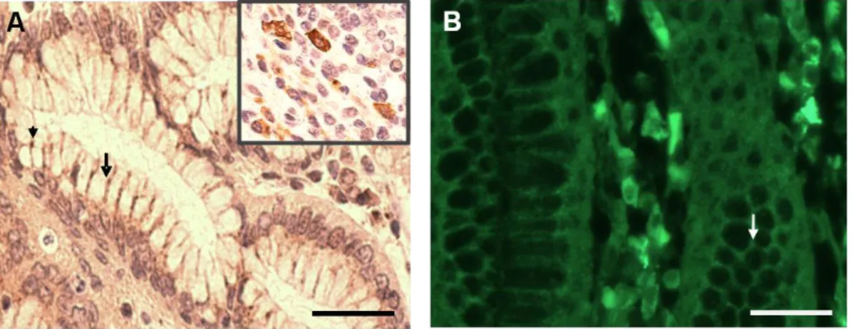
A avaliação da presença da TRIC em cortes histológicos de estômago humano em parafina foi realizada através da técnica de imunohistoquímica. Os resultados obtidos demonstraram a expressão da proteína nas junções tricelulares e bicelulares das células epiteliais gástricas, assegurando assim que estavam estabelecidas as condições experimentais óptimas para a investigação da TRIC no cérebro.
Os resultados da imunofluorescência nas células da linha celular endotelial monstraram a presença da proteína nas células endoteliais dos capilares cerebrais humanos e que a mesma é expressa nos contactos tricelulares, encontrando-se presente também nas junções bicelulares. Os ensaios de western blot evidenciaram uma banda de aproximadamente 64-kDa, correspondente à proteína TRIC. A expressão de mRNA foi também confirmada pela técnica de PCR quantitativo em tempo real. Os resultados de
western blot e PCR obtidos para as células endoteliais da linha celular foram confirmados
pelos das células da cultura primária, mas demonstram diferenças no nível da expressão, quer da proteína, quer do mRNA. Com esta técnica pareceu haver uma diminuição de expressão de aproximadamente 20-30% nas células endotelias da cultura primária, quando comparada com as células da linha celular. Este resultado pode ser justificado pela heterogeneidade das células, bem como das respectivas culturas. A presença de TRIC no endotélio dos capilares do cérebro foi corroborada através de estudos in situ feitos em cortes histológicos de rato, utilizando a técnica de imunohistoquímica. Interessantemente, os resultados evidenciaram também a expressão de TRIC nas junções tricelulares e bicelulares das células do plexo coroideu, o que seria esperado, uma vez que está descrita a presença de junções intercelulares nessas células que compõem a barreira sangue-líquido céfalo-raquidiano. Os resultados da técnica de imunofluorescência nos cortes histológicos de hipocampo humano estão de acordo com os resultados obtidos para rato. Efectivamente, os a análise in situ evidenciou a expressão da TRIC nas junções intercelulares das células endoteliais dos capilares cerebrais.
Apesar de se saber que a BHE envolve diferentes tipos de células, até hoje não foi estudada a presença da TRIC nas células neuronais e não neuronais do cérebro. Assim sendo, no presente trabalho foi estudada também a presença da TRIC em outros tipos de células que não as endoteliais, mais especificamente nos neurónios e na micróglia. A expressão de mRNA foi evidenciada em todas as amostras, parecendo haver uma menor expressão nos neurónios e na micróglia, quando comparada com as células endoteliais da
RESUMO
linha celular (aproximadamente 7% e 3%, respectivamente). De facto, a presença de proteínas de oclusão, como as claudinas e a ocludina, já foram reportadas em culturas primárias de neurónios e astrócitos. Para além disso, está descrita a presença das mesmas em cérebro humano, e sendo a TRIC uma proteína de oclusão da família da ocludina, a presença da mesma nos neurónios é um resultado provável. O mais interessante é o resultado da imunofluorescência na micróglia, que evidencia a expressão da proteína no citoplasma dessas células, sugerindo que a TRIC, de alguma forma, contribui para o sistema imune do cérebro. De facto, há estudos que descrevem a presença de TRIC, para além de outras proteínas de junção intercelular, em células dendríticas diferenciadas, as quais fazem parte do sistema imunitário. Existem ainda estudos que sugerem que as células imunocompetentes do cérebro, nomeadamente a micróglia, derivam da linhagem monocítica. Inesperadamente, não foi verificada a expressão da proteína nos neurónios. Uma possível explicação é o facto de não haver expressão proteica suficiente para que possa ser detectada por western blot e imunofluorescência. Por outro lado, duas outras hipóteses devem ser consideradas: haver expressão da proteína apenas em algumas circunstâncias, ou não haver, de todo, expressão proteica. De facto, há estudos que sugerem que a ocludina e a TRIC têm origem no mesmo gene, o qual duplicou ao longo da evolução filogenética, e tendo em conta também a presença de ocludina nos neurónios, é provável haver expressão de mRNA da TRIC nessas células. No entanto, é possível que haja expressão do mRNA sem necessariamente ocorrer a tradução do mesmo. Ainda assim, esta ausência de expressão poderá ser ultrapassada em situações pontuais, nomeadamente em patologias do SNC, em que ocorre sobre-expressão de proteínas de oclusão, como as claudinas e a ocludina. Reunindo estes dados com os resultados do presente estudo, talvez possamos especular que a função da TRIC esteja relacionada com a preservação da BHE. No entanto, são necessários mais estudos para completar os resultados obtidos.
Concluindo, este estudo revela pela primeira vez a presença da TRIC no endotélio dos capilares cerebrais humanos, indicando que esta proteína faz parte das junções intercelulares, contribuindo assim para a função de barreira entre o sangue e o encéfalo. O presente estudo demonstra também pela primeira vez a presença da TRIC em células não epiteliais e abre novos caminhos para estudos futuros, nomeadamente qual o papel da TRIC nas células nervosas.
Palavras-chave: Barreira hemato-encefálica; Unidade Neurovascular; Células endoteliais
C
HAPTER
1
1. I
NTRODUCTIONThe blood–brain barrier (BBB), a dynamic and complex interface between the blood and the central nervous system (CNS), was first identified in 1883 by Paul Ehrlich. The BBB plays a critical role in maintaining the stability of the CNS microenvironment (Li et al., 2010). The BBB function is therefore best described as a physical and metabolic barrier with selective transport systems acting as a signaling interface between brain and blood (Carvey et al., 2009). The specific structural and biochemical properties of the BBB arise from existent layers between the blood and the brain and from interactions between large varieties of cell types (Bernas et al., 2009).
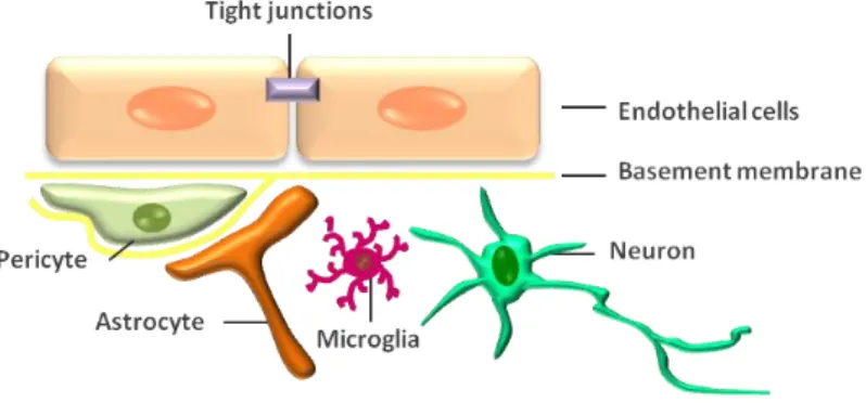
Endothelial cells (EC) of brain microvessel walls, whose apposing plasma membranes form tight junctions (TJs) that physically limit the paracellular passage to water and solutes between the blood and the brain (Romero et al., 2003) form the anatomic basis of the BBB (Cardoso et al., 2010). Together with neurons, microglia, astrocytes, as well as perivascular pericytes, there is a construct called the neurovascular unit (NVU) (Fig. 1), essential for both health and function of the CNS (Hawkins and Davis, 2005; Persidsky et al., 2006). In fact, the disruption of the BBB integrity has been shown to occur in many neurological disorders, such as Alzheimer's disease, stroke and HIV-1 encephalopathy (Huber et al., 2001; Petty and Lo, 2002), and it was recently proposed that BBB impairment contributes to the onset and progression of some neurologic disorders (Zlokovik, 2008).
Fig. 1. SiSimmpplliiffiieedd rrepeprreesseennttaattiioonn ooff tthhee nneeuurroovvaassccuullaarr uunniitt.. The brain microvessel wall consists of endothelial cells, whose apposing plasma membranes form tight junctions that seals aqueous paracellular diffusional pathway between the cells. Pericytes are adjacent to capillaries and share a common basement membrane with endothelial cells. Astrocyte endfeet form a lacework of fine lamellae closely apposed to the outer surface of the BBB endothelium and respective basement membrane. Astrocytes have a close relationship with neurons which innervate the endothelial cells and astrocytic processes. Microglia, another cell type with close contact with capillaries is the immunocompetent resident cells of the brain.
INTRODUCTION
1.1. Neurovascular unit
1. 1. 2. Neurons
There is some evidence that neurons can regulate the function of blood vessels in response to metabolic requirements by inducing expression of enzymes unique for brain microvascular endothelial cells (BMEC) (Persidsky et al., 2006). However, little is known about the role that neurons have on BBB phenotype. BMEC and astrocyte processes are directly innervated by neurotransmitters, suggesting their potential involvement in BBB regulation (Choin and Kim, 2008; Carvey et al., 2009). As neuronal function is heterogeneous throughout the brain, it follows that brain microvascular endothelia cells (BMEC) function, as well as BBB integrity and function are heterogeneous in the brain (Carvey et al., 2009).
1. 1. 3. Microglia
Microglia, another cell type in close contact with cerebral blood vessels (Cardoso et al., 2010;Correale and Villa, 2009), reside within the CNS parenchyma (Streit, 2002) and engage several important roles in the developing brain (Cuadros and Navascues, 1998; Kim and de Vellis, 2005), as well as in pathological conditions (Block and Hong, 2005; Nakajima and Kohsaka, 2004). Microglia are responsible for the immune surveillance and innate immune response in the brain and proliferate early in the embryonic environment, although most authors believe that these cells derive from the monocyte/macrophage lineage (Alliot et al., 1999; Chan et al., 2007). These cells present themselves in two forms, resting and activated microglia, changing the phenotype in response to homeostatic disturbance of the CNS. When resting, cells have small bodies and long thin process. Activated microglia, what are called “reactive microglia”, display a complexity of phenotypic alterations and assume a phagocytic morphology (Zlokovic, 2008). These cells interact with BMEC, which is thought to contribute to the BBB properties (Choin and Kim, 2008).
1. 1. 4. Astrocytes
Astrocytes are glial cells whose end-feet engulf the capillary networks of the brain (Abbot, 2002) that have long been assumed to significantly influence neurovascular structure and integrity. This is underscored by the close apposition of the astrocytic end-feet on the capillary walls and their close relationship with neurons. Thus, astrocytes are in a position to disperse vascular nutrients away from the vessels in support of neural function. They also induce and modulate the development of the BBB and BMEC unique phenotype, enhancing
INTRODUCTION
TJ and reducing gap junctional area (Abbot et al., 2006). In vitro studies suggest that BMEC grown in the presence of astrocytes or astrocyte conditioned media increases endothelial cells (EC) resistance and increases expression of TJ proteins (Abbott, 2002; Cardoso et al., 2010; Deli, 2009).
Recent studies by Carvey et al., (2009) suggest that elements of the basement membrane contributed by the astrocyte may participate in TJ formation by stabilizing the BMEC and inducing protein expression changes in the BMEC It is therefore more likely that interactions among the BMEC, astrocytes, pericytes and the basement membrane are all required to initiate complete TJ formation (Carvey et al., 2009).
1. 1. 5. Pericytes
In addition to BMEC, the pericytes is thought to be a critical component of a functional BBB, although we know little about it. Pericytes are vascular cells adjacent to capillaries that share a common basement membrane with EC and have many cytoplasmic process encircling capillaries (Shepro and Morel, 1993). These cells are sprinkled along the vascular endothelium in microvascular networks and form gap junctions with BMEC suggesting an intimate functional relationship (Fisher, 2009). Pericytes also communicate with other elements of the NVU through signaling pathways (Nakaoke et al., 2007). These actin-expressing cells have a number of vasoactive signaling receptors and release laminal proteins, suggesting their involvement in microvascular capillary flow and in the differentiation of the BBB (Dore-Duffy et al., 2006). Association of pericytes with microvascular endothelia is essential to maintain structural and junctional integrity (Braun et al., 2007). Products from pericytes may also be involved in initiating TJ formation (Kim et al., 2009) and both pericytes and EC shareTJs, adhesion plaques and soluble factors (Bagley et al., 2005).
1. 1. 6. Endothelial cells.
The cerebral endothelium forms the basis of the BBB in mammals and higher vertebrates (Hawkins et al., 2006) and the capillaries make up the primary part of the BBB (Khan, 2005). Electron microscopy studies of the BBB have identified distinct morphologic and metabolic characteristics of these particular EC, different to those present in peripheral tissues (Brightman and Reese, 1969; Lane et al., 1992). BMEC are characterized by the absence of fenestrations, a low level of pinocytic vesicles and elaborate junctional complexes that eliminate gaps or spaces between cells. Therefore, endothelial cell monolayers prevent any free diffusion of blood-borne substances into the brain parenchyma (Carvey et al., 2009;
INTRODUCTION
Chaudhuri, 2000; Zlokovic, 2008). Moreover, emdothelial cells display high transendothelial electrical resistance and cause severe restriction of the paracellular pathway for diffusion of hydrophilic solutes (Abbott, 2002). However, essential nutrients and other compounds are able to cross into the CNS by using specific membrane transporter systems (de Boer and Gaillard, 2006). The presence of elaborated junctional complexes that includes mainly TJ and adherens junction (AJ) proteins characterizes the cerebral microvasculature lining (Cardoso et al., 2010; Zlokovic, 2008).
2. Intercellular junctions
An important property of epithelial and EC is their assembly into a physical, and ion- and size-selective barrier separating tissues (Niessen, 2007). In mammals, there are elaborate structures composed of integral membrane proteins (Chiba et al., 2008; Krause et al., 2008), linker or adaptor proteins connecting them to the actin cytoskeleton, and signalling molecules (Paris et al., 2008), which form the intercellular junctions. Among these are included AJ and TJ, which play a crucial role in the formation and maintenance of epithelial and endothelial barriers (Niessen, 2007). While in epithelial barriers AJ are situated in the apical junctional complex in basolateral direction from the TJs, in brain EC TJs and AJ are intermingled (Vorbrodt et al., 2004). Another identified function of intercellular junctions is that they provide the cell not only with structural integrity but also function as landmarks, spatially confining signaling molecules and polarity cues as well as serving as docking sites for vesicles (Nelson, 2003).
2. 1. Tight junctions
TJ are networks of transmembrane proteins that are responsible for the barrier properties. These elaborate structures located on the apical region of EC, function as a seal that regulates lateral diffusion between the apical and basolateral plasma membrane domains, forming a semi-permeable barrier to paracellular flux (Cardoso et al., 2010; Hawkins and Davis, 2005; Hawkins and Egleton, 2006; Persidisky et al., 2006). Claudins, occludin (marvelD1), tricellulin (marvelD2), marvelD3 and junctional adhesion molecules (JAM) are integral membrane proteins interacting with those of neighbouring plasma membrane and forming a barrier. The transmembrane proteins of the TJ are often grouped according to the number of times they span the plasma membrane: JAM is a single-pass transmembrane protein, while occludin, claudins, tricellulin (TRIC) and marvelD3 are four-pass (Cardoso et al., 2010). The function of some of these proteins in epithelial barrier, their
INTRODUCTION
relevance in cellular and epithelial differentiation, as well in infections and inflammatory diseases have been defined in recent years (Michels et al., 2009; Reuter et al., 2009; Schulzke and Fromm, 2009; Sonoda et al., 1999; Watson et al., 2009).
2. 1. 1. Tight junction proteins
2. 1. 1. 1. Occludin
Occludin was the first component of TJ to be identified (Furuse et al., 1993). Occludin is associated with intramembranous strands, although it is not required for their assembly (Balda and Matter, 2008). It is a 60-kDa protein that modulates the barrier function, but disruption of occludin expression does not affect TJ structure (Du et al., 2010; Yu et al., 2005). This TJ is highly expressed and consistently stains in a distinct continuous pattern along the cell margins in the cerebral endothelium, whereas it is much more sparsely distributed in non-neural endothelium (Hawkis and Davis, 2005). One of its N- and C-terminal cytosolic domains is sufficient to anchor the protein in the junction and to ensure a continuous junctional distribution, but they have different functional properties. The N-terminal domain regulates neutrophil transmigration and the C-terminal domain regulates the paracellular diffusion and target the occludin to the basolateral membrane, connecting then to the cytoskeleton via accessory proteins zonula occludens (ZO)-1, ZO-2 and ZO-3 and is thought to interact with a ubiquitin ligase (Deli et al., 2010; Shimizu et al., 2008). Occludin can also recruit signal transduction molecules to TJ (Du et al., 2010). However, the underlying molecular mechanisms of many of the processes to which occludin has been linked remain to be determined and the functional relevance of most of its biochemical interactions is still unknown. (Deli et al. 2008)
2. 1. 1. 2. Claudins
Claudins, from Latin word “claudere” meaning to close was identified as a major constituent of bTJ strands. There are 24 distinct claudin family members that have been described in vertebrates (Furuse et al., 1998). Claudins range in size from 20 to 34-kDa and are integral membrane proteins that share four transmembrane domains of occludin, but do not contain any sequence homology to occludin (Wolburg and Lippoldt, 2002; Ikenouchi et al., 2005). In vitro studies have shown that claudins are capable of inducing the formation of TJ and that individual claudins vary in their ability to form a tight or leaky barrier (Furuse et al., 1998). Claudin-1, -2, -3, -5, -11, -12 and -18 are the only claudins family proteins that
INTRODUCTION
were detected in mammalian BMEC (Petty and Lo, 2002; Bélanger et al., 2007; Krause et al.,2008; Mahajan et al., 2008; Neuhaus et al., 2008.; Romanitan et al., 2009).
Claudin-1 and claudin-5 are associated with maintance of normal BBB function (Vorbrodt and Dobrogowska, 2003) and thought to be important in angiogenesis (Wolburg and Lippoldt, 2002). It was also suggested that extracellular loops of claudins create charge-selective paracellular aqueous pores that permit the passive diffusion of ions between cells (Krause et al., 2008; Van Itallie and Anderson, 2006). However, structural evidence for such pores is still missing.
2. 1. 1. 3. JAM
JAM, another integral membrane protein of TJ family, is involved in intercellular adhesion between the cells of barriers, as well as in adhesion between barriers and cells of immune system (Chiba et al., 2008; Paris et al., 2008). JAM proteins are members of the immunoglobulin superfamily and consist of JAM-1, JAM-2 and JAM-3. These adhesion molecules have 40-kDa and are expressed in TJ of EC (Vorbrodt and Dobrogowska, 2003). While JAM-1 is involved in tight junctional structure organization of cell-to-cell and contributes to barrier function and cell polarity in epithelial and EC, JAM-2 and JAM-3 interact each other and participates in spermatogenesis (Chiba et al., 2008). In addition, both JAM-2 and JAM-3 control leucocytes transmigration (Ogasawara et al., 2009; Stamatovic et al., 2008), another important aspect of barrier functions.
2. 1. 1. 4. MarvelD3
MarvelD3 is a third TJ-associated MARVEL domain protein (contains a conserved
four-transmembrane MARVEL domain – MAL and related proteins for vesicle trafficking and
membrane link). It is located on chromosome 16 and expressed by all vertebrates. Steed et al (2009) identified this novel protein by phylogenetic studies, RNA and immunofluorescence analysis.
It is known that this novel TJ protein is expressed as two isoforms (Steed et al., 2009) and is present in both bicellular and tricellular contacts (Raleigh et al., 2010). It is thought that marvelD3 is not essential for junction assembly and formation of a functional paracellular diffusion barrier. On the other hand, it is a determinant of paracellular ion permeability (Steed et al., 2009), but the mechanisms responsible for that property have to be identified. In addition, marvelD3 is able to partially compensate for occludin or TRIC loss but, they do not complement one another functionally (Raleigh et al., 2010). Maybe structural differences are
INTRODUCTION
responsible for that. In fact, the C- and N-terminal cytoplasmic domains of marvelD3 do not have the elogation factor RNA polymerase II (ELL) domain (Raleigh et al., 2010), which is present in both domains of occludin and TRIC and are important as an elongation factor that can increase the catalytic rate of RNA polymerase II transcription by suppressing transient pausing by polymerase (Zhou et al., 2009). Furthermore, the C-terminal domain is not able to interact with ZO-1. In contrast, the N-terminal domain is long and might provide a regulatory function (Raleigh et al., 2010; Steed et al., 2009). Together with occludin and TRIC proteins, marvelD3 constitute the complete TAMP (Tight junction Associate Marvel Protein) sub family of MARVEL proteins, contributing to epithelial regulation in a distinct way (Raleigh et al., 2010).
2. 1. 1. 5. Zonula occludens
TJ are formed by not only the integral membrane proteins occludins, claudins, JAMs, TRIC and marvelD3, but also many peripheral membrane proteins, including the cytoplasmic TJ accessory proteins, such as zonula occludens-1, -2 and -3 (ZO-1, ZO-2, ZO-3, respectively), which interact with each other and connect integral proteins to the actin cytoskeleton (Lee et al., 2004; Schulzke and Fromm, 2009). All these have in common the presence of a SH3 (Src homology 3) domain, a guanylate kinase homology (GK) domain (Anderson et al., 1995), and at least one PDZ (Post-synaptic density 95, Disk-large and ZO-1 proteins) domain, which recruit signaling proteins (McGee et al., 2001). It is a membrane-associated guanylate kinase protein (MAGUK‟s – modular scaffolds that organize signaling complexes at synapses and other cell junctions.), a submembranous TJ-associated protein (Persidisky et al., 2006) that plays a central role in the macromolecular assembly of TJ by interacting with integral proteins of the membrane of adjoining cells (Schulzke and Fromm, 2009), with the actin cytoskeleton, and with nuclear factors (Wolburg et al., 2009).
Human ZO-1, the first TJ-associated protein identified and characterized, is a 220-kDa phosphoprotein mostly expressed in endothelial and epithelial cells (Kniesel and Wolburg, 2000). In some specialized endothelia it is expressed as variable amounts of two related
isoforms, which originate from the alternatively spliced mRNA transcripts α+ and α– (Ciana et
al., 2010) and whose specific differential role is still unknown. This protein is not only a structural protein, but also a nuclear factor influencing gene expression (Wolburg et al., 2009). Loss or dissociation of ZO-1 from the junctional complexes is associated with increased barrier permeability (Choin and Kim, 2008). ZO-2 binds to the transmembranous proteins of the TJ and transcription factors (Persidisky et al., 2006) and ZO-3 binds occludin
INTRODUCTION
and ZO-1 directly, but not ZO-2 (Kniesel and Wolburg, 2000). Furthermore, ZO-1, ZO-2 and ZO-3 form a protein complex that can be involved in cadherin-based cell adhesion through their binding to catenin and actin filaments (Deli, 2009).
2. 1. 2. Types of tight junctions
TJ between two adjacent cells are called bicellular TJ (bTJ) whereas at tricellular contacts, where there are three epithelial cells meeting together, there is a structurally specialized organization, named as tricellular TJ (tTJ) (Ikenouchi et al., 2005). Claudins, occludin, JAM and MarvelD3 are proteins expressed at bicellular contacts. Therefore, claudins, occludin (Ikenouchi et al., 2005; Ikenouchi et al., 2008) and marvelD3 are also found in tTJ (Raleigh et al., 2010). These specialized structures were first observed between three plasma membranes by freeze-fracture electron microscopy (Friend and Gilula 1972; Staehelin et al. 1969; Staehelin 1973; Wade and Karnovsky 1974). tTJ consist of isolated sets of paired strands, which have an orientation parallel to the longitudinal axles of epithelial cells and perpendicular to the main orientation of the bicellular tight-junctional belt. At tTJ, the most apical elements of the horizontal TJ strands from both sides attach and turn together to extend vertically in the basal direction and strands of bTJ and tTJ interconnect and form one continuous complex (Staehelin, 1974). As a result, forms a vertically orientated triple pair strand structure with a 10 nm “central tube” (Staehelin, 1973; Wade and Karnovsky, 1974; Walker et al., 1994). Recently, TRIC was identified as a tricelular TJ protein located where tree cells meet (Ikenouchi et al, 2005).
3. Tricellulin
3. 1. Discovery and known localization
It was recently reported by Ikenouchi et al. (2005) the existence of a special protein of the occludin type, called TRIC at „„three corner‟‟ contact sites of simple epithelial cells of intestine, stomach and kidney, either in monolayer cell cultures or in situ. In line with the findings obtained by Ikenouchi and colleagues in mouse and dog species, we detected the presence of the protein at tricellular contacts between epithelial cells, as well as in immune cells, of the human stomach. Other studies, performed by Schlüter et al. (2007), demonstrated the presence of TRIC in epidermal and other stratified epithelial cells.
TRIC was the first protein found to be concentrated at tricellular contacts in vertebrates.
INTRODUCTION
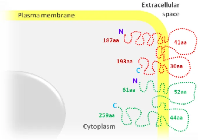
also located at tricellular contacts (Schülte et al., 2003), although these two proteins do not show any sequence similarity. TRIC is a tetraspan protein that is concentrated at the vertically oriented TJ strands of tricellular contacts of epithelial cell sheets, and contains four transmembrane domains with N- and C-terminal cytoplasmic tails, two extracelluar loops and a short intracellular loop between transmembrane domains 2 and 3 (Fig. 2.)
Fig. 2. SSiimmpplliiffiieedd mmooddeell ooff ttrriicceelllluulliinn ((TTRRIICC)) ssttrruuccttuurree aanndd mmeemmbbrraannee ffoollddiinngg.. TRIC is a four-pass transmembrane
protein, with two extracellular loops, the first with 41 aminoacids (aa) and the second with 30 aa, a short intracellular loop between transmembrane domains, a N-terminal cytoplasmatic domain with 187 aa and a C-terminal that contains 193 aa. TRIC shares some homology with occludin, which has a shorter N-C-terminal (61 aa) and a longer C-terminal domain (259 aa).
3. 2. Tricellulin regulation
It was referred by Ikenouchi et al. (2005) that the TRIC C-terminus has 32% homology with occludin. In fact, TRIC and occludin are located in tandem on human chromosome 5 and it has been speculated that they may have arisen from gene duplication during phylogenetic evolution (Steed et al., 2009). However, despite this homology, occludin and TRIC are differentially phosphorylated in their C-terminal tails: they differ in respect to serine/threonine casein kinases CK1- and CK2-mediated phosphorylation (Dörfel et al., 2009). TRIC C-terminus is not targeted by either of these kinases but also could not detect binding of the kinases to the TRIC C-terminal domain. These observations can be correlated not only with a distinct localization of protein kinase bind, but also differences in the regulation of TRIC
function (Dörfel et al., 2009). Protein kinase C (PKC) signal transduction pathways are known
to regulate epithelial barrier function via TJ (Tsukamoto et al., 1999). Phosphorylation of TJ proteins appears to play a central role in the regulation of TJ assembly and function. Different Ser/Thr- and Tyr-kinases have been identified as kinases that phosphorylate TJ components under different conditions and affect formation and stability of TJ and in consequence
INTRODUCTION
modulate barrier function and permeability properties (Feldman et al., 2005; González-Mariscal et al., 2008). Furthermore, it is known that when infective agents, free radicals, vasogenic agents and lipid mediators binds to respective receptors on the endothelial surfaces, signaling cascades are actived: protein kinases RhoA/Rho (ROCK) and mitogen-activated protein kinases (MAPK) are related with phosphorylation of TJs and AJ that cause the junction complex to dissociate from its cytoskeleton anchor, which leads to a weakened
cell-cell adhesion (Cardoso et al., 2010).
According to a recent publication by Westphal et al. (2010), a crosstalk between TRIC and occludin appears to exist, involving formation and organization of TJ strands. It was reported that in Madin-Darby canine kidney (MDCK) cell line, the TRIC C-terminal is important for the basolateral translocation of TRIC, whereas the N-terminal domain appears to be involved in directing TRIC to tricellular contacts. These observations differ from data reported by Ikenouchi et al. (2008) who showed that with both C- and N-terminal domains deleted, TRIC colocalize with claudin-1 at the cell surface. Westphal et al. (2010) demonstrated that the transport to the cell surface of TRIC with a deleted C-terminal domain was impaired and more intracellular aggregates were detectable. In contrast, the transport to the cell surface of TRIC with a deleted N-terminal domain was efficient. In addition, a construct with both C- and N-terminal domains deleted did not translocate TRIC to the cell surface and accumulated in intracellular aggregates (Ikenouchi et al., 2008; Westphal et al., 2010).
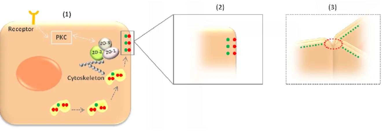
Homomeric TRIC-TRIC and heteromeric TRIC-occludin complexes were identified suggesting that both proteins are transported together to the edges of elongating bicellular junctions and get separated when tricellular contacts are formed. In this respect, it was assumed that homomeric interactions can form within endoplasmatic reticules and/or Golgi apparatus during the biosynthetic transport of the protein to the cell surface (Westphal et al., 2010). In addition with detected heteromeric TRIC-occludin complexes, Westphal et al. (2010) extend the mechanism for the tricellular localization of TRIC previously postulated by Ikenouchi et al. (2008). TRIC and occludin share a common transport pathway directing both proteins to elongation edges: TRIC and occludin form complexes in initial phases when they arrive at TJ and then separate when established tricellular contacts (Fig. 3).
INTRODUCTION
Fig. 3. SScchheemmaattiicc mmooddeell ffoorr ttrriicceelllluulliinn ((TTRRIICC)) llooccaalliizzaattiioonn aanndd rreegguullaattiioonn. (1) Protein kinase C (PKC) signal .
transduction pathway is involved in regulation of tight junctions (TJ), including TRIC. Zonula occludens proteins
(ZO-1, ZO-2 and ZO-3) interact with each other connect TRIC to actin cytoskeleton and recruit signaling proteins.
Homomeric TRIC-TRIC complexes in endoplasmatic reticules and/or Golgi apparatus during TRIC (red) protein
biosynthetic transport to the cell surface. Heteromeric TRIC-occludin (green) interactions in initial phases when they arrive at TJ. (2) Then separate when established tricellular contacts (3) and TRIC interact with another TRIC protein of opposed membrane.
Phosphorylation of TJ proteins appears to play a role in these regulations. In contrast with these data, another study reported that TRIC and occludin do not physically interact or that association is either unstable or uncommon, but shows an interaction between TRIC and marvelD3 and both occludin and marvelD3 (Raleigh et al., 2010). However, the same study shows that both TRIC and occludin mobilities were similar at bicellular and tricellular regions and they also hypothesized that TRIC and marvelD3 are delivered to the TJ immediately after synthesis, and in contrast, occludin and claudin-1 are redistributed from cytoplasmic stores to the TJ during barrier development.
3. 3. Tricellulin function
Knockdown of TRIC provided the first insight into the function of this protein. The loss of TRIC severely affected TJ organization, not only at tricellular contacts, but also bTJ were disorganized resulting in disturbed continuity of occludin distribution at bTJ with tear-drop-shaped accumulation of occludin at tTJ (Ikenouchi et al., 2008.On the other hand, in occludin knockdown MDCK cells, TRIC is mislocated to bTJ, probably to compensate occludin function (Ikenouchi et al., 2008). From these observations, it was concluded that in occludin knockout mice, TRIC may compensate at least for some of the occludin functions (Ikenouchi et al., 2005; Ikenouchi, et al., 2008). Consistent with this, TRIC and marvelD3 proteins increased in response to in vivo tumor necrosis factor (TNF) treatment; however, while
INTRODUCTION
occludin was redistributed into cytoplasmic vesicles, TRIC was enriched at bicellular and tricellular TJ, as well as within the cytoplasm (Raleigh et al., 2010).
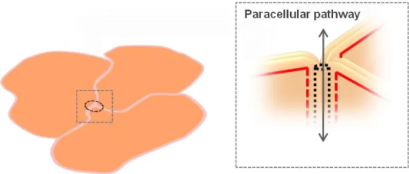
While TRIC knockdown affected TJ organization, its overexpression enhances barrier function. In fact, recent studies analyzing the physiological function of TRIC revealed that the presence of the protein in bTJ reduces strand discontinuities, increases paracellular electrical resistance and decreases permeability to ions and larger solutes, but without change in ion selectivity or charge preference (Krug et al., 2009). At tricellular contacts, TRIC specifically seals epithelial cell sheets against passage of macromolecules without significantly affecting ion permeation (Fig. 4). This property was explained by the fact that tricellular central tubes are wide enough for passage of macromolecules, but too rare to contribute significantly to ion permeability (Krug et al., 2009).
Fig. 4. SqSquueemmaattiicc ddrraawwiinngg ofof eeppiitthheelliiaall cceellllss.. In the uppermost of epithelial cells, tricellulin appears to be involved in sealing tricellular contacts, forming the central tube, which controls the paracellular flux of macromolecules. In this context, the tricellular central tubes represent an attractive target to enhance delivery of drug molecules across epithelial and endothelial barriers. It should be noted that TRIC and occludin knockdown are different from a recently published report, which showed no decreases in baseline or peak transendothelial electrical resistence (TER) (Raleigh et al., 2010). One possible explanation is the fact that Raleigh et al (2010) was used different kind of cell line for its experiments. In fact, transport and metabolic properties of cultured cells can vary due to culture conditions, seeding density, passage number, confluence, filter support, monolayer age, and stage of differentiation (Volpe, 2007). Moreover, during transport experiments, cell absorption properties can change due to the composition and pH of the transport buffer, solute concentration and solubility, temperature, additives and/or cosolvents, agitation, sampling schedule, sink conditions, and analytical methods (Volpe, 2007).
The first marker of the tTJ, TRIC, was recently identified in human nasal epithelial cells also, in vitro and in vivo, by Ohkuni et al., (2009). They described that TRIC expression is stable in human nasal epithelial cells and may play an important role for the sealing of the
INTRODUCTION
corner at tricellular contacts to prevent infiltration by various inhaled viruses and antigens. In addition, it was reported that TJ molecules in human nasal epithelial cells in part function via PKC signaling pathway. Recent study in human nasal epithelial suggest that peroxisome proliferator activated receptor (PPARγ) agonists, a nuclear hormone receptor superfamily member and a ligand-activated transcription are involved in the barrier functions and in the upregulation of TJ molecules in gastrointestinal epithelial cells, EC and urothelial cells (Varley et al., 2006; Huang et al., 2009). Investigators thought that these PPARγ agonists induces claudin-1, claudin-4, occludin and TRIC molecules, and enhances TJ barrier function. Furthermore, the PPARγ agonists affect a PKC signal transduction pathways, increasing phospo-panPKC activity (Ogasawara et al., 2010).
3. 3. 1. Mutations in tricellulin gene
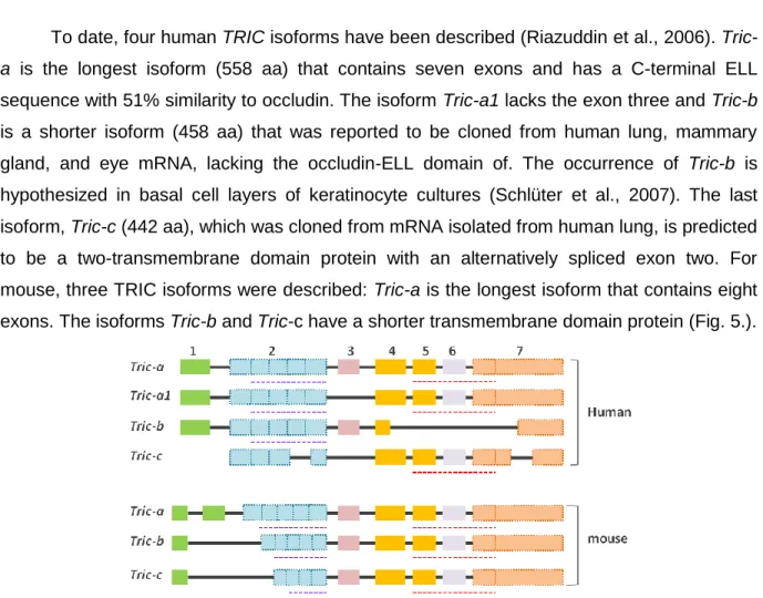
To date, four human TRIC isoforms have been described (Riazuddin et al., 2006).
Tric-a is the longest isoform (558 Tric-aTric-a) thTric-at contTric-ains seven exons Tric-and hTric-as Tric-a C-terminTric-al ELL
sequence with 51% similarity to occludin. The isoform Tric-a1 lacks the exon three and Tric-b is a shorter isoform (458 aa) that was reported to be cloned from human lung, mammary gland, and eye mRNA, lacking the occludin-ELL domain of. The occurrence of Tric-b is hypothesized in basal cell layers of keratinocyte cultures (Schlüter et al., 2007). The last isoform, Tric-c (442 aa), which was cloned from mRNA isolated from human lung, is predicted to be a two-transmembrane domain protein with an alternatively spliced exon two. For mouse, three TRIC isoforms were described: Tric-a is the longest isoform that contains eight exons. The isoforms Tric-b and Tric-c have a shorter transmembrane domain protein (Fig. 5.).
Fig. 5. AAlltteerrnnaattiivvee sspplliiccee iissooffoorrmmss ooff HHuummaann aanndd mmoouussee ttrriicceelllluulliinn ((TTRRIICC)).. The TRIC mRNA has seven exons and four alternative splice isoforms for Human (a, a1, b and c), while mouse (a, b and
Tric-c) has three. The blue dotted bars below exon 2 indicatethe localization of predicted transmembrane helices. The
INTRODUCTION
TRIC assembly at tTJ in cochlear and vestibular epithelial cells appears to play a key role in hearing, as mutations in the TRIC gene were associated with deafness (Riazuddin et al., 2006). It is known that TRIC protein is present at tricellular contacts of vestibular and cochlear epithelia and mutations of TRIC-a, the longest human isoform, have been reported to result in a human hereditary disease, nonsyndromic deafness (DFNB49). A common feature of the four DFNB49 TRIC alleles is that they encode predicted truncated proteins that lack the ability to bind to the scaffolding protein ZO-1, because of the loss of the conserved C-terminal occludin-ELL domain, and as previously described, this can interfere with the localization to TJs. In contrast, Raleigh, et al., (2010) suggested that TRIC may interact with ZO-1 at another site, because their studies show that C-terminus of TRIC was not able to bind ZO-1 GK domain. Alternatively, minor differences between the TRIC tails constructs used in both studies may explain the disparate observations. In fact, it has been speculated the relation between mutations in TJ gene and deafness. Mutations in claudin 14 gene are associated with different hearing thresholds, including profound and congenital deafness, but the role of this protein in hearing process is still unknown (Bashir et al., 2010; Ben-yosef et al., 2003). Yet, the only obvious phenotype of mutant alleles of Tric is deafness. It is possible that some other molecule compensates for the absence of wild-type TRIC in other epithelial cell types but not in the inner ear.
3. 4. Expression of tricellulin in non-epithelial cells
In spite of having reports dealing with expression of integral plasma membrane proteins specifically located at TJ like occludin, in primary cultures of neurons and astrocytes, particularly in undifferentiated state (Bauer et al., 2009), and despite the similar ectodermic origin of nervous tissue, TRIC presence in nerve cells has been underestimated, as it was only shown its expression by Northen blot analysis of mouse brain homogenates (Ikenouchi et al., 2005). Of interest is the TRIC mRNA expression in mature dendritic cells (Ogasawara et al., 2009). The mRNA expression of TRIC in human monocytes cell line THP-1, together with the presence of occludin, claudin-1 and ZO-1 in dendritic cells (Ogasawara et al., 2009), point to the establishment of TJ-like structures between dendritic and epithelial cells, which shall preserve the integrity of the epithelial barrier upon antigen sampling. The observation of such a role in the gut defense against bacteria, raises the question of whether TRIC also contributes to the brain immune system, namely through the resident immunocompetent cells, microglia.
INTRODUCTION
Barrier induction involves multiple agents and cell types (Cardoso et al., 2010; Carvey et al., 2009). However, nothing has ever been demonstrated about the influence of neuronal or non-neuronal brain cells on tricellular junctions between BMEC. On the other hand, the expression of tricelluliin brain neural cells has never been investigated, despite the several lines of evidence indicating the presence of TJ proteins in CNS cells.
Establishment of the cellular profile of TRIC expression in the brain shall provide valuable information about the neurovascular unit that ultimately shall open new opportunities for the development of new therapeutic strategies for prevention and treatment of central nervous system disorders.
4. Aim
The absence of reports about the expression of TRIC in the endothelial cell of brain capillaries, as in the other cells of the unit neurovascular, prompted us to investigate whether TRIC is present in human and in a rat brain. The precise and specific localization of TRIC in cells sites will be examined in cultures of HBMEC, as well as in the brain in situ. To establish the cell types of neurovascular unit expressing the protein, we will search the presence of
TRIC in primary culture of rat microglia and neurons, already in use in our laboratory. The
localization of TRIC will be highlighted by immunohistochemical and immunocytochemical methodologies, while the quantitative expression of the TRIC mRNA e respective protein will be determined by real-time PCR and western blot analysis, respectively.
By revealing the presence of TRIC in human brain, this study shall have an important impact in the field of cerebral vascular biology and opens new avenues for future research, namely on the role of TRIC in nerve cells.
C
HAPTER
2
2. M
ATERIAL ANDM
ETHODS 1. Human and rat samplesHuman stomach paraffin sections were obtained from the files of the department of
pathological anatomy of the Hospital São José. As human brain sample it was used discarded temporal lobe tissue obtained during neurosurgical operative procedure for treatment for intractable epilepsy, after informed consent and approval by the Ethics Commission of the Hospital Santa Maria, Lisbon.
Animal care followed the recommendations of European Convention for the Protection of Vertebrate Animals Used for Experimental and other Scientific Purposes (Council Directive 86/609/EEC) and National Law 1005/92 (rules for protection of experimental animals). All animal procedures were approved by the Institutional Animal Care and Use Committee. Every effort was made to minimize the number of animals used and their suffering.
2. Tissues samples preparation
2. 1. Human
Sections of human stomach were analyzed as a guarantee of the use of proper experimental protocols, since the presence of TRIC in the gastric epithelium was already reported (Ikenouchi et al., 2005).
Brain and stomach tissue was formaldehyde-fixed and, following macroscopic
assessment, was paraffin embedded, sectioned at 2 m and mounted on microscope slides
Superfrost® Plus (Thermo Scientific, Soeborg, Denmark) were then processed for immunohistochemical and immunofluorescence reactions.
2. 2. Rat
Wistar rats were sacrified by decapitation, under deep anesthesia. The brain was enucleated immediately, placed in a Petri dish containing ice-cold PBS and dissected. Tissue for histology was prepared as described above for human samples.
3. Cells Culture
3. 1. Primary cultures of human brain microvascular endothelial cells
Isolation and culture of HBMEC were performed as usual in our lab (Bernas et al., 2010). Briefly, the meninges and the white matter were removed from the brain tissue piece. The tissue was fragmented and homogenized, and the suspension was filtered through a
MATERIAL AND METHODS
polyester mesh with 500 µm pore-size. The collected pass-through fluid was filtered using a 30 µm mesh and the retained fragments were washed out. The microvascular fragments retained were ressuspended in isolation medium (DMEM/Ham‟s F12 supplemented with 10 % FBS, 1 % glutamax and 1 % antibiotic-antimycotic solution) and centrifuged at 30 g for 10 min. The cellular pellet was ressuspended in 4 ml of isolation supplemented medium with 50 µg/ml ECGS and 1 mg/ml heparin and then transferred to a collagen type I T-25 flask. The
flask was incubated at 37 °C in a humid atmosphere enriched with 5 % CO2. Cells were
monitored under contrast phase microscopy. At confluence, HBMEC were obtained by mild trypsinization with a trypsin-EDTA solution (0.05% trypsin, 0.02% EDTA in Hank‟s balanced salt solution) for 5 minutes at 37 ºC. All fluid containing cells was harvested and centrifuged at 30 g for 10 minutes and then the pellet was resuspended in 4 ml of isolation supplemented medium. The suspension was plated in a T-25 flask and in a 6 well plate (2 ml), previously coated with 2.5 ml or 0.8 ml collagen type I, respectively.
3. 2. Culture of human brain microvascular endothelial cell line
A cell line of HBMEC that showed to maintain morphologic and functional characteristics (Stins et al, 1997) were used. Cryopreserved cells were cultured in RPMI medium supplemented with 10 % FBS, 10 % NuSerum IV, 1 % NEAA, 1 % MEMvitamins, 1 mM sodium pyruvate, 2 mM L-glutamine and 1 % antibiotic-antimycotic solution (cell line
medium). The HBMEC line was seeded at a density of 100 x 103 cell/ml in collagen I-coated
plates and used after 48 hours in culture.
3. 3. Primary cultures of rat microglia and neurons
Mixed glial cultures were prepared from 1–2 day-old Wistar rats as previously described (McCarthy and de Vellis, 1980), with minor modifications, as usual in our lab (Gordo et al., 2006). Cells were maintained in culture until 21 days and microglia were isolated by mild trypsinization, as previously described by Saura et al. (2003). The use of 21 days in vitro (DIV) cells intents to achieve the maximal yield and purity of the cultures. Microglia were obtained for western blot, immunocytochemistry and RNA isolation for real-time PCR analysis.
Neurons were isolated from foetuses of 17–18-day pregnant Wistar rats, as previously described (Silva et al., 2002a). Neurons at 3 DIV were obtained for western blot, immunocytochemistry and RNA isolation for real-time PCR analysis.