Physical Exercise and Regulation of Intracellular Calcium in
Cardiomyocytes of Hypertensive Rats
Joel Alves Rodrigues,
1Thales Nicolau Prímola-Gomes,
1Leôncio Lopes Soares,
1Tiago Ferreira Leal,
1Clara Nóbrega,
2Danillo Laviola Pedrosa,
1Leonardo Mateus Teixeira Rezende,
1Edilamar Menezes de Oliveira,
2Antonio Jose Natali
1Universidade Federal de Viçosa (UFV),1 Viçosa, MG - Brazil Universidade de São Paulo (USP),2 São Paulo, SP - Brazil
Mailing Address: Joel Alves Rodrigues •
Rua Ana koester, 65 AP 302. Postal Code 36570-000, Centro, Viçosa, MG – Brazil E-mail: joel.a.rodrigues1@gmail.com, joel.rodrigues@ufv.br
Manuscript received September 27, 2017, revised manuscript March 15, 2018, accepted April 11, 2018
DOI: 10.5935/abc.20180113
Abstract
Background: Regulation of intracellular calcium (Ca2+) in cardiomyocytes is altered by hypertension; and aerobic
exercise brings benefits to hypertensive individuals.
Objective: To verify the effects of aerobic exercise training on contractility and intracellular calcium (Ca2+) transients
of cardiomyocytes and on the expression of microRNA 214 (miR-214) in the left ventricle of spontaneously hypertensive rats (SHR).
Methods:SHR and normotensive Wistar rats of 16 weeks were divided into 4 groups –sedentary hypertensive (SH); trained hypertensive (TH); sedentary normotensive (SN); and trained normotensive (TN). Animals of the TH and TN groups were subjected to treadmill running program, 5 days/week, 1 hour/day at 60-70% of maximum running velocity
for 8 weeks. We adopted a p ≤ 0.05 as significance level for all comparisons.
Results: Exercise training reduced systolic arterial pressure in hypertensive rats. In normotensive rats, exercise training reduced the time to 50% cell relaxation and the time to peak contraction and increased the time to 50% decay of the intracellular Ca2+ transients. In SHR, exercise increased the amplitude and reduced the time to 50% decay of
Ca2+ transients. Exercise training increased the expression of miR-214 in hypertensive rats only.
Conclusion: The aerobic training applied in this study increased the availability of intracellular Ca2+ and accelerated the
sequestration of these ions in left ventricular myocytes of hypertensive rats, despite increased expression of miR-214 and maintenance of cell contractility. (Arq Bras Cardiol. 2018; 111(2):172-179)
Keywords: Hypertension; Exercise; Rats; Calcium Signaling; Intracellular Calcium; Sensing Proteins.
Introduction
Sustained systemic arterial hypertension causes progressive myocardial remodeling. While cardiac function is increased in response to active myocyte hypertrophy in the left ventricle in the compensated phase, left ventricular (LV) remodeling is characterized by the combination of cardiomyocyte hypertrophy and proliferation of other tissues. For example, collagen deposition result in fibrosis of ventricular tissue
and consequent myocardial stiffness.1,2 This has been
reported in the initial compensatory phase of hypertension
(3-4 months), with preservation of cardiac function.1
Increased proinflammatory markers (e.g. IL-6; TNF-α) have
been found in the left ventricle of spontaneously hypertensive
rats (SHR) as young as 5-6 months.3,4
The benefits of aerobic training to hypertensive subjects
are well established in the literature.5,6 Animal models of
hypertension – SHR in the compensated stage (~ 6-month-old) – have shown the efficacy of this type of training in attenuating systolic dysfunction and restoring ventricular elasticity in female
SHR.7 Aerobic training reduced apoptosis in the myocardium
of SHR,4,8 improved contractile function of cardiomyocytes
isolated from the left ventricle, and normalized the expression of proteins involved in the regulation of intracellular calcium cycle, such as the sarcoplasmic reticulum Ca2+-ATPase
(SERCA2a) and phospholamban.9-11
In physiological or pathological cardiac hypertrophy, expression of microRNAs (miRNAs) related to cardiac
remodeling is altered.12 MicroRNA 214 (miR-214) is involved
in cardiac muscle contraction and Ca2+ sequestration, due to
its negative effect on SERCA-2a expression.13 With respect to
physical exercise, resistance training improved cardiomyocyte contractile function, with increased expression of SERCA2a and decreased miRNA-214 expression in normotensive,
infarcted rats.13,14 However, little is known about miR-214
Methods
Experimental animals
SHR and normotensive Wistar rats, 16 weeks of age, obtained from the central vivarium of the Biological Science and Health Center of Viçosa Federal University, were divided into four groups (of 13 animals each): sedentary normontensive (SN); trained normotensive (TN), sedentary hypertensive (SH) and trained hypertensive (TH). Sample size was determined by convenience. In each group, 8 animals were used for cardiomyocyte isolation, and 5 for analysis of gene expression. All procedures were carried out following the ethical principles of the Brazilian Society of Laboratory
Animal Science (COBEA, Colégio Brasileiro de Experimentação
Animal), and approved by the Ethics Committee on Animal Experimentation of Viçosa Federal University (CEUA-UFV; approval number 29/2014). The animals were housed in group cages (4 animals per cage) and allowed free access to water and chow at controlled temperature (mean of 22°C) and lighting (12:12 h light–dark cycle).
Protocol of treadmill exercise training and stress test
Before the training was initiated, animals were adapted to
the treadmill during a 5-day period, 10min/day, 0o inclination
at 5 m/min. After 48 hours, all animals were subjected to an incremental treadmill test for establishment of the maximal
running velocity (MRV), starting at 5 m/min, 0o inclination
and increments of 3m/min every 3 minutes until exhaustion. Exhaustion was defined as the time point when the animals could not run at the predetermined speed and, at this point, the test was stopped.
The TN and TH groups were subjected to an exercise training program for 8 weeks, 5 days/week (from Monday to Friday). The training started with treadmill running at 5-6 m/min
and 0o inclination for 10 minutes on the first day. In the first
week, exercise duration was increased in 5 minutes per day and the intensity was maintained. In the second week, duration continued to be increased in 5 minutes/day, but intensity was increased by 2% of MRV per day, so that from the first day of the third week to the end of the eighth week, the animals ran at 60% of MRV (~18-22 m/min) during a 60-minute period per day.
MRVs of each animal were determined before the training started and at the end of the fourth week in both TN and TH groups for measurement of the time to exhaustion (TTE) and definition of the training intensity. Forty-eight hours after the last training session, the tests were repeated in all animals for analysis of the effects of the physical training on running capacity.
During the experimental period, animals of the sedentary groups (SN and SH) were placed on the treadmill 3 days/week (Mondays, Wednesdays and Fridays), 10 minutes/day,
0o inclination at 5 m/min to subject them to similar conditions
of the trained groups.
Systolic arterial pressure (SAP) was measured in the beginning and in the end of the experimental period, i.e., 48 hours after the last exercise training session. The measurements were taken in the mornings, without anesthesia, by tail-cuff
plethysmography (LE 5001, Panlab, Harvard Apparatus, Spain). Three measurements were performed, and the intermediate value used for analysis.
Isolation of cardiomyocytes
Forty-eight hours after the last exercise session, isolation of
LV myocytes was performed as described by Locatelli et al.15
Briefly, the animals were weighed and euthanized by cervical dislocation. The heart was excised, weighed, cannulated and perfused in the Langendorff's mode with the isolation solution of the following composition (in mM): 130 Na+, 5.4 K+, 1.4 Mg+, 140 Cl−, 0.75 Ca2+, 5.0 Hepes, 10 glucose, 20 taurine and 10 creatine, pH 7.3, at room temperature. The heart was then perfused with the calcium-free solution containing 0.1 mM ethylene glycol-bis (ß-aminoethyl ether)-N, N, N’, N’-tetraacetic acid (EGTA), for 4 to 6 min. The perfusion was then changed to a solution containing 1.0 mg/mL collagenase type II (Worthington, USA) and 0.1 mg/mL protease (Sigma-Aldrich, USA) for 10-15 min. All the solutions were oxygenated (O2 100% - White Martins, Brazil) and maintained at 35°C. After perfusion, ventricles were separated from the atria and weighed. The ventricles were placed in a flask containing 5.0 mL of enzymatic solution (collagenase + protease). The flasks were briefly shaken for 5 min in a water bath at 37°C. Next, tissues were removed from the flasks and the remaining content centrifuged (3,000 rpm for 30s). The supernatant was removed, and the cardiomyocytes suspended in the isolation
solution and stored in refrigerator (5oC) until being used.
Measurements of cell contractility
Contractility of isolated myocytes was measured by evaluation of cell length using the motion edge detector (Ionoptix, Milton, MA-USA) mounted on an inverted microscope (Nikon Eclipse - TS100, Japan), as previously
described.15 Briefly, myocytes were placed in a chamber
with a glass coverslip base and bathed with a buffer solution containing (mM) 136.9 NaCl; 5.4 KCl; 0.37 NaH2PO4;
0.57 MgCl2; 5.0 Hepes = 5; 5.6 glucose and 1.8 CaCl2
(pH = 7.4 at room temperature). Cells were visualized on a monitor with a camera (Myocam, Ionoptix, at 240 Hz) attached to a microscope using an image detector system (Ionwizard, Ionoptix). External stimulation was applied at
1.0 Hz (20V) for 5 minutes at room temperature (~25oC)
via platinum electrodes and an electric field stimulator (Myopacer, Ionoptix). Motions of myocyte longitudinal borders were captured by the motion edge detector (Ionwizard, Ionoptix) and stored for posterior analysis. Only myocytes in good conditions, with clear borders and striated sarcomere, relaxed at rest and without voluntary contractions were selected for analysis. Myocyte contractions
were analyzed as previously described.15
Measurement of intracellular Ca2+ transients
Intracellular Ca2+ transients in isolated cardiomyocytes were
measured using fluorescence imaging (Ionoptix, USA), mounted on an inverted microscope (Nikon Eclipse – TS100, USA) equipped with oil immersion objective lens (S Fluor, 40x, Nikon,
Figure 1 – (A) Time to exhaustion of normotensive and hypertensive before (pre) and after (post) training. (B) Running velocity during training sessions. TN: trained
normotensive; TH: trained hypertensive. Data are expressed as mean ± SD of 8 animals in each group. * compared with TN (pre); # compared with TH (pre) (p < 0.05).
60
40
20
0
TN (pre) TN (post) TH (pre) TH (post) *#
*#
TN
TH
T
ime to exhaustion (min)
1 2 3 4 5 6 7 8
Weeks 35
30
25
15 20
A B
Running velocity (m/min)
incubated with calcium probe (5 µM for 10 minutes) (Fura-2AM, ThermoFisher, Waltham, USA). The ratio of fluorescence emission at 510 nm in response to excitation wavelengths of 340 nm to that in response to 380 nm wavelengths was
used as concentration index of intracellular Ca2+ transients.
The myocytes were electrically stimulated (Myopacer, Field Stimulator, Ionoptix, USA) by a pair of platinum electrodes with a 0.2 ms (20V) supra-threshold pulse, frequency of 1 Hz at room
temperature (~25 oC). Parameters of intracellular Ca2+ transients
were analyzed using the IonWizard software (IonWizard, 6.3, IonOptix, Milton, USA).
Gene expression analysis
For analysis of gene expression after euthanasia, LV samples
were collected and stored at -80oC. LV total RNA was isolated
in 1 mL of Trizol (Invitrogen) following the manufacturer’s
recommendations and stored at -70oC. RNA samples were
diluted 1:100 with water and analyzed by spectrophotometry at 260-280 nm.
Analysis of the miRNA-214 gene expression was performed using the Taqman MicroRNA Assays (Applied Biosystems) following the manufacturers’ recommendations. Gene expression quantification was performed in two stages: first, complementary DNA (cDNA) was obtained from reverse transcription of the total RNA sample using a stem-looped primer for reverse transcription to detect the miRNA analyzed and the TaqMan® MicroRNA Reverse Transcription Kit; second, by real-time polymerase chain reaction, PCR products were amplified from the cDNA samples previously obtained using the TaqMan® MicroRNA Assay and the TaqMan® Universal PCR Master Mix II. The U6 snRNA normalizer was used as control, and analysis was performed using the ABI 7500 Real Time-PCR Systems (Applied Biosystems).
Statistical analysis
Analysis of variance (ANOVA) assumptions regarding homogeneity of variances between the groups and normality
of observations were checked and no violations of the assumptions were detected. The variables had normal and continuous distribution. The Levene’s test, the chi-square test and the Kolmogorov test were used for analysis. Comparisons between initial and final SAP and TTE in each group were performed by the paired student’s t-test. Two-way ANOVA followed by Tukey post-hoc test was used for comparisons between the four groups. Analysis was performed using the SigmaPlot software (Systat Software, Inc., San Jose, CA, USA) and significance level was set at 5%. Results are expressed as mean ± standard deviation (SD).
Results
The physical training program increased TTE of both normotensive and hypertensive animals (Figure 1A). Figure 1B shows running velocity of these animals during training.
As compared with normotensive animals, hypertensive animals showed lower body weight (BW), similar LV weight (LVW), higher LVW/BW, whereas TH animals showed higher ventricular weight (VW)/BW (Table 1). Physical training had no effect on these parameters.
Results of SAP are presented in Figure 2. As compared with pre-training values, although the physical training program had no effect on SAP in normotensive animals, a reduction in SAP was observed in hypertensive animals (Figure 2A).
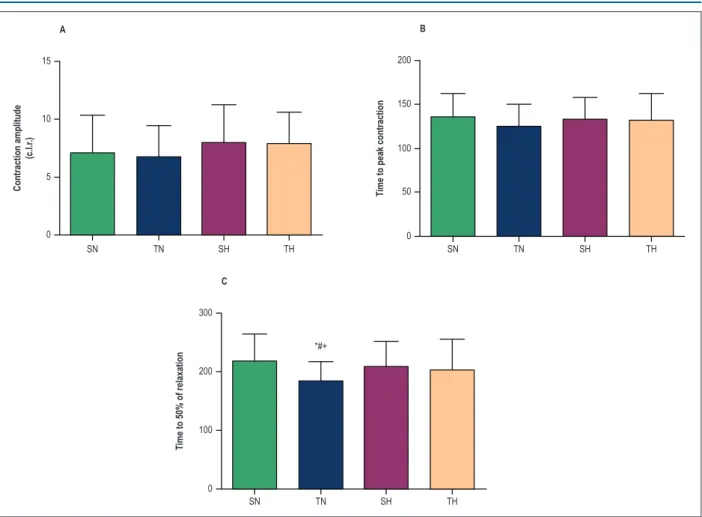
Cell contractility data are depicted in Figure 3. Contraction amplitude was not affected by hypertension or by physical training (Figure 3 A); and time to peak contraction did not change in any of the groups (Figure 3B). However, physical training reduced time to relaxation by 50% (Figure 3C) in normotensive, but not in hypertensive animals.
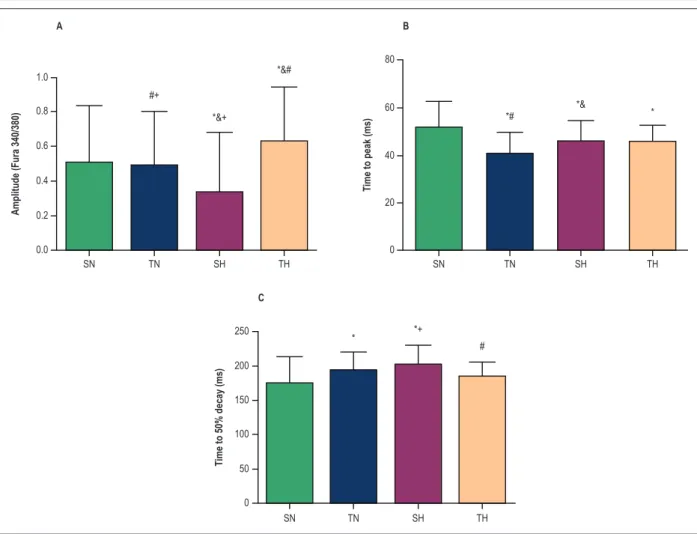
Figure 4 shows that after training, [Ca2+]
i amplitude
did not change in normotensive animals, but increased in the TH group as compared with SH animals. In addition,
physical exercise decreased the time to peak [Ca2+]
i in TN
Table 1 – Body weight and ventricular weight in the study groups
SN (n = 8) TN (n = 8) SH (n = 8) TH (n = 8) p-value
BW (g) 411.30 ± 21.51 447.10 ± 43.94 350.60 ± 21.97*& 338.90 ± 30.67*& 0.001
VW (g) 1.62 ± 0.20 1.49 ± 0.25 1.47 ± 0.17 1.66 ± 0.23 0.954
LVW (g) 1.12 ± 0.13 1.08 ± 0.13 1.18 ± 0.14 1.25 ± 0.15 0.265
VW/BW (mg/g) 3.97 ± 0.62 3.46 ± 0.47 4.19 ± 0.45 4.93 ± 0.80*& 0.000
LVW/BW (mg/g) 2.75 ± 0.42 2.42 ± 0.23 3.39 ± 0.39*& 3.72 ± 0.48*& 0.000
SN: sedentary normotensive; TN: trained normotensive; SH: sedentary hypertensive; TH: trained hypertensive; VW: ventricular weight; LVW: left ventricular weight;
n: number of animals; * compared with SN; & compared with TN (p < 0.05).
on this parameter in hypertensive animals. Longer time to
a 50% decay of [Ca2+]
i was observed in TN than SN group.
In hypertensive animals, however, this parameter was decreased after physical training (p < 0.05).
Although the physical training had no effect on mir-214 expression in normotensive animals (Figure 5), increased expression was found in TH as compared with SH and the other groups.
Discussion
In the present study, the authors evaluated the effects
of physical activity on contractility and intracellular Ca2+
transients in myocytes and miR-214 expression in the left ventricle in hypertensive rats. The results showed that the aerobic training not only reduced SAP in hypertensive animals, but also increased the amplitude of miR-214. No effect on LV myocyte contractility was observed.
The efficacy of the physical training applied in the study groups was confirmed by the higher physical capacity, indicated by the TTE, in trained animals as compared with controls. Such increase in physical capacity in response to
aerobic training has been previously demonstrated.10,17,18
More importantly, physical training reduced SAP in hypertensive animals, and such effect is well established
in the literature.11,19
Regarding LV cardiomyocyte contractility, although a reduction in the cell relaxation time in response to physical training was observed in normotensive animals, in SHR, cell contractility of trained animals was not different than that in sedentary animals.
Although cardiomyocyte contractility in SHR was not affected by exercise, higher amplitude and shorter 50%
decay time of intracellular Ca2+ transient levels were seen
in trained animals as compared with sedentary controls.
This suggests higher availability of Ca2+ in the cytosol and
faster removal of calcium from the cytosol, which in turn
brings about relaxation.20 These findings corroborate those
of another study,21 that showed increased expression of
SERCA2a, which is the main determinant of Ca2+ removal
from the cytosol into sarcoplasmic reticulum.20
With respect to miR-214, which antagonizes the effects of SERCA2a, our results contradict existing data in the literature, since the physical exercise program increased the expression of this microRNA in the left ventricle of hypertensive animals. We expected that aerobic training would reduce the expression of miR-214, which would justify de shorter time to
50% of decay in intracellular concentration of Ca2+ transients
in LV cardiomyocytes of SHR due to an expected increase in SERCA2a expression. Although SERCA2a expression in the left ventricle was not evaluated in the present study,
it was previously demonstrated14 that the left ventricle of
normotensive rats subjected to resistance training showed Figure 2 – Systolic arterial pressure (SAP) in normotensive animals and hypertensive animals. (A) SAP pre-training vs. post-training). (B) Final SAP of the groups. SN: sedentary normotensive; TN: trained normotensive; SH: sedentary hypertensive; TH: trained hypertensive. Data are expressed as mean ± SD of 8 animals in each group.
* compared with TN (pre) (A) SN (B); & compared with TN (post) (A) TN (B); # compared with TH (pre) (A) SN (B); * compared with TH-post (A) TH (B) (p < 0.05).
200
200 150
150 250
100
100
50
50
0 0
SAP
(mmHg)
SAP
(mmHg)
TN (pre) TN (post) TH (pre) TH (post) NS TN HS TH
*&+
*”&+ *&#
*”&#
Figure 3 – Cardiomyocyte contractility in normotensive and hypertensive animals. (A) Contraction amplitude expressed as percentage of change in cell length at rest (%c.l.r.) after electrical stimulation at 1HZ; (B) time to peak concentration; (C) time to 50% of relaxation; SN, sedentary normotensive; TN, trained
normotensive; SH, sedentary hypertensive; TH, trained hypertensive. Data as mean ± SD of 60-80 cells in each group. * compared with SN; # compared with SN;
+ compared with TH (p < 0.05).
A
C
B
15
10
5
0
SN TN SH TH SN TN SH TH
SN TN SH TH
Contraction amplitude
(c.l.r
.)
T
ime to peak contraction
T
ime to 50% of relaxation
200
100 150
50
0
200
100 300
0
*#+
reduced miR-214 expression and increased SERCA2a expression. These adaptations were associated with faster relaxation of myocytes isolated from the left ventricle of trained animals. Similar results showing reduction in miR-214 expression and elevated SERCA2a expression were also
reported in infarcted rats subjected to resistance training.13
Therefore, little is known about the effects of aerobic exercise on hypertensive cardiomyocytes, and further studies are needed to investigate other possible changes associated
with intracellular Ca2+ regulation in the cardiomyocytes of
hypertensive rats subjected to aerobic training.
Conclusion
The aerobic training applied in the present study increased
the availability of intracellular Ca2+ in the myocytes of the left
ventricle of hypertensive rats, despite the increased expression of miR-214 and maintenance of cell contractility.
Author contributions
Conception and design of the research: Rodrigues JA, Prímola- Gomes TN, Natali AJ; Acquisition of data: Rodrigues JA, Soares LL, Leal TF, Nóbrega C, Pedrosa DL, Rezende LMT, Oliveira EM, Natali AJ; Analysis and
interpretation of the data: Rodrigues JA, Soares LL, Leal TF, Nóbrega C, Oliveira EM, Natali AJ; Statistical analysis: Rodrigues JA, Soares LL, Leal TF, Nóbrega C, Natali AJ; Obtaining financing: Prímola-Gomes TN, Natali AJ; Writing of the manuscript: Rodrigues JA, Natali AJ; Critical revision of the manuscript for intellectual content: Natali AJ.
Potential Conflict of Interest
No potential conflict of interest relevant to this article was reported.
Sources of Funding
This study was funded by FAPEMIG (APQ-00876-14).
Study Association
This article is part of the thesis of master submitted by Joel Alves Rodrigues, from Universidade Federal de Viçosa.
Ethics approval and consent to participate
Figure 4 – Intracellular Ca2+ transient levels in cardiomyocytes of normotensive and hypertensive animals. (A) Amplitude of intracellular calcium.
(B) Time to peak intracellular calcium. (C) Time to 50% decay of intracellular calcium. SN, sedentary normotensive; TN, trained normotensive; SH, sedentary
hypertensive; TH, trained hypertensive. Data as mean ± SD of 40-50 cells in each group. * compared with SN; & compared with TN; # compared with SN;
+ compared with TH (p < 0.05).
SN TN SH TH SN TN SH TH
SN TN SH TH
Amplitude (Fura 340/380)
T
ime to peak (ms)
T
ime to 50% decay (ms)
A
C
B
#+
*# *&+
*& *&#
*
* *+
# 1.0
0.8
0.6
0.4
0.2
0.0
80
60
40
20
0
200 250
150
100
50
0
Figure 5 – MicroRNA-214 expression in the left ventricle in normotensive and hypertensive animals. SN: sedentary normotensive; TN: trained normotensive;
SH: sedentary hypertensive; TH: trained hypertensive. Data as mean ± SD of 5 animals in each group. * compared with SN; & compared with TN; # compared with SN (p < 0.05).
SN TN SH TH
*&# 800
600
400
200
0
Level of
1. Weber KT, Brilla CG. Pathological hypertrophy and cardiac interstitium. Fibrosis and renin-angiotensin-aldosterone system. Circulation. 1991;83(6):1849-65.
2. Díez J, Querejeta R, López B, González A, Larman M, Martínez Ubago JL. Losartan-dependent regression of myocardial fibrosis is associated with reduction of left ventricular chamber stiffness in hypertensive patients. Circulation. 2002;105(21):2512-7.
3. Miguel-Carrasco JL, Zambrano S, Blanca AJ, Mate A, Vázquez CM. Captopril reduces cardiac inflammatory markers in spontaneously hypertensive rats by inactivation of NF-kB. J Inflamm (Lond). 2010;7:21.
4. Huang CY, Yang AL, Lin YM, Wu FN, Lin JA, Chan YS, et al. Anti-apoptotic and pro-survival effects of exercise training on hypertensive hearts. J Appl Physiology (1985). 2012;112(5):883-91.
5. Pescatello LS, Franklin BA, Fagard R, Farquhar WB, Kelley GA, Ray CA, et al; American College of Sports Medicine. American College of Sports Medicine position stand. Exercise and hypertension. Med Sci Sports Exerc. 2004;36(3):533-53.
6. Sharman JE, La Gerche A, Coombes JS. Exercise and cardiovascular risk in patients with hypertension. Am J Hypertens. 2015;28(2):147-58.
7. Libonati JR, Sabri A, Xiao C, MacDonnell SM, Renna BF. Exercise training improves systolic function in hypertensive myocardium. J Appl Physiol (1985). 2011;111(6):1637-43.
8. Kolwicz SC, MacDonnell SM, Renna BF, Reger PO, Seqqat R, Rafiq K, et al. Left ventricular remodeling with exercise in hypertension. Am J Physiol Heart Circ Physiol. 2009;297(4):H1361-8.
9. Garciarena CD, Pinilla OA, Nolly MB, Laguens RP, Escudero EM, Cingolani HE, et al. Endurance training in the spontaneously hypertensive rat: conversion of pathological into physiological cardiac hypertrophy. Hypertension. 2009;53(4):708-14.
10. Carneiro-Júnior MA, Quintão-Júnior JF, Drummond LR, Lavorato VN, Drummond FR, Amadeu MA, et al. Effect of exercise training on Ca2+ release units of left ventricular myocytes of spontaneously hypertensive rats. Braz J Med Biol Res. 2014;47(11):960-5.
11. Carneiro-Júnior MA, Quintão-Júnior JF, Drummond LR, Lavorato VN, Drummond FR, da Cunha DN, et al. The benefits of endurance training in
cardiomyocyte function in hypertensive rats are reversed within four weeks of detraining. J Mol Cell Cardiol. 2013 Apr;57:119-28.
12. Romaine SP, Tomaszewski M, Condorelli G, Samani NJ. MicroRNAs in cardiovascular disease: an introduction for clinicians. Heart. 2015;101(12):921-8.
13. Melo SF, Barauna VG, Neves VJ, Fernandes T, Lara Lda S, Mazzotti DR, et al. Exercise training restores the cardiac microRNA-1 and −214 levels regulating Ca2+ handling after myocardial infarction. BMC Cardiovasc Disord. 2015 Dec 9;15(1):166.
14. Melo SF, Barauna VG, Júnior MA, Bozi LH, Drummond LR, Natali AJ, et al. Resistance training regulates cardiac function through modulation of miRNA-214. Int J Mol Sci. 2015;16(4):6855-67.
15. Locatelli J, Paiva NC, Carvalho SH, Lavorato VN, Gomes LH, Castro QJ, et al. Swim training attenuates the adverse remodeling of LV structural and mechanical properties in the early compensated phase of hypertension. Life Sci. 2017 Oct 15;187:42-9.
16. Natali AJ, Wilson LA, Peckham M, Turner DL, Harrison SM, White E. Different regional effects of voluntary exercise on the mechanical and electrical properties of rat ventricular myocytes. J Physiol. 2002;541(Pt 3):863-75.
17. Chen Y, Zhang H, Zhang Y, Lu N, Zhang L, Shi L. Exercise intensity-dependent reverse and adverse remodeling of voltage-gated Ca2+ channels in mesenteric arteries from spontaneously hypertensive rats. Hypertens Res. 2015;38(10):656-65.
18. Huang CC, Lin TJ, Chen CC, Lin WT. Endurance training accelerates exhaustive exercise-induced mitochondrial DNA deletion and apoptosis of left ventricle myocardium in rats. Eur J Appl Physiol. 2009;107(6):697-706.
19. Petriz BA, Almeida JA, Gomes CP, Ernesto C, Pereira RW, Franco OL. Exercise performed around MLSS decreases systolic blood pressure and increases aerobic fitness in hypertensive rats. BMC Physiol. 2015 Mar 14;15:1.
20. B e r s D M . C a r d i a c e x c i t a t i o n - c o n t r a c t i o n c o u p l i n g . N a t u r e . 2002;415(6868):198-205.
21. Carneiro-Junior MA, Primola-Gomes TN, Quintao-Junior JF, Drummond LR, Lavorato VN, Drummond FR, et al. Regional effects of low-intensity endurance training on structural and mechanical properties of rat ventricular myocytes. J Appl Physiol (1985). 2013;115(1):107-15.