Lack of detection of human papillomavirus DNA in prostate carcinomas in
patients from northeastern Brazil
Ari P. Araujo-Neto
1, Hygor Ferreira-Fernandes
1, Carolina M.M. Amaral
2, Lina G. Santos
3,
Antônio C. Freitas
2, Jacinto C. Silva-Neto
4, Juan A. Rey
5, Rommel R. Burbano
6, Benedito B. da Silva
3,
France K.N. Yoshioka
1and Giovanny R. Pinto
11
Laboratório de Genética e Biologia Molecular, Universidade Federal de Piauí, Parnaíba, PI, Brazil.
2Laboratório de Estudos Moleculares e Terapia Experimental, Departamento de Genética, Centro de
Biociências, Universidade Federal de Pernambuco, Recife, PE, Brazil.
3
Faculdade de Medicina, Universidade Federal do Piauí, Teresina, PI, Brazil.
4
Laboratório de Pesquisas Citológica e Molecular, Departamento de Histologia e Embriologia,
Centro de Biociências, Universidade Federal de Pernambuco, Recife, PE, Brazil.
5
Molecular Oncogenetics Laboratory, Unidad de Investigación, Hospital Universitario La Paz,
Paseo de la Castellana, Madrid, Spain.
6
Laboratório de Citogenética Humana, Instituto de Ciências Biológicas, Universidade Federal de Pará,
Belém, PA, Brazil.
Abstract
Prostate cancer is the second most common cancer among men in western populations, and despite its high mortal-ity, its etiology remains unknown. Inflammatory processes are related to the etiology of various types of tumors, and prostate inflammation, in particular, has been associated with prostate cancer carcinogenesis and progression. Hu-man papillomavirus (HPV) is associated with benign and malignant lesions in the anogenital tract of both females and males. The possible role of HPV in prostate carcinogenesis is a subject of great controversy. In this study, we aimed to examine the prevalence of HPV infections in prostate carcinomas of patients from northeastern Brazil. This study included 104 tissue samples from primary prostate carcinoma cases. HPV DNA was purified and then ampli-fied using MY09/11 and GP5+/GP6+ degenerate primer sets that detect a wide range of HPV types, and with specific PCR primers sets for E6 and E7 HPV regions to detect HPV 16. None of the samples showed amplification products of HPV DNA for primer sets MY09/11 and GP5+/GP6+, or the specific primer set for the E6 and E7 HPV regions. HPV infection, thus, does not seem to be one of the causes of prostate cancer in the population studied.
Keywords: human papillomavirus, prostate cancer, prevalence, HPV PCR detection, molecular epidemiology.
Received: May 15, 2015; Accepted: October 1, 2015.
Introduction
Prostate cancer is the second most common cancer among men in western populations (Siegelet al., 2014). In Brazil, the National Cancer Institute estimated that approx-imately 69,000 new cases of this disease will be diagnosed in 2015, representing an increase of approximately 24% compared to 2010 (Facina, 2014). Additionally, the mortal-ity rate of prostate cancer has been showing a worrying rise in all regions of Brazil, especially in the Northeast, where the rate increased from 3.8 in 1980 to 14.3 deaths per 100,000 men in 2010 (Conceiçãoet al., 2014). Since age is
the main risk factor associated with prostate cancer, the in-crease in incidence and mortality of this malignancy can be attributed to increased life expectancy. In Brazil, the share of seniors is expected to increase from 6.9% in 2010 to 22.5% in 2050 (Pew Research Center, 2014). Thus, the de-velopment of strategies to control age-associated diseases, such as prostate cancer, is warranted. However, the limited knowledge regarding the etiology of prostate cancer is one of the main challenges associated with the development of preventive measures against this disease (Conceiçãoet al., 2014).
Inflammatory processes are related to the etiology of various types of tumors, and prostate inflammation, in par-ticular, has been associated with prostate cancer carcinoge-nesis and progression. Infections, especially sexually trans-mitted ones and those that reach the urogenital tract, are
Send correspondence to Giovanny Rebouças Pinto. Laboratório de Genética e Biologia Molecular, Universidade Federal do Piauí, Av. São Sebastião 2819, 64202-020 Parnaíba, PI, Brazil. E-mail: pintogr@ufpi.edu.br
possible causes of intraprostatic inflammation. Infectious organisms are essential for the maintenance of local inflam-matory processes, which can cause cellular changes re-sponsible for genetic and epigenetic changes that lead to cell transformation (Sfanoset al., 2013). In this context, special emphasis is given to human papillomavirus (HPV). HPV is considered to be the most common cause of sexu-ally transmitted infections, which are implicated in about 5% of all malignancies (Heideggeret al., 2015). The carci-nogenesis of HPV depends on the integration of HPV DNA into the host genome and expression of the oncogenic viral proteins E6 and E7, which inhibit tumor suppressor pro-teins p53 and pRb, leading to increasing genomic instabil-ity, accumulation of oncogene mutations, further loss of cell-growth control, and ultimately cancer (Heidegger et al., 2015).
Interest in the study of HPV has grown steadily since HPV DNA sequences were first detected in biopsies of anogenital cancer. Epidemiological evidence shows that men who develop anal cancer have a higher risk of develop-ing prostate cancer (Rabkinet al., 1992). Previous and re-cent studies using serological and molecular technologies have suggested an association between infection with high-risk oncogenic HPV and prostate cancer (McNicol and Dodd, 1990; Anwaret al., 1992; Chen et al., 2011; Singhet al., 2015), but the results are still inconclusive, mainly because there is also evidence of the absence of this association (Effertet al., 1992; Berghet al., 2007; Gazzaz and Mosli, 2009; Yowet al., 2014)
The prevalence of HPV DNA in tumor tissue of pa-tients with prostate cancer has been poorly investigated in Brazil (Silvestreet al., 2009). Therefore, the present study examined for the first time the prevalence of HPV infec-tions in prostate carcinomas of patients from northeastern Brazil.
Subjects and Methods
This single-center study included 104 patients admit-ted to the Hospital São Marcos in Teresina, State of Piauí, in the northeastern region of Brazil. The patients were
con-secutively diagnosed with prostate cancer, with histopatho-logical confirmation between August 2011 and July 2013. All patients enrolled in the study underwent radical prosta-tectomy as a first treatment option, and fresh frozen speci-mens of primary prostate tumors were provided by the Pathology Department of the Hospital which were pre-served in RNAlater solution (Applied Biosystems, Inc. Foster City, CA) and stored at -20 °C until analysis date. The individuals signed informed consent forms, and the procedures were approved by the Universidade Federal do Piauí Ethics Committee in Research.
For DNA extraction, samples of tumor fragments (ap-proximately 20 mg) were mechanically homogenized (handheld homogenizer, model D-130, Wiggenhause SDN BHD) in nuclear lysis solution (Wizard® Genomic Kit, Promega Inc., USA) and thereafter incubated with 20 mg/ml proteinase K at 65 °C for 12 h or until the fragment was completely digested. Subsequently, proteinase K was inactivated at 95 °C for 15 min and the total product was used for DNA extraction in accordance with the recom-mendations of the kit manufacturer.
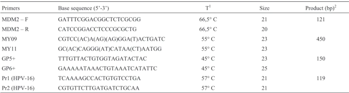
After isolation, the DNA concentration was deter-mined using a Biospec-nano spectrophotometer (Shima-dzu, Japan). The DNA obtained from the samples was submitted for amplification of the human MDM2 gene (Xiaoet al., 2010) to verify integrity and quantity of DNA in the samples. Table 1 shows the primer sequences and protocols used to amplify HPV DNA sequences.
HPV DNA detection was performed by PCR amplifi-cation using the standard nested PCR approach with the MY09/11 primer set (outer primers) and the GP5+/6+ primer set (inner primers), which are degenerate primer sets that detect a wide range of HPV types (Aghakhaniet al., 2011). Additionally, in an attempt to increase detection rate of HPV, a second inner primer set was used (Pr1/Pr2) for amplification of specific sequences of the E6 and E7 re-gions of HPV 16 (Karlsenet al., 1996). The cycling condi-tions were 94 °C for 3 min followed by 34 cycles of 94 °C for 30 s, 55 °C (for MY09/11), 45 °C (for GP5+/6+) or 57 °C (for Pr1/Pr2) for 1 min and 72 °C for 1 min, with a final
Table 1- Primers used for amplification of the region of interest, and size of the PCR product.
Primers Base sequence (5’-3’) T1 Size Product (bp)2
MDM2 – F GATTTCGGACGGCTCTCGCGG 66,5° C 21 121
MDM2 – R CATCCGGACCTCCCGCGCTG 66,5° C 20
MY09 CGTCC(AC)A(AG)(AG)GGA(T)ACTGATC 55° C 23 450
MY11 GC(AC)CAGGG(AT)CATAA(CT)AATGG 55° C 23
GP5+ TTTGTTACTGTGGTAGATACTAC 45° C 23 150
GP6+ GAAAAATAAACTGTAAATCATATTC 45° C 25
Pr1 (HPV-16) TCAAAAGCCACTGTGTCCTGA 57° C 21 119
Pr2 (HPV-16) CGTGTTCTTGATGATCTGCAA 57° C 21
1
extension step at 72 °C for 10 min. The reaction mix was prepared with 4mL DNA (200 ng), 1.6mM of each primer and 12.5mL MasterMix, in a total volume of 25mL. The PCR products were visualized on a 2% agarose gel with ethidium bromide under UV light. Each positive sample was then bidirectionally sequenced using the BigDye® Ter-minator v.3.1 Cycle Sequencing Kit.
Each batch of samples included negative controls without a DNA template, and a positive control, which con-sisted of a sample of DNA from uterine cervix cells (200 ng) known to be infected with HPV.
Results
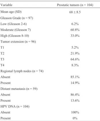
One hundred and four prostate carcinoma samples were analyzed from patients aged between 45 and 90 years (mean = 68±8.5 years). Tumors were staged using stan-dard criteria by the Gleason score and TNM staging system, according to the 7th revision of the TNM classification, 2010. No sample had a Gleason score lower than 6. Table 2 shows the cases along with their clinico-pathological char-acteristics. Because this study had a retrospective design, complete information was lacking for some patients due to a lack of information about the registration of clinical pa-thologists.
The success rate for amplification usingMDM2gene primers was 100%, indicating that a large quantity of DNA was extracted from the tumors (ranging from 410 to 1880 ng/mL, Figures S1 and S2). However, none of the samples
showed amplification products of HPV DNA for primer sets MY09/MY11 and GP5/GP6, or the specific primer set for the E6 and E7 HPV regions. In an attempt to reach a higher assay sensibility, the analyses were repeated using a GP5/GP6 auto-nested PCR approach, as described by Rem-merbachet al.(2004). However again, all samples tested negative for HPV DNA.
Discussion
Since 1990, studies in several countries have at-tempted to detect HPV DNA in prostate carcinomas using different detection methods. Some studies suggested that prostate carcinomas are related to HPV infection and have shown the presence of HPV DNA to varying degrees (from 2 to 100%), but even after several years of HPV DNA anal-ysis in benign and malignant prostate samples, it is still un-clear if HPV causes prostate carcinogenesis (Heideggeret al., 2015).
McNicol and Dodd (1990) were the first to demon-strate HPV genomic sequences in prostate tissues. They found that 14 of 15 (93.3%) benign prostatic hyperplasia (BPH) samples from Canadian patients, and all four carci-nomas (100%), tested positive for HPV 16. Subsequent studies, such as the one by Anwaret al.(1992), provided further evidence to support a relationship between HPV and prostate cancer. According to their results, HPV DNA was present in 28 (41%) out of 68 prostate carcinomas cases from Japanese patients and was not detected in any of the normal or hyperplastic prostatic tissues (10 each), which is consistent with recent findings in other popula-tions, such as the ones from Greece and India (Micho-poulouet al., 2014; Singhet al., 2015).
In contrast, several studies have found that HPV DNA is not preferentially associated with either BPH or prostate cancer. Tachezyet al.(2012) found no association between the persistence of HPV infection and the risk of developing prostate cancer, reporting the same prevalence of HPV DNA (2%) in 210 samples of tissues from 95 pa-tients with BPH and in 90 samples of 51 papa-tients with pros-tatic cancer. Other studies have detected HPV DNA sequences in approximately 12–65% of malignant and in 5–48% of benign prostatic samples, and no statistically sig-nificant difference was shown (Carozzi et al., 2004; Aghakhaniet al., 2011; Chenet al., 2011; Ghasemianet al., 2013). Based on these findings, it can be concluded that the prostate gland is a potential reservoir for transmission of HPV types with oncogenic potential. However, the HPV DNA findings in prostate tissue do not necessarily reflect active infection, or that the prostatic disease was caused by HPV; they could very well represent transient infections. Thus, while the prostate may act as a site for HPV replica-tion, it is unlikely that HPV is directly involved in the trans-formation of prostatic cells (Doddet al., 1993; Aghakhani
et al., 2011). On the other hand, the presence of high-risk HPV DNA in subsets of prostate cancers shows that at least
Table 2- Clinico-pathological characteristics of the samples
Variable Prostatic tumors (n = 104)
Mean age (SD) 68±8.5
Gleason Grade (n = 97)
Low (Gleason 2-6) 6.2%
Moderate (Gleason 7) 60.8%
High (Gleason 8-10) 33.0%
Tumor extension (n = 96)
T1 5.2%
T2 21.9%
T3 64.6%
T4 8.3%
Regional lymph nodes (n = 74)
Absent 85.1%
Present 14.9%
Distant metastasis (n = 59)
Absent 86.4%
Present 13.6%
HPV DNA (n = 104)
Absent 100%
a proportion of prostate cancers may be related to HPV in-fection.
In this study we were unable to identify the presence of HPV DNA in any of the 104 samples of prostate carci-noma in patients from northeastern Brazil. Our results are consistent with other previously and recently published re-ports. Effertet al.(1992) analyzed 30 paraffin-embedded prostate adenocarcinomas for the presence of HPV 16- or HPV 18-specific sequences by differential PCR (D-PCR) and Southern blot analysis. Despite the high sensitivity of their analytical technique, they found no evidence of HPV DNA of either type in any of the 30 primary prostate can-cers. Furthermore, Berghet al.(2007), in an investigation of 201 prostate tissue samples of patients with BPH that later progressed to prostate cancer and of 201 matched con-trols, found that all samples tested were negative for HPV, thus making it an unlikely contributing factor for subse-quent cancer development. Gazzaz and Mosli (2009) did not detect HPV DNA by hybrid capture 2 technology among 56 tested biopsies categorized into a benign lesion pattern of prostatic hyperplasia, malignant, and inflamma-tory disorders. Yowet al.(2014) achieved the same result after analyzing 195 archival paraffin-embedded tissue blocks by PCR.
Inconsistent results may be due to technical difficul-ties in HPV DNA detection. There are several possible rea-sons for false-positive or false-negative results, most notably PCR contamination, degradation, or cross-conta-mination of DNA, and tumor samples that are not represen-tative (Terris and Peehl, 1997; Adamiet al., 2003; Gazzaz and Mosli, 2009). Moreover, considering that HPV pres-ence is strongly associated with high-risk sexual behavior, including a history of sexually transmitted diseases (STDs), the number of sexual partners, and age of first sexual activi-ties, a comparison of cancers from patients with such sexual activityvs.those who did not engage in high-risk behavior may have shown different results.
The distribution of HPV varies among different popu-lations, and infection rates are influenced by geography, age, sexual history, coinfections, immune status, and ge-netic factors (Vesco et al., 2011). In Brazil, the overall prevalence of HPV infection among women, not stratified by cervical cytology results, ranged from 16.8% to 28.6% (Ayres and Silva, 2010). In men, HPV can be present in up to 72% of the samples of the male genital region, as was re-ported in a study on a population of southeastern Brazil (Freireet al., 2011). To our knowledge, the only study of the potential association between HPV and prostate cancer in Brazil was performed by Silvestreet al.(2009), in which the authors reported a 3% prevalence of HPV DNA in sam-ples of prostatic carcinoma in a northern Brazilian popula-tion. In the northeastern region of Brazil, data on HPV prevalence are scarce, both in females and in males. On the other hand, in a study in this population, Silva Jr and da Silva (2011) investigated the prevalence of HPV DNA in
79 samples of breast ductal carcinoma and reported the ab-sence of HPV types 6, 11, 16, and 18. Therefore, the results presented here, as well as those of Silva Jr and da Silva (2011) are important reports on the prevalence of HPV in neoplastic tissues in the northeastern population of Brazil.
Some limitations of this study should be taken into consideration. Although we used a relatively large sample size (n = 104) compared to other studies (McNicol and Dodd, 1990; Anwaret al., 1992; Effertet al., 1992; Carozzi
et al., 2004; Gazzaz and Mosli, 2009; Chenet al., 2011; Michopoulouet al., 2014; Singhet al., 2015), the lack of association observed in this study may have been a result of the limited number of samples analyzed. Moreover, it would be useful to use complementary approaches for the direct and indirect detection of HPV to confirm our nega-tive results. However, although serologic analysis is often used in combination with molecular analysis (Chenet al., 2011, Tachezyet al., 2012), this method is limited in that it reflects overall exposure to HPV and does not distinguish HPV infections within specific anatomical sites (Chenet al., 2011). The molecular technique used in this study (nested PCR) is one of the most sensitive for the detection of HPV DNA in clinical specimens when combined with the sequencing of positive samples, as previously included in the study design (Chaiwongkotet al., 2007). The set of primers used and the quality of DNA analyzed are the main variables that may cause errors when attempting to detect HPV DNA by PCR (Terriset al., 1997). However, the set of primers used in this study is well established in the litera-ture, and amplification of theMDM2 gene revealed that 100% of the samples had sufficient high quality DNA for subsequent reactions.
In conclusion, our study showed no evidence to sup-port the hypothesis that HPV infection is related to the de-velopment of prostate cancer in patients from northeastern Brazil, given that the prevalence of these pathogens in the population studied was null. Additional studies involving larger sample size and that take into account other con-founding variables such as sexual behavior should be car-ried out to elucidate the role of HPV in prostatic carcino-genesis.
Acknowledgments
This study was supported by the National Counsel of Technological and Scientific Development (Conselho Nacional de Desenvolvimento Científico e Tecnológico, CNPq; grant number 502193/2009-3) and by the Coordina-tion of Improvement of Higher EducaCoordina-tion (Coordenação de Aperfeiçoamento de Pessoal de Nível Superior; CAPES).
References
Aghakhani A, Hamkar R, Parvin M, Ghavami N, Nadri M, Pakfe-trat A, Banifazl M, Eslamifar A, Izadi N, Jam Set al.(2011) The role humam papillomavirus infection in prostate carci-noma. Scand J Infect Dis 43:64-69.
Anwar K, Nakakuki K, Shiraishi T, Naiki H, Yatani R and Inuzu-ka M (1992) Presence of ras oncogene mutations and human papillomavirus DNA in human prostate carcinomas. Cancer Res 52:5991-5996.
Ayres AR and Silva GA (2010) Cervical HPV infection in Brazil: systematic review. Rev Saude Publica 44:963-974. Bergh J, Marklund I, Gustavsson C, Wiklund F, Grönberg H,
Allard A, Alexeyev O and Elgh F (2007) No link between viral findings in the prostate and subsequent cancer develop-ment. Br J Cancer 96:137-139.
Carozzi F, Lombardi FC, Zendron P, Confortini M, Sani C, Bisanzi S, Pontenani G and Ciatto S (2004) Association of human papillomavirus with prostate cancer: analysis of a consecutive series of prostate biopsies. Int J Biol Markers 19:257-261.
Chaiwongkot A, Pientong C, Ekalaksananan T, Kongyingyoes B, Thinkhamrop J, Yuenyao P and Sriamporn S (2007) Evalua-tion of primers and PCR performance on HPV DNA screen-ing in normal and low grade abnormal cervical cells. Asian Pac J Cancer Prev 8:279-282.
Chen ACH, Waterboer T, Keleher A, Morrison B, Jindal S, McMillan D, Nicol D, Gardiner RA, McMillan NA and Antonsson A (2011) Human papillomavirus in benign pros-tatic hyperplasia and prospros-tatic adenocarcinoma patients. Pathol Oncol Res 17:613-617.
Conceição MB, Boing AF and Peres KG (2014) Time trends in prostate cancer mortality according to major geographic re-gions of Brazil: an analysis of three decades. Cad Saude Publica 30:559-566.
Dodd JG, Paraskevas M and McNicol PJ (1993) Detection of hu-man papillomavirus 16 transcription in huhu-man prostate tis-sue. J Urol 149:400-402.
Effert PJ, Frye RA, Neubauer A, Liu ET and Walther PJ (1992) Human papillomavirus types 16 and 18 are not involved in human prostate carcinogenesis: analysis of archival human prostate cancer specimens by differential polymerase chain reaction. J Urol 147:192-196.
Facina T (2014) Estimativa 2014 – Incidência de Câncer no Brasil. Rev Bras Cancerol 60:63.
Freire MP, Pires D, Forjaz R, Sato S, Cotrim I, Stiepcich M, Scarpellini B and Truzzi JC (2011) Genital prevalence of HPV types and co-infection in men. Int Braz J Urol 40:67-71.
Gazzaz FS and Mosli HA (2009) Lack of detection of human papillomavirus infection by hybridization test in prostatic biopsies. Saudi Med J 30:633-637.
Ghasemian E, Monavari SH, Irajian GR, Jalali Nodoshan MR, Roudsari RV and Yahyapour Y (2013) Evaluation of human papillomavirus infections in prostatic disease: a cross-sectional study in Iran. Asian Pac J Cancer Prev 14:3305-8. Heidegger I, Borena W and Pichler R (2015) The role of human
papilloma virus in urological malignancies. Anticancer Res 35:2513-2519.
Karlsen F, Kalantari M, Jenkins A, Pettersen E, Kristensen G, Holm R, Johansson B and Hagmar B (1996) Use of multiple PCR primer sets for optimal detection of human papillo-mavirus. J Clin Microbiol 34:2095-2100.
McNicol PJ and Dodd JG (1990) Detection of human papillo-mavirus DNA in prostate gland tissue by using the polymer-ase chain reaction amplification assay. J Clin Microbiol 28:409-412.
Michopoulou V, Derdas SP, Symvoulakis E, Mourmouras N, Nomikos A, Delakas D, Sourvinos G and Spandidos DA (2014) Detection of human papillomavirus (HPV) DNA prevalence and p53 codon 72 (Arg72Pro) polymorphism in prostate cancer in a Greek group of patients. Tumor Biol 35:12765-73.
Nakai Y and Nonomura N (2013) Inflammation and prostate carcinogenesis. Int J Urol 20:150-60.
Rabkin CS, Biggar RJ, Melbye M and Curtis RE (1992) Second primary cancers following anal and cervical carcinoma: evi-dence of shared etiologic factors. Am J Epidemiol 136:54-58.
Remmerbach TW, Brinckmann UG, Hemprich A, Chekol M, Kühndel K and Liebert UG (2004) PCR detection of human papillomavirus of the mucosa: comparison between MY09/11 and GP5+/6+ primer sets. J Clin Virol 30:302-8. Sarkar FH, Sakr WA, Li YW, Sreepathi P and Crissman JD (1993)
Detection of human papillomavirus (HPV) DNA in human prostatic tissues by polymerase chain reaction (PCR). Pros-tate 22:171-180.
Sfanos KS, Isaacs WB and De Marzo AM (2013) Infections and inflammation in prostate cancer. Am J Clin Exp Urol 1:3-11. Siegel R, Ma J, Zou Z and Jemal A (2014) Cancer statistics, 2014.
CA Cancer J Clin 64: 9-29.
Silva Jr RG and da Silva BB (2011) No evidence for an associa-tion of human papillomavirus and breast carcinoma. Breast Cancer Res Treat 125:261-264.
Silvestre RVD, Leal MF, Demachki S, Nahum MC, Bernardes JG, Rabenhorst SH, Smith Mde A, Mello WA, Guimarães AC and Burbano RR (2009) Low frequency of human pa-pillomavirus detection in prostate tissue from individuals from Northern Brazil. Mem Inst Oswaldo Cruz 104:665-667.
Singh N, Hussain S, Kakkar N, Singh SK, Sobti RC and Bha-radwaj M (2015) Implication of high risk human papillo-mavirus HR-HPV infection in prostate cancer in Indian pop-ulation - a pioneering case-control analysis. Sci Rep 5:e7822.
Tachezy R, Jan Hrbacek J, Heracek J, Salakova M, Smahelova J, Ludvikova V, Svec A, Urban M and Hamsikova E (2012) HPV persistence and its oncogenic role in prostate tumors. J Med Virol 84:1636-1645.
Terris MK and Peehl DM (1997) Human papillomavirus detection by polymerase chain reaction in benign and malignant pros-tate tissue is dependent on the primer set utilized. Urology 50:150-156.
Vesco KK, Whitlock EP, Eder M, Burda BU, Senger CA and Lutz K (2011) Risk factors and other epidemiologic consider-ations for cervical cancer screening: a narrative review for the U.S. Preventive Services Task Force. Ann Intern Med 155:698-705.
Xiao M, Zhang L, Zhu X, Huang J, Jiang H, Hu S and Liu Y (2010) Genetic polymorphisms of MDM2 and TP53 genes are associated with risk of nasopharyngeal carcinoma in a Chinese population. BMC Cancer 10:e147.
Detec-tion of infectious organisms in archival prostate cancer tis-sues. BMC Cancer 14:e579.
Internet Resources
Pew Research Center (2014) Attitudes about aging: a global perspective. [Data file and code book]. Washington DC, http:www.pewresearch.org (accessed Feb 12, 2015).
Supplementary Material
The following online material is available for this ar-ticle:
Figure S1 -Agarose gel electrophoresis of total DNA extracted from prostate tissue.
Figure S2 -Amplification of the DNA extracted from prostate tissue.
This material is available as part of the online article from http://www.scielo.br/gmb
Associate Editor: Carlos F. M. Menck