Original Article
Artigo Original
ISSN 2317-1782 (Online version)
This is an Open Access article distributed under the terms of the Creative Commons Attribution License, which permits unrestricted use, distribution, and reproduction in any medium, provided the original work is properly cited.
Prevalence of communication, swallowing
and orofacial myofunctional disorders in
children and adolescents at the time of
admission at a cancer hospital
Prevalência de distúrbios da comunicação,
deglutição e motricidade orofacial em crianças
e adolescentes no momento da matrícula em
um hospital oncológico
Kaliani Lima Coça1,2 Anke Bergmann3 Sima Ferman4 Elisabete Carrara de Angelis5 Márcia Gonçalves Ribeiro6
Keywords
Speech, Language and Hearing Sciences Oncology Pediatrics Communication Disorders Deglutition Disorders
Descritores
Fonoaudiologia Oncologia Pediatria Transtornos da Comunicação Transtornos da Deglutição
Correspondence address:
Kaliani Lima Coça
Instituto Nacional de Câncer José de Alencar Gomes da Silva – INCA Praça da Cruz Vermelha, nº 23, Centro, Rio de Janeiro (RJ), Brazil, CEP: 20230-130.
E-mail: [email protected]; [email protected]
Received: June 01, 2017
Accepted: September 18, 2017
Study conducted at the Instituto Nacional de Câncer José de Alencar Gomes da Silva – INCA - Rio de Janeiro (RJ), Brazil.
1 Setor de Fonoaudiologia, Instituto Nacional de Câncer José de Alencar Gomes da Silva – INCA - Rio de Janeiro
(RJ), Brazil.
2 Programa de Pós-graduação em Clínica Médica, Universidade Federal do Rio de Janeiro – UFRJ - Rio de
Janeiro (RJ), Brazil.
3 Programa de Carcinogênese Molecular, Instituto Nacional de Câncer José de Alencar Gomes da Silva – INCA
- Rio de Janeiro (RJ), Brazil.
4 Serviço de Oncologia Pediátrica, Instituto Nacional de Câncer José de Alencar Gomes da Silva – INCA - Rio
de Janeiro (RJ), Brazil.
5 Núcleo de Fonoaudiologia, Hospital Antônio Cândido de Camargo – A.C.Camargo - São Paulo (SP), Brazil. 6 Departamento de Pediatria, Universidade Federal do Rio de Janeiro – UFRJ - Rio de Janeiro (RJ), Brazil. Financial support: nothing to declare.
Conflict of interests: nothing to declare.
ABSTRACT
Purpose: Describe the prevalence of communication, swallowing and orofacial myofunctional disorders in a group of children and adolescents at the time of registration at a cancer hospital. Methods: A cross-sectional study conducted
with children aged ≥2 and adolescents, of both genders, admitted to the Pediatric Oncology Section of the Instituto
Nacional de Câncer José de Alencar Gomes da Silva (INCA) from March 2014 to April 2015 for investigation and/or
treatment of solid tumors. A protocol was used to record the sociodemographic and clinical information and findings
of the speech-language pathology clinical evaluation, which included aspects of the oral sensorimotor system, swallowing, speech, language, voice, and hearing. Results: Eighty-eight children/adolescents (41.3%) presented some type of speech-language disorder. The most frequent speech-language disorders were orofacial myofunctional disorder, dysphonia, and language impairments, whereas the less frequent ones were dysacusis, tongue paralysis, and
trismus. Site of the lesion was the clinical variable that presented statistically significant correlation with presence
of speech-language disorders. Conclusion: High prevalence of speech-language disorders was observed in children and adolescents at the time of admission at a cancer hospital. Occurrence of speech-language disorders was higher in participants with lesions in the central nervous system and in the head and neck region.
RESUMO
Objetivo: Descrever a prevalência dos distúrbios da comunicação, deglutição e motricidade orofacial em um grupo de crianças e adolescentes, no momento da matrícula hospitalar em um instituto oncológico. Método: Estudo transversal, com a inclusão de crianças com dois anos ou mais e adolescentes de ambos os gêneros, matriculados na Seção de Oncologia Pediátrica do Instituto Nacional de Câncer José de Alencar Gomes da Silva (INCA) no período de março de 2014 a abril de 2015 para investigação e/ou tratamento de tumores sólidos. Foi utilizado um protocolo
para registro das informações sociodemográficas e clínicas e os achados da avaliação clínica fonoaudiológica, que
contemplava aspectos do sistema sensório-motor oral, deglutição, fala, linguagem, voz e audição. Resultados: Oitenta e oito crianças/adolescentes (41,3%) avaliados apresentavam algum tipo de distúrbio fonoaudiológico. As alterações fonoaudiológicas mais frequentes foram o distúrbio miofuncional orofacial, a disfonia e os transtornos de linguagem. Os menos frequentes foram a disacusia, a paralisia de língua e o trismo. A variável clínica que teve associação
INTRODUCTION
Children and adolescents with benign and malignant neoplasms may present developmental speech-language disorders or impairments associated with the tumor and resulting from the treatments performed(1). The occurrence of impairments in
speech, language, and swallowing in children with central nervous
system (CNS) tumors is well described in the specific scientific
literature. Dysphagia and dysarthria, for instance, are common disorders in children with posterior fossa tumors, because the neurological structures of this region play an important role
in the accuracy and efficiency of the movements involved in
speech and swallowing(2-4).
Functional disorders in pediatric patients with head and neck (HN) tumors are common, and they vary according to the site and extent of lesion and treatment performed, as well as to individual characteristics and functions acquired until the onset of disease. Dysphonia, dysphagia, temporomandibular joint disorder, dysacusis, and facial paralysis are the most commonly observed disorders in patients with HN tumors. In most cases, the functional disorder corresponds to the impairment caused by the presence of the tumor, and it is aggravated by the treatment indicated for the disease(5,6).
Difficulties in swallowing and feeding may occur as an adverse
effect of chemotherapy and radiotherapy, involving changes in taste, food refusal, mucositis, xerostomia, and dysphagia(7).
In addition, the use of ototoxic antineoplastic agents may be a cause of hearing loss in children and adolescents with cancer(1,4).
The literature addressing the prevalence of speech-language disorders in a heterogeneous group of patients with pediatric tumors is scarce, with predominance of studies investigating
groups of children and adolescents with some specific cancer
diagnosis, especially neurological tumors(2-4). TAYLOR, WARE,
and WEIR (2012) analyzed the prevalence and severity of swallowing/feeding and communication disorders in children diagnosed with cancer and nonmalignant hematological diseases cared at a speech-language pathology service in Australia(7).
In Brazil, Gonçalves et al. (2007) reported the incidence of speech-language disorders in children and adolescents with CNS tumors and observed that 81% of the patients presented some type of disorder.
Speech-language therapists are part of the multidisciplinary pediatric oncology patient care team. Speech-language pathology practice in pediatric oncology is extensive, encompassing disorders of communication, swallowing, and orofacial motricity(1). Early
detection of speech-language disorders in pediatric patients with tumors, at the diagnosis stage or beginning of treatment, is essential for a more appropriate therapeutic management.
The present study aims to describe the prevalence of communication, swallowing and orofacial myofunctional disorders in a group of children and adolescents at the time of admission at an oncology reference center.
METHODS
This is a cross-sectional study whose inclusion criteria comprised children aged ≥2 years and adolescents, of both genders, admitted to the Pediatric Oncology Section of the Instituto Nacional de Câncer José de Alencar Gomes da Silva (INCA) between March
2014 and April 2015 for investigation and/or treatment of solid tumors. Children without written permission from parents and/or legal guardians were excluded from the study. The project was approved by the Research Ethics Committee of the aforementioned Institution (process no. 492.325/2013). All participants signed an Informed Consent Form (ICF) prior to study commencement.
Participants were assessed at the time of admission at the Institution by the researcher, exclusively. A protocol developed by the researchers was used to collect sociodemographic and clinical information, anamnesis data with a description of the speech-language complaints reported by the participants and/or
their caregivers, and findings of the clinical and speech-language
assessment, which included aspects of the oral sensorimotor system, swallowing, speech, language, voice, and hearing.
Speech-language skills were evaluated through tasks of naming and spontaneous speech registered using an audio recorder. Language assessment included phonetic-phonological, morphosyntactic, semantic-lexical and semantic-pragmatic linguistic levels.
Characteristics of phoneme production observed in the tasks of spontaneous speech and naming were recorded in a phonemic context and the phonological processes were analyzed based on the criteria established in the ABFW Child Language
Test for phonology, vocabulary, fluency, and pragmatics(8). The
morphosyntactic level involved phrase structure analysis in spontaneous speech.
The ABFW vocabulary test was applied to evaluate the semantic-lexical level(8). The semantic-pragmatic evaluation included the
observation of skills such as verbal initiative, speech acts with communicative function, speech coherence, conversation topic
maintenance, literal verbal utterance comprehension, and figurative language comprehension in participants aged ≥12 years(9).
Regarding speech, the characteristics of articulation, velocity, intelligibility, and prosody were evaluated. As for the oral sensorimotor system, posture, strength and mobility of orofacial structures and sensitivity of the face and tongue were observed.
Swallowing of saliva and food was assessed. The functional evaluation of swallowing using food involved liquid and solid
consistencies. Efficacy of lip seal, oral motor control, oral transit
time, mastication, and hyollaryngeal movement was observed, and presence of extra oral food escape, oral cavity stasis, nasal leakage, multiple swallowing, coughing, throat clearing, wet voice,
dyspnea, and cyanosis was identified. Cervical auscultation was
used during the evaluation of swallowing. The assessment also included the use of the Functional Oral Intake Scale (FOIS)(10).
Voice assessment included identification of the findings on
vocal quality, loudness, pitch (subjective feeling of frequency), vocal resonance, and pneumophonic coordination. The GRBASI (grade, roughness, breathiness, asthenia, strain, and instability) perceptual vocal scale was used(11).
With respect to the auditory function, considering that the institution where the study was conducted did not have an audiology service, reports of the participants who had undergone pure-tone audiometry less than a year before the assessment were recorded in the protocol. Frequency of dysacusis was estimated considering only the group of children and adolescents with recent hearing evaluation.
The findings observed in the clinical evaluation led to definition
presence of impairment in the functions of language and speech (speech disorder, phonetic and phonological impairment, stuttering, aphasia, apraxia, and dysarthria), voice (dysphonia), swallowing (dysphagia), orofacial motricity (orofacial myofunctional disorder, facial paralysis, trismus, and tongue paralysis), and hearing (dysacusis).
Malignant neoplasms were categorized based on the third
edition of the International Classification of Cancer in Childhood
(ICCC-3), which divides tumors into 12 major groups: I - Leukemias, myeloproliferative disease, and myelodysplastic diseases; II - Lymphomas and reticuloendothelial neoplasms; III - Central Nervous System (CNS) and miscellaneous intracranial and intraspinal neoplasms; IV - Neuroblastoma and other peripheral nerve cell tumors; V - Retinoblastoma; VI - Renal tumors; VII - Hepatic tumors; VIII - Malignant bone tumors; IX - Soft tissue and other extraosseous sarcomas; X - Germ cell tumors, trophoblastic tumors, and neoplasms of gonads; XI - Other malignant epithelial
neoplasms and malignant melanomas; XII - Other and unspecified
malignant neoplasms(12).
Descriptive statistical analyses of the quantitative and qualitative variables were performed by mean/standard deviation and absolute/relative frequencies, respectively. The Chi-squared test was applied for evaluation of the percentage differences between the outcomes and the independent variables considering
a significance level of 5%.
RESULTS
Two hundred thirteen children and adolescents (95 male and 118 female) with mean age of 9.05 years (SD±4.71) were evaluated at the time of hospital admission. Of these, 146 (68.5%) had been diagnosed with malignant neoplasm or neoplasm of uncertain biological behavior, 19 (8.9%) with benign neoplasm, and 48 (22.5%) with neoplasia.
The sample consisted mainly of patients with malignant neoplasms, but some children and adolescents who had been referred to the Institute with suspected cancer, after an investigation period, presented benign neoplasms or other diseases (absence of neoplasia) such as chronic granulomatous
inflammatory processes, epidermoid and bone cysts, dysplasias
and hyperplasias. The participants that did not present neoplasms were maintained in the study for comparison of impairment
profile with the group with neoplasia.
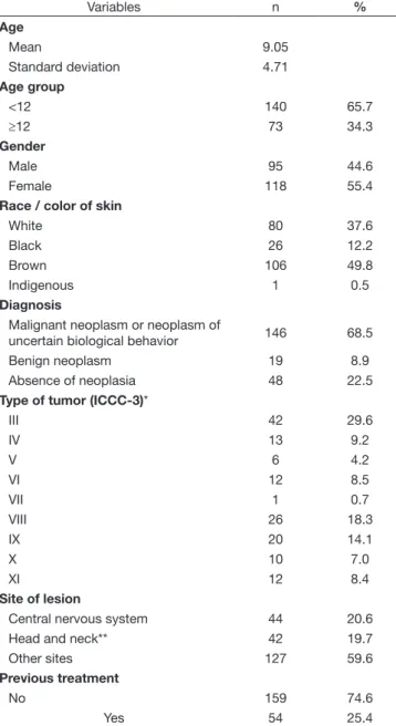
Table 1 presents the sociodemographic and clinical characteristics of the study sample. Central Nervous System (CNS) (ICCC-3 category III) and bone (ICCC-3 category VIII) tumors were the most frequently found malignant solid tumors, affecting 42 (29.6%) and 26 (18.3%) children/adolescents, respectively.
As for site of lesion, 44 (20.6%) patients presented lesions in the CNS, 42 (19.7%) in the head and neck (HN) region, and 127 (59.6%) in other sites.
At hospital registration, 54 individuals (25.4%) had already undergone some type of cancer treatment in other institutions, and 88 children/adolescents (41.3%) presented some type of speech-language disorder. Speech-language complaints associated with voice, speech, language, swallowing, and orofacial motor skills were reported by the parents/legal guardians of 69 participants.
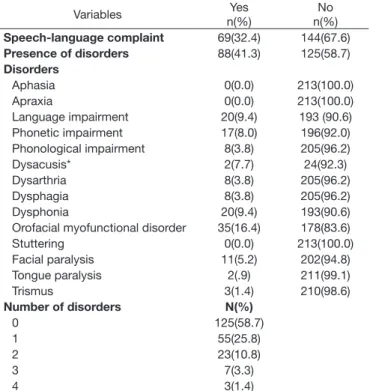
Table 2 shows a frequency description of the speech-language disorders observed.
Orofacial myofunctional disorder, dysphonia, and language impairments were the most commonly found speech-language disorders, whereas dysacusis, tongue paralysis, and trismus were the least frequently observed. Most patients with speech-language disorders presented a single impairment, and some individuals had two, three, or even four concomitant impairments.
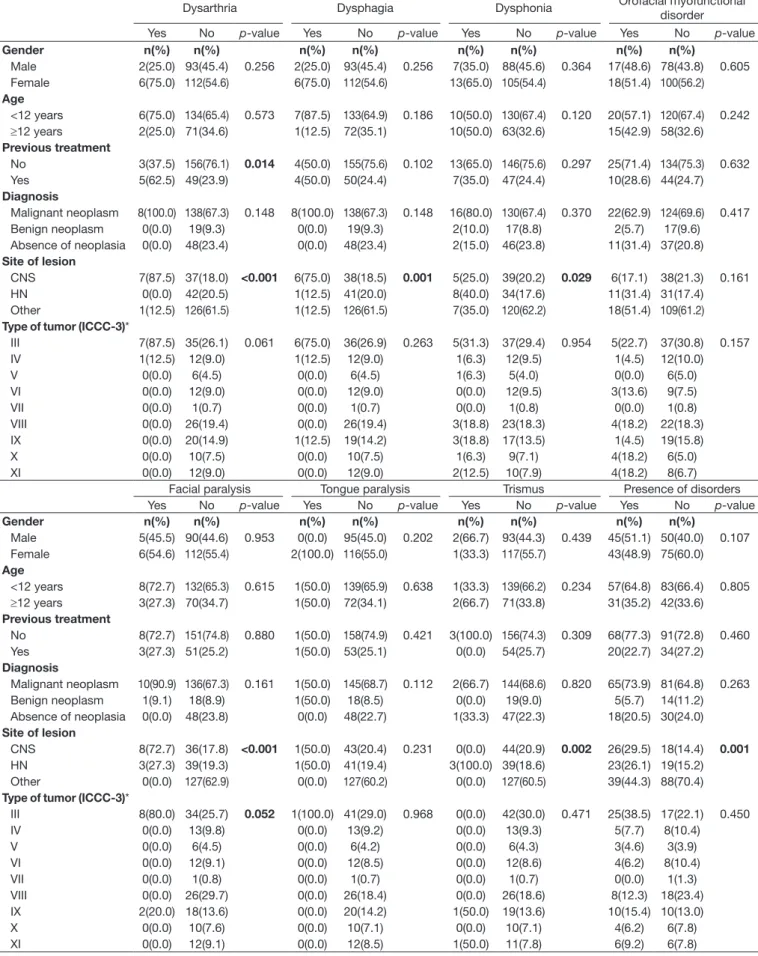
Table 3 shows a correlation between the speech-language disorders and the sociodemographic and clinical variables. Language disorders were more prevalent in males (p=0.016). Phonological impairment was only detected in children aged <12 years (p=0.037). No statistically significant correlation was found between the frequencies of phonetic impairment
Table 1. Sociodemographic and clinical characteristics
Variables n %
Age
Mean 9.05
Standard deviation 4.71
Age group
<12 140 65.7
≥12 73 34.3
Gender
Male 95 44.6
Female 118 55.4
Race / color of skin
White 80 37.6
Black 26 12.2
Brown 106 49.8
Indigenous 1 0.5
Diagnosis
Malignant neoplasm or neoplasm of
uncertain biological behavior 146 68.5
Benign neoplasm 19 8.9
Absence of neoplasia 48 22.5
Type of tumor (ICCC-3)*
III 42 29.6
IV 13 9.2
V 6 4.2
VI 12 8.5
VII 1 0.7
VIII 26 18.3
IX 20 14.1
X 10 7.0
XI 12 8.4
Site of lesion
Central nervous system 44 20.6
Head and neck** 42 19.7
Other sites 127 59.6
Previous treatment
No 159 74.6
Yes 54 25.4
and orofacial myofunctional disorder and the variables gender, age, previous treatment, diagnosis, site of lesion, and type of tumor (ICCC-3).
Dysacusis and tongue paralysis occurred concomitantly in a patient with CNS tumor and in one with HN tumor, but they were not observed in participants with tumors in other sites.
Dysarthria was diagnosed in seven patients with CNS tumors (p<0.001) and in only one patient with lesion in a different site. Most of the children/adolescents in whom dysarthria was detected at the time of hospital admission had already received some type of treatment in other institutions (p=0.014).
Prevalence of dysphagia varied according to the site of lesion,
with statistical significance (p=0.001). Most participants with dysphagia (n=6) had lesions in the CNS, whereas presence of dysarthria and dysphagia was only observed in individuals with malignant neoplasms.
Dysphonia was most frequently found in patients with HN tumors (p=0.029). It was observed in 19% (8/42) of the children/adolescents with lesions in the HN region, in 11.4% (5/44) of the patients with lesions in the CNS, and in 5.5% (7/127) of the individuals with lesions in other sites.
All of the patients with trismus (n=3) had HN lesions (p=0.002): two with malignant neoplasms and one with absence of neoplasia (benign lesion). Most of the participants with Table 2. Prevalence of speech-language disorders
Variables Yes
n(%)
No n(%) Speech-language complaint 69(32.4) 144(67.6) Presence of disorders 88(41.3) 125(58.7) Disorders
Aphasia 0(0.0) 213(100.0)
Apraxia 0(0.0) 213(100.0)
Language impairment 20(9.4) 193 (90.6) Phonetic impairment 17(8.0) 196(92.0) Phonological impairment 8(3.8) 205(96.2)
Dysacusis* 2(7.7) 24(92.3)
Dysarthria 8(3.8) 205(96.2)
Dysphagia 8(3.8) 205(96.2)
Dysphonia 20(9.4) 193(90.6)
Orofacial myofunctional disorder 35(16.4) 178(83.6)
Stuttering 0(0.0) 213(100.0)
Facial paralysis 11(5.2) 202(94.8) Tongue paralysis 2(.9) 211(99.1)
Trismus 3(1.4) 210(98.6)
Number of disorders N(%)
0 125(58.7)
1 55(25.8)
2 23(10.8)
3 7(3.3)
4 3(1.4)
*Considering patients with recent hearing assessment (n=26)
Table 3. Speech-language disorders x sociodemographic and clinical variables
Language impairment Phonetic impairment Phonological impairment Dysacusis (n=26) Yes No p-value Yes No p-value Yes No p-value Yes No p-value
Gender n(%) n(%) n(%) n(%) n(%) n(%) n(%) n(%)
Male 14(70.0) 81(42.0) 0.016 5(62.5) 90(43.9) 0.229 8(47.1) 87(44.4) 0.832 1(50.0) 11(45.8) 0.910 Female 6(30.0) 112(58.0) 3(37.5) 115(56.1) 9(52.9) 109(55.6) 1(50.0) 13(54.2) Age
<12 years 14(70.0) 126(65.3) 0.672 13(76.5) 127(64.8) 0.331 8(100.0) 132(64.4) 0.037 1(50.0) 10(41.7) 0.819
≥12 years 6(30.0) 67(34.7) 4(23.5) 69(35.2) 0(0.0) 73(35.6) 1(50.0) 14(58.3) Previous treatment
No 18(90.0) 141(73.1) 0.097 12(70.6) 147(75.0) 0.688 8(100.0) 151(73.7) 0.093 2(100.0) 19(79.2) 0.473 Yes 2(10.0) 52(26.9) 5(29.4) 49(25.0) 0(0.0) 54(26.3) 0(0.0) 5(20.8) Diagnosis
Malignant neoplasm 14(70.0) 132(68.4) 0.303 12(70.6) 134(68.4) 0.898 6(75.0) 140(68.3) 0.736 1(50.0) 18(75.0) 0.499 Benign neoplasm 0(0.0) 19(9.8) 1(5.9) 18(9.2) 1(12.5) 18(8.8) 1(50.0) 4(16.7) Absence of neoplasia 6(30.0) 42(21.7) 4(23.5) 44(22.4) 1(12.5) 47(22.9) 0(0.0) 2(8.3) Site of lesion
CNS 4(20.0) 40(20.7) 0.470 5(29.4) 39(19.9) 0.098 1(12.5) 43(21.0) 0.649 1(50.0) 2(8.3) 0.049 HN 2(10.0) 40(20.7) 6(35.3) 36(18.4) 1(12.5) 41(20.0) 1(50.0) 3(12.5) Other 14(70.0) 113(58.5) 6(35.3) 121(61.7) 6(75.0) 121(59.0) 0(0.0) 19(79.2) Type of tumor (ICCC-3)*
III 4(28.6) 38(29.7) 0.105 5(41.7) 37(28.5) 0.203 1(16.7) 41(30.1) 0.158 1(100.0) 2(11.1) 0.229 IV 2(14.3) 11(8.6) 0(0.0) 13(10.0) 2(33.3) 11(8.1) 0(0.0) 0(0.0)
V 3(21.4) 3(2.3) 0(0.0) 6(4.6) 0(0.0) 6(4.4) 0(0.0) 0(0.0)
VI 1(7.1) 11(8.6) 3(25.0) 9(6.9) 0(0.0) 12(8.8) 0(0.0) 0(0.0)
VII 0(0.0) 1(0.8) 0(0.0) 1(0.8) 0(0.0) 1(0.7) 0(0.0) 0(0.0)
VIII 2(14.3) 24(18.7) 0(0.0) 26(20.0) 0(0.0) 26(19.1) 0(0.0) 10(55.6) IX 1(7.1) 19(14.8) 2(16.7) 18(13.8) 1(16.7) 19(14.0) 0(0.0) 1(5.6)
X 0(0.0) 10(7.8) 0(0.0) 10(7.7) 0(0.0) 10(7.4) 0(0.0) 3(16.6)
XI 1(7.1) 11(8.6) 2(16.7) 10(7.7) 2(33.3) 10(7.4) 0(0.0) 2(11.1)
Dysarthria Dysphagia Dysphonia Orofacial myofunctional disorder
Yes No p-value Yes No p-value Yes No p-value Yes No p-value
Gender n(%) n(%) n(%) n(%) n(%) n(%) n(%) n(%)
Male 2(25.0) 93(45.4) 0.256 2(25.0) 93(45.4) 0.256 7(35.0) 88(45.6) 0.364 17(48.6) 78(43.8) 0.605 Female 6(75.0) 112(54.6) 6(75.0) 112(54.6) 13(65.0) 105(54.4) 18(51.4) 100(56.2) Age
<12 years 6(75.0) 134(65.4) 0.573 7(87.5) 133(64.9) 0.186 10(50.0) 130(67.4) 0.120 20(57.1) 120(67.4) 0.242
≥12 years 2(25.0) 71(34.6) 1(12.5) 72(35.1) 10(50.0) 63(32.6) 15(42.9) 58(32.6) Previous treatment
No 3(37.5) 156(76.1) 0.014 4(50.0) 155(75.6) 0.102 13(65.0) 146(75.6) 0.297 25(71.4) 134(75.3) 0.632 Yes 5(62.5) 49(23.9) 4(50.0) 50(24.4) 7(35.0) 47(24.4) 10(28.6) 44(24.7) Diagnosis
Malignant neoplasm 8(100.0) 138(67.3) 0.148 8(100.0) 138(67.3) 0.148 16(80.0) 130(67.4) 0.370 22(62.9) 124(69.6) 0.417 Benign neoplasm 0(0.0) 19(9.3) 0(0.0) 19(9.3) 2(10.0) 17(8.8) 2(5.7) 17(9.6) Absence of neoplasia 0(0.0) 48(23.4) 0(0.0) 48(23.4) 2(15.0) 46(23.8) 11(31.4) 37(20.8) Site of lesion
CNS 7(87.5) 37(18.0) <0.001 6(75.0) 38(18.5) 0.001 5(25.0) 39(20.2) 0.029 6(17.1) 38(21.3) 0.161 HN 0(0.0) 42(20.5) 1(12.5) 41(20.0) 8(40.0) 34(17.6) 11(31.4) 31(17.4) Other 1(12.5) 126(61.5) 1(12.5) 126(61.5) 7(35.0) 120(62.2) 18(51.4) 109(61.2) Type of tumor (ICCC-3)*
III 7(87.5) 35(26.1) 0.061 6(75.0) 36(26.9) 0.263 5(31.3) 37(29.4) 0.954 5(22.7) 37(30.8) 0.157 IV 1(12.5) 12(9.0) 1(12.5) 12(9.0) 1(6.3) 12(9.5) 1(4.5) 12(10.0)
V 0(0.0) 6(4.5) 0(0.0) 6(4.5) 1(6.3) 5(4.0) 0(0.0) 6(5.0)
VI 0(0.0) 12(9.0) 0(0.0) 12(9.0) 0(0.0) 12(9.5) 3(13.6) 9(7.5)
VII 0(0.0) 1(0.7) 0(0.0) 1(0.7) 0(0.0) 1(0.8) 0(0.0) 1(0.8)
VIII 0(0.0) 26(19.4) 0(0.0) 26(19.4) 3(18.8) 23(18.3) 4(18.2) 22(18.3) IX 0(0.0) 20(14.9) 1(12.5) 19(14.2) 3(18.8) 17(13.5) 1(4.5) 19(15.8)
X 0(0.0) 10(7.5) 0(0.0) 10(7.5) 1(6.3) 9(7.1) 4(18.2) 6(5.0)
XI 0(0.0) 12(9.0) 0(0.0) 12(9.0) 2(12.5) 10(7.9) 4(18.2) 8(6.7)
Facial paralysis Tongue paralysis Trismus Presence of disorders Yes No p-value Yes No p-value Yes No p-value Yes No p-value
Gender n(%) n(%) n(%) n(%) n(%) n(%) n(%) n(%)
Male 5(45.5) 90(44.6) 0.953 0(0.0) 95(45.0) 0.202 2(66.7) 93(44.3) 0.439 45(51.1) 50(40.0) 0.107 Female 6(54.6) 112(55.4) 2(100.0) 116(55.0) 1(33.3) 117(55.7) 43(48.9) 75(60.0) Age
<12 years 8(72.7) 132(65.3) 0.615 1(50.0) 139(65.9) 0.638 1(33.3) 139(66.2) 0.234 57(64.8) 83(66.4) 0.805
≥12 years 3(27.3) 70(34.7) 1(50.0) 72(34.1) 2(66.7) 71(33.8) 31(35.2) 42(33.6) Previous treatment
No 8(72.7) 151(74.8) 0.880 1(50.0) 158(74.9) 0.421 3(100.0) 156(74.3) 0.309 68(77.3) 91(72.8) 0.460 Yes 3(27.3) 51(25.2) 1(50.0) 53(25.1) 0(0.0) 54(25.7) 20(22.7) 34(27.2) Diagnosis
Malignant neoplasm 10(90.9) 136(67.3) 0.161 1(50.0) 145(68.7) 0.112 2(66.7) 144(68.6) 0.820 65(73.9) 81(64.8) 0.263 Benign neoplasm 1(9.1) 18(8.9) 1(50.0) 18(8.5) 0(0.0) 19(9.0) 5(5.7) 14(11.2) Absence of neoplasia 0(0.0) 48(23.8) 0(0.0) 48(22.7) 1(33.3) 47(22.3) 18(20.5) 30(24.0) Site of lesion
CNS 8(72.7) 36(17.8) <0.001 1(50.0) 43(20.4) 0.231 0(0.0) 44(20.9) 0.002 26(29.5) 18(14.4) 0.001 HN 3(27.3) 39(19.3) 1(50.0) 41(19.4) 3(100.0) 39(18.6) 23(26.1) 19(15.2) Other 0(0.0) 127(62.9) 0(0.0) 127(60.2) 0(0.0) 127(60.5) 39(44.3) 88(70.4) Type of tumor (ICCC-3)*
III 8(80.0) 34(25.7) 0.052 1(100.0) 41(29.0) 0.968 0(0.0) 42(30.0) 0.471 25(38.5) 17(22.1) 0.450
IV 0(0.0) 13(9.8) 0(0.0) 13(9.2) 0(0.0) 13(9.3) 5(7.7) 8(10.4)
V 0(0.0) 6(4.5) 0(0.0) 6(4.2) 0(0.0) 6(4.3) 3(4.6) 3(3.9)
VI 0(0.0) 12(9.1) 0(0.0) 12(8.5) 0(0.0) 12(8.6) 4(6.2) 8(10.4)
VII 0(0.0) 1(0.8) 0(0.0) 1(0.7) 0(0.0) 1(0.7) 0(0.0) 1(1.3)
VIII 0(0.0) 26(29.7) 0(0.0) 26(18.4) 0(0.0) 26(18.6) 8(12.3) 18(23.4) IX 2(20.0) 18(13.6) 0(0.0) 20(14.2) 1(50.0) 19(13.6) 10(15.4) 10(13.0)
X 0(0.0) 10(7.6) 0(0.0) 10(7.1) 0(0.0) 10(7.1) 4(6.2) 6(7.8)
XI 0(0.0) 12(9.1) 0(0.0) 12(8.5) 1(50.0) 11(7.8) 6(9.2) 6(7.8)
*Included in this classification are patients with malignant neoplasms and neoplasms with uncertain biological behavior contemplated in the ICCC-3 (n = 140; n = 19 for dysacusis); Captions: CNS = Central Nervous System; HN = HN. Chi-squared test
facial paralysis (n=8) presented CNS tumors, and some had HN tumors (n=3). No patients with tumors in other sites had facial paralysis (p<0.001).
Site of lesion was the clinical variable that had statistically
significant influence on the occurrence of speech-language
disorders (p=0.001). Of the patients with lesions in the CNS, 59% (26/44) presented impairments; of those with lesions in the HN region, 54.8% (23/44) had some disorder; of those with lesions in other sites, 30.7% (39/127) showed some impairment.
DISCUSSION
This is the first study reporting the occurrence profile of
communication, swallowing, and orofacial myofunctional disorders in a heterogeneous group of children, admitted at a cancer institute for research and/or treatment of solid tumors, through a description of the prevalence of these functional impairments in the analyzed population.
Child and juvenile cancer encompasses a group of diseases that have in common the uncontrolled proliferation of abnormal cells that can occur anywhere in the body. Tumors of the central nervous system (CNS) correspond to 8-15% of pediatric tumors, and they are the most common solid tumors in the pediatric age group. They occur mainly in children under 15 years of age, with a peak at the age of 10(13). In the group of children and
adolescents with solid malignant neoplasm evaluated in this study, CNS tumors were the most frequent, which is compatible with the distribution in the Brazilian population, affecting 29.6% of the cancer patients(13).
In the literature, there are few studies addressing the frequency of speech-language disorders in a heterogeneous group of patients with pediatric tumors. Most studies involve
groups with some specific oncological diagnosis(2-4,6), which
hinders the comparison of the results of this research with those of other similar surveys in the literature.
Gonçalves et al.(4) described the incidence of speech-language
disorders in 190 children and adolescents with CNS tumors. The results showed that 81% of the patients presented some type of disorder. Among the disorders observed, 23% corresponded to impairments in the orofacial muscular system, 16% to
difficulties in speech (stuttering, dysarthria, and articulation
disorders), 17% to dysphagia, 14% to dysphonia, 14% to language impairment, 9% to hearing impairments, and 6% to facial paralysis(4). A retrospective investigation conducted in
Australia(7) was the first study to describe the prevalence and
severity of swallowing/feeding and communication disorders in children diagnosed with cancer and nonmalignant hematological diseases cared at a Speech-language Pathology and Audiology Service. Of the 70 patients analyzed, 44.3% had swallowing disorders, 34.3% presented receptive language impairment, 38.6% showed expressive language impairment, 31.4% had speech disorders, and 5.7% presented voice dysfunction. In the present study, it was possible to observe that 41.3% of the patients had one or more speech-language disorders at the time of hospital admission; some disorders were developmental whereas others were acquired, associated with the disease.
In this research, during the speech-language pathology assessment performed at the time of hospital registration, some typical developmental disorders possibly unrelated to disease onset were found, but other observed impairments, which are not common in healthy individuals, appeared in the group analyzed. Language, phonetic and phonological impairments, dysphonia, and orofacial myofunctional disorder occurred in the groups with malignant and benign neoplasms and absence of neoplasia. In contrast, dysacusis, dysarthria, dysphagia, and facial and tongue paralysis occurred only in patients with neoplasia, which suggests tumor-related involvement of the structures responsible for neurological control or execution of speech, orofacial mobility, swallowing, and hearing, highlighting that the appearance of impairments coincided with the initial symptoms of disease, according to information provided by the caregivers.
Language impairments were observed in 20 (9.4%) patients, with higher frequency in the male gender. A language impairment
or disorder is characterized by difficulties in the acquisition and
use of language, and it may involve aspects associated with language form (phonology, morphology, and syntax), language content (semantics), and/or language function in communication (pragmatics)(14). The literature reports higher prevalence of
language impairments in the male gender(15).
Phonological impairment was detected in eight (3.8%) patients, only in the group aged <12 years. Phonological impairment is a linguistic dysfunction characterized by the inappropriate use of sounds, according to age and regional variations, which may involve errors in their production, perception, or organization. It occurs with moderate to severe grade in approximately 2-3% of children aged 6-7 years, and its occurrence is higher in milder grades(16).
Studies on the Brazilian population have recorded varied prevalence of phonological impairment/speech disorders in children: 11.3%(17), 9.17%(18), 33.7%(19). Variation in prevalence values, with
respect to speech disorders of phonetic and phonological origin, is associated with the different methodologies used for diagnosis and with the differences in sample size and nomenclatures used. Phonetic impairment, involving articulation disorders in speech, was observed in 17 patients (8.0%) in this study.
In this survey, prevalence of orofacial myofunctional disorder was 16.4%, similar to that reported in the study by Rabelo et al.
(17), which revealed prevalence of 17.1% of orofacial motricity
alterations in a sample composed of schoolchildren.
Dysphagia, which is a dysfunction involving changes in the dynamics of deglutition(20), and dysarthria, which corresponds
to a group of speech disorders caused by a neurological lesion, resulting from disturbances in the muscle control of the speech mechanisms(21), were more frequently observed in patients with
CNS tumors. The association of these tumors, especially those localized in the posterior fossa, with occurrence of dysphagia and dysarthria is well described in the literature(2-4,22,23).
role in the neurological control of speech and swallowing(2).
Eight (3.8%) patients evaluated in the present study presented
dysphagia at the time of hospital admission, and five of them
had tumors in the posterior fossa. Considering only the group with CNS tumors, of the 44 patients with tumors in this site, six (13.6%) presented dysphagia.
Mei e Morgan(2) investigated 27 children with posterior fossa
tumors and reported that 15% of them presented dysarthria preoperatively and 30% postoperatively. As for dysphagia, they observed 11% of pre-surgical cases and 33% of post-surgical cases. In the research conducted by Morgan et al.(22) with patients
with posterior fossa tumors, none had dysphagia before surgery and 73% (8/11) of them presented it after surgery. Dysarthria was detected in eight patients investigated in this study, with seven of them belonging to the group of CNS tumors. Most of the
patients with dysarthria (five individuals) had already undergone
previous surgical treatment in other institutions; therefore, the occurrence of this impairment may be related to the presence of the tumor or to the manipulation of neurological structures during surgery(2).
One patient with dysarthria had a tumor in a site other than the CNS, and belonged to group IV in the ICCC-3, which corresponds to neuroblastoma. In addition to dysarthria, the patient also had dysphagia. It was a case of opsoclonus-myoclonus syndrome, a rare autoimmune condition characterized by cerebellar degeneration. It occurs most often as a paraneuplastic syndrome, when a cancer distant from the brain induces cerebellar dysfunction that is not associated with metastases. Many cases occur in children with neuroblastoma (24,25). The neoplastic cells produce substances that
are toxic to the cerebellar neurons. This systemic, neurological disease is clinically manifested with opsoclonus (rapid and uncoordinated movements of the eyes), myoclonus, and ataxia
often long before its main etiology is identified. Patients may
still present dysphagia, dysarthria, hypotonia, and lethargy as signs of cerebellar damage(25).
Regarding the patients with dysphonia observed in this study, most had tumors in the head and neck (HN) region. Dysphonia may be present in children with CNS and HN tumors in cases of vagus nerve injury, usually characterized by hypernasality, hoarseness, and breathiness(4,5). Voice disorders are common in
childhood and may result from a modified vocal physiology,
including changes in the pattern of vocal fold vibration, resonance and/or articulation. In children who do not present organic causes of dysphonia, i.e., tumors, most have vocal abuse as its main etiological factor(26).
In the present study, patients with dysacusis, facial paralysis, and tongue paralysis belonged to the groups with malignant and benign tumors, localized in the CNS or HN region, and these disorders were not detected in the group with absence of neoplasia and with lesions in other sites. This suggests that the presence of these disorders is associated with the tumors,
localized in specific regions (CNS and HN), and is not related
to other possible causes in the cases analyzed.
Some studies have reported presence of hearing impairment in children with cancer, especially those related to the ototoxicity
of some chemotherapeutics, mainly cisplatin(4,27). The two
patients who were diagnosed with dysacusis had not received any treatment at the time of assessment, thus hearing loss could be associated with the presence of the tumor, because of the site of lesions they presented (CNS and HN), with involvement of the auditory pathways somewhere along the central or peripheral path.
Prevalence of dysacusis was estimated considering only the group of patients who had undergone recent hearing evaluation (pure-tone audiometry), within the year before - 26 patients. The frequency of auditory disorders may have been underestimated because the evaluations did not include the whole sample, which is a limitation of this study.
Facial paralysis was observed in 11 patients: 72.7% with tumors in the CNS and 27.3% in the HN region. Patients with tumors and in the brain stem and in the region of the cerebellar angle may present facial paralysis resulting from injury of the VII cranial nerve(28,29). Facial paralysis is an abnormality also
frequently found in patients with rhabdomyosarcoma in the HN region. Rhabdomyosarcoma is the most common type of soft tissue sarcoma in children, accounting for 50% of these tumors. The most common primary site for this tumor in children and adolescents is the HN region(6,30).
Trismus was observed in three patients in the sample - all
with HN lesions. Trismus is characterized by difficulty in opening
the mouth and, in patients with HN tumors, may be caused by tumor invasion and expansion, peripheral injury of the trigeminal nerve, surgical trauma, or by the effects of radiotherapy(5).
Site of lesion was the clinical variable that presented statistically
significant correlation with presence of speech-language disorders.
The higher frequency of communication, swallowing, and orofacial myofunctional disorders observed in the groups with lesions in the CNS and HN region is easily associated with the role that the structures of these sites play in both the neurological control and performance of the functions assessed(2-5,22).
The results of this study indicate the importance of the presence of a speech-language therapist in the multidisciplinary team of pediatric oncology, prioritizing early detection of disorders, especially in the groups of patients with CNS and HN tumors, in which speech-language disorders are more prevalent. Detection of disorders in the initial phase of patient monitoring in an oncological institution favors a more adequate speech-language pathology management throughout the treatment. Longitudinal studies with patient monitoring over time are recommended, because they would enable an understanding of the evolution of the disorders detected at the initial phase and the investigation of emergence of new disorders associated with disease and treatment.
CONCLUSION
REFERENCES
1. Gonçalves MIR, Dishtchekenian A, Iório MCM. Oncologia pediátrica: atuação fonoaudiológica. In: Malagutti W, editor. Oncologia pediátrica, uma abordagem mutiprofissional. São Paulo: Martinari; 2011. p. 253-256. 2. Mei C, Morgan AT. Incidence of mutism, dysarthria and dysphagia associated
with childhood posterior fossa tumor. Childs Nerv Syst. 2011;27(7):1129-36. PMid:21442268. http://dx.doi.org/10.1007/s00381-011-1433-x. 3. Cornwell PL, Murdoch BE, Ward EC, Morgan A. Dysartria and dysphagia
as long-term sequelae in a child treated for posterior fossa tumor. Pediatr Rehabil. 2003;6(2):67-75. PMid:14534043. http://dx.doi.org/10.1080/13 63849031000139289.
4. Gonçalves MIR, Radzinsky TC, Silva NS, Chiari BM, Consonni D. Speech-language and hearing complaints of children and adolescents with brain tumors. Pediatr Blood Cancer. 2008;50(3):706-8. PMid:17534932. http:// dx.doi.org/10.1002/pbc.21209.
5. Arakawa-Sugueno L. Fonoaudiologia e paciente pediátrico com tumor de cabeça e pescoço. In: Malagutti W, editor. Oncologia pediátrica, uma abordagem mutiprofissional. São Paulo: Martinari; 2011. p. 243-251. 6. Durve DV, Kanegaonkar RG, Albert D, Levitt G. Paediatric rhabdomyosarcoma
of the ear and temporal bone. Clin Otolaryngol Allied Sci. 2004;29(1):32-7. PMid:14961849. http://dx.doi.org/10.1111/j.1365-2273.2004.00764.x. 7. Taylor OD, Ware RS, Weir KA. Speech pathology services to children
with cancer and nonmalignant hematological disorders. J Pediatr Oncol Nurs. 2012;29(2):98-108. PMid:22472483. http://dx.doi. org/10.1177/1043454212438963.
8. Andrade CR, Befi-Lopes DM, Fernandes FD, Wertzner HF. Teste de linguagem infantil nas áreas de fonologia, vocabulário, fluência e pragmática. São Paulo: Pro Fono; 2000.
9. Mousinho R, Deschamps B, Coça K, Schuemk D, Marchi A, Rufino B. Aquisição da linguagem figurada. Rev Psicopedagogia. 2009;26(80):200-6. 10. Crary MA, Groher ME. Initial psychometric assessment of a functional
oral intake scale for dysphagia in stroke patients. Arch Phys Med Rehabil. 2005;86(8):1516-20. PMid:16084801. http://dx.doi.org/10.1016/j. apmr.2004.11.049.
11. Piccirillo JF, Painter C, Haiduk A, Fuller D, Fredrickson JM. Assessment of two objective voice function indices. Ann Otol Rhinol Laryngol. 1998;107(5 Pt 1):396-400. PMid:9596217. http://dx.doi.org/10.1177/0003489498107 00506.
12. Steliarova-Foucher E, Stiller C, Lacour B, Kaatsch P. International classification of childhood cancer, third edition. Cancer. 2005;103(7):1457-67.
13. INCA: Instituto Nacional do Câncer José Alencar Gomes da Silva. Estimativa 2016: incidência de câncer no Brasil. Rio de Janeiro: Inca; 2015. 14. ASHA: American Speech-Language-Hearing Association. Definitions of
communication disorders and variations. Rockville: ASHA; 1993 [citado 2016 outubro 4]. Disponível em: www.asha.org/policy
15. Law J, Boyle J, Harris F, Harkness A, Nye C. Prevalence and natural history of primary speech and language delay: findings from a systematic review of the literature. Int J Lang Commun Disord. 2000;35(2):165-88. PMid:10912250. http://dx.doi.org/10.1080/136828200247133. 16. Wertzner HF. Fonologia: desenvolvimento e alterações. In: Ferreira LP,
Befi-Lopes DM, Limongi SCO, editores. Tratado de fonoaudiologia. São Paulo: Roca; 2004. p. 772-86.
17. Rabelo AT, Campos FR, Friche CP, Silva BS, Friche AAL, Alves CR, et al. Alterações fonoaudiológicas em crianças de escolas públicas em Belo Horizonte. Rev Paul Pediatr. 2015;33(4):453-9. PMid:26300524. http:// dx.doi.org/10.1016/j.rpped.2015.02.004.
18. Cavalheiro LG, Brancalioni AR, Keske-Soares M. Prevalência do desvio fonológico em crianças da cidade de Salvador, Bahia. Rev Soc Bras Fonoaudiol. 2012;17(4):441-6. http://dx.doi.org/10.1590/S1516-80342012000400013. 19. Caldeira HJM, Antunes SLNO, Rossi-Barbosa LAR, Freitas DA, Barbosa MR, Caldeira AP. Prevalência de alterações de fala em crianças por meio de teste de rastreamento. Rev Cefac. 2013;15(1):144-52. http://dx.doi. org/10.1590/S1516-18462012005000039.
20. Jotz GP, Carrara-de Angelis E, Barros APB. Tratado da deglutição e disfagia. Rio de Janeiro: Livraria e Editora Revinter; 2009.
21. Ortiz KZ. Alterações da fala: disartrias e dispraxias. In: Ferreira LP, Befi-Lopes DM, Limongi SCO, editores. Tratado de fonoaudiologia. São Paulo: Roca; 2004. p. 304-14.
22. Morgan AT, Sell D, Ryan M, Raynsford E, Hayward R. Pre and post-surgical dysphagia outcome associated with posterior fossa tumor in children. J Neurooncol. 2008;87(3):347-54. PMid:18209951. http://dx.doi.org/10.1007/ s11060-008-9524-6.
23. Tomita T, Grahovac G. Cerebellopontine angle tumors in infants and children. Childs Nerv Syst. 2015;31(10):1739-50. PMid:26351227. http:// dx.doi.org/10.1007/s00381-015-2747-x.
24. Pang KK, Sousa C, Lang B, Pike MG. A prospective study of the presentation and management of dancing eye syndrome/opsoclonus–myoclonus syndrome in the United Kingdom. Eur J Paediatr Neurol. 2010;14(2):156-61. PMid:19423368. http://dx.doi.org/10.1016/j.ejpn.2009.03.002. 25. Scarff JR, Iftikhar B, Tatugade A, Choi J, Lippmann S. Opsoclonus
myoclonus. Innov Clin Neurosci. 2011;8(12):29-31. PMid:22247816. 26. Maia AA, Simões-Zenari M, Azevedo R. Distúrbio vocal infantil. In:
Marchesan IG, Silva HJ, Tomé MC, editores. Tratado das especialidades em fonoaudiologia. São Paulo: Guanabara Koogan; 2014. p. 153-61. 27. Grewal S, Merchant T, Reymond R, McInerney M, Hodge C, Shearer P.
Auditory late effects of childhood cancer therapy: a report from the children’s oncology group. Pediatrics. 2010;125(4):938-950. PMid:20194279. http:// dx.doi.org/10.1542/peds.2009-1597.
28. Donaldson SS, Laningham F, Fisher PG. Advances toward an understanding of braistem gliomas. J Clin Oncol. 2006;24(8):1266-72. PMid:16525181. http://dx.doi.org/10.1200/JCO.2005.04.6599.
29. Kunert P, Smolarek B, Marchel A. Facial nerve damage following surgery for cerebellopontine angle tumours. Prevention and comprehensive treatment. Neurol Neurochir Pol. 2011;45(5):480-8. PMid:22127944. http://dx.doi. org/10.1016/S0028-3843(14)60317-0.
30. Moretti G, Guimarães R, Oliveira KM, Sanjar F, Voegels RL. Rhabdomyosarcoma of the head and neck: 24 cases and literature review. Braz J Otorhinolaryngol. 2010;76(4):533-7. PMid:20835543. http://dx.doi. org/10.1590/S1808-86942010000400020.
Author contributions