ISSN 2448-0479
Abstract - Mouthwashes are the most common antiseptic used for buccal hygiene. However, some studies have shown that these compounds containing alcohol can induce genotoxic and carcinogenic effects. The human buccal micronucleus cytome assay (BMCyt) has been used widely to investigate biomarkers for DNA damage, cell death, cytockinesis defects and basal cell frequency in buccal cells. Thus, the aim of this study was to assess whether the mouthwashes could induce genetic damage in exfoliated cells of the buccal epithelium by BMCyt. Sixty individuals participated of this study, divided in two groups: one, exposed to mouthwashes (n = 30), and another, not exposed (control group; n = 30). Nuclear damage (micronucleus and nuclear buds), incomplete cytokinesis (binucleated cells), cell death (condensed chromatin, karyorrhectic, pyknotic and karyolytic cells) and basal cells were evaluated in epithelium cells using image analysis at x1,000 magnification. The results showed a significant increase of nuclear buds, pyknotic and basal cells in exposed group. We observed that the composition of alcoholic products can be related to the micronucleus and nuclear bud cells frequencies, according to the following order: Listerine® > Colgate® > Oral B®. However, studies with a larger numbers of individuals are need to support the preliminary findings presented here.
Keywords - Mouthwash. Micronucleus. Cellular death. Genotoxicity. BMCyt Assay.
Resumo - Os enxaguatórios bucais são os antissépti-cos mais utilizados para a higiene bucal. No entanto, alguns estudos demonstraram que estes compostos que contém álcool podem induzir efeitos genotóxi-cos e carcinogênigenotóxi-cos. O ensaio de micronúcleos em células da mucosa bucal (BMCyt assay) de humanos tem sido amplamente utilizado para investigar bio-marcadores para avaliação de danos no DNA, morte celular, defeitos na citocinese e frequência de células basais em células bucais. Assim, o objetivo deste es-tudo foi avaliar se os enxaguatórios poderiam induzir danos genéticos em células esfoliadas do epitélio bucal pelo ensaio BMCyt. Participaram do estudo 60 indi-víduos, divididos em dois grupos: um grupo exposto aos enxaguatórios (n = 30) e outro não exposto (grupo controle, n = 30). Os danos nucleares (micronúcleos e brotos nucleares), citocineses incompletas (células bi-nucleadas), morte celular (células com cromatina con-densada, cariorréxis, picnóticas e cariolíticas) e células basais foram avaliados em células de epitélio usando análise de imagem com aumento de 1.000x. Os resul-tados mostraram um aumento significativo de células com brotos nucleares, picnóticas e basais no grupo ex-posto. Observamos que a composição de enxaguantes contendo álcool pode estar relacionada às frequências de micronúcleos e de células com brotos nucleares, de acordo com a seguinte ordem: Listerine®> Colgate®> Oral B®. No entanto, estudos com um maior número de indivíduos são necessários para sustentar os resulta-dos preliminares aqui apresentaresulta-dos.
Assessment of genetic damage of mouthwashes by
buccal micronucleus cytome assay: a preliminary study
1Karolina Cardoso Hernandes, 2Camila Alves da Silva, 3Vanessa Kristine de OliveiraSchmidt, 4Jane Marlei Boeira
1Universidade Federal do Rio Grande do Sul, Instituto de Ciência e Tecnologia de Alimentos, ICTA. Av. Bento Gonçalves, 9500 – Campus do Vale – Prédio 43.212, CEP: 91501-970, Porto Alegre, RS. Email: [email protected] 2Universidade Federal do Rio Grande do Sul /HCPA. Rua Ramiro Barcelos, 2350, CPE, 2° andar, Laboratório de Câncer
e Neurobiologia, CEP 90035-903, Porto Alegre, RS. Email:[email protected]
3 Universidade Estadual do Rio Grande do Sul - Uergs, Unidade Novo Hamburgo, Av. Inconfidentes, 395, B. Primavera, CEP 93340-140, Novo Hamburgo, RS. Email: [email protected]
4Universidade Estadual do Rio Grande do Sul - Uergs, Unidade Novo Hamburgo, 395, B. Primavera, CEP 93340-140, Novo Hamburgo, RS. Email: [email protected]
Palavras-chave: Enxaguatório bucal. Micronúcleos. Morte celular. Genotoxicidade. Ensaio BMCyt
Recebido em: 04 de setembro de 2016.
Aprovado em: 12 de dezembro de 2016.
1 INTRODUCTION
Mouthwashes are the most common antiseptic used for buccal hygiene being mostly composed of a mixture of substances containing antibacterial, water, alcohol, surfactants, humectants and flavors (BUGNO et al., 2006).
A number of studies have been published suggesting a possible connection between the daily use of alcohol-based mouthwashes and the development of oropharyngeal cancer, and that has led researchers to question the safety of using alcohol as a component of mouthwashes (WINN et al., 2001; McCULLOUGH; FARAH, 2008; LACHENMEIER et al. 2009). However, other studies (GANDINI et al., 2012; ROS-LLOR; LOPEZ-JORNET, 2014) have found no evidence that mouthwashes containing alcohol can increase the risk of oral cancer.
In the present study, we used the micronucleus (MN) test because of their advantages for the screening of DNA damage caused by mutagens. This assay is a simple, fast, low cost and direct technique for assessing genotoxic effects. Micronuclei (MN) are fragments or entire chromosomes that were not included in the main body of the daughter cells during cell division and appear in the cytoplasm as extra nuclear DNA (LUCERO et al., 2000; REMOR et al., 2009). MN are originate during anaphase and can be induced by various chemical agents as result of chromosome broken off usually due delay in mitotic spindle (BORTOLI; AZEVEDO; SILVA., 2009).
The use of the MN test in exfoliated buccal mucosa cells has substantially increased and it considered a useful biomarker of genotoxic effects in populations exposed to genotoxicants, through direct contact with ingested or inhaled compounds.
The sensitivity of the MN assay for epithelial tissue can be improved if performed according to the protocols published by Tolbert, Shy and Allen (1992) and subsequently standardized by Thomas et al. (2009) and Bolognesi et al. (2013). Additional scoring of all of the cells types and other nuclear anomalies were incorporated into the analysis of the
test. (BOLOGNESI et al., 2013). Although these structures can be characteristics of an epithelium in constant renewal, when founded in excess, can be indicative of cell death, cytokinesis defects or arrest and nuclear damage. For that reason, the MN test in exfoliated cells from the buccal mucosa has been denominated “Buccal Micronucleus Cytome Assay” – BMCyt, which has been widely applied in genotoxicity studies (THOMAS et al., 2009; BOLOGNESI et al., 2013). Additionally, the buccal mucosa cells can be collected in a minimally invasive manner.
The MN and other nuclear abnormalities in the buccal mucosa outer layer cells reflect chromosomal aberrations started in the basal layer of the epithelium (THOMAS et al., 2009). The proportion of basal cells and differentiated cells in cellular death is an indication of tissue renovation capacity. However, 90 % of all known cancers seem to have epithelial origin (BOLOGNESI et al., 2013), indicating that this proportion may become an unbalanced front of exposure to genotoxic agents. Thus, the buccal mucosa cells can be used for monitoring the first genotoxic event as result of potential carcinogens that reach the tissues (HOLLAND et al., 2008; BOLOGNESI et al., 2013).
Therefore, the aim of this study was to evaluate whether the use of mouthwashes induce genomic instability, cell death and changes in cell renewal capacity by the frequency of basal cells, using the
BMCyt assay.
2 METHODOLOGY 2.1 Study population
This study included 60 individuals (male and female). According to the experimental design proposed by Meireles et al. (2006) they were divided into two groups: a) control group with 30 individuals
who had no history of exposure to chemicals or other potentially genotoxic substances; b) exposed group with 30 individuals using mouthwash with or without alcohol in its composition. The group exposed used the mouthwashes for a period of at least 30 days. Table 1 shows the commercial name and chemical composition of mouthwashes used by the exposed group of this study.
The sample consisted of college individuals and their families from the same geographical area. Smokers were removed from our study, as well as those who reported exposure to others genotoxic agents.
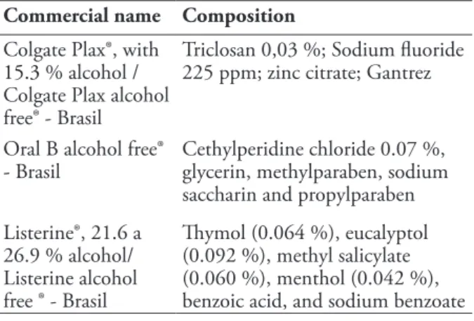
Table 1 - Mouthwashes used in this study by subjects.
Commercial name Composition
Colgate Plax®, with
15.3 % alcohol / Colgate Plax alcohol free® - Brasil
Triclosan 0,03 %; Sodium fluoride 225 ppm; zinc citrate; Gantrez Oral B alcohol free®
- Brasil Cethylperidine chloride 0.07 %, glycerin, methylparaben, sodium saccharin and propylparaben Listerine®, 21.6 a 26.9 % alcohol/ Listerine alcohol free ® - Brasil Thymol (0.064 %), eucalyptol (0.092 %), methyl salicylate (0.060 %), menthol (0.042 %), benzoic acid, and sodium benzoate
2.2 Ethical aspects
This study was approved by the Committee on Research Ethics of Public Health School of Porto Alegre, RS, Brazil (Comitê de Ética em Pesquisa da Escola de Saúde Pública de Porto Alegre - CAAE: 23153513.1.0000.5312) and were conducted in accordance with the Declaration of Helsinki.
Prior to this study, all individuals gave informed consent and answered an Individual Questionnaire about age, gender, occupation, alcohol consumption, tobacco and dietary habits, use of mouthwashes, including time, exposure to genotoxics agents and health problems. The buccal mucosa cell samples were obtain following the procedure described below, and samples were further manipulated in accordance with ethical standards.
2.3. Cells and Buccal Micronucleus Cytome Assay
(BMCyt)
Exfoliated buccal cells were collected from each subject by a single researcher, according Thomas et al. (2009). Before collecting the exfoliated buccal cells of subjects, their mouth was rinsed with water to remove saliva, food particles or any other debris.
Cells of each subject were obtained by gently rubbing the inside of the inner cheek by a cytological brush. After, the material was immersed into cold saline solution (NaCl 0.9 %) in a conic tube and it was transported, under refrigeration, to the Biotechnology Laboratory (Universidade Estadual do Rio Grande do Sul, UERGS, Novo Hamburgo, RS) for processing and preparation of the slides (BMCyt assay).
Buccal cell samples in cold saline solution were centrifuged for 10 min at 1,500 rpm, fixed in Carnoy solution (acetic acid:ethanol, 1:3) and were washed twice with Carnoy solution, under the same
centrifugation conditions. The cells suspension was dropping onto previously frozen microscope slides. Next, the slides were placed in HCl 5 N solution for 20 min at room temperature for the hydrolysis of organic material. After washing in running water (10 min), the slides were dried and stained with Schiff´s Reactive (Sigma®) for 90 min at room temperature and counterstained for 1 min with acid Fast Green (Sigma®) (Fast Green 0.1 g and acetic acid 1 %). The slides were washed twice in distilled water and they were dried and stored until the optical microscope analysis. Three slides were prepared for each subject.
2.4. Microscopic analysis
All analysis were performed in blind test and at the end of this study the data from the questionnaire and the exposure records were linked to code number for data analysis. About 2,000 epithelial cells of buccal mucosa of each subject were considered adequate for analysis and were computed distinctly individualized cells with or without changes. Only non-overlapping cells and with intact cytoplasm were counted.
MCyt assay was used to measure biomarkers such as DNA damage (micronuclei and nuclear buds), cytokinesis defects (binucleated cells), cell death (karyorrhexis, condensed chromatin pyknosis and karyolysis) and proliferative potential (basal cells) according to Thomas et al. (2009) and Bolognesi et al. (2013).
Bolognesi et al. (2013) adopted the MN identification criteria and other nuclear changes. MN are considered rounded and distinctly separate nucleus structures with well-defined boundaries, measuring about 1/3 to 1/5 the size of the nucleus and presenting chromatin and similar coloration structure, besides being visualized in the same plane.
Nuclear buds are structures analogous to MN. They are rounded bodies with similar coloration to the nucleus, but appear connected to it by a chromatin filament.
Binucleated cells are cells containing two main nuclei instead of one; the nuclei are usually very close and may touch each other and usually have the same morphology as the one observed in normal cells.
Karyorrhexis are identified by the presence of many Feulgen positive corpuscles, showing the occurrence of nuclear fragmentation.
abnormally distributed chromatin (around the inner surface of the nucleus or concentrated in the center disposed in coarse granules).
Pyknotic cells are identified as extreme chromatin condensation, associated with substantial reduction of the size of the nucleus.
Karyolytic cells are terminally differentiated cells that appear to be lacking a nucleus. Their nuclei have been completely depleted of DNA and therefore appear as Feulgen-negative ghost-like images.
Basal cells were identified as small cells with large oval nuclei uniformly stained, slightly angled cytoplasm with nuclear-cytoplasmic ratio larger than the differentiated cells.
2.5. Statistical analysis.
Results of microscopic analysis were showed as mean ± standard error. The genetic damage data between the control and exposed group (users of mouthwash) were analyzed using the Mann-Whitney test (nonparametric). When data were compared between two or more groups, these were evaluated by the Kruskal-Wallis test, according to Gattás (2001). Student t-test was used to compare age and gender of the exposed and control groups. Data with p < 0.05 was considered significant. All tests were analyzed using Graph Pad Prism Software v.6.
3 RESULTS AND DISCUSSION
This study evaluated the genotoxicity induced by mouthwash through Buccal Cytome Micronucleus assay (BMCyt assay). Table 2 shows the characteristics of subjects who participated of this study. The ages of the men and women of the exposed group compared to the control group showed no significant differences (p = 0.10 and p = 0.09, respectively). The frequency of use of mouthwash ranged from 1 to 2 times per day (18 subjects) and 1 to 4 times per week (12 subjects). Regarding to mouthwash usage time, 11 subjects reported to use for less than 12 months and 19 reported to use mouthwashes for 12 months or more. Among the exposed, 17 subjects reported using the mouthwashes with alcohol, while 13 used the product without alcohol. The analysis from questionnaires showed that subjects did not use continuously medication or foods that could increase genotoxicity. All subjects with smoking habit were excluded from this study and none of the subjects reported regularly alcohol ingestion.
Table 2 - General profile of subjects from this study.
a Control group = do not use mouthwashes; b Exposed group =
mouthwashes users.
Characteristic Control a Exposed b
Number of Subjects 30 30
Males 10 10
Age (mean in years ± SE) 26.50 ± 2.14 33.00 ± 3.14
Females 20 20
Age (mean in years ± SE) 26.05 ± 1.90 31.10 ± 2.24 Mouthwash use frequency
Daily (1 – 2 times) - 18 1-4 times per week - 12 Time of mouthwash use
Less than 12 months - 11 More than 12 months - 19 Type of mouthwash
Alcoholic - 17
Non-alcoholic - 13
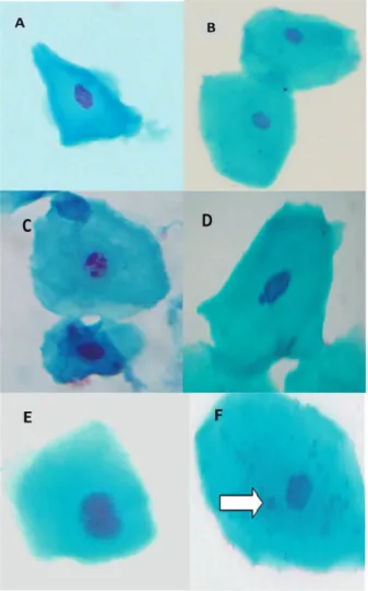
The Figure 1 shows same cells by BMCyt assay after the use of mouthwashes.
Figure 1 - Photomicrograph of exfoliated buccal mucosa cells: A) Normal basal cell; B) Normal differentiated cell; C) Karyorrhexic cell; D) Cell with Nuclear Bud (NB); E) Binucleated cell (BN); F)
The Table 3 shows the results of the genotoxicity induced by mouthwashes. All changes evaluated in this study showed frequencies increased in the exposed group. We observed that there was a significant increase (p = 0.0004; U = 217.00) of basal cells in the exposed group compared with control group indicating an intense proliferation of exfoliated epithelial tissue. These results are in agreement with Feliciano (2011) that showed a significant increase in basal cells in patients with oral cancer. In our study, the subjects reported no oral cancer, which leads us to believe that the increased frequency of these cells may indicate an event induced by exposure to mouthwashes genotoxic components, manifested by increased tissue renewal.
There was also significant increase in differentiated cells with nuclear buds and pyknotic cells compared to the control group. Nuclear buds have been considered as a DNA damage event that precedes the MN formation (THOMAS et al. 2009). Their structure is suggestive of a budding process involved in the elimination of excess nuclear material such as unresolved DNA repair complexes or amplified DNA following its segregation to the periphery of the nucleus (JAGGI; YADAV, 2015; BOLOGNESI et al. 2013). Importantly, there was an increase of cells with MN, though not significant in individuals who used mouthwashes. The significant increase observed in pyknotic cells in the exposed group represents an alternative mechanism of nucleus disintegration, characteristic of the cell death process in epithelial tissue (THOMAS et al., 2009). These observations suggest that the mouthwashes may be
related to genotoxic damage increase observed in buccal mucosa of exposed subjects.
Some results found in literature on genotoxicity of mouthwashes have been controversial. Wiltgen (2007) did not find significant difference of MN in exposed group compared to the control. In contrast, Freitas et al. (2005) and Dórea (2008) observed a higher incidence of MN in mouthwashes users, however the authors evaluated a smaller number of subjects including users of tobacco and/or alcohol. Based on these findings, we suggest that the results of these authors may not reflect the damage induced only by mouthwashes. In our study, subjects with smoking habit or exposed to other genotoxic agents were not included. All participants reported they do not drink alcohol.
Most known mouthwashes containing different concentrations of alcohol, which has the purpose of emphasizing the flavor, solubilize the active ingredient and improve the product conservation.
The alcohol content in mouthwashes evaluated in this study ranged from 15.2 to 26.9 % (table 1). Because of this, the cell damage of buccal mucosa in individuals who used mouthwash with alcohol (n = 17) and without alcohol (n = 13) were compared (unpublished data). However, there were no significant differences in DNA damage or cell death. Some studies have discussed that the alcohol in mouthwash would be responsible for the increased DNA damage in buccal mucosal cells (FREITAS et al., 2005; DÓREA, 2008). Feliciano (2011) also suggested that ethanol allows the penetration of carcinogenic compounds, which induce DNA damage
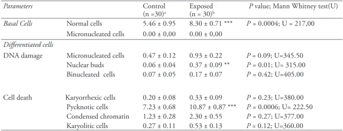
Table 3 - Nuclear changes (mean ± standard error) in basal and differentiated cells in the buccal mucosa epithelial cells of exposed (that use mouthwashes) and did not use (control).
Parameters Control
(n =30)a Exposed (n = 30)b P value; Mann Whitney test(U) Basal Cells Normal cells 5.46 ± 0.95 8.30 ± 0.71 *** P = 0.0004; U = 217,00
Micronucleated cells 0.00 ± 0,00 0.00 ± 0,00
Differentiated cells
DNA damage Micronucleated cells 0.47 ± 0.12 0.93 ± 0.22 P = 0.09; U=345.50
Nuclear buds 0.06 ± 0.04 0.37 ± 0.09 ** P = 0.01; U= 315.00
Binucleated cells 0.07 ± 0.05 0.17 ± 0.07 P = 0.42; U=405.00
Cell death Karyorrhexic cells 0.20 ± 0.08
0.33 ± 0.09 P = 0.23; U=380.00
Pycknotic cells 7.23 ± 0.68 10.87 ± 0,87 *** P = 0.0006; U= 222.50
Condensed chromatin 1.23 ± 0.28 2.30 ± 0.55 P = 0.27; U=377.00
Karyolitic cells 0.27 ± 0.11 0.53 ± 0.13 P = 0.12; U=360.00
to epithelial tissue, since alcohol has the capacity to dissolve the lipid portion of cell membranes promoting increased permeability. Unlike our study, some researchers (ZAMORA-PEREZ et al., 2013) have found significant differences in MN and other nuclear changes in individuals using mouthwash with alcohol compared to those without alcohol, indicating that such compounds containing alcohol are more genotoxic.
Comparing basal cells and DNA damage frequencies induced by different products (Colgate®, Listerine® and Oral B®; Figure 2), we observed an increase in basal cells (Figure 2A) in subjects who used the Oral B® (11.33 ± 1.83) compared to Colgate® (6.50 ± 0.56) and Listerine® (9.00 ± 1.38), although significant, only in relation to Colgate® (p = 0.023), assessed by Kruskal-Wallis test. Regarding BN cells (Figure 2B), Listerine® had the lowest frequencies [Listerine® (0.10 ± 0.10) < Oral B® (0.17 ± 0.16) < Colgate® (0.21 ± 0.11)]. However, we observed that Listerine® showed higher frequencies of MN (1.60 ± 0.56) and NB (0.60 ± 0.16), although not significant (p > 0.05) compared with the other products [Colgate®: MN (0.64 ± 0.19); NB (0.28 ± 0.12); Oral
B®: MN (0.50 ± 0.22); NB (0.17 ± 0.16)] (Figure 2C and Figure 2D). Some studies have revealed that Listerine® did not induce significant increase of MN (TURKEZ; TOGAR; ARABACI, 2011). In our study, the slight increase in the frequency of MN and NB found in Listerine® occurred probably due to the higher concentration of alcohol in this product (ranging from 21.6 to 26.9 %) compared to Colgate® (15.3 %), while Oral B® is alcohol free. Thus, we observed that alcoholic products could been related to the MN and NB frequencies, according to the following order: Listerine® > Colgate® > Oral B®.
Alterations indicative of cell death (karyorrhectic, pyknotic, condensed chromatin and karyolytic cells) showed no significant differences between mouthwashes and, due to this, the data were not presented. These results agree with those found by Ros-Llor and Lopes-Jornet (2014), who also observed no significant differences in assessing the death damage induced by mounthwash Listerine® (thymol 0.064 %), Bexident® (Triclosan 0.15 %) and Cariax sgingival® (Chlorhexidine digluconate 0.12 %). Only the latter differs from the composition of mouthwashes used in our study.
Figure 2 - Basal cells (A) and DNA damage frequencies: binucleated cells (B), nuclear buds (C) and micronucleated cells (D) induced by mouthwashes (Colgate®, Listerine® and Oral B®), evaluated by Kruskal Wallis test.
When we compared the age effects, we observed again a significant increase (p < 0.05) of basal cells in the group > 29 years old (n = 14; 10.00 ± 1.08) comparing to subjects with age 18 - 29 years (n = 16; 6.1 ± 0.78). In addition, these groups did not differ statistically in the MN frequencies in differentiated cells (data not shown). Bloching et al. (2007) and Holland et al. (2011) found a relationship between age and increased MN frequencies. These observations could be explained by cellular senescence, since the increase of MN frequencies with age occurs probably due to a combination of effects that include mutations accumulation acquired in genes involved in DNA repair, segregation of chromosomes, checkpoints cycle mobile, numerical and structural aberrations in chromosomes caused by exposure to endogenous and exogenous genotoxins (FENECH; BONASSI, 2011). Nevertheless, in our study, only basal cells frequencies were increased in older group, probably due to renewal capacity of the tissue exposed to mouthwashes.
Regarding gender, there is no consensus among researchers. Some studies (THOMAS et al., 2011) reported that women have higher MN rates than men. However, in our study, there were not significant differences in MN frequencies and other abnormalities by comparing women and men. Towards time and frequency of mouthwash use, there were also no significant differences in damage between subjects. In a study proposed by Winn et al. (1991), oral cancer risks were related to mouthwash use in 40 % of men and 60 % of women. These authors related these risks between the genders regarding the duration and frequency of mouthwashes use. In our study, these differences were not observe probably due to the fact that the exposed and the control groups presented a smaller number of individuals than the study of Winn et al. (1991), which included 866 cancer patients and 1249 controls. In another study, Pithon et al. (2011) evaluated the cytotoxicity of Listerine® in gingival fibroblasts cultures L929 and observed that the damage induced by this mouthwash was directly proportional to the exposure time.
In a recent study, Rocha et al. (2014) observed a significant increase of MN and apoptotic cells (karyorrhexis, condensed chromatin and pyknosis) in subjects using mouthwash and those that using the mouthwash and drank alcohol, indicating that mouthwash alone or in combination with alcohol induced genotoxic effects.
In our study, the Buccal Cytome Micronucleus Assay was performed in volunteers exposed to mouthwashes with or without alcohol to evaluate in vivo cellular response. The buccal mucosa is a barrier to xenobiotics, mainly because of their cell layers of compacted surface and intracellular material. Due to these characteristics of the epithelium, the detection of DNA damage and cell death in desquamated epithelial cells requires that the genotoxic agent overcome the permeability barrier reaches of the basal layer and induce lesions in DNA that are converted into MN during cell division. Such cells must then migrate to the surface to be collected for the MN assay.
4 CONCLUSION
This research found a significant increase in basal cell frequencies, nuclear buds and pyknotic cells in individuals who used the mouthwash. These damages were correlated to alcoholic composition of products suggesting a decreasing order: Listerine® > Colgate® > Oral B®. However, more studies are needed with larger numbers of participants using mouthwashes to ensure the safety of these products.
ACKNOWLEDGMENTS
The authors express their gratitude to all the individuals who volunteered to participate in this study and the granting agencies for research fellowships (Fundação de Amparo à Pesquisa do Estado do Rio Grande do Sul – FAPERGS – to K.C.H. and Conselho Nacional de Desenvolvimento Científico e Tecnológico – CNPq – to C.A.S.).
REFERENCES
BLOCHING, M. et al. Micronucleus rate of buccal mucosal epithelial cells in relation to oral hygiene and dental factors.
Oral Oncology. v. 2008, n. 44, p. 220–226, 2007.
BOLOGNESI, C. et al. The HUMNxl scoring criteria for different cell types and nuclear anomalies in the buccal micronucleus cytome assay – An update and expanded photogallery. Mutation Research, v.753, p.100–113, 2013. BORTOLI , G. M., AZEVEDO, M. B., SILVA, L. B. Cytogenetic biomonitoring of Brazilian workers exposed to pesticides: Micronucleus analysis in buccal epithelial cells of soybean growers. Mutation Research, v.675, p.1-4, 2009.
BUGNO, A. et al. Enxaguatórios bucais: avaliação da eficácia antimicrobiana de produtos comercialmente disponíveis. Revista do Instituto Adolfo Lutz, São Paulo, v. 65, n. 1, p. 40-45, 2006.
DÓREA, L.T.M. Danos Cromossômicos e Apoptose em
células esfoliadas do epitélio bucal: associação do habito de fumar e as lesões pré malignas e malignas do epitélio oral. 2008. Dissertação (Mestrado em Saúde Coletiva) - Departamento de Saúde da Universidade Estadual de Feira de Santana. Feira de Santana, 2008.
FELICIANO, Luciana Maria. Alterações citogenéticas
em portadores de desordens potencialmente malignas e carcinoma oral. 2011. 52 f. Dissertação (mestrado) - Universidade Estadual Paulista, Instituto de Biociências de Botucatu, 2011. Disponível em: <http://hdl.handle. net/11449/92461>.
FENECH, M.; BONASSI, S. The effect of age, gender, diet and lifestyle on DNA damage measured using micronucleus frequency in human peripheral blood lymphocytes. Mutagenesis, v. 26, p.43–49, 2011.
FREITA, V.S. et al. Efeitos genotóxicos de fatores considerados de risco para o câncer bucal. Revista Baiana
de Saúde Publica, v. 29, n.2, p.189-199, 2005.
GANDINI, S. et al. Mouthwash and oral cancer risk – quantitative meta-analysis of epidemiologic studies.
Annals of Agricultural and Environmental Medicine, v.19, n.2, p. 173-180, 2012.
GATTÁS, G.J.F. et al. Frequency of oral mucosa micronuclei in gas station operators after introducing methanol. Occupational Medicine, v. 51, n. 2, p. 107-113, 2001.
HOLLAND, N. et al. The micronucleus assay in human buccal cells as a tool for biomonitoring DNA damage: The HUMN project perspective on current status and knowledge gaps. Mutation Research, v. 659, p. 93–108, 2008.
HOLLAND, N. et al. Micronuclei in neonates and children: effects of environmental, genetic, demographic and disease variables. Mutagenesis, v. 26, n.1, p. 51– 56, 2011.
LACHENMEIER, D.W. et al. Salivary acetaldehyde increase due to alcohol-containing mouthwash use: A risk factor for oral cancer. International Journal of Cancer, v. 125, n. 3, p. 730–735, 2009.
LUCERO, L. et al. Cytogenetic biomonitoring of Spanish greenhouse workers exposed to pesticides:
micronuclei analysis in peripheral blood lymphocytes and buccal epithelial cells. Mutation Research, v. 464, p. 255–262, 2000
MERELES, J.R.C. et al. Apoptosis in exfoliated cells from the oral mucosa of individuals occupationallyexposed to mutagenic and carcinogenic agents. Revista Brasileira de
Cancerologia, v. 52, n. 4, p. 337-343, 2006.
McCULLOUGH, M.J.; FARAH, C.S. The mouthwash question: authors’ reply. Australian Dental Association, v.54, p. 78-81, 2009.
PITHON, M.M. et al. Avaliação in vitro da citotoxicidade de enxaguatórios bucais Listerine®. Revista Cirurgia e
Traumatologia Buco-Maxilo-Facial, v.11, p. 83-88, 2011. REMOR, A.P. et al. Occupational exposure of farm workers to pesticides: Biochemical parameters and evaluation of genotoxicity. Environment International, v . 3 5 , p . 2 7 3 - 2 7 8 , 2 0 0 9 .
ROCHA, R.S.; MEIRELES, J.R.C; CERQUEIRA, E.M.M. Chromosomal damage and apoptosis analysis in exfoliated oral epithelial cells from mouthwash and alcohol users. Genetics and Molecular Biology, v. 37, n. 4, p. 702-707, 2014.
ROS-LLOR, I.; LOPEZ-JORNET, P. Cytogenetic analysis of oral mucosa cells, induced by chlorhexidine, essential oils in ethanolic solution and triclosan mouthwashes.
Environmental Research, v. 132, p. 140–145, 2014. THOMAS, P. et al. Buccal micronucleus cytome assay,
Nature Protocols, v. 6, p. 825- 837, 2009.
THOMAS, P. et al. Effect of dietary intervention on human micronucleus frequency in lymphocytes and buccal cells.
Mutagenesis, v.26, n. 1, 69-76, 2011.
TOLBERT, P.E.; SHY, C.M.; ALLEN, J.W. Micronuclei and other nuclear anomalies in buccal smears: methods development. Mutation Research, v. 271, p. 69-77, 1992. TURKEZ, H.; TOGAR, B.; ARABACI, T. Evaluation of genotoxicity after application of Listerine® on human lymphocytes by micronucleus and single cell gel electrophoresis assays. Toxicology and Industrial Health, v. 28, n. 3, p. 271–275, 2011.
WILTGEN, A. Investigação do Potencial Genotóxico de
Anti-sépticos Bucais – in situ. 45 f. 2007. Dissertação (Mestrado em Diagnóstico Genético e Molecular), Universidade Luterana do Brasil, Canoas, 2007.
in the Risk of Oral and Pharyngeal Cancer. Cancer
Research, v. 51, p. 3044-3047, 1991.
WINN, D.M. et al. Mouthwash in the etiology of oral cancer in Puerto Rico. Cancer Causes Control, v. 12, n. 2, p. 419-429, 2001.
YADAV, A.S.; JAGGI, S. Buccal Micronucleus Cytome Assay: A Biomarker of Genotoxicity. Journal of Molecular Biomarkers &Diagnosis, v. 6, p. 1-6, 2015.
ZAMORA-PEREZ, A.N. et al. Increased number of micronuclei and nuclear anomalies in buccal mucosa cells from people exposed to alcohol-containing mouth wash.
Drug and Chemical Toxicology, v. 36, n. 2, p. 255–260, 2013.