UNIVERSIDADE DE LISBOA
FACULDADE DE CIÊNCIAS DA UNIVERSIDADE DE LISBOA
DEPARTAMENTO DE BIOLOGIA VEGETAL
ACTIVITIES OF AN AQUEOUS PLANT EXTRACT AGAINST HERPES SIMPLEX VIRUS
TYPE 2 (HSV 2): ROLE IN VIRUS REPLICATION AND PROGENY YIELD
Ana Rita Mota Mendes
Trabalho orientado pela Professora Doutora Maria Filomena Caeiro
(CESAM-FCUL) e pela Doutora Sílvia Lopo Esteves (INSA)
Mestrado em Biologia Molecular e Genética
Dissertação
ACTIVITIES OF AN AQUEOUS PLANT EXTRACT AGAINST HERPES SIMPLEX VIRUS
TYPE 2 (HSV 2): ROLE IN VIRUS REPLICATION AND PROGENY YIELD
Ana Rita Mota Mendes
2015
This thesis was mostly performed at Centro de Estudos do Ambiente e do Mar da Universidade
de Aveiro – Pólo FCUL (CESAM FCUL) under the direct supervision of Professor Maria Filomena
Caeiro, and partially performed at Instituto Nacional de Saúde Dr. Ricardo Jorge (INSA) under
the supervision of Doctor Sílvia Lopo Esteves.
Table of Contents
Acknowledgements ... i Abstract ... ii Resumo ... iii Abbreviations ... vi 1. Introduction ... 11.1. Herpes Simplex Virus ... 2
1.1.1. Classification ... 2 1.1.2. Virion Properties ... 2 1.1.3. Replication ... 3 1.1.4. Clinical Manifestations ... 6 1.1.5. Therapeutic ... 6 1.2. Medicinal Plants ... 7 1.2.1. Solidago virgaurea ... 7 1.3. Objectives ... 8
2. Materials and Methods ... 9
2.1. Cell Culture ... 9
2.2. Aqueous extract stock preparation ... 9
2.3. Virus ... 10 2.3.1. Virus Production ... 10 2.3.2. Virus Titration ... 10 2.4. Cytotoxicity Assays ... 11 2.5. Virucidal Effect... 11 2.6. Effect on HVS-2 replication ... 12
2.6.1. Extract effect on viral plaques formation ... 12
2.6.2. Extract effect on virus yield ... 12
2.6.3. IC50 and IC90 determination... 13
2.6.4. Determination of the affected stage during the virus replication ... 13
2.6.5. Comparison with acyclovir inhibition ... 13
2.7. Visualization of treated and non-treated infected cells and virus suspensions by Transmission Electron Microscopy (TEM) ... 14
2.7.1. Infected cells – treated and control – observation by TEM ... 14
2.8. Kinetics of HSV-2 infection in treated and non-treated cells ... 15
3. Results ... 17
3.1. Cytotoxicity assays ... 17
3.1.1. Determination of the extract CC50 and MNCC ... 17
3.1.2. Correlation between FBS concentration in the culture medium and cytotoxic effect of the extract ... 18
3.2. Virucidal effect... 18
3.3. Extract effect on viral plaques formation ... 19
3.4. Extract effect on virus yield ... 20
3.4.1. IC50 and IC90 determination... 20
3.4.2. Determination of the affected stage of virus replication ... 21
3.4.3. Comparison with ACV inhibition... 21
3.4.4. Visualization of treated and non-treated infected cells and virus suspensions by Transmission Electron Microscopy (TEM) ... 22
3.4.5. HSV-2 Infection Kinetics ... 22
3.4.6. Quantitative real-time PCR (qPCR) ... 24
4. Discussion ... 25
4.1. Extract cytotoxic effect ... 25
4.2. Virucidal effect... 26
4.3. Extract effect on plaque formation ... 26
4.4. Extract effect on virus yield ... 27
4.5. Further Perspectives ... 29
5. References ... 30
6. Supplementary Data ... 33
6.1. Extract Cytotoxic Effect ... 33
6.2. Extract Virucidal Effect ... 33
6.3. Extract effect on viral plaques formation ... 34
i
Acknowledgements
À minha orientadora, Professora Doutora Filomena Caeiro, pelos melhores e mais úteis conhecimentos transmitidos, por toda a disponibilidade e ajuda.
À minha co-orientadora Professora Doutora Sílvia Lopo, pela ajuda prestada e pela disponibilidade.
À Professora Doutora Lia Ascensão e ao Professor Doutor António Pedro de Matos, pela disponibilidade prestada.
À Professora Doutora Luísa Serralheiro, pela generosidade e disponibilidade.
À Teresa Granja, Rute Miguel e Manuela Lucas, por todos os esclarecimentos, ajuda e disponibilidade.
À Mónica Barra e Nadieny Barbosa, pelos concelhos e esclarecimentos prestados.
À Egídia Azevedo e Marta Figueiredo, pelo apoio e boa disposição.
À Marta Costa, por toda a ajuda e disponibilidade constante.
ii
Abstract
Herpes simplex virus type 2 (HSV-2) is widely distributed through the human population, infecting more than 500 million people globally. Although causing generally mild infections this virus may cause severe symptoms occasionally, mainly in immunocompromised patients. Presently, there are a number of systemic antiviral agents against herpesvirus, the most commonly used being acyclovir (ACV) and its prodrugs. However, long term treatments with these drugs may result in the development of resistance, especially in immunocompromised patients, which leads to a continuous search for new and better therapeutic alternatives. According to the World Health Organization (WHO) plants would be the best sources for obtaining a wide variety of drugs. In fact, in the last decades many pharmacological and chemical studies have focused on medicinal plants and the discovery of new natural therapeutic compounds. In this study the anti-herpetic action of an aqueous extract was evaluated. The product was obtained by decoction of stem and leaves from Solidago virgaurea, a perennial herb member of the Asteraceae family. The study of the aqueous extract activity included a preliminary evaluation of its cytotoxicity in Vero cells – by the MTT assay - in the same conditions that are applied for viral production. Extract direct effect on viral particles – virucidal effect – was also assessed, having proved null. Anti-herpetic activity was investigated through two kinds of experiments: treatment of infected cells during virus production that revealed a mean yield reduction of 94 % in treated relatively to non-treated cells and an IC50 of 35.1 μg/mL; and treatment of infected cells during
virus titration, which revealed slighter inhibition but significant size differences between virus plaques formed in treated and control conditions (smaller in treatment conditions). To further evaluate the mechanisms that mediate the aqueous extract inhibitory effect, infected cells – treated and non-treated - and virus particles – produced in non-treated and non-non-treated cells - were visualized through Transmission Electron Microscopy (TEM), revealing less damage due to infection in treated cells and a reduced amount of viral particles in HSV-2 suspensions produced in treated cells, relatively to controls. A kinetic of the first hours of the infection was performed with and without treatment, to assess possible differences in DNA production. Extracted samples were subjected to qPCR and results showed that the amount of viral DNA raises significantly slower in treated versus non-treated infected cells, throughout the infection. This is consistent with the effective reduction of the extract when added at later infection times - 4-6 h p.i. - when DNA replication is already in an advanced stage. Our results suggest that the aqueous extract inhibits HSV-2 replication, when present at the beginning of the infection, possibly by interfering with the viral DNA synthesis.
iii
Resumo
O vírus Herpes simples tipo 2 (HSV-2) é um vírus de simetria icosaédrica e grandes dimensões, pertencente à família Herpesviridae, subfamília Alphaherpesvirinae da ordem Herpesvirales. Infecta mais de 500 milhões de pessoas em todo o mundo, surgindo, em cada ano, mais de 20 milhões de casos. A sua prevalência é geralmente mais elevada entre mulheres do que homens e varia substancialmente entre diferentes regiões do globo, alcançando os mínimos na Europa Ocidental - 18 % para mulheres e 13 % para homens – e percentagens máximas na África Subsaariana – 70 % para mulheres e 55 % para homens. O HSV infecta as células da mucosa oral ou genital e tem a capacidade de infetar as dendrites e os gânglios sensoriais que enervam estes tecidos onde estabelece latência. Estas infeções causam normalmente patologias ligeiras – gengivite, herpes labialis ou herpes genitalis – podendo até ser assintomáticas. Porém, infeções com HSV-2 podem causar complicações sérias a nível ocular - sendo a primeira causa de cegueira devida a infeção no mundo Ocidental -, neurológico - causando meningites e encefalites, especialmente comuns em neonatos – e dermatológico. Estas patologias mais severas, associadas à infeção com HSV, ocorrem com maior frequência em pacientes imuno-comprometidas, nomeadamente em indivíduos HIV (Vírus da Imunodeficiência Humana) positivos. Aliás, a infeção com HSV-2 aumenta 3 a 4 vezes a probabilidade de adquirir HIV e a sua disseminação, sendo esta relação recíproca, uma vez que pacientes HIV positivos têm maior probabilidade de adquirir HSV-2 e sofrem mais reativações da infeção. Atualmente, existem vários agentes com atividade antiviral contra HSV, como o Aciclovir (ACV) e as suas pró-drogas, no entanto, tratamentos prolongados com estes compostos podem resultar no desenvolvimento de resistência. De acordo com a Organização Mundial de Saúde (OMS) as plantas são a melhor fonte para a obtenção de novas drogas e, nas últimas décadas, as plantas medicinais têm sido alvo de vários estudos químicos e farmacológicos. Neste estudo foi avaliado o efeito anti-herpético do extrato aquoso de Solidago virgaurea, uma planta perianal, membro da família Asteraceae.
O extrato utilizado foi obtido por decocções de caules e folhas de S. virgaurea e a solução stock foi preparada a 100 mg/mL em DMSO. Antes de ser sujeito a testes de actividade anti-herpética foi ensaiado o efeito do extracto aquoso na viabiliade de células hospedeiras não infectadas, através do método do MTT – um ensaio colorimétrico que se baseia na capacidade de reduzir metabolicamente o sal de tetrazolio das células viáveis. Para isso culivaram-se células Vero em placas de 96 poços – em meio de cultura com FBS (Soro Fetal Bovino) a 10 % – até à sub-confluência. O meio de cultura foi depois substituido por meio com FBS a 2 % e diferentes concentrações de extracto (25 - 250 μg/mL), ou meio com FBS a 2 % sem adição de extrato no caso dos testemunhos, e as placas foram incubadas 24 ou 48 horas a 37 °C. No final do período de tratamento fez-se o teste do MTT para determinação da viabilidade das culturas tratadas, por comparação com as culturas não tratadas. Os conteúdos de cada
iv poço da P96 foi analisado espectofotometricamente num leitor de placas Elisa. Para caracterizar a actividade citotóxica do extracto foram comparadas as absorvâncias obtidas em poços com células tratadas e relação a células não tratadas e determinaram-se os parametros CMNC (Concentração Máxima Não Citotóxica) - 150 μg/mL - e CC50 (Concentração que inviabiliza 50 % das células) – 177,3
μg/mL.
O efeito directo do extracto nas partículas de HSV-2 foi testado através da comparação dos títulos de suspensões virais previamente tratadas com 100 μg/mL de extrato em meio com FBS a 2 % com os títulos de suspensões não tratadas. O extrato não demonstrou efeito virucida sobre partículas virais em suspensão, uma vez que os títulos das suspensões tratadas foram muito semelhantes aos títulos dos testemunhos.
Para testar o efeito do extrato na formação de placas virais infetaram-se células, em placas de 24 poços, com diluições seriadas de HSV-2. Depois da entrada do vírus – 30 minutos pós infeção – os inóculos foram removidos e foi adicionado meio de cultura com FBS a 2 % com ou sem 100 μg/mL de extrato, às culturas tratadas e testemunho, respetivamente. No final do período de tratamento – 3 a 4 horas pós infeção (h p.i.) – foi adicionado meio com FBS e Sephadex a 2 %. A redução de título em culturas tratadas foi 71,6 ± 26,8 %, em relação às não tratadas. Foi ainda evidente uma redução no diâmetro das placas produzidas nas culturas tratadas.
Foram também levadas a cabo várias experiências envolvendo a produção de vírus em condições de tratamento, em que culturas celulares sub-confluentes foram infetadas com cerca de 3 pfu/célula e tratadas com extrato em diferentes concentrações e durante diferentes períodos da infeção. Os títulos das suspensões produzidas em condições de tratamento foram comparados com os títulos dos testemunhos para o cálculo das percentagens de inibição. A redução média de título – com concentrações de extrato não citotóxicas – foi de 93,65 ± 8 %, relativamente a vírus produzido na ausência do extrato. Para determinar os parâmetros IC50 e IC90 (concentrações que reduzem o
rendimento viral em 50 e 90 %, respetivamente) foram produzidas suspensões de HSV-2 em meio com FBS a 2 % e diferentes concentrações de extrato (10-175 μg/mL) que foram posteriormente tituladas. Os IC50 e IC90 foram calculados através de regressão linear da curva dose-resposta e foram 35,1 e 128,1
μg/mL, respetivamente. Para investigar a etapa da infeção afetada pelo extrato produziram-se suspensões de HSV-2 sob tratamento - com 100 μg/mL de extrato – iniciado em diferentes horas pós infeção. Foi observável uma redução da percentagem de inibição do extrato para cerca de 50 % em tratamentos começados entre as 3 e as 6 h p.i. e para 0 % a partir das 15 h p.i. Células infetadas tratadas e não tratadas, e partículas virais produzidas em ambas as condições, foram observadas por Microscopia Eletrónica de Transmissão (TEM), revelando a existência de menos danos, devidos à
v infeção, em células infetadas tratadas do que em não tratadas e uma quantidade significativamente menor de partículas de HSV-2 em suspensões virais produzidas em células tratadas, apesar dos viriões produzidos sob tratamento não apresentarem defeitos morfológicos. Por fim foi executada uma cinética da infeção em condições de tratamento (e uma cinética testemunho, em condições normais de produção viral), para tal, células infetadas – tratadas e não tratadas - foram raspadas para o meio de cultura a diferentes horas pós infeção – 2 a 7. O DNA de cada cultura infetada recolhida foi extraído, quantificado e submetido a PCR para a deteção de DNA viral. O resultado não permitiu visualizar diferenças significativas na quantidade de DNA de HSV-2 em células tratadas e não tratadas, mas mostrou um aumento considerável na quantidade de DNA a partir das 5 horas pós infeção. As mesmas amostras foram depois utilizadas para PCR em tempo real (qPCR) para avaliar possíveis diferenças na quantidade de DNA viral. O resultado mostrou que, embora nas horas iniciais – 3-4 h p.i. – as diferenças na quantidade de DNA sejam mínimas, entre amostras de DNA extraído de células tratadas e não tratadas, é visível um aumento muito mais lento no número de cópias de DNA viral ao longo da infeção em culturas tratadas do que em não tratadas, indicando que a replicação de DNA é afetada durante o tratamento com extrato aquoso de S. virgaurea.
vi
Abbreviations
ACV - Acyclovir
ATCC – American Type Culture Collection
bp – Base pair
CC10 - 10 % Cytotoxic Concentration
CC50 – 50 % Cytotoxic Concentration
CC90 - 90 % Cytotoxic Concentration
CNS – Central Nervous System
D-MEM – Dulbecco’s Modified Eagle Medium
Da - Dalton
DMSO - Dimethyl sulfoxide
DNA - Deoxyribonucleic Acid
dsDNA – Double stranded DNA
EtBr – Ethidium Bromide
FBS - Fetal Bovine Serum
FCV - Famciclovir
FDA – Food and Drug Administration
FOS - Foscarnet
g – Gravitational acceleration
HIV – Human immunodeficiency virus
HK – Herpes Keratitis
h p.i. – Hours post infection
vii
HSV – Herpes simplex virus
HSV-1 – Herpes simplex virus type 1
HSV-2 – Herpes simplex virus type 2
HVEM – Herpesvirus Entry Mediator
IC10 – 10 % Inhibiting Concentration
IC50 – 50 % Inhibiting Concentration
IC90 – 90 % Inhibiting Concentration
ICTV - International Committee on Taxonomy of Viruses
IDU - Idoxuridine kDa - Kilodalton kb – Kilobase mg – Milligram mL – Milliliter mM – Millimolar
MNCC - Maximum Non-cytotoxic Concentration
MTT – 3-(4,5-dimethylthiazol-2-yl)-2,5-diphenyltetrazolium bromide
ng - Nanogram
nM – Nanomolar
nm - Nanometer
PBS - Phosphate Buffered Saline
PCR – Polymerase Chain Reaction
pfu – Plaque forming units
viii
RNA – Ribonucleic Acid
mRNA – Messenger Ribonucleic Acid
rpm – Revolutions per minute
RT-PCR - Reverse Transcriptase Polymerase Chain Reaction
SI – Selective Index
TFT – Trifluorothymidine
TNF – Tumor Necrosis Factor
T25, T75 – Culture flasks with 25 or 75 cm2 area
UL – Unique long sequence
US – Unique short sequence
UV – Ultraviolet
VACV – Valaciclovir
vhs – Virus host shut off proteins
VP – Virion Polypeptide
VZV – Varicella zoster virus
WHO - World Health Organization
α -TIF – α-transinducing factor
μg – Microgram
1
1. Introduction
Herpes simplex viruses (HSV) have been interacting with humans for thousands of years [1] and occur
worldwide, without seasonal variation [2]. HSV-2 infects more than 500 million people globally, and,
each year, more than 20 million new cases arise. HSV-2 prevalence increases with age, peaking at 35-39 years of age, and is generally higher among females than males. It varies substantially by region, ranging from about 18 % among women and 13 % among men in Western Europe to 70 % and 55 % in Sub-Saharan Africa [3].
Herpetic lesions at the oro-labial area are typically caused by HSV-1 and genital lesions are commonly associated to HSV-2, but, up to 10 % of primary oro-facial herpes infections could be caused by HSV-2
[4] and, on the other hand, HSV-1 seems to be emerging as the most common cause of primary genital
herpes [5]. Even though, recurrences of infection by HSV-1 represent less than 5 % of the total cases of
genital herpes [6].
Although HSV typically causes mild diseases it may provoke severe infections in immunocompromised patients like HIV seropositives and recipients of solid organ or bone marrow transplants [7]. In fact, an
important finding in the last decades is that infection with HSV-2 is associated with high susceptibility to HIV, and increases its shedding [8, 9]. This relationship is reciprocal, as infection with HIV also
significantly increases the probability of acquiring HSV-2 and its reactivation has shown itself more frequent in HIV-1 positive patients [10].
Currently, there are several systemic antiviral agents, against herpesvirus, like acyclovir (ACV) and its prodrugs – valaciclovir (VACV), famciclovir (FCV), ganciclovir, valganciclovir, cidofovir and foscarnet (FOS) – approved by the US Food and Drug Administration (FDA) [11], but, long-term prophylaxis and
treatment with ACV, VACV or FCV can result in the development of resistance, especially in the immunocompromised patients group[7,12] where ACV-resistant HSV prevalence varies between 3.5 %
and 10 % [7].
Plants are still an important source of widely used pharmaceutics [13-19] and Asteraceae family has been
one of the most studied lately, and specifically its inhibitory activity against HSV was even the subject for a few recent works [17, 19-21]. Therefore, and considering the serious negative clinical implications
that HSV-2 infections cause globally, it’s highly significant to further evaluate anti-viral activities of plants of the Asteraceae family, inclusively the anti-herpetic proprieties of Solidago virgaurea, the object of this study.
2
1.1. Herpes Simplex Virus
1.1.1. Classification
Herpesvirus classification was addressed for the first time in 1971 by the International Committee on Taxonomy of Viruses (ICTV) where the genus Herpesvirus was established [23]; this genus was later
elevated to the family Herpesviridae[23].A first attempt of grouping herpesvirus into subfamilies has
been done based in biological criteria[24]; this division was successful but contained a few
misclassifications, therefore, further distribution of the subfamilies into genera was based in molecular data – genome characteristics, such as size and structure - to a greater extent than before[25].
Nowadays, the former family Herpesviridae has been elevated to a new order, the Herpesvirales, which accommodates three subfamilies - Alphaherpesvirinae, Betaherpesvirinae and Gammaherpesvirinae - 17 genera and 90 species [26].
1.1.2. Virion Properties
HSV are large viruses, and their viral particles have complex and characteristic structures. The spherical virion includes four major elements: core, capsid, tegument and envelope [27, 28].
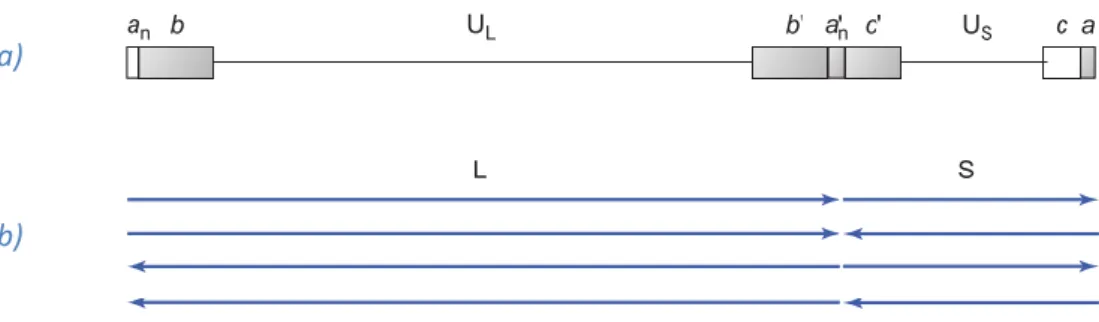
The core consists of the viral genome packed as a linear double-stranded DNA molecule - 150 kpb - into the icosahedral capsid. Herpes simplex virus genome has an unusual structure that consists of two covalently linked components, long (L) and short (S), and each of these components contains a stretch of unique sequences, UL and US, flanked by inverted repeats (Fig. 1 a). The two components can invert
relative to one another, creating four different types of DNA molecule that differ only in the orientation of DNA sequences (Fig. 1 b). The sequence repeat at the left end of UL is designated ab, and the internal
inverted repeat is designated b’a’. The inverted repeats flanking US are designated a’c’ and ca. Most
viral genes are present in only one copy per genome; the UL region contains 65 protein coding
sequences, and the US region contains 14 [28].
Therefore, HSV contain at least 84 different genes. Presently, most herpes simplex genes are numbered sequentially from one end of the unique sequence in which they are found. Thus, genes UL1 to UL56
are located in the L unique segment and US1 to US12 are located in the S unique segment, and some
genes lying within the b and c segments have names related to their time of expression or other properties. HSV genes can be classified in at least three groups – immediate early (α), early (β) and late (γ) - based on the time of their expression during the replication cycle [2, 28].
3 Fig. 1 - Genome of herpes simplex virus. a) Organization of unique and repeat sequence elements. Repeat sequence elements aare shown in small letters (a,b,c) and elements shwon with a prime are inverted. Unique sequence elements are shown in capital letters (UL, US). b) Four isomeric forms of viral DNA with different orientations of the long (L) and (S) components due to frequent recombination at the repeated sequence elements. (Source: Acheson 2007)
The bilayered capsid (100-110 nm in diameter) is constructed with six proteins, of which the major one, VP5 (150 kDa), makes up the 162 capsomers, that are arranged as 12 pentamers at the vertices and 150 hexamers on the faces and edges of the icosahedron. Another capsid protein, VP26, is located at the tips of the capsomers. The capsid is enclosed in an envelope that contains at least ten different glycoproteins (gB to gM) and several other non-glycosylated membrane proteins [28, 29].
The virus envelope has a typical appearance and is thought to be derived from patches of host cell nuclear membrane modified by the insertion of virus glycoprotein spikes [29].
Unlike in most enveloped viruses, there is a large amorphous mass between the capsid and the envelope, called the tegument, which contains about 14 viral proteins like virus host shutoff (vhs), VP16 (α-TIF, ICP25), VP11-12, VP13-14 and VP1-2. These proteins are introduced into the cell upon infection by herpesviruses, and some of them carry out crucial functions in the virus growth cycle like activation of transcription and shutoff of host macromolecular synthesis [28, 29].
1.1.3. Replication
This virus is characterized by a short – 18 to 24 hours – and cytolytic replicative cycle. Initial attachment and penetration of the host cell is mediated via envelope glycoprotein gB that binds to heparan sulfate proteoglycans at the cell surface. Afterwards, envelope glycoprotein gD binds to at least two alternative receptors: nectin 1 – member of the nectin family of intercellular adhesion molecules – and herpesvirus entry mediator (HVEM) – member of the tumor necrosis factor (TNF) receptor family. Receptor binding triggers a pH-independent fusion of the viral envelope with the cellular membrane, which requires three conserved glycoproteins – gB, gH and gL [28, 29].
a)
4 Fusion results in the introduction of tegument proteins and viral nucleocapsid into the cell cytoplasm and the rapid shut down of host macromolecular metabolism. Host DNA synthesis ceases, protein synthesis declines rapidly, ribosomal RNA synthesis is reduced and host protein glycosylation ceases. Most tegument proteins are believed to act directly or indirectly to produce early shutoff of the host macromolecular synthesis and to contribute to the early events of replication, like nucleocapsid attachment to nuclear pores and DNA release [29].
Nucleocapsids, along with some of the tegument proteins, are transported to the nuclear pores by dynein motors along the microtubular network of the cell and, at the nuclear pore, DNA is released and enters the nucleus. Empty capsids remain at the cytoplasmic side of nuclear pores for several hours until they disintegrate. Viral DNA in the nucleus is circularized, either by direct ligation of the ends or by recombination between the a sequences at the ends [28].
HSV gene expression is regulated and sequentially ordered as a cascade (α→β→γ). Transcription of HSV immediate early (α) genes is carried out in the host nucleus, during the first several hours after infection, by cellular RNA polymerase II. Five of the six α gene products are regulatory proteins, involved in the regulation of viral and cellular gene expression, and their expression is required for the production of all subsequent polypeptide groups. To initiate transcription, tegument protein α-TIF or VP16, known as α-transinducing factor, forms a complex with at least two key cellular proteins - Oct-1 and HCF-1 - which binds to specific response elements in viral DNA that have a central consensus sequence – TAATGARATT. These elements, along with binding sequences for other cellular transcription factors, are found upstream of the promoters of all α genes, and act as transactivators to enhance the transcription of these genes. Transcripts are then transported to the cytoplasm where they are translated into five proteins – ICP0, ICP4, ICP22, ICP27 and ICP47 – that are transported back to the nucleus where they exert regulatory and activating functions relatively to β genes expression [28, 29].
Expression of early – or β - genes results in the production of proteins that work together to carry out essential steps in DNA replication. Maximum synthesis rates of these proteins are observed at 5-7 h p.i., and when sufficient levels have accumulated within the infected cell, viral DNA replication starts. Like other DNA viruses, herpesviruses make their own DNA polymerase that is encoded by a single major RNA and has its synthesis during β phase. This enzyme can begin DNA synthesis at any one of three viral origins of DNA replication on the herpes simplex virus genome. oriL is located in the middle of UL region, and the other two – oriS – are in the c inverted repeats flanking the US region. Replicating
DNA in infected cells is found as high molecular weight, branched head-to-tail multimers called concatemers, suggesting that DNA is made by a rolling circle mechanism, which can produce multimers
5 from a circular template. However, herpesvirus initially sets up bidirectional replication on the circular viral DNA, and only later shifts to rolling circle replication [28, 29].
Although γ1 genes can be transcribed in the absence of viral DNA replication their optimal expression
requires viral DNA synthesis, presumably because of the increase in the number of template DNAs available for transcription; in contrast, the transcription of γ2 genes is totally dependent on viral DNA
synthesis. Late mRNAs enter the cytoplasm for translation into late proteins which include the capsid structural proteins that travel back to the nucleus, envelope proteins that are produced in the endoplasmic reticulum (ER) and become incorporated in the nuclear membrane and other structural and packaging related proteins [28].
Immature capsids lacking DNA are assembled in the nucleus by accumulation of the major capsid protein VP5 and several other proteins around a scaffold formed by three late proteins – VP21, pre-VP22a and VP24. Viral DNA - in the form of head-to-tail concatemers - is cleaved and stuffed into the preformed capsids by the interaction of several viral late proteins with high conserved packaging sites – pac1 and pac2 – located in the a sequence near the termini of the DNA. Entry of viral DNA into the capsids is carried out by a multimers of UL6 protein located at one vertex of the capsid and involves
several other nonstructural proteins. DNA packaging is complete when a sufficient length of the DNA is introduced and when the packaging machinery encounters the next a sequence along the DNA in the same orientation. The packaged DNA is cleaved from the concatemers at a specific site within a 20-nt direct repeat flanking the terminal a sequence The DNA filled capsids proceed to bud trough the modified inner nuclear membrane, into the lumen between the two nuclear membranes, acquiring an envelope and a layer of tegument proteins. Virions are then transported from the lumen to the outside of the cell [28, 29].
Three distinct theories account for the mechanism by which virus particles exit the cell. According to one theory, virions retain their envelopes and are transported to the Golgi membranes within vesicles that bud from the outer nuclear membrane. According to another theory, virions lose their envelope by fusion with the outer nuclear membrane, releasing nucleocapsids into the cytoplasm; these nucleocapsids subsequently reacquire an envelope by budding into Golgi membranes. In either case, the envelope proteins would be glycosylated within the Golgi, and further transport of mature virions to the extracellular space is via secretory vesicles. A third theory is based on the observation that nuclear pores become enlarged and the Golgi membranes are fragmented during viral replication; according to this hypothesis, nucleocapsids exit through the enlarged nuclear pores without an envelope and become enveloped at multivesicular bodies derived from fragmented Golgi membranes
6
1.1.4. Clinical Manifestations
HSV infections cause usually mild pathologies – gingivostomatitis, herpes labialis and herpes genitalis – and may be, many times, asymptomatic [28, 30]. Transmission occurs by intimate contact with an
infected individual either with evident infection or asymptomatic excretion of viral particles, and, after primary HSV replication, at the oral or genital mucosa, the virus is able to infect neuronal dendrites of sensory ganglia that innervate these tissues and establishes long-term latency, from which it can reactivate episodically [31, 32].
Besides common herpetic lesions HSV-1 and HSV-2 infections can also result in other skin diseases such as herpetic whitlow, which manifests as lesions at the finger tips [33], and eczema herpeticum in patients
underlying atopic dermatitis – the most common chronic inflammatory skin disease - which may be life-threatening for children [34].
These viruses are also linked with a variety of ocular complications, mostly epithelial and stromal keratitis. HSV can cause stromal opacification and is estimated to be the first cause of blindness due to infection in the Western world [35].
In addition to establish latent infection in the human nervous system these neurotropic viruses can also infect the central nervous system (CNS) causing diseases as meningitis and encephalitis. These kinds of infections more often occur in neonates as a result of congenital infection from the mother to the new born at birth - even when the mother shows no clinical signs of infection - and can lead to severe neurological damage as well as death [36].
An important finding in the last decades is the fact that infection with HSV-2 increases 3-4 times the likelihood of acquiring HIV [8] and considerably increases the dissemination of HIV in the genitalia of
co-infected patients [9].Interestingly this is a reciprocal relationship, since infection with HIV also
significantly increases both HSV-2 reactivations and the probability of acquiring it [10].
1.1.5. Therapeutic
The antiviral therapies for herpes were first approached at Yale, USA in 1959 where a team synthesized idoxuridine (IDU) - the first Food and Drug Administration approved antiviral drug - followed by trifluorothymidine (TFT), both used to treat Herpes Keratitis (HK) [11].
Vidarabine was the first antiviral drug selective enough to be used for systemic treatment of herpesvirus (HSV and Varicella zoster virus, VZV) [11, 37]. This drug was later abandoned for reasons
7 including relative insolubility in aqueous medium and rapid deamination, but, most of all, by being surpassed by much safer and potent drugs [11].
In December 1977 [38] and April 1978 [39] it was announced the discovery of acyclovir
(9-(2-hydroxyethoxymethyl guanine)), a guanosine nucleoside analog that once internalized into infected cells is processed into a monophosphorylated form – by HSV thymidine kinase - that is subsequently phosphorylated into acyclovir triphosphate by host kinases. In this form ACV thriphosphate is incorporated by the viral DNA polymerase disrupting the viral genome replication by a chain termination mechanism [40 -42].
In vitro, ACV is most potent against HSV-1, approximately half as potent against HSV-2, 1/10th as potent
against VZV and Epstein-Barr virus (EBV) and least potent against human cytomegalovirus (HCMV)[7].
Its main limitation resides in its rather poor oral bioavailability, which led to the development of valacyclovir (VACV), an L-valyl-ester prodrug of acyclovir [43, 44].
Drug-resistant strains of HSV frequently develop following therapeutic treatment, particularly in immunocompromised patients group [7, 12] where its prevalence was reported between 3.5 % and 7 %
in HIV patients, 2.5 % and 10 % in solid organ transplant patients and 4.1 % and 10.9 % in hematopoietic stem cell transplant (HSCT) patients [7].
1.2. Medicinal Plants
Evidence for the use of medicinal plants dates back to 60,000 years ago [45] and, in the last decades,
many pharmacological and chemical studies concerning medicinal plants have been made, with the discovery of new therapeutic compounds as a goal [13]. In fact, according to the World Health
Organization (WHO), these would be the best sources for obtaining a wide variety of drugs and could benefit a large population [14]. It has been estimated that more that 50 % of available drugs have, in
some way, originated from plants [15, 16], examples being morphine, ementine, vincristine and colchicine [13].
1.2.1. Solidago virgaurea
Solidago virgaurea is a perennial herb member of the Asteraceae family, one of the largest plant families with more than 1,600 genus and 24,000 to 30,000 species [46, 47]. This family has a wide
distribution, existing in every continent, except Antarctic [17, 18]. Solidago L. genus includes about 125
8 Goldenrod, being the single member of this genus native in Europe [47, 48]. This species is recognizable
by its elongated and branched golden yellow flower heads that bloom in summer and has been traditionally used to treat urinary tract, nephrolithiasis and prostate diseases [49, 50].
1.3. Objectives
Considering the high prevalence of this virus and the serious clinical implications that HSV-2 infections may cause to seropositive patients, along with the cooperative relation it can establish with HIV and the increasing occurrence of ACV-resistant strains, especially in imunodeficient individuals, the search for new and effective treatments is an extremely important matter. As two previous studies [17, 19] had
concluded that S. virgaurea aqueous extract exhibited anti-herpetic activity, this work aims to further characterize its inhibitory effect and investigate the associated mechanisms.
9
2. Materials and Methods
Working with virus and, therefore, with cell cultures demands really strict sterility conditions. Thus, most procedures described were prepared in a class II laminar flow cabinet (Biohazard), irradiated with U.V. light for 30 minutes before and after each utilization. Most materials used were sterile, either disposable or glass, the last ones were sterilized in an autoclave (121 ºC, 15 minutes). All culture media were supplemented with antibiotic and kept at 4 ºC.
2.1. Cell Culture
Cells used were Vero – a continuous lineage isolated from the renal epithelium of the green African monkey (Cercopithecus aethiops) – and were obtained from the American Type Culture Collection (ATCC) with the reference CCL81. These cells form a monolayer adherent to the flask surface and are widely used in virology studies for being permissive for a large variety of virus.
Cultures were grown in T25 and T75 flasks (Nunc) at 37 ºC in CO2 Independent Medium (Gibco) or
Dulbecco’s Modified Eagle Medium (D-MEM, Gibco) supplemented with 10 % Fetal Bovine Serum (FBS, Gibco), 0.1 % gentamicin sulfate (50 μg/mL) and glutamax (5 μg/mL) with or without a 5 % CO2
atmosphere, respectively for each medium. The cultures were observed through an inverted optic microscope (Zeiss IM).
Cells were transferred to a new flask when cultures reached 90 % confluence. The culture medium was removed and the cultures washed, twice, with 2.5 mL of PBS (Phosphate Buffered Saline, Gibco), then the cultures were trypsinized with 1 mL of trypsin (Gibco) at 37 ºC for 5 minutes. After that, 5 mL of 10 % FBS culture medium – D-MEM or CO2 Independent Medium - were added to the flask, to disable
trypsin and homogenize the suspension. Then the cell suspension was distributed accordingly to the flask/plate size.
2.2. Aqueous extract stock preparation
The aqueous extract used in this study was obtained from steams and leafs of the Portuguese Asteraceae family member Soligado virgaurea L. The lyophilized extract was supplied by Prof. Dr. Luísa Serralheiro from CQB (Centro Química e Bioquímica), FCUL (Faculdade de Ciências da Universidade de Lisboa) and kept at -20 ºC. Stock solution was prepared at 100 mg/mL concentration in DMSO
10 (Dimethyl sulfoxide) and kept at -20 ºC, the work solutions were prepared at 10 mg/mL in PBS and kept at 4 ºC.
2.3. Virus
The viruses used in this study were acquired from ATCC, and are wild strains of HSV-2 (Herpes simplex type 2 HD) and HSV-1 (Herpes simplex type 1 SC 16). Viruses were kept in suspension with 2 % FBS culture medium (D-MEM or CO2 Independent Medium) at 4 ºC.
2.3.1. Virus Production
The virus suspensions were produced in T75 flasks with sub confluent cell cultures (about 107 cells).
Culture medium was removed and cells were infected with, approximately, 106 pfu, in a 1 mL volume.
After the adsorption period – 30 to 60 minutes at 37 ºC – 12 mL of 2 % FBS culture medium (D-MEM or CO2 Independent Medium) were added. Flasks were kept at 37 ºC until total cytopathic effect –
characterized by severe morphological changes in the culture and loss of adherence by most or all the cells - could be observed, what takes, usually, 48 to 72 hours of infection.
Virus suspensions were collected in 15 mL Falcon tubes and centrifuged at 3,000 g for 5 minutes, the supernatants were transferred to new, similar, tubes and the pellets were discarded. Produced viruses were kept at 4 ºC.
2.3.2. Virus Titration
To determine the virus titer – the number of plaque forming units (PFU) per mL – each virus suspension was serially diluted (1:10) in 2 % FBS culture medium (D-MEM or CO2 Independent Medium) to 10-6.
Then, in 24 or 48 well plates with sub confluent Vero cell cultures, the 10% FBS culture medium was removed and 0.1 mL of each viral dilution was inoculate in duplicates.
The plates were then incubated for 30 minutes at 37 ºC, to allow viral entry, before the addition of 500 μL of titration medium – 2 % FBS culture medium with 2 % Sephadex G-75 (Pharmacia Fine Chemicals). Plates were kept, absolutely still, at 37 ºC for 2 days – HSV-2 – or 3 days – HSV-1.
To end the titration, the infected cultures were fixated with 0.5 mL of 10 % (v/v) formaldehyde for 30 minutes, at room temperature, with stirring. After that, formaldehyde was removed and the plates
11 washed with tap water. The cells were then dyed with 0.2 % crystal violet for 15 minutes, with stirring, then the stain was removed and the plates were washed with tap water and dried in an oven at 37 ºC.
All countable viral plaques were counted and virus titer was calculated by the formula:
𝑉𝑖𝑟𝑢𝑠 𝑡𝑖𝑡𝑒𝑟 (𝑝𝑓𝑢/𝑚𝐿) = #𝑝𝑙𝑎𝑞𝑢𝑒𝑠 × ( 1
𝐷𝑖𝑙𝑢𝑡𝑖𝑜𝑛) × (
1
𝐼𝑛𝑜𝑐𝑢𝑙𝑒 (𝑚𝐿))
2.4. Cytotoxicity Assays
The aqueous extract cytotoxicity was determined by the MTT (3-(4,5-dimethylthiazol-2-yl)-2,5-diphenyltetrazolium bromide) method, which is a colorimetric assay that relies on viable cells ability to metabolically reduce tetrazolium salt - through mitochondrial enzyme succinic dehydrogenase – into purple formazan crystals that accumulate inside the cells. [51]
Cells were grown in 96 well plates, at 37 ºC, with 10 % FBS culture medium until sub confluence was reached. Afterwards, the 10 % FBS culture medium was replaced by 200 μL of 2 % FBS medium with different extract concentrations (25 – 250 μg/mL). Wells with 2 % FBS culture medium were kept as control. The cell cultures were then incubated at 37 ºC, and regularly observed, for 24 or 48 hours. When the incubation periods were concluded all inoculums were removed and the wells were washed with 100 μL of PBS.
Then 100 μL of 0.5 mg/mL MTT solution (Sigma) in 2 % FBS culture medium was added to each well and the plate was incubated at 37 ºC for 2 hours, to allow MTT reduction. After that, the solution was carefully removed and the formazan crystals were dissolved with the addiction of 100 μL of DMSO. Following 30 minute incubation at room temperature the wells content was spectrophotometrically analyzed in a microplate reader (Tecan Sunrise) with a 570 nm wavelength and a 630 nm reference filter.
To characterize the extract cytotoxicity parameters as MNCC (maximum non-cytotoxic concentration), CC10, CC50 and CC90 (cytotoxic concentration that destroy 10, 50 and 90 % of the cells, respectively)
were defined, by means of comparison of treated and control wells average absorbance values.
2.5. Virucidal Effect
The virucidal effect of a compound is its ability to directly inactivate a viral particle, outside or inside the host cell. To determine whether or not the aqueous extract exhibit this effect on HSV-2 particles,
12 viral suspensions of 100 – 200 μL (~106 pfu) were treated, in 1.5 mL micro tubes, with different extract
concentrations for 1-3 hours at room temperature with stirring. A control suspension, with the addition of the same volume of PBS, was prepared in parallel to each treated suspension.
Following the treatment each viral suspension was titrated as well as the respective control. After titers determination, the percentage of virus inactivation by each extract concentration was calculated by the formula:
𝑉𝑖𝑟𝑢𝑠 𝑖𝑛𝑎𝑐𝑡𝑖𝑣𝑎𝑡𝑖𝑜𝑛 = (1 − ( 𝑇𝑟𝑒𝑎𝑡𝑒𝑑 𝑣𝑖𝑟𝑢𝑠 𝑡𝑖𝑡𝑒𝑟
𝑁𝑜𝑛 − 𝑡𝑟𝑒𝑎𝑡𝑒𝑑 𝑣𝑖𝑟𝑢𝑠 𝑡𝑖𝑡𝑒𝑟)) × 100
2.6. Effect on HVS-2 replication
To uncover the extract influence in HSV-2 replication cycle, two kinds of approaches were employed: HSV-2 titration under treatment and HSV-2 production under treatment.
2.6.1. Extract effect on viral plaques formation
HSV-2 was serially diluted (1:10) to 10-6, in 2 % FBS culture medium (D-MEM or CO
2 Independent
Medium) and, in 24 (or 6) well plates with sub confluent Vero cell cultures, the 10 % FBS culture medium was removed and 0.1 mL (or 0.4 mL) of each viral dilution was inoculated in duplicates. After a 30 minute incubation - to allow viral entry – 0.15 (or 0.7 mL) of culture medium with or without 100 μg/mL extract were added, respectively, to each treated and control infected cells.
Plates were then incubated for 10 minutes to 4 hours at 37 ºC, before the addition of 0.5 (or 1.5 mL) of titration medium – 2 % FBS culture medium with 2 % Sephadex G-75 – with or without 100 μg/mL extract, respectively to treated and control infected cells.
2.6.2. Extract effect on virus yield
Sub confluent cell cultures, grown in 6 well plates, were inoculated with 3 to 30 pfu/cell of HSV-2 and incubated at 4 ºC for 1 to 1.5 hours at a plate rocker to allow viral adsorption to cell membrane. After the adsorption period, plates were incubated at 37 ºC for 30 minutes, for viral entry, and inoculums were removed. Infected cells were then treated with 500 to 900 μL of 2 % FBS culture medium with or without extract (100 μg/mL), and incubated at 37 ºC, in the presence of the extract, for 2 to 24 hours. The content of all wells was replaced by 1.4 mL of 2 % FBS culture medium, following the treatment
13 period. Produced HSV-2 were subsequently titrated in 24 well plates as described before, and treated suspensions titers were compared to control titers for evaluation of the yield reduction percentage.
2.6.3. IC
50and IC
90determination
Sub confluent cell cultures, grown in a 12 well plate, were inoculated with 3 pfu/cell of HSV-2 and incubated at 4 ºC for 1 hour at a plate rocker. After that, plates were incubated at 37 ºC for 30 minutes and then inoculums were removed. Infected cells were then treated with 550 μL of 2 % FBS culture medium with or without extract – at variable concentrations -, and incubated at 37 ºC, in the presence of the extract, for 4.5 hours. The content of all wells was replaced by 1.4 mL of 2 % FBS culture medium following the treatment period. Produced HSV-2 were subsequently titrated in 24 well plates, as described before, and treated suspensions’ titers were compared to control titers for evaluation of the yield reduction percentage.
Concentration that reduces viral yield in 50 % (IC50) and concentration that reduces viral yield in 90 %
(IC90) were calculated through linear regression of the dose-response curve.
2.6.4. Determination of the affected stage during the virus replication
Sub confluent cell cultures, grown in 6 well plates, were inoculated with 3 pfu/cell of HSV-2 and incubated at 4 ºC for 1 hour at a plate rocker. After the adsorption period plates were incubated at 37 ºC for 30 minutes and inoculums were removed. 800 μL of 2 % FBS culture medium without extract were added to each well and, at each treatment starting point – 0.5 to 20 h p.i. -, 800 μL of 2 % FBS culture medium with extract (200 μg/mL) were added to treated cultures. Four controls were kept - two for treatment starting at 0.5 h p.i., one for 6 h p.i. and one for 20 h p.i. – where 800 μL of 2 % FBS culture medium without extract were added at the indicated times. Plates were incubated at 37 ºC, in the presence of the extract, for 24 hours. Produced HSV-2 were subsequently titrated in 24 well plates, as described before, and treated suspensions titers were compared to control suspensions titers for evaluation of the yield reduction percentage.
2.6.5. Comparison with acyclovir inhibition
Sub confluent cell cultures, grown in 6 well plates, were inoculated with 3 pfu/cell of HSV-2 incubated at 4 ºC for 30 minutes at a plate rocker. After adsorption plates were incubated at 37 ºC for 30 minutes and inoculums were removed. Infected cells were then treated with 1.5 mL of 2 % FBS culture medium
14 with or without extract/ACV (100 μg/mL), and incubated at 37 ºC, for 48 hours. Products were present throughout the whole infection, unlike in most experiments, once ACV requires continuous presence to exert maximum inhibitory effect. Produced HSV-2 were subsequently titrated in 24 well plates, as described before, and treated suspensions titers were compared to control titers for the evaluation of yield reduction percentage.
2.7. Visualization of treated and non-treated infected cells and virus
suspensions by Transmission Electron Microscopy (TEM)
2.7.1. Infected cells – treated and control – observation by TEM
Sub confluent Vero cell cultures, grown in 6 well plates, were inoculated with 140 pfu/cell of HSV-2 and incubated at 4 ºC for 1 hour at a plate rocker. After the adsorption period plates were incubated at 37 ºC for 30 minutes, for viral entry. Then, inoculums were removed and each culture was washed with 500 μL of PBS. Infected cells were subsequently treated with 1 mL of 2 % FBS culture medium with or without extract (100 μg/mL) and incubated at 37 ºC, in the presence of the extract, for 3 hours. The content of all wells was replaced by 1.4 mL of 2 % FBS culture medium following the treatment period.
Infected cells – treated and control – were collected at 14 hours post infection, by scraping to the medium with a policemen, to 1.5 mL microtubes and centrifuged at 3,000 g for 5 minutes. Supernatants were rejected and pellets were resuspended in 800 μL of 3 % glutaraldehyde in sodium cacodylate buffer (0.1 M, pH 7.3), which was replaced, 24 hours later, by cacodylate buffer. Samples were kept at 4 ºC until they were sent to Dr. A. P. Alves de Matos (CESAM and Centro de Investigação Interdisciplinar Egas Moniz), being then processed and analyzed as described before [52].
2.7.2. Virus suspensions – treated and control – Negative Staining for TEM
Sub confluent cell cultures, grown in T75 flasks, were inoculated with 1 pfu/cell of HSV-2 in 2.5 mL of virus suspension, and incubated at 4 ºC for 1 hour at a plate rocker. After the adsorption period flasks were incubated at 37 ºC for 30 minutes and inoculums were removed. Infected cells were then treated with 5 mL of 2 % FBS culture medium with or without extract (100 μg/mL), and incubated at 37 ºC, in the presence of the extract, for 2.5 hours. The content of both flasks was replaced by 13 mL of 2 % FBS culture medium without extract, following the treatment period. Treated and control suspensions were collected 28 hours post infection.
15 Virus suspensions – treated and control – were collected at 28 h p.i. to 15 mL Falcon tubes and centrifuged at 3,000 g for 5 minutes. Supernatants were collected in centrifuge tubes and centrifuged at 19,000 g for 5 hours, for virus concentration. Supernatants were rejected and the pellet (concentrated virus) was resuspended in 100 μL of PBS.
Samples were kept at 4 ºC until they were sent to Dr. A. P. Alves de Matos (CESAM and Centro de Investigação Interdisciplinar Egas Moniz), being then processed and analyzed as described before [52].
2.8. Kinetics of HSV-2 infection in treated and non-treated cells
To determine the extract effect on viral DNA replication, sub confluent cultures grown in 12 well plates, were infected with 3 pfu/cell of HSV-2, incubated at 4 ºC for 1 hour at a plate rocker and at 37 ºC for 30 minutes. Then, the viral inoculums were removed and 1 mL of culture medium with or without 100 μg/mL of extract was added to each treated or control well.
Each treated and respective control culture was scrapped into the culture medium and collected in a 1.5 mL microtube, at different hours post-infection – 2 to 7 h p.i. Collected suspensions were centrifuged at 3,000 g for 5 minutes, supernatants were rejected and kept at 4 ºC until DNA purification.
DNA was purified using JETQUICK® protocol for the purification of DNA from up to 1 mL of whole blood
[53] as per manufacturer’s instructions and eluted in 200 μL of DNase free water. Purified DNA samples
were observed thru electrophoresis in a 0.7 % agarose gel in TBE with 0.005 % EtBr (ethidium bromide), to confirm their integrity. DNA concentration was determined for every sample using a NanoDrop® Spectrophotometer (Thermo Scientific ND-1000), and 19.41 ng of each DNA sample was used for polymerase chain reaction (PCR), to detect HSV-2 DNA.
Each sample was amplified with HV/Cons primers [54], and reactions were carried out in a total volume
of 5 µL prepared in ultrapure water with 2.5 µL of 2X MyTaq™ Red Mix (Bioline), 0.5 µM of each primer and 1 µL of DNA sample, following manufacturer’s instructions. The amplification program consisted of an initial 3 min denaturation at 95 ºC followed by 30 cycles of denaturation – 95 ºC for 30 seconds, annealing – 45 ºC for 30 seconds – and extension – 72 ºC for 1 minute. The final extension was carried out at 72 ºC for 5 minutes. DNA samples extracted from HSV-2 particles, with JETQUICK® protocol for the purification of DNA from up to 1 mL of whole blood [53], were used as PCR positive controls. PCR
products were visualized by electrophoresis in a 0.7 % agarose gel in TBE with 0.005 % EtBr using 1Kb Plus DNA Ladder (Gibco) as a molecular weight marker. The expected amplified fragment size should be about 500 bp [54].
16 To further detect and quantify the amount (copy number) of virus DNA, in treated and non-treated cells, 2 ng – in 10 µ of each DNA sample were subjected to real-time PCR (qPCR). qPCR was performed with HSV1 HSV2 VZV R-gene® kit from ARGENE® [55] in an Applied Biosystems™ 7500 Instrument,
according to manufactures instructions - except in the primers addition step, where only HSV-2 amplification premix was added to the reactions.
17
3. Results
3.1. Cytotoxicity assays
Results shown were obtained through the MTT colorimetric assay.
3.1.1. Determination of the extract CC
50and MNCC
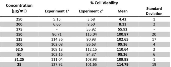
Cell viability (Fig. 2, Tab. 1 and Tab. S. 1 in supplementary data) was evaluated by comparison of measured absorbance in wells with treated cells – maintained in 2 % FBS medium with different extract concentrations - and control cells – maintained in 2 % FBS medium without extract. Cytotoxic concentration that destroys 50 % of the cells (CC50) and maximum non-cytotoxic concentration (MNCC)
were calculated through linear regression of the dose-response curve.
Fig. 2 - Graphic representation of the extract cytotoxicity. Values represent the mean ± SD of four independent experiments. The black arrow indicates the CC50.
According to the calculations the extract CC50 and MNCC are 177.3 and 150.3 μg/mL, respectively.
0 % 20 % 40 % 60 % 80 % 100 % 120 % 0 50 100 150 200 250 Ce ll Viab ili ty Extract Concentration (μg/mL) Extract Concentration (μg/mL) % Cell Viability 250 4.4 ± 1 200 8.1 ± 2 *175 *55.9 150 100.9 ± 20 125 102.7 ± 17 100 99.4 ± 4 62.50 110.6 ± 2 50 98.26 ±6 31.25 110.0 ±1 25 114.8 ±19
Tab. 1 – Cell viability percentages obtained in MTT assays post 24 or 48 hours of incubation with the extract. Values represent the mean ± SD of two independent experiments (except *) with 4 replicates.
18
3.1.2. Correlation between FBS concentration in the culture medium and
cytotoxic effect of the extract
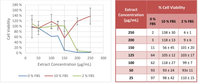
To investigate possible dissimilarities of the extract cytotoxic effect when it is diluted in medium with different serum concentrations, sub-confluent cell cultures were treated with different extract concentrations in medium with different FBS concentrations - 0, 2 or 10 % - and controls (cultures maintained in medium with the same FBS concentrations, without extract). The extract exhibited extremely variable cytotoxicity (Fig. 3, Tab. 2) with variations in the FBS concentration in the culture medium.
Fig. 3 - Graphic representation of the extract cytotoxicity in different FBS concentrations. Values represent the mean ± SD of 1, 3 and 4 experiments – respectively to 0, 10 and 2 % FBS culture medium – with 4 replicates.
Extract CC50 and MNCC in medium with 2 % FBS were 177.3 and 150.3 μg/mL, respectively, and 123.2
and 21.2 μg/mL in serum free medium. These values could not be determined in the assays with 10 % serum medium.
3.2. Virucidal effect
Virucidal effect results were obtained by titration of previously treated (with 100 µg/mL of extract) and control viral suspensions and comparison of the obtained titers. As noticeable in Fig. 4 and Tab. S. 2 (in supplementary data) the extract revealed no meaningful effect against viral particles outside the host cell, with a 3.44 ± 2.37 % (mean of 3 experiments) reduction on the virus titer of treated suspensions – 7.67 × 10⁶ pfu/mL – relatively to non-treated ones – 7.94 × 10⁶ pfu/mL.
0 % 20 % 40 % 60 % 80 % 100 % 120 % 140 % 160 % 180 % 0 50 100 150 200 250 300 Ce ll Viab ili ty Extract Concentration (μg/mL) 0 % FBS 10 % FBS 2 % FBS Extract Concentration (μg/mL) % Cell Viability 0 % FBS 10 % FBS 2 % FBS 250 2 138 ± 30 4 ± 1 200 3 118 ± 13 9 ± 6 150 11 56 ± 45 101 ± 20 125 64 105 ± 12 103 ± 17 100 62 118 ± 27 99 ± 7 50 93 93 ± 24 93± 11 25 97 98 ± 42 110 ± 15
Tab. 2 - Cell viability percentages obtained in MTT assays after 24 or 48 hours of incubation with the extract diluted in culture medium with 0, 2 or 10 % FBS.
19 Fig. 4 – HSV-2 suspensions – treated and control - titration results.
3.3. Extract effect on viral plaques formation
In order to test the extract effect on HSV-2 plaque formation, cell cultures, grown in 24 or 6 well plates were infected with serial dilutions of the virus. After the viral adsorption and entry periods, the inoculums were removed and 2 % FBS culture medium – with or without 100 μg/mL of extract – was added to treated and control wells. Following the treatment period, 2 % Sephadex titration medium was added to each well.
The extract inhibitory effect was evaluated by comparison of treated and control titers and plaques’ diameters.
Control
Treated
10
-610
-510
-4Treated
Control
10
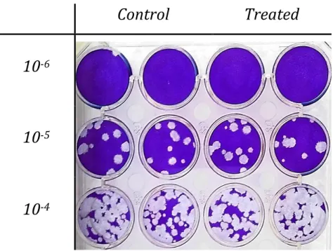
-710
-620 When added during titration the extract exhibit a 71.6 ± 26.8 % (mean of 3 experiments, see Tab. S. 3 in supplementary data) reduction in titer and a visible decrease in plaque diameter (Fig. 5) of treated versus control conditions.
3.4. Extract effect on virus yield
Several experiments, involving HSV-2 production under treatment, were performed, to uncover the extract effect on virus yield and the involved infection stages.
It’s noteworthy that a mean yield reduction of 93.65 ± 8 % was calculated using data obtained in 13 experiments (see Tab. S. 4 in supplementary data) performed in “standard” treatment conditions – 2 % FBS medium with 100 µg/mL of extract at 0.5 h p.i. during 2 to 4.5 hours.
3.4.1. IC
50and IC
90determination
To determine the extract IC50 and IC90, HSV-2 was produced in culture medium with different extract
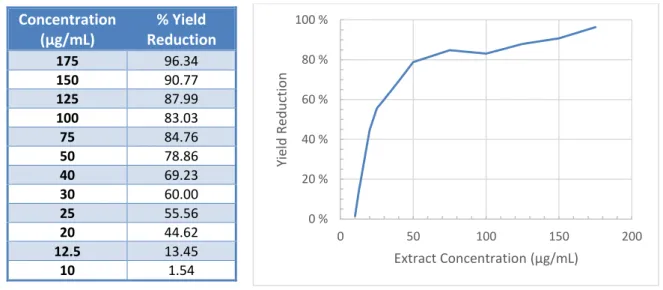
concentrations, and controls were produced in culture medium without extract. Produced suspensions were then titrated, and viral titers (in treated and control cultures) were compared to obtain the percentage of yield reduction attributed to each concentration (Tab. 3, Fig. 6). Concentration that reduces viral yield in 50 % (IC50) and concentration that reduces viral yield in 90 % (IC90) were calculated
through linear regression of the dose-response curve.
Fig. 6 - Graphic representation of HSV-2 yield reduction when cell cultures were treated with different extract concentrations. Values were obtained in one experiment.
According to the calculations the extract IC50 and IC90 are 35.1 and 128.1 μg/mL, respectively.
0 % 20 % 40 % 60 % 80 % 100 % 0 50 100 150 200 Yie ld Re d u ctio n Extract Concentration (μg/mL) Concentration (μg/mL) % Yield Reduction 175 96.34 150 90.77 125 87.99 100 83.03 75 84.76 50 78.86 40 69.23 30 60.00 25 55.56 20 44.62 12.5 13.45 10 1.54
Tab. 3 - HSV-2 yield reduction percentage when cell cultures were treated with different extract concentrations.
21
3.4.2. Determination of the affected stage of virus replication
HSV-2 was produced under treatment conditions - in culture medium with 100 μg/mL of extract – with different treatment starting times, and a control suspension was produced in culture medium without extract. Yield reduction percentage was determined by titration of the produced virus suspensions and comparison of the titers obtained in treated and control conditions.
Fig. 7 - Graphic representation of HSV-2 yield reduction by the extract (100 μg/mL) in different treatment starting points. Values were obtained in one experiment.
There is a noticeable decrease (Tab. 4, Fig. 7 and Tab. S. 4 in supplementary data) of the extract effectiveness when added between 3 to 6 h p.i. and no effect when the treatment is started at 15 h p.i. or later (data not shown).
3.4.3. Comparison with ACV inhibition
To evaluate the mechanisms which mediate the virus yield reduction by the aqueous extract, HSV-2 was produced under treatment conditions with both compounds – aqueous extract and ACV – at the same concentration – 100 μg/mL. Control suspensions were produced in culture medium without extract. Each compound yield reduction percentage was determined by titration of the produced virus suspensions and comparison of titers obtained in treated and control conditions.
0 % 20 % 40 % 60 % 80 % 100 % 0 2 4 6 8 10 12 14 Yie ld Re d u ctio n Treatment (h p.i.) Treatment Start (h p.i.) % Yield Reduction 0.5 91.27 1 78.55 3 64.53 6 46.27 14 28.74
Tab. 4 - HSV-2 yield reduction percentage at different treatment starting times (h p.i.).
95.89 % 97.40 % 0 % 20 % 40 % 60 % 80 % 100 % ACV Extract
Virus Yield Reduction
Fig. 8 - Graphic representation of HSV-2 yield reduction by the extract and ACV (100 μg/mL). Values were obtained in one experiment with 2 replicates.
22 Fig. 8 shows HSV-2 yield reduction percentages in virus produced under treatment with ACV – 95.89 % - or S. virgaurea aqueous extract – 97.40 % - , relatively to non-treated virus. The extract exhibited a very similar inhibition to the one observed in ACV treatment.
3.4.4. Visualization of treated and non-treated infected cells and virus
suspensions by Transmission Electron Microscopy (TEM)
Cell cultures were infected with HSV-2 and treated with 2 % FBS culture medium with or without 100 μg/mL of extract. Infected cells were then collected at 14 hours post infection and observed by TEM. Treated and control viral suspensions were concentrated and observed by negative staining.
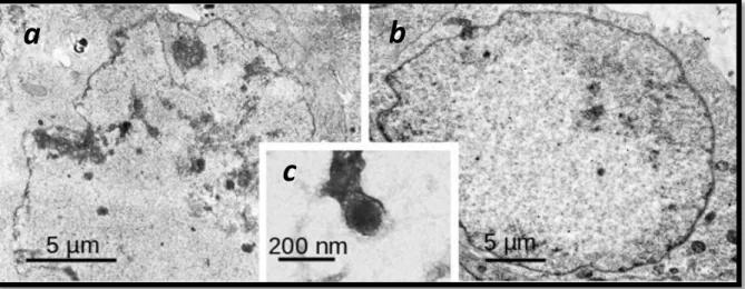
Fig. 9 – a-b) Thin section TEM micrographs of HSV2 infected Vero cells processed for TEM at 14 hours p.i. a) Non-treated (control). b) Cells treated with 100 μg/mL of the extract. c) HSV-2 virus particle negatively stained with 1 % aqueous uranyl acetate. Photos were kindly provided by Dr. A.P. Alves de Matos.
Negative staining showed a highly reduced amount of virus produced under treatment, as compared to controls, with no significant morphogenetic defects (Fig. 9 c), while fine sections with treated infected cells (Fig. 9 b) exhibited fewer morphological deficiencies, due to infection, than control infected cells (Fig. 9 a).
3.4.5. HSV-2 Infection Kinetics
DNA was extracted from Vero cell cultures infected with HSV-2 – in treatment and control (non-treatment) conditions – at 14 hours post-infection (h p.i.) and was observed by gel electrophoresis (Fig. 10). DNA concentration of
Infection Time (hours) DNA concentration (ng/µL) Treated Control 3 26.35 20.34 4 24.18 19.41 5 22.39 25.93 6 22.40 26.74 7 34.24 31.25
Tab. 5 - Infected cells - treated and non-treated - DNA quantification.
a
b
23 each sample was determined (Tab. 5) using a NanoDrop® and was equalized to 19.41 ng/µL in every sample, with DNase free water. PCR was performed to 26 ng of each sample, in order to verify the presence of HSV-2 DNA.
Sample
L
Treated Controls (Non-Treated)
h p.i. 2 3 4 5 6 7 2 3 4 5 6 7
Fig. 10 – DNA from treated and non-treated infected cells (from 2 to 7 h p.i.) DNA observed trought gel electrophoresis.
All extracted DNAs showed similar bands when observed in gel electrophoresis (Fig. 10), indicating that only cellular DNA is visible.
Sample Treated Controls (Non-Treated)
L
PCR Controls
h p.i. 2 3 4 5 6 7 2 3 4 5 6 7 + + -
Fig. 11 – Results of PCR for the detection of HSV-2 DNA in infected cells – treated and controls – at different times post infection. White arrows indicate the size of two molecular weight marker bands that surround the PCR product bands. Although no differences are evident between treated and non-treated PCR products (Fig. 11) this experiment shows that, in the applied experimental conditions, viral DNA replication is started between 4 and 5 h p.i.
1018 bp
24 The PCR products visualization (Fig. 11) showed that the amplified fragment size was slightly above 506 bp but under 1018 bp, which fits the expected size.
3.4.6. Quantitative real-time PCR (qPCR)
To evaluate possible differences between the amounts of viral DNA in treated and non-treated cell cultures at different hours post infection, qPCR was performed with 2 ng of the extracted DNA samples (Fig. 10).
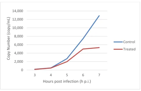
Fig. 12 - Quantification of HSV-2 DNA (copy/mL) in infected cells (treated and non-treated) at different hours post infection (h p.i.).
While at the beginning of infection – 3-5 h p.i. – viral DNA quantities are similar with or without treatment, it’s noticeable a slower increase of the copy number in treated relatively to non-treated samples, that gets more evident as the infection progresses.
0 2,000 4,000 6,000 8,000 10,000 12,000 14,000 3 4 5 6 7 Cop y N u m b er (c o p y/m L)
Hours post infection (h p.i.)
Control Treated