UNIVERSIDADE DE LISBOA
Faculdade de Medicina Veterinária
Development of specific recombinant single-domain antibodies
against gp120 HIV-1 glycoprotein and their selection by
Phage Display
Fabiana Carvalho Marques
CONSTITUIÇÃO DO JÚRI:
Doutor Luís Manuel Morgado Tavares Doutora Ana Isabel Simões Pereira Duarte Doutor Frederico Nuno Castanheira Aires da Silva
ORIENTADOR:
Doutor Frederico Nuno Castanheira Aires da Silva
CO-ORIENTADOR:
Doutora Solange Judite Roque Coelho Alves Gil
2018 Lisboa
UNIVERSIDADE DE LISBOA
Faculdade de Medicina Veterinária
Development of specific recombinant single-domain antibodies
against gp120 HIV-1 glycoprotein and their selection by
Phage Display
Fabiana Carvalho Marques
DISSERTAÇÃO DE MESTRADO INTEGRADO EM MEDICINA VETERINÁRIA
CONSTITUIÇÃO DO JÚRI:
Doutor Luís Manuel Morgado Tavares Doutora Ana Isabel Simões Pereira Duarte Doutor Frederico Nuno Castanheira Aires da Silva
ORIENTADOR:
Doutor Frederico Nuno Castanheira Aires da Silva
CO-ORIENTADOR:
Doutora Solange Judite Roque Coelho Alves Gil
2018 Lisboa
ACKNOWLEDGEMENTS
Esta dissertação de mestrado representa o culminar de 6 anos de trabalho árduo que foram percorridos ao lado de muitas pessoas que tiveram um papel fundamental na minha evolução. Os meus sinceros agradecimentos:
Ao meu orientador de estágio, o Dr. Frederico Aires da Silva, por me ter integrado na sua equipa da melhor maneira possível e por me ter permitido experienciar o dia-à-dia de um investigador. Obrigada pelo apoio, disponibilidade e simpatia demonstrados e por todos os conhecimentos transmitidos.
Às “meninas” da equipa “FAS”: Ana, Joana e Sandra, muito obrigada por me terem recebido tão bem. Constituem uma equipa espetacular. O vosso apoio foi constante e perfeito. O vosso espírito de equipa é incrível e tenho muito orgulho em ter testemunhado o vosso trabalho. São excelentes profissionais que contribuem em muito para a evolução da medicina humana e veterinária em Portugal. Ana foste, sem dúvida, o meu braço direito no desenvolvimento prático deste trabalho e sem ti, sem a tua calma e partilha de experiência teria sido muito mais difícil e frustrante conseguir chegar ao fim com sucesso.
À minha co-orientadora, Dra. Solange Gil, por ter depositado confiança em mim, por me ter guiado até à equipa do Dr. Frederico, por ter ouvido todas as minhas ideias e dentro da realidade de um curto estágio curricular me ter encontrado a solução.
Um agradecimento especial ao professor João Barcelos. Sem dúvida um excelente professor, tão dedicado ao ensino da Medicina Veterinária. Obrigada por me ter dado as palavras certas no momento certo e por ter cultivado em mim a procura do conhecimento e profissionalismo constante.
Aos meus amigos Carlitos, Élia, Luísa, Patrícia e Pedro pelo apoio ao longo de todos estes anos e pelas palavras de coragem e motivação.
Às minhas queridas amigas e futuras veterinárias Juliana e Xica e ao meu querido Xico por terem sido a minha família nos, que pareciam intermináveis, dois anos na ilha Terceira e por serem aqueles que eu levo com tanto carinho deste terrível mas maravilhoso curso. Juliana, estiveste sempre ao meu lado, penaste comigo, viste as mesmas desgraças que eu, revoltaste-te da mesma forma, salvaste da mesma forma, partilhámos momentos inesquecíveis e importantes para o nosso desenvolvimento como seres humanos sensíveis e por isso orgulho-me imenso.
À Ana Soraia e à Rita por me terem acolhido e apoiado. Espero ser um dia a vossa “vet” de eleição!
À minha tia Teresa e à minha prima Odete por me incentivarem a continuar sempre com muita força e por me ajudarem a ver a luz ao fundo do túnel.
À Lesley, porque mesmo tão longe sempre me apoiou ao longo destes anos.
Ao meu querido Rui por ser o meu melhor amigo, por estar sempre ao meu lado, por me incentivar a ser melhor, por contribuir imenso para o meu crescimento como futura médica veterinária e por fazer com que tudo pareça mais fácil. Onze *
Aos meus pais e à minha irmã pelo apoio incondicional e por demonstrarem tanto orgulho em mim.
Este projeto foi financiado pelo programa Gilead GÉNESE no âmbito do projeto PGG/015/2016 - "Desenvolvimento de anticorpos biespecificos anti-gp120/gp40 para o tratamento da infeção do VIH-1".
ABSTRACT
Development of specific recombinant single-domain antibodies against gp120
HIV-1 glycoprotein and their selection by Phage Display
Human immunodeficiency virus (HIV) is the causative agent of acquired immunodeficiency syndrome (AIDS), a condition in which progressive failure of the immune system allows life-threatening opportunistic infections to succeed. One of the most important factors in the worldwide spread of HIV is its enormous genetic variability and rapid evolution. The revealing of all stages of HIV replication cycle led to the identification of potential therapeutic targets in order to decrease the replicative process. However, the acquisition and transmission of HIV drug resistance still poses a major risk to the success of antiretroviral therapy. Thus, new targets and more promising strategies must emerge quickly to improve treatment options for patients who are infected with viruses already resistant to currently available antiretrovirals. The inhibition of HIV entry into the host cell is an extremely promising strategy, and glycoprotein gp120 plays a central role in this process. In this context, the aim of this project consisted in the development and selection, by Phage Display, of specific single-domain antibodies (sdAbs) against the surface HIV gp120 glycoprotein. To achieve this goal, rabbit antibody immunized libraries were generated and a pool of VH and VL sdAbs were selected by Phage Display. This approach allowed the construction of highly diverse sdAbs libraries and a posterior specific selection of VH and VL sdAbs with high binding activity to gp120. With the results obtained in this project, the potential of rabbit derived sdAbs as therapeutic molecules were once again demonstrated. At this stage the results are promising and further studies will be implemented in the future, to characterize the selected antibodies and to obtain more data regarding the binding activity to the target molecule and their potential to neutralize HIV infection.
Keywords: Human Immunodeficiency Virus, gp120, rabbit immunization, recombinant single domain antibodies, Phage Display
RESUMO
Desenvolvimento de anticorpos recombinantes de pequeno domínio contra a
glicoproteína gp120 do VIH-1 e sua seleção por Phage Display
O vírus da imunodeficiência humana (VIH) é o agente causador da síndrome da imunodeficiência adquirida, uma condição na qual a falha progressiva do sistema imunitário permite que infeções oportunistas, potencialmente letais, se instalem. Um dos fatores mais importantes na disseminação mundial do VIH é a sua enorme variabilidade genética e rápida evolução. O conhecimento de todas as etapas do ciclo replicativo do VIH levou à identificação de possíveis alvos terapêuticos a fim de diminuir o processo replicativo. No entanto, a aquisição e transmissão de resistência aos fármacos contra o VIH ainda representa um grande risco para o sucesso da terapia antirretroviral. Assim, novos alvos e estratégias mais promissoras devem emergir rapidamente para melhorar as opções de tratamento dos pacientes infetados com vírus resistentes aos antirretrovirais atualmente disponíveis. A inibição do processo de entrada do VIH é uma estratégia extremamente promissora, e a glicoproteína gp120 desempenha um papel central neste processo. Neste contexto, o objetivo deste projeto consistiu no desenvolvimento e seleção, por Phage Display, de anticorpos específicos contra a gp120. Para alcançar este objetivo, foram criadas bibliotecas imunizadas de anticorpos de coelho e foi selecionado um conjunto de anticorpos de domínio pequeno (anticorpos no formato VH e VL) por Phage Display. Esta abordagem permitiu a construção de bibliotecas altamente diversificadas e uma posterior seleção específica de anticorpos com elevada afinidade de ligação à gp120. Com os resultados obtidos neste projeto, o potencial, como moléculas terapêuticas, dos anticorpos de pequeno domínio derivados do coelho foi novamente demonstrado. Nesta fase, os resultados são promissores e mais estudos serão implementados no futuro de modo a caracterizar os anticorpos selecionados e obter mais dados sobre a afinidade de ligação à molécula alvo e o seu potencial de neutralização da infeção pelo VIH.
Palavras-chave: Vírus da Imunodeficiência Humana, gp120, imunização de coelhos, anticorpos recombinantes de pequeno domínio, Phage Display
INDEX
Acknowledgements ... i
Abstract ... iii
Resumo ... iv
Index ... v
List of figures ... vii
List of tables ... ix
List of abbreviations ... x
1. Internship Report ... 1
2. Introduction ... 2
2.1 Human Immunodeficiency Virus ... 2
2.1.1 HIV Taxonomy ... 3
2.1.2 HIV Structure and Genome ... 3
2.1.3 Genetic diversity in HIV ... 6
2.1.4 The host-cells and HIV interaction ... 8
2.1.5 HIV Transmission ... 10
2.1.6 HIV Replication Cycle ... 10
2.2 Current and emerging therapies for HIV ... 14
2.2.1 HIV entry inhibitors ... 16
2.3 gp120 HIV-1 glycoprotein ... 20
2.3.1 Structure and Function of gp120 HIV-1 glycoprotein ... 20
2.3.2 gp120 HIV-1 glycoprotein as a therapeutic target ... 22
2.4 Antibodies ... 24
2.4.1 Immune system ... 24
2.4.2 Characteristics and properties of Antibodies ... 26
2.4.3 Novel therapeutic antibodies ... 27
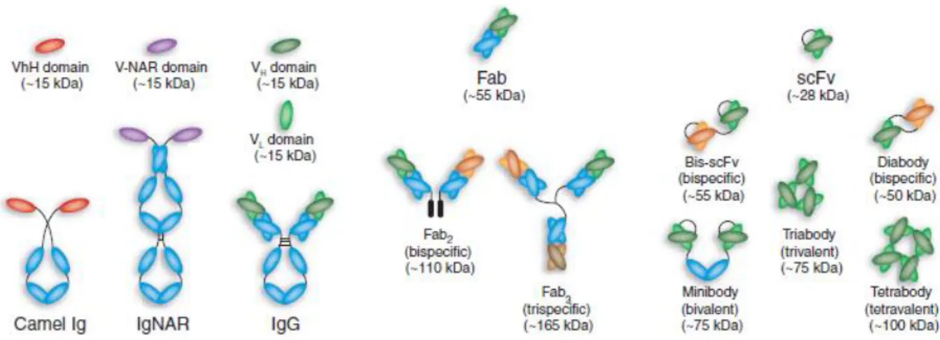
2.4.3.1 Antibody Fragments ... 28
2.4.3.2 Single-Domain Antibodies ... 28
2.5 Selection of monoclonal antibodies ... 31
2.5.1 Antibody Libraries ... 31
2.5.2 Rabbit Immunizations ... 32
2.5.3 Tissue source ... 33
2.5.4 Phage Display technology ... 34
2.6 Objectives ... 38
3. Material and Methods ... 39
3.1 Rabbit immunizations ... 39
3.2 sdAbs immune library construction ... 39
3.2.1 Isolation of Total RNA ... 39
3.2.2 First strand cDNA synthesis ... 40
3.2.4 Purification of PCR products ... 41
3.2.5 Precipitation of PCR products ... 42
3.2.6 Digestion of vector DNA and PCR products with restriction enzyme SfiI ... 42
3.3 Phage Display library ... 43
3.3.1 Cloning of PCR products into pComb3x vector ... 43
3.3.2 Confirmation of library insert efficiency and diversity ... 44
3.3.3 Selection of specific sdAbs against gp120 by Phage Display ... 45
3.3.4 Phage Titration ... 46
3.4 Characterization of sdAbs against gp120... 47
3.4.1 Sequencing of anti-gp120 clones ... 47
4. Results ... 49
4.1 Rabbit immunizations ... 49
4.2 sdAbs immune library construction ... 50
4.2.1 Isolation of Total RNA and cDNA synthesis ... 50
4.2.2 PCR amplification and purification of single domains antibody genes ... 51
4.2.3 Digestion of vector DNA and PCR products with restriction enzyme SfiI ... 54
4.3 Phage Display library ... 56
4.3.1 Cloning of PCR products into pComb3x vector, confirmation of library insert efficiency and diversity and clones profile analysis ... 56
4.3.2 Selection of specific sdAbs against gp120 by Phage Display ... 59
4.3.2.1 Input and Output Phage Titering and characterization of sdAbs against gp120……….59
5. Discussion ... 62
5.1 Rabbit immunizations ... 62
5.2 sdAbs immune library construction ... 63
5.3 Phage Display library ... 65
6. Conclusion and Future Perspectives ... 67
LIST OF FIGURES
Figure 1 - HIV prevalence………..………. 3
Figure 2 - Schematic view of the HIV-1 particle……….…. 4
Figure 3 - Schematic model of the HIV-1 and HIV-2 full-length genome……… 5
Figure 4 - Global distribution of HIV-1 subtypes and recombinant forms………... 8
Figure 5 - HIV-1 tropism………..……… 9
Figure 6 - Schematic overview of the HIV-1 replication cycle……….…….. 12
Figure 7 - Schematic representation of the constituent domains of HIV-1 Env….…………. 20
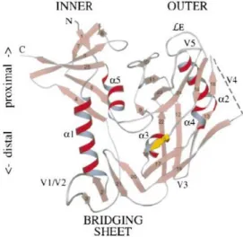
Figure 8 - Structure of the HIV-1 SU gp 120 core……….……….. 21
Figure 9 - Steps in HIV entry into host cells……….……… 22
Figure 10 - Basic IgG structure………..……….... 26
Figure 11 - Schematic representation of different antibody formats………...…………. 29
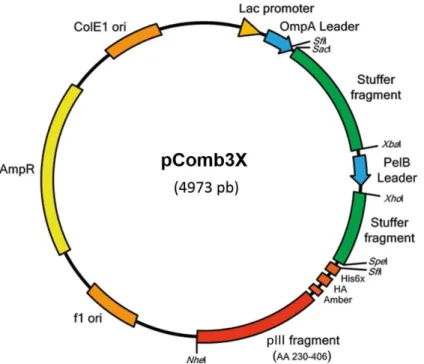
Figure 12 - pComb3X phagemid vector………..….. 35
Figure 13 - Schematic representation of a Phage Display selection round panning)……... 36
Figure 14 - Schematic representation of the methodology used in the development of the recombinant sdAbs in the present work……… 38
Figure 15 - Primer combination for the generation of VH and VL antibody fragments by PCR……….…… 41
Figure 16 - Schematic representation of a selection round for Phage .isplay……… 46
Figure 17 - Schematic representation of a) input and b) output phage titering……….. 47
Figure 18 - Titration of serum antibodies from rabbit R8……… 49
Figure 19 - RNA and cDNA from rabbit R8……….…. 50
Figure 20 - PCR amplification of VH family from Sp and M cDNA………... 51
Figure 21 - PCR amplification of VL family from Sp and M cDNA……… 52
Figure 22 - Purification of VH families in a 2% low melting point agarose gel after amplification……….……….. 53
Figure 23 - Purification of VL families in a 2% low melting point agarose gel after amplification………... 54
Figure 24 - Purification of VH and VL domains, after Sfi I restriction, in a 2% low melting point agarose gel………...……... 55
Figure 25 - Restriction a) and purification b) of phagemid pComb3X-ss, after Sfi I restriction, in a 0,8% low melting point agarose gel………..…. 55
Figure 26 - Confirmation of transformation efficiency for VH and VL libraries……… 56 Figure 27 - Guide trees……….………...……… 57 Figure 28 - Alignment of the sequences obtained for each clone of a) VH and b) VL domains……….. 58 Figure 29 - Variable domain antibody families of the built immune library……….. 59 Figure 30 - Selection of VH and VL domains by Phage Display……….. 60 Figure 31 - Alignment of the VH Output sequences obtained from the third panning……... 61
LIST OF TABLES
Table 1 - Antiretroviral therapies available………. 15 Table 2 - Rabbit immunization schedule………. 40 Table 3 - Primers sequences………...………. 42 Table 4 - Conditions used for VH and VL digestion by Sfi I restriction endonuclease…… 43 Table 5 - Name and sequence of the primers used to confirm the presence of the genes encoding VH and VL sdAbs cloned in pComb3x vector………... 44 Table 6 - Conditions used in each panning……… 46 Table 7 - Quantification of mRNA and can from the Sp and BM of R8……….. 50 Table 8 - Quantification of VHs and VLs families from Sp and BM of immunized rabbit after amplification and purification……… 53 Table 9 - Grouping, by antibody families, of the selected and sequenced clones…….….. 58 Table 10 - Input and Output phage titering……….... 60
LIST OF ABBREVIATIONS
AIDS Acquired immune deficiency syndrome ART Antiretroviral treatment
BM Bone Marrow
bnAbs Broadly neutralizing antibodies
C Conserved regions
CA Capsid protein
cDNA complementary DNA
CDR Complementary Determining Region
CD4i CD4 induced site
CH Constant domain of heavy-chain CHR C-terminal heptad repeat
CL Constant domain of light-chain CRFs Circulating Recombinant Forms DNA Deoxyribonucleic Acid
E. coli Escherichia coli
ELISA Enzyme- Linked Immunosorbent Assay
ENV Envelope
ER Endoplasmic Reticulum
Fab Fragment antigen binding
Fc Constant Fragment
FDA Food and Drug Administration FIV Feline Immunodeficiency Virus
FRs Framework residues
FR1 First framework region
Fv Variable fragment
Gag Group Specific Antigen
gp Glycoprotein
H Heavy
HAART Highly active antiretroviral therapy
HCDR3 heavy chain CDR3
HIV Human Immunodeficiency Virus
Ig Immunoglobulin
iMed.ULisboa Center for Molecular Pathogenesis and Research Institute of Medicine
IN Integrase
L Light
LB+amp LB-agar medium with Ampicillin
M Major
MA Matrix protein
mAbs Monoclonal antibodies
MHC Major histocompatibility complex M-tropic Macrophage-tropic
N Nonmajor and Nonoutlier
NC Nucleocapsid protein
Nef Negative factor
NHR N-terminal heptad repeat
NNTI Non-nucleoside reverse transcriptase inhibitors NRTI Nucleoside/nucleotide reverse transcriptase inhibitors
O Outlier
ORFs Open Reading Frames
PBS Phosphate Buffered Saline XI PCR Polymerase Chain Reaction
PEG Polyethylene Glycol
PI Protease inhibitors
PIC Pre-Integration Complex
Pol Polymerase
PR protease
Pr 55Gag Gag precursor
P-TEFb Positive Transcription Elongation Factor b Rev Regulator of virion expression
RNA Ribonucleic Acid RNA Pol II RNA polymerase II rpm Rotations per minute
RRE Rev Response Element
RT Reverse transcriptase
R5 tropism M-tropic strains
R6 Rabbit 6
R8 Rabbit 8
R5X4 tropism Dual-tropic strains
scFv Single-chain variable fragment sdAbs Single Domain antibodies
SIVs Simian Immunodeficiency Viruses
Sp Spleen
SU Surface glycoproteins
Tat Transcriptional transactivator TCL-tropic T cell line–tropic
TM Transmembrane protein
U Units
URFs Unique Recombinant Forms
UV Ultra violet
V Variable regions
VH Variable domain of heavy-chain Vif Virion Infectivity Factor
VIH Vírus da imunodeficiência humana VL Variable domain of light chain Vpr Viral Protein Regulatory
Vpu Viral protein U
Vpx Viral protein X
VV Vaccinia virus
X4 tropism TLC-tropic strains
1. INTERNSHIP REPORT
The curricular internship to obtain a Master's Degree in Veterinary Medicine was carried out at the Laboratory of Immunology and Virology at Faculty of Veterinary Medicine of Lisbon University, between September 11, 2017 and January 15, 2018, with a total of 620 hours. The internship was in the area of laboratory research where the goal was to construct a rabbit derived single domain antibody immune library and whose results contributed to the development of the final dissertation.
During the internship, I was able to learn and execute several laboratory techniques, such as:
Titration of antibodies through enzyme-linked immunosorbent assay (ELISA);
Synthesis of cDNA by reverse transcription of RNA;
Amplification of DNA through Polymerase Chain Reaction (PCR);
Quantification of RNA and DNA;
Electrophoresis in agarose gel;
DNA precipitation;
DNA purification;
Digestion of DNA using restriction enzymes;
Ligation reactions between target DNA and a pComb3x vector;
Bacteria transformation by electroporation;
DNA extraction from bacteria;
Culture of bacteria in liquid and solid mediums;
Preparation of culture mediums;
Maintenance of cell cultures;
Phage culture;
Phase precipitation;
Phage titration;
Infection of bacteria with phage;
Phage Display technology for antibody selections
DNA sequencing;
2. INTRODUCTION
2.1 Human Immunodeficiency Virus
Human immunodeficiency virus (HIV) is a lentivirus that causes HIV infection and over time can cause AIDS, a condition in which progressive failure of the immune system allows life-threatening opportunistic infections to thrive (World Health Organization (WHO), 2017). HIV infection per se affects in complex ways every organ system, at all ages from the neonate to the elderly (Lucas & Nelson, 2015). Like many viruses, HIV has the ability to mutate and change over time - within the main types of HIV there are many genetically distinct subgroups (Taylor, Sobieszczyk, McCutchan & Hammer, 2008). There are two major types of HIV: HIV-1 (the most common) and HIV-2 (relatively uncommon and less infectious). Both virus are associated with the same mode of transmission and cause AIDS and similar opportunistic infections, although, HIV-2 is prevalent in West Africa and some countries in Western Europe, while HIV-1 is prevalent worldwide and is responsible for the global HIV pandemic (Clavel et al., 1987; Clavel et al., 1986).
The earliest direct evidence of HIV infection in humans was found retrospectively in a serum sample and a lymph node biopsy specimen stored in 1959 and 1960, respectively, in Kinshasa, Democratic Republic of the Congo (Worobey et al., 1998; Zhu et al., 1998).
HIV-1 in humans resulted from at least four cross-species transmissions of simian immunodeficiency viruses (SIVs) from chimpanzees and gorillas in West Central Africa, while HIV-2 viruses resulted from at least eight independent transmissions of SIVs infecting sooty mangabeys in West Africa only (Peeters, Jung & Ayouba, 2013). Cross-species transmission probably occurred through bloodborne transmission in the process of hunting and butchering of primates for bushmeat and the capture, trade and keeping of monkeys as pets (Hahn, Shaw, De Cock & Sharp, 2000).
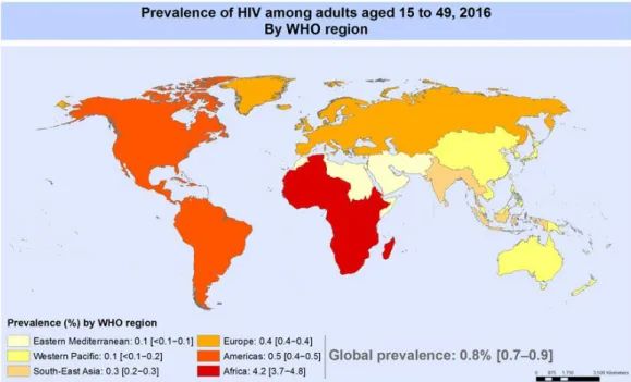
Enormous progress against AIDS for the past 15 years has inspired a global commitment to end the epidemic by 2030. In 2016 there were: 36,7 million people living with HIV, 19,5 million people living with HIV on antiretroviral therapy, 1,8 million people newly infected with HIV and 1 million people died from AIDS-related illnesses worldwide, compared to 1.9 million in 2005 and 1.5 million in 2010 (Figure 1) (UNAIDS, 2017).
With the development of effective antiretroviral treatment (ART), the life expectancy for people with HIV is now approaching the one seen in the general population. As ART is given to growing proportions of individuals with HIV infection, and is life-extending but not curative, interest has been directed on to the outcomes of the interactions of HIV, anti-retroviral therapy and the ageing process, and also on how to manage the growing number of persons with life-long HIV infection (Smith et al., 2014).
Figure 1. HIV prevalence.
Total number of existing cases of adults and adolescents, aged 15-49, infected with HIV by 2016. Adapted from WHO, 2017.
2.1.1 HIV Taxonomy
HIV is a member of the genus Lentivirus, which belongs to the family Retroviridae (Turner & Summers, 1999). The family Retroviridae is classified currently into two subfamilies (Orthoretrovirinae and Spumaretrovirinae) and seven genera. The family includes many viruses of importance in veterinary and human medicine, and to biomedical science in general. The prefix “Retro” refers to reverse and is due to the reverse transcriptase (RT), a particular enzyme which characterizes this family (Maclachlan & Dubovi, 2011). Like all retroviruses, HIV's genome is encoded by ribonucleic acid (RNA), which is reverse-transcribed to viral deoxyribonucleic acid (DNA) by the viral RT upon entering a new host cell. Other examples of lentiviruses include the simian immunodeficiency virus, visna virus, equine infectious anemia virus (Turner & Summers, 1999) and feline immunodeficiency virus (FIV) (Sparger et al., 1989). Since the 1980s, retroviruses are demonstrated to cause a number of important human diseases, including lymphomas, leukemias, and AIDS, which has further catalyzed intensive investigation of both human and animal retroviruses (Maclachlan & Dubovi, 2011).
2.1.2 HIV Structure and Genome
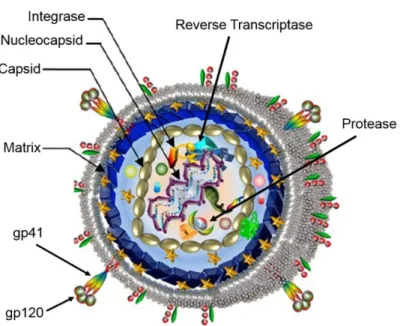
Lentivirus genus includes retroviruses that possess complex genomes and exhibit cone-shaped capsid core particles (Figure 2). The HIV virus is enveloped by a lipid bilayer that is derived from the membrane of the host cell (Turner & Summers, 1999) and contains several
cellular membrane proteins, including major histocompatibility antigens, actin and ubiquitin also derived from the host cell (Arthur et al., 1992). The virus particle contains an inner core containing the viral genome as well as enzymes that are required for early replication events. The inner core is protected by a protein capsid which, in turn, is surrounded by the lipid bilayer membrane. Approximately 2000 copies of virus matrix protein (MA, p17) is inserted into the inner surface of the membrane forming the matrix shell. A protein membrane, also called the glycoprotein envelope (Env), protrudes through the membrane and forms the outer surface of the virus particle (Haseltine, 1991). A conical capsid core particle comprising about 2000 copies of the capsid protein (CA, p24) is located in the center of the virus. The capsid encapsidates two copies of the unspliced viral genome, which is stabilized as a ribonucleoprotein complex with approximately 2000 copies of the nucleocapsid protein (NC, p7), and also contains three essential virally encoded enzymes: integrase (IN), RT and protease (PR) (Turner & Summers, 1999).
Figure 2. Schematic view of the HIV-1 particle.
The Env precursor polyprotein (gp160) is synthesized in the endoplasmic reticulum (ER) using the spliced env mRNA gene as the message. The protein appears to oligomerize to a trimeric structure in the ER, and is heavily glycosylated. Env is posttranslationally modified in the ER and Golgi apparatus
and is cleaved to produce the non-covalently associated TM-SU trimeric glycoprotein complex.
Adapted from Li et al., 2015.
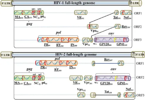
The virus genome is diploid, consisting of a homodimer of two single-stranded RNA approximately 9 kb in length and contains 9 open reading frames (ORFs). The ends of each HIV RNA strand contain an RNA sequence called the long terminal repeat (LTR). Regions in the LTR act as “switches” to control production of new viruses and can be triggered by proteins from HIV or from the host cell. The largest three reading frames contains the three
major genes, which encode several proteins that have essential roles during the HIV life cycle (Freed, 2015; Turner & Summers, 1999; Frankel & Young, 1998) (FIGURE 3):
Gag (group specific antigen), which encodes for structural proteins (Ma, CA, NC and p6);
Pol (polymerase), which encodes for viral enzymes such as PR, RT and IN. The most important enzyme is RT, which has different functions depending on the part of the protein envolved in the biological process, being an RNA-dependent DNA polymerase, a DNA-dependent DNA polymerase and an RNase;
Env (envelope) which encodes for specific surface protein (SU) gp120 and transmembrane protein (TM) gp41, determining cell tropism and contributing for pathogenicity. The superficial cone-shaped capsid core particles, also called envelope spikes, comprise trimers of non-covalently linked heterodimers consisting of the SU gp120 and the TM gp41. When triggered, these spikes initiate a cascade of conformational changes that culminates in fusion between the viral and host cell membranes and release of the viral core into the cytoplasm (Engelman & Cherepanov, 2012; Turner & Summers, 1999).
Figure 3. Schematic model of the HIV-1 and HIV-2 full-length genome. Adapted from Li et al., 2015.
The remaining genes code for regulatory proteins (Tat - Transcriptional transactivator and Rev - Regulator of virion expression) that control the ability of HIV to infect cells, produce new copies of virus, or cause disease (Frankel & Young, 1998); and accessory proteins (Vif - Virion Infectivity Factor; Vpu - Viral protein U, Vpx - Viral protein X, Vpr- Viral Protein
Regulatory; and Nef - Negative factor) (Malim & Emerman, 2008). Vpu is found exclusively in HIV-1, whereas Vpx is carried by HIV-2 (Li et al., 2012). Virus particles package the accessory proteins, Nef, Vif and Vpr however, the three additional accessory proteins that function in the host cell, Rev, Tat and Vpu, do not appear to be packaged (Turner & Summers, 1999).
Vpu promotes degradation of CD4 when there is the formation of complexes of Env glycoproteins with newly synthesized CD4 molecules in the ER, thus allowing Env transport to the cell surface for assembly into viral particles (Frankel & Young, 1998). Nef, like Vpu, reduces the levels of cellular CD4, serving as a direct bridge between CD4 and the cellular endocytic machinery. By downregulating CD4, Nef may enhance Env incorporation into virions, promote particle release, and possibly affect CD4 T-cell signaling pathways. It can also downregulate expression of Major Histocompatibility Complex (MHC) class I molecules, which may help protect infected cells from cytotoxic T cells (Schaeffer, Geleziunas & Greene, 2001). Vif has an important role in the production of highly infectious mature virions by blocking the innate antiviral activity of a cytidine deaminase APOBEC3G (CEM15) that induces guanine to adenine hypermutation in the viral genome (Mariani et al,. 2003; Frankel & Young, 1998). Vpr intervenes in the transport of the viral genome from the cytoplasm to the nucleus (Frankel & Young, 1998). Vpx enhances HIV-2 replication by neutralizing the host factor SAMHD1 activity. SAMHD1 restricts HIV-2 replication by depleting the cytoplasmic pool of deoxynucleoside triphosphates needed for viral DNA production (Lahouassa et al., 2012). Tat and Rev have a regulatory function and are essential for HIV replication by activating the transcriptional process and regulating the expression of viral proteins, respectively (Frankel & Young, 1998).
Although HIV genome code for only 16 viral proteins (Li et al., 2012), a great number of physical interactions between pairs of HIV proteins, so-called HIV pairwise protein interactions, as well as interactions between HIV and human proteins provide essential mechanisms for HIV to achieve efficient viral replication at different stages of the HIV life cycle (Li, 2014).
2.1.3 Genetic diversity in HIV
One of the most important factors in the worldwide spread of HIV is its enormous genetic variability and rapid evolution (Hamelaar, 2012). This genetic variability of HIV results from the high mutation and recombination rates of the RT enzyme, together with high rates of virus replication (Roberts, Bebenek & Kunke, 1988; Ho et al., 1995). The RT of HIV lacks proofreading activity, which is the ability to confirm that the DNA transcript it makes is an accurate copy of the RNA code, and confers a mutation rate of approximately 3.4×10⁻⁵ mutations per base pair, per cycle of replication (Mansky & Temin, 1995). Recombination is also one of the reasons for high variability of HIV, which involves shuttling of mutations
between viral genomes and leads to major antigenic shifts or alterations in virulence (Burke, 1997). Recombination occurs when there is dual infection. Dual infection is detected in 10– 20% of HIV-infected individuals in regions in Africa where different variants cocirculate (Powell, Urbanski, Burda, Kinge & Nyambi, 2009). Dual infection can be the result of either the simultaneous infection with two heterologous strains that are multiplying in the same cell (co-infection) or sequential infection, in which a second infection with a heterologous strain occurs after seroconversion to the initial infection (superinfection) (Gottlieb et al., 2004; Grobler et al., 2004).
Phylogenetic analysis of HIV-1 from nonhuman primates suggests that, early in the 20th century, three independent transmission events gave rise to three HIV-1 groups: major (M) outlier (O), and nonmajor and nonoutlier (N). Strains related to the M and N groups have been found in chimpanzees, however, there is evidence that suggests that group O may have originated in gorillas (Keele et al., 2006; Van Heuverswyn et al., 2006). Group M is the predominant circulating HIV-1 group (90% of reported HIV/AIDS cases) and viral envelopes have diversified so greatly that this group has been subclassified into nine clades including A–D, F–H, J and K, which in turn are divided by sub-subtypes denoted with numerals (Taylor et al., 2008). Intra-subtype genetic variation can be 15 to 20%, whereas inter-subtypes variation is usually 25 to 35% (Korber et al., 2001).
Recombinants between different HIV-1 group M subtypes are designated as either circulating recombinant forms (CRFs) or as unique recombinant forms (URFs). These are the result of recombination between subtypes within a dually infected person, from whom the recombinant forms are then passed to other people. If fully sequenced and found in three or more epidemiologically unlinked individuals, recombinants are classified as CRFs; if not meeting these criteria, recombinants are classified as URFs (Robertson et al., 2000). Over 20 different CRFs have been defined within group M alone, where the subtypes A and C account for the majority of HIV cases in the pandemic (Klimas, Koneru & Fletcher, 2008). Differential characteristics of viral subtypes and their interactions with the human host may influence HIV transmission and disease progression. For example, HIV strains capable of using the chemokine coreceptor CCR5 are more frequently transmitted than strains that use the CXCR4 coreceptor; and the latter emerge later in infected patients and are associated with more rapid disease progression (Berger et al., 1998).
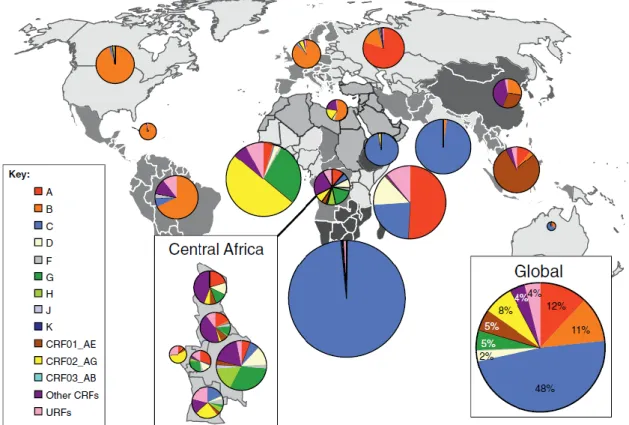
The global distribution of subtypes and recombinant forms (Figure 4) reflects the molecular epidemiological complexity of HIV (Taylor et al., 2008). In 2004–2007, subtype C accounted for 48% of all infections worldwide, followed by subtypes A (12%) and B (11%), the CRFs CRF02_AG (8%) and CRF01_AE (5%), subtype G (5%) and D(2%). The Subtypes F, H, J and K together cause fewer than 1% of global infections. All recombinant forms taken together were responsible for 20% of infections worldwide. (Hamelaar, Gouws, Ghys & Osmanov, 2011).
The extensive genetic variation and rapid evolution that has been observed in the HIV genome, makes HIV one of the fastest evolving organisms. Therefore, The HIV diversity has a major impact on HIV diagnosis, pathogenesis, transmission, clinical management and vaccine development (Hamelaar, 2012; Freed, 2001).
Figure 4. Global distribution of HIV-1 subtypes and recombinant forms.
Pie charts representing the distribution of HIV-1 subtypes and recombinants from 2004 to 2007 in each region are superimposed on the regions. Adapted from Hamelaar, 2012.
2.1.4 The host-cells and HIV interaction
CD4+ T cells and monocytes/macrophages are the two major cell types infected by HIV-1 in vivo (Rice, 2015). The state of differentiation of host cells has an important effect on the replication level of the virus. In contrast to activated CD4+ T cells and differentiated macrophages, resting CD4+ T cells and monocytes are non-permissive for HIV-1 replication. CD4+ T cells must be activated to become highly permissive for viral replication, whereas monocytes must undergo the macrophage differentiation process (Chiang & Rice, 2012).
HIV-1 particles bind specifically to cells bearing CD4, a protein that normally functions in immune recognition. Binding occurs via interactions between the gp120 and the amino-terminal immunoglobulin domain of CD4. However this interaction is sufficient for binding but not for fusion of the viral and the cellular membranes and consecutive virus entry and host
infection (Turner & Summers, 1999). Therefore, in addition to CD4, HIV requires a coreceptor for entry into target cells. The chemokine receptors CXCR4 and CCR5 are members of the G protein-coupled receptor superfamily and have been identified as the principal coreceptors for HIV fusion process. The abilities of the corresponding Env to use CXCR4 and/or CCR5, and the expression patterns of these coreceptors on different CD4 target cells are the two considerations that can explain the tropism of different HIV-1 strains (Figure 5). HIV-1 isolates that show efficient infectivity for continuous CD4 T cell lines, but poor infectivity for primary macrophages are designated T cell line–tropic (TCL-tropic) and they induce the formation of syncytia in the infected cells. On the other hand, HIV-1 strains that show the opposite preference, infecting primary macrophages much more efficiently than continuous T cell lines are designated macrophage-tropic (M-tropic) or nonsyncytium-inducing. There are also dual-tropic isolates that replicate efficiently in both target cell types. Continuous T cell lines abundantly express CXCR4, primary macrophages express CCR5, and primary T cells express both coreceptors. Moreover, M-tropic strains prefer CCR5 (R5 tropism), TCL-tropic strains preferentially use CXCR4 (X4 tropism) and dual-tropic strains can use both coreceptors (R5X4 tropism) (Berger, Murphy &Farber, 1999).
Figure 5. HIV-1 tropism.
HIV strains can be broadly divided into three categories: T cell line–tropic (TCL-tropic), macrophage-tropic (M-macrophage-tropic) and dual-macrophage-tropic. TCL-macrophage-tropic strains are specific for CXCR4 coreceptor and can infect continuous CD4 T cell lines and primary CD4 T cells. M-tropic strains are specific for CCR5 coreceptor and can infect macrophages and primary CD4 T cells. Dual-tropic strains can use both CXCR4 and CCR5 coreceptors, and can infect continuous CD4 T cell lines, macrophages, and primary T cells. Adapted from Berger et al., 1999.
Early in the course of HIV infection, the M-tropic strain viruses predominate. However, as the infection progresses to AIDS, TCL-tropic isolates can be isolated from many patients. These
strains typically display higher cytopathic effects in vitro, suggesting that they may have a particularly important role in the decline of CD4 T cells in vivo, which is the hallmark of AIDS (Berger et al., 1999).
2.1.5. HIV Transmission
The mainly known routes for HIV-1 transmission are: sexual contact across mucosal surfaces, maternal-infant exposure and percutaneous exposure (Shaw & Hunter, 2012). During sexual intercourse, HIV can cross the mucosal barrier of the vagina, vulva, penis, and rectum by first coming into contact with immune cells (dendritic cells) that then carry the virus across the mucosa (Lekkerkerke, Kooyk & Geijtenbeek, 2006). Factors such as concurrent sexually transmitted diseases, rough sex, or a partner with a very high viral load are known to increase the risk of transmission (Atkins, Carlin, Emergy, Griffiths & Boag, 1996). Generally women are at a higher risk of acquiring HIV during heterosexual intercourse due to female physiological characteristics, such as the large amount of mucosal surface area that is exposed to seminal fluid (Klimas et al., 2008).
Maternal-infant exposure can lead to infection of the fetus or newborn during pregnancy, delivery or by breastfeeding (Klimas et al., 2008). The mother-to-child transmission of HIV decreased in the last 20 years due to great prophylactic strategies. In places where one could implement all the prophylactic measures, the transmission rate reduced from 25-42% without any interventions to 1% or less. Prophylactic measures include the use of antiretroviral protocols during pregnancy, labor and delivery, and postnatally to the infant; and elective cesarean delivery before amniotic membrane rupture if HIV load is still detectable in late pregnancy (Panel on Treatment of HIV-Infected Pregnant Women and Prevention of Perinatal Transmission, 2018; WHO, 2013).
HIV transmission can also occur by contact with infected blood, where the most important way is through the practice of reusing and sharing syringes and needles for drug administration (Chitwood et al., 1990).
The transmission of HIV in healthcare workers is a reality, accidental needlestick and mucosal splash with contaminated blood are the mainly routes (Klimas et al., 2008).
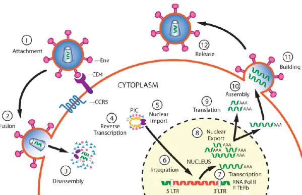
2.1.6 HIV Replication Cycle
The HIV replication cycle (Figure 6), closely resembles that of other retroviruses and can be divided into an early and a late phase. The early phase is initiated with recognition of target cells by the mature virion and encompasses the entire process leading to and including integration of the genomic DNA into the host cell genome (Turner & Summers, 1999). This includes virus binding to cell surface receptors, cell entry, reverse transcription of the viral RNA to DNA, uncoating of the viral capsid, nuclear import of viral DNA, and DNA integration (Freed, 2015). The HIV-1 replication cycle begins when the virion Env binds to the CD4
receptor and CCR5 or CXCR4 co-receptor on the surface of CD4+ T cells or monocytes/macrophages (Rice, 2015). Binding with the CCR5 coreceptor typically predominates in initial infection. However, as infection progresses, mutations in the viral envelope allows the virus to use the CXCR4 coreceptor instead of or in addition to CCR5. The CCR5:CXCR4 ratio on CD4+ cells is 1:9; thus the switch from CCR5 to CXCR4 as a coreceptor allows infection of a much greater number of CD4+ cells and is associated with accelerated HIV disease progression (Doms, 2004). The gp120 subunit binds to the CD4 cell receptor, witch induces conformational changes in the glycoprotein envelope, exposing a previously inaccessible highly conserved domain that binds to the co-receptor. Co-receptor binding induces, in turn, conformational changes at the gp41 subunit level, resulting in the fusion of viral and host cellular membranes (Doms, 2004).The membrane fusion reaction enables the viral core to gain access to the cytoplasm and is thus central to the infection process. Into the cytoplasm, the viral core begins a regulated process of disassembly. In this process, the viral capsid is disrupted and the viral RT enzyme is fully activated converting the two RNA molecules in the virion to a linear double-stranded DNA, which is then transported to the nucleus as part of a pre-integration complex (PIC). This complex appears to include the IN, MA, RT, and Vpr proteins. After active transport to the nucleus, the viral DNA is covalently integrated into the host genome by the catalytic activity of IN (Freed, 2001; Turner & Summers, 1999). HIV integration is strongly favored in active transcription units, which may promote efficient viral gene expression after integration (Craigie & Bushman, 2012). The integrated viral genome is termed provirus. From the integrated provirus it is possible to synthesize mRNAs that encode structural, regulatory and accessory proteins necessary for viral replication (Freed, 2001).
Integration marks the transition from the early to late phase of HIV-1 replication. The late phase of the virus life cycle begins with the regulated expression of the integrated proviral genome and involves all processes up to and including virus budding and maturation (Tuner et al., 1999). As a result of the natural metabolism of the host cell, if the cell is activated, the DNA, containing the viral genome, is transcribed. This process is catalyzed by the cellular enzyme RNA polymerase II. If host cells are not activated, it is possible for the virus to persist in a latent stage for many years (Potter et al., 2007). Transcription requires the viral Tat, witch recruits the general transcriptional elongation factor P-TEFb to the Pol II complex, for efficient elongation. The transcription process gives rise to viral mRNAs of a variety of alternatively spliced species that can be divided into three major classes: unspliced RNAs (mRNAs for the Gag and Gag-Pol polyprotein precursors), partially spliced mRNAs (encoding the Env, Vif, Vpu, and Vpr proteins) and small multiple spliced mRNAs (enconding Rev, Tat, and Nef) (Tazi et al., 2010, Freed, 2001). After HIV mRNA is processed in the cell's nucleus it is transported to the cytoplasm. The smaller messages are exported readily from the nucleus, whereas the unspliced and singly spliced mRNAs require the action of Rev
protein. Rev binds to the Rev Response Element (RRE), located within the env mRNA coding region, resulting in the formation of a complex capable of interacting with the cellular nuclear export machinery (Daugherty, Liu & Frankel, 2010).
Figure 6. Schematic overview of the HIV-1 replication cycle.
The infection begins when Env glycoprotein spikes engage the receptor CD4 and the membrane co-receptor CCR5, leading to fusion of the viral and cellular membranes and entry of the viral particle into the host cell. In the host cell, reverse transcription yields the pre-integration complex (PIC). After import into the cell nucleus, PIC-associated integrase carries out the formation of the integrated provirus. Proviral transcription is mediated by host RNA polymerase II (RNA Pol II) and positive transcription elongation factor b (P-TEFb), and yields viral mRNAs of different sizes, which are then exported to the cytoplasm. mRNAs serve as templates for protein production and RNA genome is incorporated into viral particles with protein components. Viral-particle budding and release from the host cell is accompanied or soon followed by protease-mediated maturation to create an infectious viral particle. Steps in the viral replication cycle are numbered. LTR - long terminal repeat. Adapted from Rice, 2015
Once the proteins necessary for viral replication are synthesized, the process of formation of new virions begins. Thus, to assemble a virus, the structural proteins must be produced first. For HIV- 1, the genomic RNA is also the mRNA that codes for the Gag precursor (Pr 55Gag) and the Gag-Pol fusion protein (PR 160Gag-Pol). Pr 55Gag is as polyprotein precursor that can be processed into MA (p17), CA (p24), NC (p7) and p6. Pr 55Gag plays a key role in the new virions formation process since it contains determinants that target it to the plasma membrane, bind the membrane itself, promote Gag-Gag interactions, encapsidate the viral RNA genome, associate with the viral Env glycoproteins, and stimulate budding from the cell.
The genes encoding the viral enzymes PR, RT and IN are translated as part of the fusion Gag-Pol polyprotein. The Env gp160 is synthesized in the ER and is modified, after translation, in the ER and Golgi apparatus and then cleaved by a host protease to produce the two envelope glycoproteins gp41 and gp120. These are transported to the plasma membrane of the host cell where gp41 anchors the Env complex in the host membrane and associates non-covalently with gp120. The Gag and Gag-Pol polyproteins also associate with the inner surface of the plasma membrane along with the HIV genomic RNA as the forming virion begins to bud from the host cell. As the particle buds and is released from the cell surface coated with gp120 and gp41, the virion undergoes a morphologic change known as maturation, involving proteolytic processing of the Gag and Gag-Pol polyproteins by viral protease. The mature virion is then ready to infect the next cell and start a new infection cycle (Freed, 2001).
HIV disseminates rapidly in the absence of preexisting immune pressures, leading to a burst of viremia manifested in a substantial proportion of patients by an acute HIV syndrome (Moir, Chun & Fauci, 2011). In the initial infection, there is massive systemic lympho-reticular infection by HIV with loss of CD4+ T cells, most severe in the gut mucosa, which never recovers its pre-infection lymphoid population (Lucas & Nelson, 2015). With migration of infected T lymphocytes or virions into the bloodstream, secondary amplification in the gastrointestinal tract, spleen, and bone marrow results in massive infection of susceptible cells. Early after infection, a pronounced depletion of activated as well as memory CD4+ T cells, located in the gut associated lymphoid tissues, has been seen in individuals (Simon, Ho & Karim, 2006). The mechanisms of CD4+ cell depletion are complex and they include apoptosis of productively infected and activated cells via activation of caspase 3 and pyroptosis triggered by abortive viral infection, via activation of caspase 1, which may account for the great majority of T cells lost (≥ 95%). Therefore, this death pathways links CD4 T-cell depletion and chronic inflammation, which are the two signature events in HIV infection, and creates a vicious pathogenic cycle where dying CD4 T-cells release inflammatory signals that attract more cells to die. (Doitsh et al, 2014). Since HIV-infected T helper cells are lysed and their production and maturation are simultaneously inhibited by Nef and Tat viral proteins, a gradual decline of these cells is observed and part of the newly produced T helper lymphocytes do not develop normal functions. After a long-lasting HIV infection the continuous loss of T helper lymphocytes results in immunodeficiency, since they play a central role in the immune response, signaling other cells such as the cytotoxic T cell and the B cells to perform their functions (Zeng et al., 2012). Depletion of susceptible CD4+ T cell targets, together with the evolution of an incompletely effective HIV-specific immune response, leads to the establishment of a plasma viral set point and to qualitative changes within each immune cell population that ultimately affect overall immunologic competence (Moir et al., 2011).
2.2. Current and emerging therapies for HIV
The revealing of all stages of HIV replicative cycle led to the identification of potential therapeutic targets in order to decrease the replicative process. This resulted in tremendous scientific progress in the drug discovery and development process (Pau & George, 2014). Approximately 30 different antiretroviral drugs, targeting four different steps in the replicative cycle of the virus, have been developed and approved for use in the treatment of HIV. There are six classes of ART available that interrupt viral replication (Table 1): nucleoside/nucleotide reverse transcriptase inhibitors (NRTI) and non-nucleoside reverse transcriptase inhibitors (NNRTI), which target the reverse transcription step that converts the viral genomic RNA into linear double-stranded DNA; protease inhibitors (PI), which inhibit the protease activity that is critical for the maturation of viral particles which bud from infected cells; CCR5 antagonists and fusion inhibitors, which block entry of the virus into new target cells by thwarting either the interaction between gp120 and the co-receptor CCR5 or the formation of the six-helix bundle of gp41, thus blocking fusion between the viral and cellular membranes; and finally, integrase inhibitors, which blocks the strand transfer activity of integrase required for insertion of viral DNA into the host cell chromosome (Englemen et al., 2012; Panel on Antiretroviral Guidelines for Adults and Adolescents, 2011).
Zidovudine, a NRTI, was the first approved antiretroviral agent for the treatment of HIV in 1987. With the administration of NTRIs, in a monotherapy regimen, the patients showed a reduction in viral load, delay in disease progression and prolonged survival, however, the use of a single agent did not provide sustained viral suppression (Pau & George, 2014). With the development of drugs with different spectra of activity, the possibility of making therapeutic combinations emerged, which dramatically changed the course of the HIV epidemic. Current treatment consists of highly active antiretroviral therapy (HAART), that is, at least three drugs belonging to two classes of antiretroviral agents. Normally, these classes are two NRTIs plus either a PI or a NNRTI or an integrase inhibitor (Menendez-Arias, 2013). HAART is effective in >70% of patients in bringing down blood viral loads to undetectable levels (<50 virus particles/ml blood) and enabling the blood CD4+ T cell count to rise to nearer normal levels (>500 cells/mm3 blood) (Lucas & Nelson, 2015).
Drug adherence, tolerability and long-term toxicity constituted major limitations to the clinical use of HAART. However, improvements in potency, safety and dosage simplification have alleviated those problems. Nevertheless, the acquisition and transmission of HIV drug resistance still poses a major risk to the success of ARTs (Menendez-Arias, 2013). The combinatorial approach to drug treatment significantly suppresses the probability of selection for, and resulting outgrowth of, resistant HIV-1 strains that quickly arise during monotherapy (Englemen et al., 2012). HIV resistance to ART can be divided into two categories: primary resistance, which reflects acquisition of a drug-resistant strain by a newly infected person;
and secondary or acquired resistance, which develops after a period of HIV treatment and which results from drug selection pressure accelerating specific mutations in the virus (Taylor et al., 2008). It should be considered that HIV-1 group O, HIV-2 and some HIV-1 group M subtypes already have natural resistance against NNRTIs and PI (Charpentier et al., 2015; Menendez-Arias, 2013). Therefore, a genotypic resistance test should be done prior to treatment in order to avoid a reduced effectiveness of the chosen therapy (Menendez-Arias, 2013).
Table 1. Antiretroviral therapies available. Adapted and modified from Lucas & Nelson, 2015.
Class Action point on HIV
life cycle Examples Specific toxicities
Nucleoside/Nucleotide reverse transcriptase inhibitors Inhibits reverse transcriptase activity, preventing the
conversion of HIV RNA into dsDNA Zidovudine Lamivudine Abacavir Didanosine Tenofovir Mitochondrial toxicity Muscle and nerve damage Lactic acidosis
Liver steatosis Hepatitis
Hypersensitivity reaction Kidney tubular damage Non-nucleoside reverse transcriptase inhibitors Inhibits reverse transcriptase activity, preventing the
conversion of HIV RNA into dsDNA
Efavirenz Nevirapine
Liver necrosis
Stevens–Johnson syndrome Toxic epidermal necrolysis
Protease inhibitors Inhibits the assembly line of new HIV viruses
Ritonavir Darunavir Atazanavir Dyslipidaemias Lipodystrophy Liver damage Integrase inhibitors
Blocks HIV integrase, preventing integration of its genes into the host cell DNA
Raltegravir Uncommon
Fusion inhibitors
Interfere with HIV’s ability to fuse to gp120 receptors on the outer surface of the cell, and blocking entry
Enfuvirtide Uncommon
Entry inhibitors
Interfere binding of HIV with chemokine
receptors, blocking entry
Maraviroc
Ibalizumab Uncommon
The limitations of HAART and the difficulty encountered to develop an effective vaccine against HIV-1 infection have require a high demand for the development of new therapeutic approaches for AIDS treatment. Thus, the HIV research community continue to explore novel therapeutic strategies, including those that target steps in the viral replication cycle that are not disrupted by currently available drugs (Freed, 2015). More promising targets need to emerge quickly, with the aim of blocking further steps in the virus replication cycle and countering the more resilient forms of HIV (Menendez-Arias, 2013). Increased knowledge
about the virus entry process, as well as its participants, has helped to discover and design new molecules capable of inhibiting the infection process. Entry into host cells is an essential step in HIV replication cycle and inhibition of this stage would complement approaches targeting other aspects of the viral replication cycle, such as inhibition of the viral RT, PR and IN enzymes (Acharya et al., 2015; Doms, 2004). Thus, the entry process is quite promising to develop new therapeutic molecules and the entry inhibitors represent a novel approach in prevention and antiretroviral therapy. New classes of drugs such as biologic small-molecule entry inhibitors (antibodies) will improve treatment options and clinical prospects particularly for patients who are infected with viruses already resistant to currently available antiretrovirals (Adamson & Freed, 2008). The development of this type of therapeutic molecules, targeting the virus entry process, represents the focus of this work.
2.2.1. HIV entry inhibitors
The unfolding elucidation of viral entry into cells has paved the way for the development of entry inhibitors. Since there are several protein molecules involved in the entry process, both at the virus and at the host cell level, there are multiple potential therapeutic targets. Unlike most currently used drugs that act only after infection has occurred, entry inhibitors stop the virus before it infects the host cell. Therefore, they may be useful as prophylactic agents, creating a barrier to the initial infection event, and all the knowledge gained in developing these molecules can provide useful leads for effective HIV-1 vaccines (Eckert & Kim, 2001). Viral entry inhibition additionally has the advantage of blocking the progress of the infection at the early steps, preserving an uninfected population of target cells (Flores & Quesada, 2013). Since each step of the viral entry pathway is a potential target for antiviral agents, depending on which step entry inhibitor block, they can be divided into three major classes: CD4-gp120-binding inhibitors; CCR5- or CXCR4-based inhibitors; and fusion inhibitors (Adamson & Freed, 2008).
CD4-gp120-binding inhibitors act on the binding between the gp120 viral glycoprotein and the CD4 cell receptor, which means that they prevent the first step of the virus entry process in the host cell (O’Hara & Olson, 2002). A variety of different candidate molecules with different mechanisms of action have been studied. These include PRO-542, BMS- 378806, BMS-488043, BMS-663068 and Ibalizumab (Adamson & Freed, 2008). Early attempts to develop specific inhibitors of CD4-gp120-binding focused on the design and testing of recombinant soluble CD4 molecules that lack the transmembrane and cytoplasmic domains, but retain the ability to bind gp120, thus functioning as molecular decoys. These molecules showed good in vitro activity against tissue culture-adapted strains of HIV-1, but activity in early phase clinical trials was disappointing (Henrich & Kuritzkes, 2013). The conjugation of soluble CD4 molecules to Pseudomonas aerugniosa exotoxin PE40 to create an immunotoxin (sCD4-PE40) led to similarly disappointing results (Davey et al., 1994).
Preliminary studies of PRO 542 generated more promising data. PRO 542 is a fusion protein in which the gp120 binding site of the tetrameric structure of the CD4 receptor is bound to the constant region of an immunoglobulin, with the aim to mimic the CD4 receptor. In patients with advanced HIV disease it was observed a modest reduction in plasma HIV-1 RNA levels in a phase 1-2 trial of PRO 542 (Jacobson et al., 2004). However, no additional studies of PRO 542 were developed (U. S. National Library of Medicine, 2018).
Small molecule inhibitors, such as BMS-378806 and BMS-488043, that bind to a specific region within the CD4 binding pocket of gp120 and block the gp120-CD4 interaction showed greater promising. BMS-378806 showed a potent activity in vitro against HIV-1 subtype B, but was less active against other subtypes and inactive against HIV-2 (Lin et al., 2003). Although there has been a reduction in plasma HIV-1 RNA in treatment- naïve subjects with BMS-488043, additional development of this molecule was discontinued due to suboptimal pharmacokinetics
(Henrich & Kuritzkes, 2013). However this limitation has been overcome
with the development of BMS-663068 (a prodrug of the attachment inhibitor BMS-626529), which demonstrated improved pharmacokinetics and increased potency against a greater range of HIV-1 subtypes (Nowicka-Sans et al., 2012). BMS-663068 has not been approved by FDA and there are no ongoing clinical trials (U. S. National Library of Medicine, 2018). Ibalizumab (formerly TNX-355) is an anti-CD4 humanized monoclonal antibody of murine origin that has displayed promising antiviral activity and safety in early clinical trials. Ibalizumab binds between domain 1 and domain 2 of the extracellular region of CD4. In contrast to attachment inhibitors, ibalizumab does not prevent gp120 binding to CD4, but is thought to decrease the flexibility of CD4, thereby hindering access of CD4-bound gp120 to CCR5 or CXCR4. Its mechanism of action allows antiviral activity regardless of chemokine receptor tropism and suggests that cross-resistance with other classes of antiretrovirals is unlikely. Since ibalizumab binding site is located away from the binding site for major histocompatibility complex class II molecules, it does not appear to interfere with immunological functions that involve antigen presentation (Henrich & Kuritzkes, 2013; Bruno & Jacobson, 2010; Song et al., 2010; Burkly et al., 1992). On March 2018, the U.S. Food and Drug Administration (FDA) approved ibalizumab for adult patients infected with HIV who were previously treated with multiple HIV drugs and whose HIV infections are resistant to currently available therapies (U.S. Department of Health and Human Services, 2018).Another strategy to inhibit HIV-1 entry is aimed at blocking the interaction between gp120 and the CCR5 or CXCR4 coreceptors. These molecules target the cellular rather than the viral component of the interaction (Adamson & Freed, 2008). Chemokine receptors are attractive targets because they are static targets not prone to mutation, in contrast to viral targets (Eckert & Kim, 2001). The small molecule CCR5 antagonists have been given generic names with the suffix “-viroc”, an abbreviation for viral receptor occupancy (Kuritzkes et al., 2009). Four of these compounds—aplaviroc, maraviroc, vicriviroc and INCB009471
have been developed, but only maraviroc is currently approved for clinical use (FDA, 2018; Henrich & Kuritzkes, 2013). CCR5 is an especially attractive target, since the natural genetic absence of surface-expressed CCR5 in Δ32 protects homozygous genotype individuals from infection with R5 strains of HIV-1 and heterozygous individuals from rapid disease progression, with little apparent impact on their immune status or general health. This epidemiological evidence together with the fact that CCR5 is the coreceptor for the most commonly transmitted HIV-1 strains, which predominate during the early stages of infection and remain the dominant form in more than 50% of late stage HIV-1-infected patients, encouraged researchers towards developing CCR5 ligands as a means to inhibit viral entry (Adamson & Freed, 2008; Dorr et al., 2005). However, the finding that CXCR4 knockout mice suffer from severe disorders suggests that drugs targeting CXCR4 coreceptor may be less well tolerated, and as a result, the development of CXCR4 inhibitors has been more challenging (Henrich & Kuritzkes, 2013; Adamson & Freed, 2008).
Given the clinical efficacy of maraviroc, its relatively low toxicity profile, and its ability to antagonize viral entry, there has been much interest in using the drug for antiretroviral treatment intensification (Henrich & Kuritzkes, 2013). By targeting exclusively CCR5 coreceptor, maraviroc does not inhibit X4 or dual tropic viral isolates (Dorr et al., 2005). Moreover, the possibility that treatment with CCR5 antagonists would promote detrimental selection for viruses with altering tropism and accelerating disease progression (such as X4 viruses), is a significant concern (Henrich & Kuritzkes, 2013). Virologic failure to maraviroc was associated with emergence of CXCR4 tropism in 57% of subjects in whom a repeat tropism test was obtained at the failure time-point (Fatkenheuer et al., 2008). Hence, FDA approval of maraviroc stipulates that it is used as therapy for treatment of adult patients in which only R5 tropic HIV-1 is detectable by baseline coreceptor screening (Adamson & Freed, 2008).
PRO 140 is a humanized anti-CCR5 monoclonal antibody that potently inhibits HIV-1 entry at concentrations that do not affect CCR5's chemokine receptor activity (Trkola et al., 2001). Unlike maroviroc which is a small molecule CCR5 inhibitor with agonist activity, PRO 140 binding to CCR5 does not result in agonist activity but instead in competitive activity (allosteric inhibitor). PRO 140 has completed 7 human clinical trials to determine its therapeutic impact on HIV infection and is currently in two US FDA approved phase IIb/III clinical trials in HIV infected patients (Burger, Parker, Guinta & Lindner, 2018).
Fusion inhibitors act on the gp41 viral protein, in particular on the intermediary states that originate from the conformational changes resulting from the entry process. By binding to gp41, they prevent the conformational changes that are necessary to the success of the fusion process between the cell membranes of the virus and the host cell (O’Hara & Olson, 2002). There are three classes of fusion inhibitors: Class 1 fusion inhibitors bind to the trimeric N-terminal heptad repeat (NHR); class 2 fusion inhibitors bind to the C-terminal
heptad repeat (CHR); and class 3 fusion inhibitors promote the disruption of the trimeric structure of the NHR region (Hrin et al., 2008). To date, the only FDA-approved fusion inhibitor is enfuvirtide (T-20), which is a class 1 fusion inhibitor and was the first entry inhibitor approved for clinical use (Henrich & Kuritzkes, 2013). Enfuvirtide is a 36-amino-acid synthetic peptide, whose sequence corresponds to that of the HR-2 region of the HIV-1 envelope gp41 subunit. Enfuvirtide is thought to block the fusion process by binding to HR1, thereby competitively inhibiting the HR1-HR2 interaction and blocking the six-helix bundle formation (Henrich & Kuritzkes, 2013; Adamson & Freed, 2008). Despite enfuvirtide being the only membrane fusion inhibitor available for treatment of viral infection, its use entails some drawbacks: it requires high doses of administration and has a low genetic barrier for resistance (Ding et al., 2017). Other limitations associated with T20 treatment include the high cost of the peptide manufacturing process and its mode of delivery, which entails twice a day subcutaneous injection (Adamson & Freed, 2008). Searching for fusion inhibitors with higher efficacy and less limitations was a logical next step. Thus, a synthetic peptide with 36 amino acid residues, called Sifuvirtide, was designed based on the crystal structure of gp41 (Wang et al., 2009). Sifuvirtide covers the deep pocket of gp41 so that it has higher binding activity to gp41 and thus, comparing to efuvirtide, is more efficient in inhibiting HIV fusion. It shows potent activity against both primary and laboratory-adapted HIV-1 isolates, and is also highly effective against enfuvirtide resistant HIV-1 variants (Miyamoto & Kodama, 2012). Sifuvirtide can be injected once daily and has entered early phase human clinical studies in Asia (Henrich & Kuritzkes, 2013).
F63 is a recently engineered VL sdAb with elongated CDRs which was selected by Phage Display technology from a synthetic library. The antibody showed to broadly and potently inhibit HIV-1 and HIV- 2 primary isolates infection by targeting a different sequence within the highly conserved hydrophobic pocket of HR1 (Cunha-Santos et al., 2016).
Although several entry inhibitors have already been developed, only three have been approved by FDA. Enfuvirtide was the first drug to be used in clinical practice, followed by maraviroc and ibalizumab. However, none of these agents offers a complete solution to the inhibition of HIV entry (Nowicka-Sans et al., 2012). The discovery and development of novel
agents directed towards other targets of the entry process of the replicative cycle of the virus,