2017
Dynamics of laminin niches during muscle development and
disease
Doutoramento em Biologia Biologia do Desenvolvimento
Andreia Marcelino Nunes
Tese orientada por:
Sólveig Thorsteinsdóttir e Marianne Deries
2017
Dynamics of laminin niches during muscle development and
disease
Doutoramento em Biologia Biologia do Desenvolvimento
Andreia Marcelino Nunes
Tese orientada por:
Sólveig Thorsteinsdóttir e Marianne Deries
Júri: Presidente:
● Maria Margarida de Mello dos Santos Reis Gutterres da Fonseca Vogais:
● José António Henriques Conde Belo ● Maria José Cardoso Oliveira
● Diana Esperança dos Santos Nascimento André ● Maria Leonor Tavares Saúde
● Edgar Rodrigues Almeida Gomes ● Sólveig Thorsteinsdóttir
Documento especialmente elaborado para a obtenção do grau de doutor
The work presented in this dissertation was developed with the support from the Fundação para a Ciência e Tecnologia, through a PhD Fellowship (SFRH/BD/86985/2012) and the Project Grant (PTDC/SAU-BID/120130/2010), Association Française contre les Myopathies Téléthon (contract nº 19959), Cure CMD, Struggle Against Muscular Dystrophy (SAM) and NIH/NIAMS Project Grant (R01AR064338-01A1).
Nota Prévia
Na elaboração da presente dissertação, e nos termos do nº1 do Artigo 45, do Regulamento de Estudos Pós-Graduados da Universidade de Lisboa, publicado no Diário da República, 2ª série, nº 65 de 30 de Março de 2012, foi usado integralmente um artigo científico publicado numa revista científica internacional indexada. Uma vez que os trabalhos referidos foram efectuados em colaboração com outros investigadores, o autor desta dissertação esclarece que participou integralmente na concepção e execução do trabalho experimental, na análise, interpretação e discussão dos resultados, assim como na redação dos manuscritos.
To my family and friends!
“Here's to the crazy ones. The misfits. The rebels. The troublemakers. The round pegs in the square holes. The ones who see things differently. They're not fond of rules. And they have no respect for the status quo. You can quote them, disagree with them, glorify or vilify them. About the only thing you can't do is ignore them. Because they change things. They push the human race forward. And while some may see them as the crazy ones, we see genius. Because the people who are crazy enough to think they can change the world, are the ones who do.” Rob Siltanen
i Em primeiro lugar, gostaria de agradecer à Sólveig por ter-me dado uma oportunidade quando mais precisava. Por alguma razão acreditaste em mim e eu nunca vou esquecer isso. Agradeço-te por toda a generosidade, preocupação e amizade. Não são precisas muitas palavras para dizer que foi sempre com um sorriso que fui trabalhar para o laboratório e que podia contar com o teu apoio e ajuda quando algo corria menos bem. Foste sempre uma presença serena que me trasmitiu entusiasmo e gosto por fazer Ciência. Tive uma enorme sorte de seres a minha mentora e ter aprendido contigo. Vou levar comigo tudo o que aprendi! Para finalizar, não quero deixar de agradecer todas as conversas sobre os mais variados temas e todo o carinho que sempre me transmitiste.
I would also like to thank Marianne, my co-supervisor, for your always encouraging and optimistic words, and for all the advice that you gave me throughout my PhD. Thank you for the fun times and for always bringing an easygoing mood into the lab.
I would like to thank Dean for everything he has done for me and for believing in me. Your lab is really the reflection of your personality. It was always so much fun to work in your laboratory. There was always time to go out for lunch to celebrate the good news or to go to movies in the morning session. I always felt that I was part of the group even if I was officially a collaborator.
Tive sempre imensa sorte de poder trabalhar em grupos animados e cheios de boa disposição. Gostava de agradecer em primeiro lugar ao meu grupo em Portugal. Quero agradecer a todos os membros passados do grupo com quem convivi: Raquel Vaz, Pedro Rifes, Gonçalo Pinheiro e psôr Gabriel. Quero também agradecer aos membros presentes do grupo. À Gabriela, companheira de gargalhadas nas mais disparatadas ideias, principalmente durante os lab meetings! Quero agradecer ao André e ao Luís por terem sempre piadas na carteira para manter os almoços animados. Luís, surpreendes-me sempre com a tua cultura nos mais variados temas :p... André, contemporâneo nesta saga do PhD, obrigado por teres sempre contigo esse espírito relaxado. Quero também agradecer à Marta, um membro não efectivo do nosso grupo, mas uma BDiana de coração. A diversão durante os almoços também não seria a mesma sem ti. Obrigada pela amizade e por poder contar sempre contigo para qualquer problema. Quero ainda agradecer à Inês por todo o companheirismo e amizade. Estes útimos meses foram duros, mas tu fizeste com que fossem mais fáceis e tornaste tudo mais divertido e especial com a tua companhia e carinho. Não trocava a “minha” mestranda por nenhum outro estudante e quero manter-te sempre presente, vá para onde for! Por fim, mas não de todo em último lugar, queria agradecer à Patrícia. Não só porque fazes parte do meu dia-a-dia, mas também porque fazes parte do grupo de amigos próximos. És e serás sempre
ii
20 ou vários milhares de quilómetros de distância, porque o Skype estará lá sempre para dar uma mãozinha...
I would also like to thank my american group. Merica! Everyone made me feel at home, even if I was miles away from home. The joy of everyone is contagious and I had an amazing time with everyone. I would like to thank Apurva, Ryan, Pam, Paul, Becky, Tatiana, Pamela, Vivian, Ashley, Keely and Suzann. I would like to thank Ryan for being a mentor to me and for teaching me so much in a few months. And ah, for all your attempts to mock me too... I would also like to thank Pam for her generosity and for all the help in the lab. I would like to thank Paul for his OCD with the hood and for protecting us from everything in the lab. And for creating a jungle in the lab too… I would like to thank Becky for all the help in the lab and outside of the lab. You were always ready for a good laugh in the free times. I would like to thank to my “girls”. Vivian for being a sweet and caring friend. Tats, my Portuguese carago fellow, for your energy, partnership and help. And to Pamelita, my mexican chica, for always helping and taking care of me (even in awkward situations...). Thank you for being awesome lab mates and friends. Finally, I would like to thank to my buddy Apurva for the amazing friend she is. You made so much for me that I can’t thank you enough. You truly made my experience in the States unforgettable and I want to cherish your friendship forever.
Gostava ainda de deixar um agradecimento a todas as pessoas da FCUL que sempre estiveram dispostas a ajudar quando precisei. Quero agradecer em especial à Sara Carona, Mónica Silva, Pedro Simões, Ana Neto e Inês Fragata.
Felizmente tenho uma grande lista de amigos a quem agradecer. Quero agradecer aos meus amigos Andreia Rodrigues, Ana Farinha, Alexandra, Joana, Tomás, Inês Albuquerque, Catarina Nabais, Eduardo, Catarina Dourado, Lucas, Rachel, Nelson, Raquel Silva, Andreia Margarido, Sofia Carvalho, Maggie, Diana, Inês Fragata e Josefina. Sou uma sortuda por poder contar com a vossa companhia e amizade. Agradeço à Ana Farinha pelas suas palavras sábias e meigas, à Xana pelo prazer que é estar na tua companhia e por me salvares das dorminhocas em Espanha. À Joana ornitorrinca por seres sempre uma querida e estares presente quando mais preciso. À Inês Albuquerque, pesinho de lama, por seres sempre uma presença calma, confortante e inspiradora. Catarina Nabais, por seres a minha parceira de aventura. A tua energia e espírito são contagiantes, mas mais que isso, sempre estiveste lá quando precisei de apoio. Quero agradecer ao senhor Eduardo Borboleta Marabuto, pela boa disposição e pelas suas maluqueiras! Lucas, obrigado pela tua presença dissimulada, mas atenta e generosa com que posso contar. Rachel, pela tua visão prática nos momentos em que precisei de uma perspectiva diferente. Por seres uma amiga leal e por teres estado sempre presente nos momentos
iii lá no momento seguinte! Obrigado à Sofia, por mudares o penteado de cada vez que nos vimos e pelo teu estilo cómico e peculiar. À Maggie, o mini caracolinho coração de manteiga, por poder contar sempre com o teu apoio preciso e prático, mesmo não estando lá todos os dias. Obrigado ao Tomás, camarada benfiquista, por todo o apoio e companhia. Finalmente, agradeço à Andreia, por seres uma amiga carinhosa e pelas conversas quotidianas e pelo apoio constante mesmo a várias centenas de quilómetros.
Por fim, não poderia deixar de agradecer à minha família que me atura desde pequena e que tem estado presente em todos os momentos da minha vida. Por terem sempre acreditado que conseguia alcançar os meus objectivos e pelas reuniões familiares animadas. Um agradecimento especial aos meus pais por terem sempre apoiado nas minhas decisões e por terem ensinado que nada se consegue na vida sem esforço e trabalho. Ensinaram-me os valores mais importantes da vida. Sou a pessoa que sou graças a tudo o que me deram e ensinaram. Em especial, um obrigado à minha mãe por ter dado tudo de si para eu poder chegar onde cheguei. Sem ti, nunca tinha chegado aqui. Agradeço também aos meus irmãos Vanessa e Jorge por estarem presentes com com o seu espírito brincalhão e com as suas “tontices”. Aos meu avós Matilde e Manuel, aos meus primos Sara, João e Inês, e aos meus tios: Zé, “o brincalhão caneleiro” sempre na calha para a próxima piada, por manteres sempre a malta animada; Belinha, por seres sempre uma madrinha preocupada e por estares sempre pronta a ajudar; Nela, por teres estado sempre na linha da frente com os teus conselhos para ajudar a empurrar-me para a frente e pela confiança de que eu conseguia chegar ao fim; Nanda, o poço de ternura e mel que nunca mais acaba, sempre disposta a distribuir mimos; Xana, porque uma presença muita calma sempre com energias positivas; e ao Nelo, “o rockeiro coração de manteiga” sempre a observar, mas atento para emergências! E ao resto da família que é gigante mesmo aquela que não vejo tão frequentemente: Nazaré, Maísa, tias Elvira, Natália e Henriqueta, tios Zé e Henrique e primos Nuno, Ana Rita, Ana Luísa, Mário, entre muitos outros familiares.
v
Abstract ix
Resumo xi
Resumo alargado xiii
List of abbreviations and acronyms xix
Chapter 1: Introduction 1
I. State of the art 3
1. Introduction 3
2. Extracellular matrix 3
2.1. Laminins 4
2.1.1. Laminin composition and structure 4
2.1.2. Laminin assembly into the basement membrane 6
2.1.3. Laminin remodeling in the basement membrane 8
2.2. Laminin receptors 10
2.2.1 Integrins 10
2.2.1.1. Integrin structure and activation 10
2.2.1.2. Integrins as mediators of interactions
between laminins and the cytoskeleton 14
2.2.2. Dystroglycan 14
3. Cardiac development 16
3.1. Specification of first and second heart field progenitors 16
3.2. Heart tube formation and heart looping 18
3.3. Chamber specification and fetal myocardial growth 20
3.4. Laminins during cardiac development 22
4. From somite to skeletal muscle 23
4.1. Somite formation 23
vi
4.2.3. Myotome: The first differentiated skeletal muscle 26
4.2.4. Syndetome: linking bone and muscles 27
5. Skeletal muscle development 28
5.1. Myogenic regulatory factors 28
5.2. Epaxial muscle development 29
5.2.1. Formation of the epaxial myotome 29
5.2.2. The formation of the definitive epaxial muscle masses 30
5.3. Hypaxial Myogenesis 32
5.3.1. Formation of the hypaxial myotome 32
5.3.2. Limb myogenesis 32
5.4. Primary myogenesis 34
5.5. Secondary myogenesis 35
5.6. Postnatal myogenesis 37
5.7. Laminins and laminin receptors during myogenesis 37
6. Adult muscle and satellite cells 39
6.1. Satellite cells: drivers of regeneration with a characteristic
profile 39
6.2. Self-renewal versus differentiation of satellite cells 41
6.2.1. Symmetric vs asymmetric divisions: polarized
segregation of intracellular components 41
6.2.2. Extracellular environment of satellite cells: myofibers
and myoblasts 42
6.2.3. Extracellular environment of satellite cells: the
interstitial cells 45
7. Merosin-deficient congenital muscular dystrophy type 1A (MDC1A) 46
vii
deficient congenital muscular dystrophy 69
Chapter 3: Building laminin matrices during skeletal muscle
development 97
Chapter 4: Behind the curtain of muscle development: techniques to
unveil the role of laminin 211 during fetal muscle development 127
Chapter 5: Dynamics of laminin matrices during cardiac muscle
development 147
Chapter 6: Discussion 185
6.1. Laminin diversity during cardiac and skeletal muscle
development 187
6.2. Laminins within skeletal and cardiac muscle tissues 193
6.2.1. On the specificity of laminin isoforms during cardiac
and skeletal muscle development 193
6.2.2. “Skeletal” laminins versus “cardiac” laminins 196 6.3. New paths for Merosin-deficient congenital muscular
dystrophy type 1A 198
6.3.1. Insights into the mechanism underlying MDC1A 198
6.3.2. What’s next? New avenues for the treatment of
MDC1A 200
6.4 Final considerations 202
ix start early in embryogenesis. Skeletal muscle cells of the trunk, limbs and tongue arise from the somite-derived dermomyotome. The myotome, the first differentiated skeletal muscle, is formed when the dermomyotome precursors enter the myogenic program and delaminate from the dermomyotome. Following stages of skeletal muscle development are marked by the formation of the embryonic and fetal myofibers. Cardiac muscle development starts when the cardiogenic progenitors in the splanchnic mesoderm are brought to the midline to form the heart tube. The heart tube then undergoes several developmentally regulated rearrangements, originating the four cardiac chambers and a myocardium capable of pumping the blood.
The main goal of this thesis was the characterization of laminin niches during muscle development. In chapter 2 and 3, we addressed the dynamics of laminin synthesis and assembly during skeletal muscle development. Our results reveal a complex assembly dynamics, which generate specific microenvironments during different phases of muscle development. We then focused our analysis on the role of laminin niches during the onset of the Merosin-deficient congenital muscular dystrophy type 1A (MDC1A) in the dyW
mouse model, and demonstrated that absence of laminin 211 in the myofiber basement membrane leads to impaired fetal muscle growth. In Chapter 4 we show the progress in the development of different techniques to unveil the mechanism by which laminin 211 signals during skeletal muscle development. In Chapter 5, we characterized the dynamics of laminin assembly during cardiac development and found that different phases of cardiac development are marked by specific laminin niches.
Together, the results presented in this thesis provide a detailed framework on laminin matrices during muscle development. This thesis also unveils an important role of laminin 211 during the fetal development of skeletal muscles.
xi desenvolvimento embrionário. Os músculos esqueléticos do tronco têm origem no dermamiótomo que contém os progenitores musculares. O miótomo forma-se quando os progenitores entram no programa miogénico e delaminam do dermomiótomo. As fases seguintes da miogénese são marcadas pela formação das miofibras embrionárias e fetais. O desenvolvimento do músculo cardíaco inicia-se quando os progenitores cardíacos na mesoderme esplancnica são levados para a linha mediana do embrião para formar o tubo cardíaco. Este tubo sofre depois vários rearranjos que permitem desenvolver um coração com quatro câmaras cardíacas.
O principal objectivo desta tese foi caracterizar os nichos de laminina ao longo do desenvolvimento do músculo esquelético e cardíaco. Nos Capítulos 2 e 3, estudámos a dinâmica da síntese e montagem das matrizes de laminina durante o desenvolvimento do músculo esquelético. Os resultados obtidos revelam a existência de microambientes de laminina específicos ao longo do desenvolvimento do músculo. Os nossos resultados demonstram ainda que a matriz de laminina 211 tem um papel no despoletar da Distrofia muscular congénita merosina negativa (MDC1A) no modelo dyW de ratinho, uma vez que
a ausência desta laminina na membrana basal da miofibra resulta num défice de crescimento do músculo.
No Capítulo 4 apresentamos o progresso no desenvolvimento de diferentes técnicas que permitam compreender o mecanismo de sinalização da laminina 211 durante o desenvolvimento do músculo esquelético. No Capítulo 5, caracterizámos as dinâmicas de montagem de matrizes de laminina ao longo do desenvolvimento do músculo cardíaco, e descobrimos que diferentes fases do seu desenvolvimento são marcadas por nichos de laminina específicos.
xii
e evidenciam o papel da laminina 211 no desenvolvimento fetal dos músculos esqueléticos.
Palavras-chave: Laminina, Desenvolvimento do Músculo Esquelético, Desenvolvimento
xiii dos animais, mas também para o correcto funcionamento de órgãos vitais, como o coração. Os músculos esqueléticos do tronco, dos membros e da língua têm origem embrionária na mesoderme paraxial, mais precisamente nos sómitos que se formam periodicamente em ambos os lados do tubo neural. A maturação dos sómitos dá origem ao dermamiótomo, um epitélio onde os progenitores musculares são mantidos. Quando a formação dos músculos, ou miogénese, é induzida, os progenitores activam o programa miogénico e delaminam para o espaço subjacente para formar os miócitos que constituem o miótomo. Durante esta fase inicial da miogénese, o miótomo cresce até ocorrer a dissociação do dermamiótomo. Posteriomente, os miócitos fundem com os mioblastos primários para formar as fibras embrionárias. Estas funcionam como suporte para a formação das fibras fetais, aquando de uma nova onda de fusão dos mioblastos fetais entre si para formar as fibras fetais. Durante esta vaga de fusões, os mioblastos fetais fundem também com as fibras embrionárias e fetais para que estas cresçam.
O desenvolvimento do músculo cardíaco é induzido quando os progenitores cardíacos localizados na mesoderme esplâncnica de ambos os lados do embrião se aproximam e se juntam na linha mediana para formar o tubo cardíaco. O tubo cardíaco sofre depois uma remodelação extensa, durante a qual o epicárdio reveste a camada exterior do miocárdio e o coração é sujeito a um processo de morfogénese até adquirir a organização final do coração adulto. Durante esta fase, o músculo cardíaco, ou miocárdio, é remodelado em trabéculas e zona compacta. As fases que se seguem englobam o desenvolvimento de todo o sistema que suporta o funcionamento do coração, tais como o desenvolvimento da vasculatura coronária, o desenvolvimento dos septos que separam as diversas câmaras cardíacas, e o desenvolvimento do sistema eléctrico de excitação cardíaca.
xiv
ainda pouco conhecido, e os poucos estudos realizados não fornecem uma perspectiva global das matrizes de laminina presentes durante o desenvolvimento destes dois tipos de músculo. Apesar destas lacunas, alguns destes estudos revelaram que as matrizes de laminina desempenham um papel importante durante desenvolvimento do miótomo, na homeostase do músculo esquelético no adulto, bem como durante o desenvolvimento do coração.
Nesta tese pretendemos compreender as dinâmicas de montagem das matrizes de laminina durante o desenvolvimento dos músculos esquelético e cardíaco, a fim de perceber de que forma estas matrizes modulam o desenvolvimento destes tipos de músculo durante o estabelecimento das membranas basais nos músculos. Abordamos diferentes perspectivas da montagem das matrizes de laminina, desde a expressão dos genes que codificam para as três cadeias das lamininas até à sua incorporação na membrana basal. Analisamos também o papel de lamininas durante o início da Distrofia muscular congénita merosina negativa (MDC1A) in utero, na qual o gene LAMA2 se encontra mutado e a consequente ausência de lamininas com a cadeia α2. Por fim, descrevemos ainda o progresso no desenvolvimento de duas técnicas que visam fornecer diferentes metodologias para o estudo do papel das lamininas durante o desenvolvimento do músculo.
Numa primeira abordagem, esta tese teve como objectivo compreender a dinâmica dos ciclos de montagem das matrizes de laminina durante o desenvolvimento dos músculos esqueléticos epaxiais, bem como a relação desta dinâmica com o desencadear da distrofia muscular MDC1A que ocorre na ausência de lamininas 211/221. Os resultados apresentados no capítulo 2 demonstram que o desenvolvimento do músculo esquelético ocorre em paralelo com diferentes ciclos de montagem de matrizes
xv de lamininas 211/221 no modelo dy de ratinho para MDC1A não interfere com o decorrer miogénese no miótomo que parece prosseguir sem defeitos significativos. Isto pode possivelmente dever-se à presença da laminina 111, que parece compensar a ausência de laminina 211 no músculo adulto. Verificámos que a miogénese primária prossegue sem defeitos significativos na ausência de lamininas 211/221, o que correlaciona com uma fase da miogénese onde as matrizes de laminina não são montadas pelas células miogénicas. Em conjunto com o capítulo 3, revela que a ausência de lamininas incorporadas na membrana basal no músculo durante a miogénese primária não corresponde à ausência de expressão dos genes de laminina. Pelo contrário, a expressão destes genes é mantida durante toda a miogénese primária. Os dados reportados no capítulo 2 revelam que o efeito da ausência de laminina 211 na membrana basal que contacta com a miofibra e com as células estaminais musculares Pax7-positivas faz-se notar durante a miogénese fetal, quando o músculo começa a apresentar défices de crescimento, em particular no crescimento por hipertrofia. Este defeito correlaciona com a redução do número de células Pax7-positivas e Miogenina-positivas que, de acordo com os nossos dados, não conseguem assim contribuir devidamente para o normal crescimento do músculo durante esta fase do seu desenvolvimento. Este defeito correlaciona também com a activação excessiva da sinalização por STAT3, induzida provavelmente através da falta de sinalização por laminina 211-integrina α7β1, e com a redução da sinalização por Miostatina. A combinação dos efeitos da alteração destas sinalizações no contexto da ausência de laminina 211 parece inibir a expansão das células Pax7-positivas e dos mioblastos em diferenciação, o que resulta num défice de células a contribuir para o crescimento das fibras. Durante o desenvolvimento pós-natal, o defeito no tamanho das massas musculares torna-se mais evidente, com as massas deficientes para laminina 211 a evidenciarem uma incapacidade de crescimento. A sobreactivação da
xvi
capítulo 2 indicam ainda que a composição das matrizes de laminina influenciam a “idade” das células miogénicas. O nosso modelo de sinalização por laminina 211 sugere que a ausência de laminina 211 na membrana basal em contacto com as miofibras e as células estaminais musculares fornece os sinais característicos de fases de desenvolvimento mais tardias e, por isso, uma acumulação progressiva dos sinais que promovem a diferenciação em detrimento dos sinais que induzem a proliferação celular.
No capítulo 3 demonstramos que ao longo da miogénese, as células estaminais musculares podem construir o seu nicho. Os nosso resultados evidenciam que as células estaminais musculares no dermamiótomo constroem de forma autónoma a membrana basal do dermamiótomo. Durante a miogénese fetal, a matriz de laminina que cobre as miofibras e as células estaminais musculares parece ser construída por células mononucleadas adjacentes às miofibras, provavelmente células estaminais musculares e/ou mioblastos em diferenciação. De facto, as células estaminais musculares produzem matrizes de laminina no início da miogénese secundária. A população de células estaminais musculares parece ser bastante heterogénea dado que algumas células montam matrizes de laminina com as cadeias α2 e α5 em simultâneo, enquanto que outras produzem apenas um tipo de matriz de laminina, e outras não produzem qualquer laminina. Esta heterogeneidade sugere que células com um repertório diferente de lamininas podem reagir de formas distintas à ausência de laminina 211 no seu nicho.
O estudo do desenvolvimento do músculo in utero coloca diversas barreiras técnicas que impedem a realização de alguns estudos funcionais. No capítulo 4, apresentamos o progresso no desenvolvimento de diferentes técnicas com vista a poder compreender melhor o mecanismo de sinalização por laminina 211 que opera durante o desenvolvimento fetal do músculo. Os resultados exploratórios apresentados neste
xvii adulto, como prova de princípio de que as lamininas 211/111 permitem recuperar o defeito fetal. Esta metodologia poderá ser utilizada em estudos futuros para estudar os efeitos da sinalização por laminina 211.
No capítulo 5 evidenciamos a dinâmica de construção das matrizes de laminina durante o desenvolvimento do coração in utero e nas fases pós-natal e adulta. Neste capítulo demonstramos a existência de matrizes de laminina muito dinâmicas nas membranas basais cardíacas. A membrana basal na interface entre o endocárdio e o miocárdio é incialmente constituída pela combinação das matrizes de laminina 111/121, 411/421 e 511/521. Contudo, esta vai sendo progressivamente reduzida à matriz de laminina 521. Após o epicárdio envolver o coração, a membrana basal subepicardial é composta pelas matrizes de lamininas 111 e 511, sendo no entanto posteriormente substitutídas pela matriz de laminina 521. Por fim, os nosso resultados revelam a presença das matrizes de laminina 211 e 511 aquando da formação da membrana basal dos cardiomiócitos, sendo estas depois gradualmente substituídas pelas matrizes de lamininas 221 e 521 perto do fim do desenvolvimento fetal do coração.
Os capítulos desta tese ilustram a diversidade de matrizes de laminina construídas nas membranes basais presentes durante o desenvolvimento do músculo esquelético e cardíaco. O nosso estudo demonstra ainda em particular que a matriz de laminina 211 desempenha um papel fucral durante o desenvolvimento fetal dos músculos esqueléticos.
Palavras-chave: Lamininas, Desenvolvimento do músculo esquelético, Desenvolvimento
xix
ADAM- Metalloproteinase with thrombospondin motifs ANF- Atrial Natriuretic Factor
bHLH- Basic Helix-Loop-Helix
CXCR4- C-X-C Chemokine Receptor type 4 BDNF- Brain-Derived Neurotrophic Factor BMP- Bone Morphogenetic Protein
DGC- Dystrophin- associated Glycoprotein Complex DML- Dorsomedial Lip
DNA- Deoxyribonucleic Acid E(n)- Embryonic day (number) ECM- Extracellular Matrix EDL- Extensor Digitorum Longus
EMT- Epithelium to Mesenchyme Transition EGF- Epidermal Growth Factor
EGFR- Epidermal Growth Factor Receptor EPDC- Epicardium-Derived Cell
ERK- Extracellular signal–Regulated Kinases FAK- Focal Adhesion Kinase
Fgf- Fibroblast Growth Factor FKRP- Fukutin-Related Protein FLK1- Fetal Kinase Liver 1 HGF- Hepatocyte Growth Factor ILK- Integrin-Linked Kinase IPP- ILK, PINCH and Parvin
JAK-STAT- Janus Kinase/Signal Transducers and Activators of Transcription LAMA2-CMD- Laminin-α2 Congenital Muscular Dystrophy
xx
MyHC- Myosin Heavy Chain
MyHC-emb- Embryonic Myosin Heavy Chain MyHC-β/slow- Myosin Heavy Chain-β/Slow MRF- Myogenic Regulatory Factor
N-CAM- Neural Cell Adhesion Molecule NICD- Notch Intracellular Domain P(n)- Postnatal day (number) PCP- Planar Cell Polarity
PINCH- Particularly Interesting Cys-His-rich
PECAM- Platelet Endothelial Cell Adhesion Molecule PDGFR- Platelet-derived Growth Factor Receptor POMT1- Protein O-MannosylTransferase 1 POMT2- Protein O-MannosylTransferase 2
POMGnT1- Protein O-Linked Mannose N-AcetylGlucosaminylTransferase 1 PSM- Presomitic Mesoderm
RGD- Arginine-Glycine Aspartic acid SDF1- Stromal Cell-Derived Factor 1 Shh- Sonic Hedgehog
TAL1- T-Cell Acute Lymphocytic Leukemia 1 TBX- T-BoX
TGFβ- Transforming Growth Factor Beta
UGC- Utrophin- associated Glycoprotein Complex V-CAM1- Vascular Cell Adhesion Molecule 1 VEGF- Vascular Endothelial Growth Factor
VEGFR- Vascular Endothelial Growth Factor Receptor VLL- Ventrolateral Lip
3
I. State of the art
1. Introduction
Striated muscles evolved during the early diversification of the animal kingdom from an ancestral actinomyosin machinery that predates muscle evolution (Steinmetz et al., 2012). They drive the blood circulation and enable the locomotion of the body. Laminins are major components of basement membranes, a specific type of extracellular matrix (ECM), which are in close contact with a number of organized tissues in the body, namely epithelia, endothelia, nerves, adipose cells and muscles (LeBleu et al., 2007; Durbeej, 2010; Domogatskaya et al., 2012). The ancestral form of trimeric laminins is believed to have arisen before the separation of the cnidaria and bilateria (Zhang et al., 2002). Laminins are crucial for the assembly of basement membranes and the absence of all laminins is incompatible with life in most, if not all, bilaterian animals (Domogatskaya et al., 2012). In this chapter I will introduce laminins and their role in basement membrane assembly and function. Moreover, I will review the development of the two types of striated muscle, cardiac and skeletal, as well as the relevance of studying laminins in these tissues.
2. Extracellular matrix
During development in multicellular organisms, individual cells interact with each other via cell-cell communication events by receiving signals from neighboring cells through paracrine factors, by engaging with adjacent cells through cell-surface bound molecules and by contacting with the cell-generated extracellular matrix. The ECM is a complex network that provides both a physical scaffold and signaling cues to cells. It comprises many different types of molecules, including glycoproteins such as laminins, fibronectin, collagens and tenascins, and polysaccharide-rich molecules such as
4
glycosaminoglycans and proteoglycans. These molecules are produced by a variety of cell types and are assembled extracellularly into a supramolecular matrix that connects back to cells through specific cell surface receptors (Frantz et al., 2010; Thorsteinsdóttir et al., 2011). The ECM can be divided into interstitial matrices and pericellular matrices. Interstitial matrices surround mesenchymal cells and constitute the connective tissue matrix. These matrices are composed of collagens, proteoglycans, glycosaminoglycans and contain numerous other glycoproteins and proteins such as fibronectin, tenascin and elastins among others (Frantz et al., 2010; Thorsteinsdóttir et al., 2011). Pericellular matrices are matrices that are in close proximity to cells and the most common form of pericellular matrix are the basement membranes. Basement membranes are mainly composed of laminins, collagens type IV, perlecan and nidogens. They line epithelia and endothelia, and surround neurons, fat and muscle cells (LeBleu et al., 2007; Thorsteinsdóttir et al., 2011).
2.1. Laminins
2.1.1. Laminin composition and structure
Laminins are trimeric glycoproteins composed of an α, a β and a γ chain, and are the basic building blocks of basement membranes (LeBleu et al., 2007; Durbeej, 2010; Yurchenco et al., 2015). The laminin trimer is composed of an α-helical coiled coil domain containing the three chains and three short arms corresponding to each chain (Durbeej, 2010; Hohenester and Yurchenco, 2013; Holmberg and Durbeej, 2013; Fig. 1). The short arms of α, β and γ chains vary in length but are all composed of laminin N-terminal (LN) domains separated by tandem repeats of laminin type epidermal growth factor-like (LE) domains. The C-terminal globular domain of the α chain contains five laminin G-like (LG) subdomains (Durbeej, 2010; Domogatskaya et al., 2012; Hohenester and Yurchenco, 2013;
5 Holmberg and Durbeej, 2013; Fig. 1). The
α3A and α4chains are distinct from other α chains because their short arms retain only one LE domain subdomain (Durbeej, 2010; Domogatskaya et al., 2012; Hohenester and Yurchenco, 2013; Holmberg and Durbeej, 2013).
Different combinations of α, β and γ chains generate at least 16 different laminin isoforms which are assembled into basement membranes by several cell types in multiple tissues. The current nomenclature is based on the chain composition, e.g., laminin 211 is composed of α2, β1 and γ1 chains (Aumailley et al., 2005; Table 1). The first described laminin was laminin 111 identified in the basement membrane of the murine Engelbreth-Holm-Swarm
(EHS) sarcoma (Orkin et al., 1977; Timpl et al., 1979). However, the ancestral form of trimeric laminin is present in the radiata Hydra, and is assembled in the basement membrane separating the endoderm from the ectoderm (Zhang et al., 2002; Domogatskaya et al., 2012). The trimeric composition of Hydra laminin is not yet completely uncovered and doubts arise about whether the trimer is a combination of α, β and γ chains, or if the combination includes one α and two β chains (Zhang et al., 2002; Domogatskaya et al., 2012). With the appearance of Bilateria, α chain diversification led to the appearance of two branches of ancestral α chain. In the particular case of the
Figure 1- Laminin trimer structure. Schematic representation of laminin 111. The coiled coil domain contains the three different chains, in contrast to each N-terminal short arm which is established by each chain. The short arms are responsible for laminin self-assembly. The α chain C-terminal end consists of five globular domains which interact with cell surface receptors. Tandem LE domains are depicted in the short arm separating the LN domains. The binding sites for other basement membrane components and cell receptors are also depicted. Figure from Yurchenco, 2015.
6
Vertebrata, the ancestor α1,2 originated α1 and α2 chains, whereas the α3,5 ancestor gave
rise to α3 and α5 chains (Domogatskaya et al., 2012). Diversification of β and γ chains are likely to have occurred during non-vertebrate Deuterostomia evolution (Domogatskaya et al., 2012).
2.1.2. Laminin assembly into the basement membrane
The construction of basement membranes is a multi-step process, where laminin and collagen IV matrices are assembled separately and then incorporated into a supramolecular network (Yurchenco, 2015). Laminin trimer secretion is a two-step process, where ionic interactions first promote the formation of a βγ dimer and the subsequent addition of the α chain stabilizes the previously formed dimer (Kumagai et al., 1997; Yurchenco et al., 1997; Yurchenco, 2015). The addition of the α chain is the
Table 1- Laminin nomenclature and composition. Adapted from Thorsteinsdóttir et al., 2011. Current laminin nomenclature Old laminin nomenclature Chain composition Genes
Laminin 111 Laminin- 1 α1β1γ1 Lama1 Lamb1 Lamc1 Laminin 121 Laminin- 3 α1β2γ1 Lama1 Lamb2 Lamc1 Laminin 211 Laminin- 2 α2β1γ1 Lama2 Lamb1 Lamc1 Laminin 221 Laminin- 4 α2β2γ1 Lama2 Lamb2 Lamc1 Laminin 213 Laminin- 12 α2β1γ2 Lama2 Lamb1 Lamc3 Laminin 212 (existence proposed) - α2β1γ2 Lama2 Lamb1 Lamc2 Laminin 222 (existence proposed) - α2β2γ2 Lama2 Lamb2 Lamc2 Laminin 311 Laminin- 6 α3Aβ1γ1 Lama3A Lamb1 Lamc1 Laminin 321 Laminin- 7 αA3β2γ1 Lama3A Lamb2 Lamc1 Laminin 332 Laminin- 5 α3Aβ3γ2 Lama3A Lamb3 Lamc2 Laminin 3B32 Laminin- 5B α3Bβ3γ2 Lama3B Lamb3 Lamc2
Laminin 333 - α3Aβ3γ3 Lama3A Lamb3 Lamc3
Laminin 411 Laminin- 8 α4β1γ1 Lama4 Lamb1 Lamc1 Laminin 421 Laminin- 9 α4β2γ1 Lama4 Lamb2 Lamc1 Laminin 423 Laminin- 14 α4β2γ3 Lama4 Lamb2 Lamc3 Laminin 511 Laminin- 10 α5β1γ1 Lama5 Lamb1 Lamc1 Laminin 521 Laminin- 11 α5β2γ1 Lama5 Lamb2 Lamc1 Laminin 522 (existence proposed) - α5β2γ2 Lama5 Lamb2 Lamc2 Laminin 523 Laminin- 15 α5β2γ3 Lama5 Lamb2 Lamc3
7 determinant step as the trimer is only secreted when the α chain is added to the βγ dimer (Kumagai et al., 1997; Yurchenco et al., 1997).
Once secreted to the extracellular space, laminin is believed to polymerize according to the “three-arm interaction model” (Yurchenco et al., 1985; Hohenester and Yurchenco, 2013; Fig. 2). This model is supported by several in vitro reports demonstrating that laminin trimers self-assemble into polymers through a calcium dependent process, involving ternary interactions between the LN terminal domains of the α, β and γ short arms, which are subsequently stabilized by disulphide bridges (Yurchenco et al., 1985; Schittny and Yurchenco, 1990; Yurchenco and Cheng, 1993; McKee et al., 2007; Hussain et al., 2011; Hohenester and Yurchenco, 2013). These interactions promote the assembly of laminin molecules into homopolymers, but different laminin isoforms can also assemble into mixed heteropolymers (Cheng et al., 1997). Laminin isoforms in which the α chains lack most of the short arm domain, such as laminins 3A11, 3A21, 411 and 421, cannot self-assemble (Cheng et al., 1997; Domogatskaya et al., 2012).
In vivo laminin assembly seems to depend on the binding to cell surface receptors which interact with the five LG domains at the C-terminal of the α chains (McKee et al., 2007; McKee et al., 2009). However, the dependence of laminin assembly on cell surface receptors and the mechanism underlying their cross-talk is still controversial (McKee et al., 2007; McKee et al., 2009). Laminin polymerization was shown to induce clustering of integrins and dystroglycan in the cell membrane, and aggregation of intracellular cytoskeletal components (Colognato et al., 1999; Li et al., 2005a). In other studies, the production of a gal-sulfatide receptor by Schwann cells precedes laminin accumulation, and hydrolyzation of sulfatides prevents the accumulation of laminin on the cell surface (Li et al., 2005a). Conversely, dystroglycan null embryoid bodies develop a basement membrane (Li et al., 2002). Thus, these results suggest that different cell surface receptors might have different impacts on laminin assembly.
8
Basement membrane maturation and stability depends on the proper interconnection between different components. Laminins and collagen IV are the only basement membrane components capable of self-assembly, but the addition of perlecan and nidogen bridges allow the link between the laminin and the collagen IV networks (LeBleu et al., 2007; McKee et al., 2007; Behrens et al., 2012; Hohenester and Yurchenco, 2013). The first step in the construction of basement membranes seems to be the assembly of the laminin matrix as it precedes collagen IV accumulation on the cell surface, and occurs in the absence of assembled collagen (Smirnov et al., 2002; Li et al., 2002; Tsiper and Yurchenco, 2002; McKee et al., 2007; Yurchenco, 2015; Fig.2).
2.1.3. Laminin remodeling in the basement membrane
The ECM is modified by metalloproteinases with proteolytic activity, which cleave ECM components. The two major metalloproteinase families targeting the ECM are the matrix metalloproteinase (MMP) family that is composed of 23 different proteinases, and Figure 2- Laminin self-assembly into the basement membrane. Self-assembly of laminin according to the “three-arm interaction model” (left) and its incorporation into the supramolecular network of the basement membrane through interaction with other ECM components such as collagen IV, perlecan, agrin and nidogen (right). Adapted from Hohenester and Yurchenco, 2013.
9 the metalloproteinase with thrombospondin motifs (ADAM) family which includes 19 members (Lu et al., 2011). MMPs are usually secreted molecules constituted by the prodomain, the catalytic domain and the hemopexin-containing ancillary domain. However, certain MMPs display extra domains, such as MMP2 and MMP9 which contain fibronectin type II inserts in the catalytic domain, and MMPs 14, 15, 23 and 24 which are membrane-type MMPs with a transmembrane domain and a cytoplasmic tail (Page-McCaw et al., 2007; Lu et al., 2011). Several proteinases are synthesized in an inactive form and need further cleavage of the prodomain, which normally inhibits the activity of the catalytic domain (Page-McCaw et al., 2007; Lu et al., 2011). This processing is performed by other proteinases which in the case of MMPs include the proteinases tryptase and plasmin, but also other MMPs (Lu et al., 2011). Further tuning of MMP and ADAM activity is performed by tissue inhibitor of metalloproteinases (TIMP) molecules (Sternlicht and Werb, 2001; Lu et al., 2011). Each MMPs targets specific ECM components, albeit different MMPs sharing common targets. Laminins are cleaved by MMPs 2, 3, 10, 11, 12, 13, 14, 15 and 25 (Lu et al., 2011).
ECM degradation does not solely affect the composition of the ECM, but can also release ECM fragments with biological activity as well as ECM-bound paracrine growth factors which impact cell behavior (Streuli, 1999; Page-McCaw et al., 2007; Mott and Werb, 2004). For instance, cleavage of laminin 111 into several fragments with biological activity, combined with MMP9-mediated release of VEGF (Vascular endothelial growth factor), strongly affects tumor growth and metastasis formation (Mott and Werb, 2004; Kikkawa et al., 2013). In another example, fragments generated from laminin 332 cleavage were shown to interfere with mammary gland epithelial cell migration (Koshikawa et al., 2000).
10
2.2. Laminin receptors
Laminins interact with several cell surface receptors through their LG domains in the C-terminal ends of the α chains. Laminins display binding affinity to integrins, dystroglycan, syndecans and Lutheran blood group glycoproteins, also called BCAM (basal cell adhesion molecule) (Durbeej et al., 2010; Hohenester and Yurchenco, 2013; Yurchenco, 2015). Different laminins have different degrees of affinity for each receptor. For example, α4-laminins bind to cell surface-bound heparin sulfate proteoglycans and sulfatides, but these laminins are poor ligands for dystroglycan and integrins (Talts et al., 2000; Nishiuchi et al., 2006). Integrin receptor binding requires the LG1-3 domains, whereas LG4-5 domains are generally responsible for binding to α-dystroglycan, heparan sulfates and sulfatides (Timpl et al., 2000; Wizemann et al., 2003; Yu and Talts, 2003; Ido et al., 2004; Suzuki et al., 2005; Durbeej, 2010; Gawlik et al., 2010; Hohenester and Yurchenco, 2013). However, α-dystroglycan also displays binding affinity to the LG1-3 domains of the α2 chain (Talts et al., 1999; Smirnov et al., 2002).
2.2.1 Integrins
2.2.1.1. Integrin structure and activation
Integrins are allosteric proteins that form heterodimers composed of non-covalently associated α and β subunits, which act as mechanosensors and mechanotransducers of the extracellular environment (Hynes, 2002; Ingber, 2006; Barczyk et al., 2010). In vertebrates, the integrin family is composed of 18 α subunits and 8 β subunits that can assemble into 24 different heterodimers (Barczyk et al., 2010). The α subunit determines integrin-ligand specificity, whereas the β subunit connects to the cytoskeleton and other intracellular proteins (Barczyk et al., 2010). Integrins can generally be grouped into collagen receptors, RGD (Arginine-Glycine Aspartic acid)-domain receptors, laminin receptors and leucocyte-specific receptors (Humphries et al., 2006; Barczyk et al., 2010). They are structurally divided into the head region and the leg region.
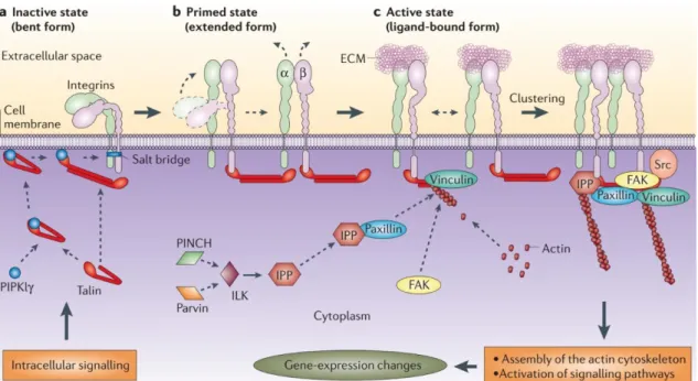
11 The head region includes the β-propeller domain and the thigh domain from the α-subunit, and the βA domain, the hybrid domain and the PSI domain from the β-subunit (Askari et al., 2009; Fig. 3). The leg region comprises the calf1 and calf2 domains from the α-subunit, and the EGF (Epidermal growth factor) domains and the β-tail domain from the β-subunit (Askari et al., 2009; Fig. 3). The intracellular β-tail domain of the β-subunit interacts directly and indirectly with several proteins which mediate the integrin-cytoskeleton linkage. These include talin, α-actinin, kindlin, FAK (focal adhesion kinase), vinculin and the IPP complex formed by ILK (integrin-linked kinase), PINCH (particularly interesting Cys-His-rich protein) and parvin proteins (Legate et al., 2006; Legate et al., 2009).
Figure 3- Integrin structure. Schematic representation of the integrin head and leg regions. The head region is formed by the α-subunit β-propeller and the thigh domains, and the βA, hybrid and PSI domains which are part of the β-subunit. The leg region includes the α-subunit calf1 and calf2 domains, and the EGF and β-tail domains of the the β subunit. Adapted from Askari et al., 2009.
12
Integrins are thought to adopt three major conformations which correlate with certain activation states: “inactive” of low affinity, “primed” or “active” of high affinity, and ligand occupied (Askari et al., 2009; Legate et al., 2009). The requirement of the physical extension of the integrin for its activation is still a matter of debate, but it is generally accepted that integrin activation requires the separation of the α and β cytoplasmic tails, which in turn allows the β-hybrid domain to move outwards. This rearrangement alters the βA domain conformation from a low affinity to a high affinity state (Mould et al., 2003; Rocco et al., 2008; Askari et al., 2009). Integrin activation is modulated bidirectionally through inside-out activation, which sets integrins into the “primed” or “active” high affinity state, and through outside-in activation, which is induced upon binding with their ligand and triggers the activation of multiple signaling pathways (Hynes, 2002; Askari et al., 2009; Legate et al., 2009; Fig. 4). Integrins are believed to be maintained in an inactive state, but integrin independent signals can trigger the
Figure 4- Integrin “inside-out” activation. Integrins can adopt different conformation/activation states: “inactive” of low affinity, “primed” or “active” of high affinity, and ligand occupied. Intracellular signaling is responsible for priming the integrins into a more extended form which allows their activation once the intracellular mediators of integrin activation such as vinculin, paxillin, the IPP complex and FAK form a complex that binds to integrins. After activation integrins bind to their ligands which induces outside-in signaling that can involve several signaling pathways. Figure from Legate et al., 2006.
13 recruitment of talin and kindlin to the β integrin cytoplasmic tails (Askari et al., 2009; Legate et al., 2009). Talin and kindlin mediate the separation of α and β cytoplasmic domains (Hynes, 2002; Askari et al., 2009; Legate et al., 2009). Talin also links directly to the actin cytoskeleton, but the binding of vinculin and α-actinin to the talin-actin complex is crucial to strengthen this interaction (Miyamoto et al., 1995; Gallant et al., 2005; Legate et al., 2009). Other molecules such as paxillin, FAK and Src, and the IPP complex, are also recruited to the integrin complex (Miyamoto et al., 1995; Geiger et al., 2001; Legate et al., 2006; Schwartz and DeSimone, 2008). The aggregation of all these proteins into an integrin binding complex then induces a conformational change in the integrin protein, setting it for an “active” high affinity state (Hynes, 2002; Askari et al., 2009; Legate et al., 2009; Fig. 4). The following clustering of integrins allows the exponential propagation of integrin “outside-in” activation upon the binding of the ligand. This activation is then translated into the activation of multiple signaling pathways, including that of MAPK (Mitogen-activated protein kinases), ERK (Extracellular signal–regulated kinases), Wnt (Wingless/integrated family members) and Akt associated signaling pathway, which are involved in several cellular processes such as migration, proliferation, survival and apoptosis (Hynes, 2002; Legate et al., 2006; Legate et al., 2009).
Another level of complexity in integrin signaling is provided by the cross-talk between integrins and paracrine growth factors where bidirectional signaling impacts cellular processes regulated by both signaling systems (Comoglio et al., 2003; Danen and Sonnenberg, 2003). For instance, integrin aggregation was shown to induce phosphorylation and activation of the receptor tyrosine kinases PDGFR (Platelet-derived growth factor receptor), EGFR (Epidermal growth factor receptor), and VEGFR (VEGF receptor) (Miyamoto et al., 1996; Moro et al., 1998), and to act synergistically with EGF to induce strong activation of ERK signaling (Chen et al., 1996; Renshaw et al., 1997). Additionally, the direct interaction between αvβ6 integrin and TGFβ (Transforming growth
14
factor β) was shown to induce FAK and paxillin phosphorylation in parallel with TGFβ1 activation in lung epithelial cells (Munger et al., 1999).
2.2.1.2. Integrins as mediators of interactions between laminins and the cytoskeleton
The major laminin binding integrins are the α3β1, α6β1, α7β1 and α6β4 integrins which present different ligand specificities and thus, different signaling outputs (Nishiuchi et al., 2006). For example, α6β1 integrin has the highest affinity for laminins 111, 332 and 511/521, while α7X1β1 isoform has high affinity for laminins 511/521 and α7X2β1 isoform displays high affinity for laminins 111 and 211/221 (Nishiuchi et al., 2006; von der Mark et al., 2007; Domogatskaya et al., 2012). Although these integrins bind to the α chain LG1-3 domain (Smirnov et al., 2002; Suzuki et al., 2005; Durbeej et al., 2010; Domogatskaya et al., 2012), β and γ chains also mediate laminin-integrin binding (Ido et al., 2008; Taniguchi et al., 2009).
2.2.2. Dystroglycan
The laminin receptor dystroglycan consists of an extracellular α subunit and a transmembrane β subunit which are non-covalently associated. α-dystroglycan is responsible for binding to the ligand, whereas the C-terminal domain of β-dystroglycan binds to the cysteine-rich domain of dystrophin, which is in turn linked to F-actin (Ervasti et al., 1993; Jung et al., 1995; Kobayashi and Campbell, 2012). The dystroglycan receptor is the product of the DAG1 gene, which is transcribed and translated into an αβ pro-peptide, then cleaved post-translationally into the extracellular α subunit and the transmembrane β subunit (Ibraghimov-Beskrovnaya et al., 1992; Holt et al., 2000; Domogatskaya et al., 2012; Holmberg and Durbeej, 2013).
15 Laminin binding requires extra post-translational modifications within the mucin domain of α-dystroglycan, which are employed by the glycosyltransferase LARGE inducing glycosylation of the O-mannosyl core (Kanagawa et al., 2004; Yoshida-Moriguichi et al., 2010; Kobayashi and Campbell, 2012). Other post-translation modifications are performed by POMT1 (Protein O-Mannosyltransferase 1), POMT2 (Protein O-Mannosyltransferase 2), POMGnT1 (Protein O-Linked Mannose N-Acetylglucosaminyltransferase 1), Fukutin, and FKRP (Fukutin-related protein) glycotransferases (Kobayashi and Campbell, 2012). Compared to integrins, α-dystroglycan displays a narrower binding spectrum and presents high affinity only for laminin α1 and α2 chains (Durbeej, 2010). As mentioned previously, the laminin LG4-5 domains are the α-dystroglycan binding domains by excellence, but the α2 chain contains additional binding sites in the LG1-3 domains (Timpl et al., 2000; Suzuki et al., 2005; Durbeej, 2010; Holmberg and Durbeej, 2013).
Dystroglycan is part of the dystrophin-associated glycoprotein complex (DGC) which also includes the sarcoglycans (SG-α, β, δ and γ), the dystrobrevins, sarcospan, and the syntrophins (Allikian and McNally, 2007; Townsend, 2014; Fig.5). In addition,
β-Figure 5- Dystrophin associated glycoprotein complex structure. The DGC is
composed of α and β dystroglycan subunits, the sarcoglycan complex(SG-α, β, δ and γ), the dystrobrevins, sarcospan, and the syntrophins. Figure from Kobayashi and Campbell, 2012.
16
dystroglycan associates with the utrophin-associated glycoprotein complex (UGC) where utrophin replaces dystrophin in the linkage to the actin cytoskeleton (Rafael and Brown, 2000). Together with α7β1, the DGC and the UGC are the major laminin receptor complexes in adult skeletal muscle (Ervasti et al., 1993; Burkin and Kaufman, 1999; Rafael and Brown, 2000).
3. Cardiac development
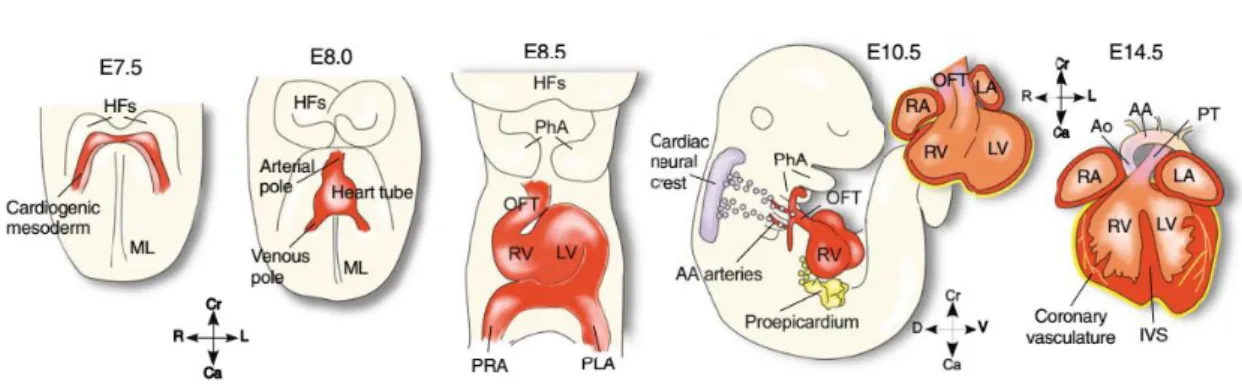
3.1. Specification of first and second heart field progenitors
The heart is the first organ to be fully functional during embryogenesis. The presumptive myocardial cells are generated early during development, from the epiblast cells that ingress into the primitive streak to form the mesoderm (Kirby and Waldo, 2007). Cardiac development starts around E7.5 (embryonic day 7.5), when the cardiac progenitors, specified by the transcription factors Nkx2.5 and Mesp (Mesoderm posterior) 1, enter the cardiac myogenic program in the primitive streak and move antero-laterally to constitute two cardiogenic plates or cardiogenic fields, named first and second heart fields (Saga et al., 1999; Yang et al., 2002; Kirby and Waldo, 2007; Wu et al., 2008; Savolainen et al., 2009; Vincent and Buckingham, 2010; Kelly et al., 2014). They can be distinguished by Myosin Light Chain 2a, Tbx5 (T-box 5) and Hcn4 expression in the first heart field, and Islet1, Tbx10, Fgf8 (Fibroblast growth factor) and Fgf10 in the second heart field (Cai et al., 2003; Meilhac et al., 2004; Laugwitz et al., 2008; Vincent and Buckingham, 2010; Kelly et al., 2014; Meilhac et al., 2014). The first heart field progenitors are the first to migrate along the endoderm to form the heart tube (Kirby and Waldo, 2007; Kelly et al., 2014; Meilhac et al., 2014; Fig. 6). Afterwards, the second heart field progenitors migrate along the pharyngeal endoderm to the anterior and posterior poles of the heart tube (Vincent and Buckingham, 2010; Kelly et al., 2014; Meilhac et al., 2014; Fig. 6). Second
17 heart field progenitors are
maintained in a
proliferative state in the cardiogenic field, and differentiate once they enter the heart tube (Vincent and Buckingham, 2010). The activation of the cardiomyogenic program is mediated by the activation of Gata4, Mefc2 and Tbx5, in response to signals such as Shh (Sonic hedgehog)
and BMP (Bone
morphogenetic protein) 2/Fgf 4 produced by the
endoderm (Kirby and Waldo, 2007; Vincent and Buckingham, 2010). While the first heart field progenitors are the source of cells constituting the left ventricle, the second heart field progenitors will originate the atria, outflow tract, right ventricle and inflow region (Kirby and Waldo, 2007; Vincent and Buckingham, 2010; Fig. 6). These cardiogenic fields provide the progenitors for cardiomyocytes, endocardium cells and the conduction system, also termed conduction fibers or Purkinje fibers (Kirby and Waldo, 2007; Vincent and Buckingham, 2010; Meilhac et al., 2014). The heart is also colonized by neural crest cells which contribute to the outflow tract and septa that separates aorta from the pulmonary trunk (Waldo et al., 2005; Hutson and Kirby, 2007; Vincent and Buckingham, 2010).
Figure 6- First and second heart field progenitor contribution to heart tube formation. First heart field progenitors form the heart tube which subsequently grows with the contribution of second heart field progenitors. The heart tube forms most of the future left ventricle, whereas the second heart field progenitors contribute mostly to the formation of outflow tract, left and right atria and right ventricle. Adapted from Laugwitz et al., 2008.
18
3.2. Heart tube formation and heart looping
The beating heart tube is formed by E8.0 (Kirby and Waldo, 2007; Savolainen et al., 2009), when the cardiogenic fields formed bilaterally are brought to the midline with the inward movement of the splanchnic mesoderm to form a double layered heart tube (Kirby and Waldo, 2007; Savolainen et al., 2009; Vincent and Buckingham, 2010; Kelly et al., 2014; Fig. 6 and 7). The myocardium is composed of cardiomyocytes, which are responsible for the contraction capability of the heart tube (Kirby and Waldo, 2007; Meilhac et al., 2014). The inner layer, the endocardium, is composed of endothelial cells that cover the interior of the myocardial heart tube and early in development, these endocardial cells are discriminated from the myocardium by the synthesis of TAL1 (T-Cell Acute Lymphocytic Leukemia 1), Flk1 (Fetal kinase liver 1), PECAM
(Platelet endothelial cell adhesion molecule), CD34 and VE-cadherin (Drake and Fleming, 2000). In the interface between the endocardium and the myocardium is the cardiac jelly, a layer of ECM secreted by the myocardium (Kirby and Waldo, 2007; Savolainen et al., 2009). The cardiac jelly is digested by the metalloproteinase ADAMTS1 between E12.5 and E14.5 (Stankunas et al., 2008; Cooley et al., 2012; Paige et al., 2015).
The heart changes its linear anterior-posterior conformation into the right-left conformation between E8.5 and E10.5 through a progression of bending, rotation and
Figure 7- Heart tube formation. The heart tube is formed when the cardiogenic fields are brought to the midline with inward movement of the splanchnic mesoderm during the formation of the foregut pocket. Figure from Gilbert, 2013.
19 torsion processes, collectively called heart looping (Kirby and Waldo, 2007; Fig. 8). During these rearrangements, the heart tube changes its linear conformation into the “C-shape” and then into the final “S-shape” (Kirby and Waldo, 2007). The initial C-shape is achieved with bending and rightward rotation of the heart tube, and then its elongation and convergence of inflow and outflow poles. Finally, during the following S-shape looping, the aorta rotates and stays between the atrioventricular valves (Kirby and Waldo, 2007). Different intrinsic and extrinsic factors are involved in the heart looping. The rightward rotation and convergence of inflow and outflow rearrangements are dependent on asymmetric divisions which allow the left side to grow faster than the right side (Linask et al., 2005; Kirby and Waldo, 2007). The cardiomyocyte proliferation during this process is in part regulated by FAK (Doherty et al., 2010). Additionally, ECM assembly and modification by MMP2, highly expressed by the endocardium, are crucial for the proper looping of the heart (Linask et al., 2005).
From E9.0 until E11.0, the proepicardium derived from the septum transversum mesenchyme in the inflow pole, starts migrating to eventually cover the external surface of the heart to form the epicardium (Moore et al., 1999; Sengbusch et al., 2002; Kirby and Waldo, 2007; Zhou et al., 2008; Vincent and Buckingham, 2010). The proepicardial and
Figure 8- Cardiac development in the mouse. Cardiac development starts with the establishment of the heart fields and posterior formation and growth of the heart tube. Subsequent cardiac myogenesis and heart remodeling occurs with the heart looping, epicardium development, chamber specification and trabeculation, and development of the cardiac cushions and conduction system. Figure from Laugwitz et al., 2008.
20
epicardial cells are characterized by the expression of Nkx2.5, Islet-1, Wt1 (Wilms Tumor 1) and Tbx18 (Zhou et al., 2008; Vincent and Buckingham, 2010). Soon after the epicardium completely covers the outer myocardium, epicardial cells de-epithelialize and form a population of mesenchymal epicardium-derived cells (EPDCs). These cells invade the interior of the heart to originate myocardial fibroblasts and also the endothelial and smooth muscle cells of the coronary vasculature (Moore et al., 1999; Sengbusch et al., 2002; Kirby and Waldo, 2007; Zhou et al., 2008; Vincent and Buckingham, 2010). Strikingly, Wt1-positive epicardial cells also differentiate into cardiomyocytes during mid embryonic and fetal cardiac development (Zhou et al., 2008). The epicardium and the myocardium share a common subepicardial matrix in the interface between these two layers, which is composed of ECM molecules such as fibronectin, collagens, elastin, tenascin-X and laminins among other components (Wessels and Perez-Pomares, 2004).
3.3. Chamber specification and fetal myocardial growth
Around E10.5, the “secondary genetic program”, involving factors such as ANF (Atrial natriuretic factor), Chisel, Cxn43 and Irx5, is activated to promote the development and growth of the myocardium into four different heart chambers: left and right atria, and left and right ventricle (Christoffels et al., 2000; Fig. 6 and 8). The activation of the secondary program leads to the activation of the chamber specific program, where atria are determined by the expression of myosin light chain isoform Mlc2a, and ventricles are specified by myosin light chain isoform Mlc2v expression (Small and Krieg, 2004). During this phase, the differentiation of second heart field progenitors in the myocardium is ceased and cardiomyocyte proliferation becomes the main process contributing to myocardium growth (Sedmera and Thompson, 2011; Kelly et al., 2014). The morphological changes occurring along this period are mediated by myocardial growth in the outer curvature of the heart through ballooning morphogenesis (Christoffels et al., 2000; Kelly,
21 et al., 2014; Paige et al., 2015). In parallel, the development of septa, cardiac cushions and Purkinje fibers allows the proper coordination between cardiac contractions and blood flow through the different chambers (Kelly et al., 2014).
After the heart loops, the myocardial wall is remodeled into the highly proliferative compact zone and the trabeculae. The development of trabeculae, or trabeculation, is crucial to increase the myocardial surface and the oxygenation of the internal wall of the myocardium (Savolainen et al., 2009; Samsa et al., 2013; Paige et al., 2015). Different signals such as Notch and neuregulin-1 from the endocardium, retinoic acid and Fgf from the epicardium, and BMP10 expressed in the trabecular myocardium, have been shown to regulate ventricular trabeculation and myocardial proliferation (Chen et al., 2004; Martin-Puing et al., 2008; Meilhac et al., 2014; Paige et al., 2015; Fig. 9). During fetal stages, the compact zone is further expanded through the proliferation of cardiomyocytes, but these start losing proliferative capacity during postnatal maturation (Martin-Puig et al., 2008; Sedmera and Thompson, 2011).
Figure 9- Myocardium trabeculation. Molecular regulation of myocardium trabeculation in the ventricle. Figure from Paige et al., 2015.
22
3.4. Laminins during cardiac development
Several studies have highlighted the role of different ECM components during cardiac development and repair (Dobaczewski et al., 2010; Kirby and Waldo, 2007). Laminin matrix is known to be remodeled during cardiac infraction, even though the exact role laminins play during this process is still controversial (Yabluchanskiy et al., 2013). In homeostatic conditions, laminins are assembled around cardiomyocytes (Kim et al., 1999), and seem to play critical roles during cardiac development as Lama4 null mice and mutations in LAMA4 in human display cardiomyopathy (Wang et al., 2006; Knöll et al., 2007). However, the role of different laminin isoforms during cardiac development remains largely unknown. At gestation week 8 during human cardiac development, β1 and β2 containing laminins can be found in the cardiomyocyte matrix (Kim et al., 1999; Roediger et al., 2010). Interestingly, α6 and α7 integrins were found to be synthesized in specific regions of the myocardium during cardiac development. In a mouse embryo stage E8.5-E9.5, integrin α6 mRNA and protein are enriched in the atrial chamber compared to the ventricle (Thorsteinsdóttir et al., 1995). During fetal stages, α6-integrins are produced by the atrial myocardium and compact myocardium of the ventricles, but is absent from the trabecular myocardium (Hierck et al., 1996). Conversely, integrin α7 mRNA (Itga7) is expressed in both atrial and ventricular myocardium at E13.0, but by E18, this mRNA becomes localized only at the base of trabeculae (Hierck et al., 1996). One possible outcome of the regionalized allocation of integrins is the assembly of different laminin isoforms in specific regions of the myocardium as well. Nevertheless, as noted above, information about laminin matrices in the cardiac muscle is still scarce and this hypothesis has not been formally tested.
23
4. From somite to skeletal muscle
4.1. Somite formation
Somites are transient embryonic segments which give rise to the axial skeleton, tendons, dermis of the back, axial vessels, brown fat cells and to all skeletal muscles of the body and the tongue (Dubrulle and Pourquié, 2004; Christ et al., 2007). They are spherical structures derived from the presomitic mesoderm (PSM), which lie on both sides of the neural plate and notochord. Somites are generated rhythmically from the anterior PSM and are composed of a layer of epitheloid cells surrounding a core of mesenchymal cells (Scaal and Christ, 2004; Martins et al., 2009). The rhythmic budding off of somites is thought to involve a posterior to anterior maturation process of the PSM cells and a molecular oscillator, the embryonic clock (Palmeirim et al., 1997; Bessho et al., 2003; Aulehla and Herrmann, 2004; Dubrulle and Pourquié, 2004; Andrade et al., 2007).
4.2. Somite derivatives
Somites undergo a process of maturation that leads to the formation of four different compartments known as sclerotome,
dermomyotome, myotome and
syndetome, which contain the cells responsible for the formation of the axial musculoskeletal system (Brent and Tabin, 2002; Fig. 10).
Figure 10- Somite derivatives. Scheme depicting the dermomyotome (dm; green), the myotome (m; red), the sclerotome (yellow) and the syndetome (blue). Adapted from Andrade et al., 2007.