University of the Algarve
Faculty of Sciences and Technology
Integrated multi-scale architecture of the
cortex with application to computer vision
Ph.D. Thesis in Electronic and Computer Engineering, Major in Computer Science
Jo˜
ao Miguel Fernandes Rodrigues
Supervisor: Johannes Martinus Hubertina du Buf, Faculty of Science and Technology, University of the Algarve, Portugal
Juri: President:
Jo˜ao Pinto Guerreiro, Reitor, University of the Algarve, Portugal Vogals:
Rolf Wuertz, Institut fur Neuroinformatik, University of Bochum, Germany
Gustavo Deco, Department of Technology, University Pompeu Fabra, Barcelona, Spain Aur´elio Campilho, Faculty of Engineering, University of Porto, Portugal
Alexandra Reis, Faculty of Human and Social Sciences, University of the Algarve, Portugal Johannes du Buf, Faculty of Science and Technology, University of the Algarve, Portugal Miguel Castelo-Branco, Faculty of Medicine, University of Coimbra, Portugal
Hamid Shahbazkia, Faculty of Science and Technology, University of the Algarve, Portugal Marina Gra¸ca, School of Education, University of the Algarve, Portugal
Faro (July, 2007)
University of the Algarve
Faculty of Sciences and Technology
Integrated multi-scale architecture of the
cortex with application to computer vision
Ph.D. Thesis in Electronic and Computer Engineering, Major in Computer Science
Jo˜
ao Miguel Fernandes Rodrigues
Faro (July, 2007)
I
Acknowledgments
First many thanks go to Prof. Hans du Buf, for directing my attention to the challenging but also inspiring field of human vision, for his excellent supervision based on his broad experience, for providing a stimulating and cheerful research environment in his Vision Laboratory, for his en-couraging support and his way to always find time for discussions, and last but not the least for the numerous hours that he spent in correcting my English.
Many thanks go also to my laboratory colleagues, to Roberto Lam who implemented Fig. 2.3, and to Samuel Nunes, Daniel Almeida, Vera Brito and Jo˜ao Carvalho for all the work that contributed to Chapter 7 and the related papers on painterly rendering.
To all the “inhabitants” of the laboratory, permanent or visiting, espe-cially those with whom I have worked with, almost on a daily basis, discussing scientific and technical problems, but also almost all problems in the world.
A special thanks to the Escola Superior de Tecnologia at UAlg and my colleagues at the Department of Electrical Engineering, for reducing my teaching duties during the last 3 years.
Some Portuguese words for my parents: “um obrigado especial para os meus pais, Fl´avia e Jos´e Rodrigues, pelo apoio, carinho e motiva¸c˜ao que sempre me deram ao longo de toda a minha vida acad´emica, sem os quais nunca teria sido poss´ıvel chegar at´e aqui.”
To my own family, first to my wife Celia Ramos, for all the love and support, encouragements and suggestions, and for the home environment that allowed me to work many times until late into the night, but also to my daughter Joana, who was born at about the same time that this work started, for all the affection, not forgetting the interesting talks about how she saw the world as a two- and three-year old child. Many of these talks were very inspiring and contributed to the ideas about object categorizations.
III
NOME: Jo˜ao Miguel Fernandes Rodrigues
FACULDADE: Faculdade de Ciˆencias e Tecnologia
ORIENTADOR: Professor Doutor Johannes Martinus Hubertina du Buf DATA: Julho de 2007
TITULO DA TESE: Arquitectura integrada multi-escala do c´ortex visual com aplica¸c˜oes na vis˜ao por computador
Resumo
O foco principal desta disserta¸c˜ao ´e compreender o desenvolvimento e a funcionalidade do c´ortex visual atrav´es de modelos computacionais. Na camada de entrada V1 do c´ortex visual, existem c´elulas simple, complex e end-stopped. Estas permitem uma representa¸c˜ao multi-escala de ob-jectos ou de cenas em termos de linhas, arestas e pontos-chave. Nesta disserta¸c˜ao, s˜ao combinados os progressos mais recentes no desenvolvi-mento de modelos computacionais destas e de outras c´elulas com os pro-cessos que decorrem em ´areas superiores do c´ortex V2, V4 etc. Trˆes desafios pertinentes s˜ao estudados: (i) o reconhecimento de objectos em-bebido numa arquitectura cortical; (ii) a percep¸c˜ao do brilho, e (iii) a renderiza¸c˜ao de pinturas usando a vis˜ao humana. Aspectos espec´ıficos s˜ao Foco-de-Aten¸c˜ao baseado em mapas de saliˆencia criados a partir de pontos-chave, o reencaminhamento dinˆamico de atributos a partir de V1 para ´areas superiores do c´ortex de forma a obter invariˆancia `a transla¸c˜ao, `
a rota¸c˜ao e ao tamanho, e a constru¸c˜ao de modelos can´onicos das vis-tas dos objectos na mem´oria visual. As nossas simula¸c˜oes mostram que as representa¸c˜oes multi-escala podem ser integradas numa arquitectura cortical, de forma a modelar os seguintes passos: segrega¸c˜ao, diferentes n´ıveis de categoriza¸c˜ao e o reconhecimento final de objectos. Relativa-mente ao processamento cortical real, o sistema come¸ca com a informa¸c˜ao das escalas grosseiras, refina a categoriza¸c˜ao usando escalas interm´edias, e utiliza todas as escalas para o reconhecimento. Tamb´em apresenta-mos um modelo de brilho em 2D, baseado na representa¸c˜ao simb´olica de linhas e arestas, combinado com um canal passa-baixo e com fun¸c˜oes de transferˆencia n˜ao lineares, de tal forma que o reconhecimento de objectos e a percep¸c˜ao de brilho s˜ao processos integrados e baseados na mesma informa¸c˜ao. O modelo de brilho consegue prever efeitos tais como ban-das Mach, a ilus˜ao Craik-O’Brien-Cornsweet e a indu¸c˜ao de gratings e de brilho, mais concretamente os efeitos opostos de assimila¸c˜ao (efeito White) e contraste simultˆaneo de brilho. Por fim, introduzimos uma nova aplica¸c˜ao: a renderiza¸c˜ao da pintura tem estado ligada `a vis˜ao computacional, mas n´os propomos a liga¸c˜ao desta com a vis˜ao humana, porque a percep¸c˜ao e a pintura s˜ao dois processos interligados.
PALAVRAS-CHAVE: C´ortex visual, Foco-de-Aten¸c˜ao, categoriza¸c˜ao, reconhecimento, brilho, renderiza¸c˜ao.
V
Integrated multi-scale architecture of the cortex with application to computer vision
Abstract
The main goal of this thesis is to try to understand the functioning of the visual cortex through the development of computational models. In the input layer V1 of the visual cortex there are simple, complex and end-stopped cells. These provide a multi-scale representation of objects and scene in terms of lines, edges and keypoints. In this thesis we combine recent progress concerning the development of computational models of these and other cells with processes in higher cortical areas V2 and V4 etc. Three pertinent challenges are discussed: (i) object recognition em-bedded in a cortical architecture; (ii) brightness perception, and (iii) painterly rendering based on human vision. Specific aspects are Focus-of-Attention by means of keypoint-based saliency maps, the dynamic routing of features from V1 through higher cortical areas in order to obtain translation, rotation and size invariance, and the construction of normalized object templates with canonical views in visual memory. Our simulations show that the multi-scale representations can be integrated into a cortical architecture in order to model subsequent processing steps: from segregation, via different categorization levels, until final object recognition is obtained. As for real cortical processing, the system starts with coarse-scale information, refines categorization by using medium-scale information, and employs all medium-scales in recognition. We also show that a 2D brightness model can be based on the multi-scale symbolic representation of lines and edges, with an additional low-pass channel and nonlinear amplitude transfer functions, such that object recognition and brightness perception are combined processes based on the same in-formation. The brightness model can predict many different effects such as Mach bands, grating induction, the Craik-O’Brien-Cornsweet illusion and brightness induction, i.e. the opposite effects of assimilation (White effect) and simultaneous brightness contrast. Finally, a novel application is introduced: painterly rendering has been linked to computer vision, but we propose to link it to human vision because perception and paint-ing are two processes which are strongly interwoven.
KEYWORDS: Visual cortex, Focus-of-Attention, categorization, recognition, brightness, rendering.
Contents
Acknowledgments I
Resumo III
Abstract V
1 Introduction 1
1.1 Scope of the thesis . . . 1
1.1.1 Object recognition . . . 3
1.1.2 Modeling brightness perception . . . 5
1.2 Overview of the thesis . . . 6
2 Overview: cortex, architecture and functionality 7 2.1 Introduction . . . 7
2.2 The visual cortex . . . 9
2.2.1 Cortical areas . . . 10
2.2.2 Cells: simple, complex, end-stopped and more . . . 11
2.2.3 Modeling simple cells by Gabor functions . . . 14
2.3 Invariance . . . 15
2.3.1 View-based approach . . . 16
2.4 Initial conclusions . . . 17
3 Multi-scale keypoints in V1 and beyond 19 3.1 Introduction . . . 19
3.2 Basic cell models and NCRF inhibition . . . 22
3.3 Keypoint detection with NCRF inhibition at fine scale . . . 24
3.4 Multi-scale keypoint representation . . . 26
3.5 Object segregation . . . 27
3.6 Automatic scale selection . . . 29
3.7 Focus-of-Attention by saliency maps . . . 30
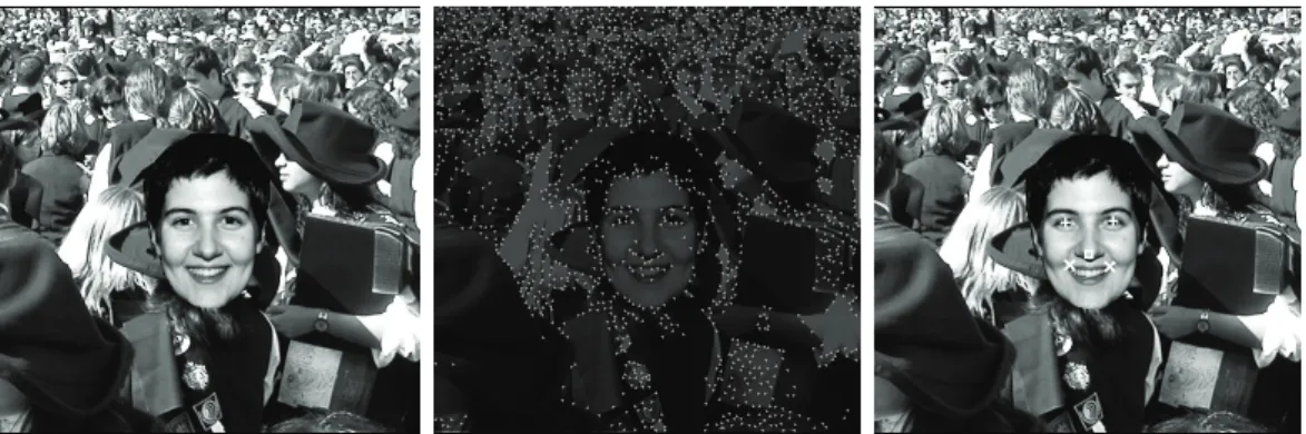
3.8 Application: face detection . . . 33
3.9 Discussion . . . 35
4 Multi-scale lines and edges in V1 and beyond 39 4.1 Introduction . . . 39
4.2 Line and edge detection and classification . . . 41
4.2.1 Multiple scales . . . 46
4.3 Visual reconstruction . . . 47
4.4 Object segregation . . . 50 VII
VIII
4.5 Automatic scale selection . . . 51
4.6 Object categorization . . . 53 4.6.1 Pre-categorization . . . 54 4.6.2 Categorization . . . 55 4.7 Face recognition . . . 56 4.8 Disparity estimation . . . 59 4.9 Discussion . . . 61 5 Integrated architecture 65 5.1 Introduction . . . 65 5.2 Recognition models . . . 67
5.3 Partial and global saliency maps and face recognition . . . 69
5.3.1 Results . . . 71
5.4 Invariant object recognition . . . 74
5.4.1 The creation of group templates . . . 77
5.4.2 Results . . . 79
5.5 Integrating the architecture . . . 81
5.6 Discussion . . . 84
6 Modeling brightness perception using line and edge representations 87 6.1 Introduction . . . 87
6.2 Brightness model . . . 89
6.2.1 The blocks of the model . . . 92
6.2.2 Model calibration . . . 94
6.3 Experiments . . . 98
6.3.1 Mach bands . . . 98
6.3.2 Brightness induction . . . 101
6.3.3 Craik-O’Brien-Cornsweet . . . 106
6.3.4 Other patterns and effects . . . 108
6.4 General discussion . . . 110
7 Application: painterly rendering using human vision 113 7.1 Introduction . . . 113
7.2 From perception to rendering . . . 114
7.2.1 Lines, edges and brightness . . . 115
7.2.2 Keypoints, saliency and FoA . . . 117
7.3 Color constancy . . . 118 7.4 Painterly rendering . . . 118 7.5 Discussion . . . 121 8 Concluding remarks 127 8.1 Summary . . . 127 8.2 Achievements . . . 128
8.3 Directions for further research . . . 129
Appendix 130
IX
Chapter 1
Introduction
Abstract: This chapter introduces the scope of the thesis as well as the two major problems that will be studied, namely object recognition and brightness perception.
1.1
Scope of the thesis
Imagine: you are going to see a movie with your daughter Joana, you are in the line in front of the entrance of the theater talking to one of your friends, and she enters the room first taking with her the tickets with the seat numbers. When you enter you don’t know where she is. A small embedded system in your coat connected to a few button-sized cameras tells you “Joana is third to the right,” “partly occluded by blond woman.” When you start walking towards her, the light is dimmed and the system alerts you “attention handbag on floor,” “attention cane between seats,” “attention popcorn bag on seat.”
From an engineering point of view, you will think that the implementation of such a system involves methods from Computer Vision (CV). When you try to join all the pieces, you find that even state-of-the-art CV methods, which are very good at solving restricted problems like object detection (floor, seats), categorization (face, handbag, cane) and iden-tification (daughter Joana), are not able to categorize all types of objects in complex scenes nor recognize individual objects like faces when partly occluded, especially with additional complications like different illuminations and viewpoints etc. Just imagine for instance the same scene as above but at an airport lounge or in a disco. Not surprisingly, such a general and flexible system still belongs to science fiction. Nevertheless, we know very well one sys-tem that can cope with all such complications—our visual syssys-tem. So, HV (Human Vision) will provide a solution, or CV based on HV. There’s only one small problem left: we need to know first how HV works.
When analyzing the performance of contemporary systems based on HV—often referred to as biologically inspired systems—one must conclude that they fall behind CV systems, despite some very promising results. This means that “science fictional” systems based on HV have a long road to go, but we are certain that they will work at the end, simply because we see them working every day. In addition, all information exchanged between vision researchers, neurophysiologists, psychologists and engineers may also help to treat
2
vision deficiencies and diseases, and to create new experiments to better understand how our brain works.
There are many reasons for creating an HV model, but they can be summarized as follows: 1. The human visual system is the best vision system “on the market,” not claiming that other systems of birds or mammals or other primates are inferior; we know what we see and we can only guess what a chimp sees. (After reading the following chapters of this thesis the reader should understand that the last part of the last sentence should read “we think we know what we see”).
2. We believe that enough computational ideas and experimental data are now available. On the basis of these it is possible to begin the development of an integrated theory of the ventral and dorsal data streams in the brain, focusing on an explanation of visual object recognition. This theory as a whole or parts of it may be incorrect, but at least it represents a skeletal set of claims and ideas which can be tested, confirmed or rejected, and an integrated architecture will be modular such that parts can be replaced or improved.
3. With ever increasing performance of modern computers (Moore’s Law) we are going to have the necessary power to create realistic models. To give an idea: using two graphics boards with GPUs optimized for vectorized multiplication and accumulation (multiply-add or MADD) operations, one can obtain a performance of 1 TFLOPS on a normal personal computer. 1 TFLOPS means 1012 or one million million of floating
point operations per second. Our entire brain counts 1012neurons. This does not mean that it will be possible to create a dynamic model of the entire brain at intervals of one second, because most neurons have between 100 and 1000 interconnections. Apart from this limitation, the real bottleneck is still storage capacity, both memory and disk space (and disk access time).
Of course, instead of modeling the entire brain or “only” the entire visual system we intend to focus on a few cortical areas and a relatively small group of cells in the early layers, because (1) the HV system is far too complex to try to model everything in a single step (this even applies to the biggest research groups), (2) cortical areas V1 and V2 etc. are the major processing areas, (3) they are the best known and investigated areas, and (4) like a house has to be constructed starting with its foundations, a cortical model/architecture has to be started from the best known and lowest layers, and with time more cells and layers can be added.
Despite the above arguments, many questions remain open. For instance: how is the in-formation really processed in the cortex? Is it only done bottom-up, also called data-driven? Or are there also top-down feedback loops, and if so, only a few or many? Between which layers and/or which areas? What do they serve for? Even the interconnections between neurons are not well known, i.e., there is no consensus between the principal groups working in this field about a neural architecture that could unify all processing steps into a single structure. For example, at the lowest level one can ask how and when each cell is activated in each layer, which cells combine within one single layer, and which cells activate the next layer. At the highest level the same questions are related to how object representations are stored in visual memory, to when and how scene and object recognition start, etc.
Since there are so many open questions, we are convinced that many groups will be working on cortical models in the future, each time trying to take the knowledge one step
3
further, but progress will be slow because right now only few groups are developing cortical cell models, less groups are developing an integrated cortical architecture, and even fewer groups are combining object recognition with other aspects like brightness perception. That there are not more groups developing an integrated architecture has two main reasons: it is a very interdisciplinary field and it does not return “excellent” results very fast (excellent in terms of data suitable for publications in a “publish or perish” academic society).
The research groups working on cortical models or on HV are all looking for the Holy Grail: an approach, probably multi-scale, that can yield a complete characterization of an image. There are many practical applications that can benefit from advanced cortical mod-els: object and face recognition, texture analysis, image segmentation, motion and depth prediction, and image enhancement and coding. In addition, a good two-dimensional bright-ness model can replace human observers, for example in estimating image quality in coding, by comparing the (subjective) brightness of a coded-decoded image with the (subjective) brightness of the original image. There also are other scientific and technological areas where HV-related knowledge can be applied, from engineering (better ways to recognize per-sons) to medicine (how to treat some illnesses), from education (more efficient ways to teach the brain) to arts (new ways to study paintings and painters).
Specifically, the main focus of this thesis is on the visual cortex, exploring a possible integrated architecture, but always having in mind practical applications. Main topics are:
• the development of computational cell models on the basis of cortical simple, complex
and end-stopped cells for the explicit extraction of lines, edges and keypoints,
• to incorporate these models into a multi-scale approach,
• to extract the most accurate and reliable information by coarse-to-fine scale processing, • to study multi-scale approaches for object categorization and recognition,
• to complement multi-scale image representations for object recognition with brightness
perception, and
• to propose an integrated model or architecture of the cortex, relating features with
cells, cell layers and with the information pathways of the visual system.
In the next sections, the modeling of object recognition and brightness perception are dis-cussed in more detail and the structure of the thesis will be presented.
1.1.1
Object recognition
Object recognition is a classical problem which is addressed in any book on computer vision, image processing and machine vision. It can be loosely defined by determining whether or not an image (or video) contains one or several specific objects, features or activities. Despite this very generic definition, it is quite difficult to define the term recognition in the context of HV, because each author applies his own definition, and it even may change within one publication. For instance, we can refer to recognition as to recognize one or several pre-specified or learned objects or object classes (e.g. this is a coffee mug), it may be the identification of a specific object (this is Paul’s coffee mug), or even detection (there are two coffee mugs in this image). It must follow from the context what is really meant, detection, categorization or identification. The same applies to this thesis.
4
Computationally, recognition is one of the most difficult tasks, but the difficulty depends on the task: it is quite easy for an average computer-vision student to detect and recognize objects after a very few lessons, i.e., if we put a few and very distinct objects on the top of a white table with good illumination. But if we put more and less distinct objects, some objects partly occluding other objects, and cover the table with a cloth with some complex texture, the task becomes more difficult, recognition performance decreases, and much more effort will be required to boost performance to an acceptable level.
Most real-world applications are not trivial, even the ones that appear relatively easy. For instance, counting how many people there are waiting on a sidewalk to automate the control of a zebra crossing, or reading number plates of cars passing a toll gate at 100 km/h, are already quite complicated. And then there are the very complicated applications, like recognizing a person after changing the hair style, after growing or shaving a beard, or after having grown old and gray. The extreme case is spotting in CCTV video, in real-time or in logged video, someone who does not want to be recognized, who therefore may use all disguisement tricks and even plastic surgery.
In neurosciences the concept of object recognition is even more difficult since it involves several levels of understanding, from the information processing or computational level to the level of circuits and cellular and biophysical mechanisms. After decades of research effort, neuroscientists working on functions in striate and extrastriate cortical areas have produced a huge and still rapidly increasing amount of data, and the emerging picture of how cortex performs object recognition is in fact becoming too complex for any simple model [Serre et al., 2005]. Recognition turns out to be a delicate compromise between selectivity and invariance. Therefore, the key computational issue in object recognition is the specificity-invariance trade-off: the system must be able to finely discriminate between different objects or object classes, while at the same time be tolerant to sometimes big object transformations which include scaling, translation and (2D) rotation, also changes of illumination, (3D) viewpoint, context and clutter, non-rigid transformations such as a change of facial expression and, in the case of categorization, also shape variations within a class [Serre et al., 2005].
Another problem that increases difficulty in modelling “biological recognition” is the def-inition of the instant when it all starts. Psychologists and psychophysicists, who study how we perceive patterns and images, used to think that, before the processes of object recogni-tion and categorizarecogni-tion could begin, the brain must first isolate a figure in the image—such as a tree or a piece of fruit—from its background (this process is called object segregation). However, recent research suggests that we actually categorize objects before we have segre-gated them, or that both processes occur in parallel. This means that by the time you realize that you are looking at something, your brain already knows what that thing is [Oliva and Torralba, 2006]. Such topics even relate to consciousness, which will not be stressed in this thesis.
Grill-Spector and Kanwisher [2005] tested three types of visual recognition by briefly flashing images before the eyes of human observers. The first type, object detection, was tested by showing images that may or may not have contained figures. Participants had to quickly judge whether or not there was a figure present against a background. The second type concerned categorization, where participants were shown images of figures and they had to indicate what type of figure they saw, such as bird, car, or food. In the third part of the test, more specific images were shown in order to test identification. Participants had to identify figures within categories, such as parrot or pigeon in the category “bird.” It turned out that the participants were as fast and accurate in naming the category that an object belonged to as they were at saying whether or not they had seen an object at all. The ability
5
of the subjects to process the images in such a short time proved that, by the time they knew an image contained some sort of object, they already knew its category.
Grill-Spector and Kanwisher [2005] concluded that “There are two main processing stages in object recognition: categorization and identification, with identification following catego-rization,” also “Overall, these findings provide important constraints for theories of object recognition,” and “Rapid categorization obviously facilitates our survival and interaction with the environment on an everyday level.” This built-in human process of rapid catego-rization before identification restricts the brain’s search for a match between the visual input (the picture you looked at) and internal category-relevant representations (stored images of other objects you have seen and identified prior to today).
From these conclusions it follows that recognition/identification should not be studied or modeled as a single-level task, but as a multi-level task where one or several levels of categorization should be performed. In addition, categorization should start at the same time as detection or segregation.
1.1.2
Modeling brightness perception
Visual psychophysics is a scientific area concerned with developing a complete understanding of how it works: from the physical input (the light flux entering the eye) to the output (the subjective image that we perceive). There are many aspects like brightness, contrast, color, shape, shading and texture [du Buf, 2001]. One chapter of this thesis concentrates on brightness, i.e., the relation between (physical) luminance and (subjective) brightness of many spatial patterns. The goal is the construction of a generally applicable brightness model, which can predict most if not all known brightness effects.
Developing brightness models is perhaps one of the most difficult aspects of quantitative visual psychophysics, and this subject has not been very popular in vision research [du Buf and Fischer, 1995]. It requires knowledge about published data and experiments, as there is no standardized database that joins all available experimental results, also knowledge about signal and image processing, a lot of programming and, again, really fast computers.
du Buf [2001] proposed that semantic processing, wherever it may be done, obtains input from lower-level syntactical processing layers, probably providing a multi-scale line, edge and vertex representation. This actually is the same representation that will be exploited in this thesis for object recognition. In other words, object recognition and brightness perception are related processes which can be integrated: seeing an object implies seeing its brightness pattern but also knowing what it is. This, again, relates to consciousness: we open our eyes and we see the world around us, we become conscious of the world and our position in it. Here there are four main observations: (1) the entire idea is based on the fact that simple cells do not allow to discriminate between lines and ramp edges, which explains the appearence of Mach bands at ramp edges (see Chapter 6), (2) brightness perception being related to multi-scale detection and processing of lines and edges in area V1 and beyond, higher-level cognitive effects such as change blindness imply that brightness is based on low-and high-level processing, (3) the previous point implies that consciousness too is a holistic process which may involve the entire brain, and (4) the image that we perceive is not a straightforward reconstruction because we think in terms of semantics, where objects are not represented any more by some sort of stored “pictograms” but in the form of functional descriptors. Some of these points lead to a nice paradox: opening our eyes is like switching on a TV set, but where are the electrons and the phosphor atoms?
6
been published recently, either as computational models or as theoretical explanations (see e.g. [du Buf and Fischer, 1995; Blakeslee et al., 2005; Logvinenko and Ross, 2005]). There are good reasons to pursue research in this field: (1) There are real applications in areas like image processing and computer graphics, such as image enhancement for pattern detection in medical imaging. (2) A model based on psychophysical data can be tested against these and model predictions, in particular inaccurate ones, lead to a better insight into the process of visual perception, i.e., feedback leading to additional psychophysical experiments concerning unclear aspects of spatial interactions in brightness perception. (3) A good brightness model can serve as the basis for codecs (coding/decoding) schemes with high compression rates be-cause those may be-cause image deformations that are more natural and therefore more difficult to perceive if compared to standard codecs based on straightforward subband decomposition and quantization schemes.
1.2
Overview of the thesis
Chapter 2 presents a small overview of the visual cortex, its architecture and functionality. It explains generically most known cells, the most significant visual areas and visual pathways. It finalizes by presenting some initial conclusions that will guide us towards developing an invariant object categorization and recognition architecture.
Chapter 3 introduces the multi-scale keypoint representation. It shows that this provides very important information for object and face detection. It also shows that saliency maps for Focus-of-Attention can be constructed on the basis of this representation, and that such maps can be employed for the detection of facial landmarks and faces.
Chapter 4 introduces the multi-scale line and edge representation. It illustrates visual re-construction, and how object segregation can be achieved with coarse-to-fine-scale groupings. A two-level object-categorization scenario is tested and also a multi-scale object-recognition model. A new disparity model based on the multi-scale line and edge coding is presented, such that depth from stereo can be attributed to lines and edges.
Chapter 5 extends the multi-scale representations into an integrated, invariant architec-ture with dynamic routing of object feaarchitec-tures throughout the cortex and the construction of normalized object and group templates.
Chapter 6 presents a two-dimensional brightness model. This model is calibrated using psychophysical data, and it is shown that it can predict many brightness effects such as Mach bands, White’s effect, simultaneous brightness contrast, grating induction and the Craik-O’Brien-Cornsweet illusion.
Chapter 7 presents a specific application: painterly rendering using human vision. Com-pletely automatic rendering is obtained by applying the multi-scale line and edge represen-tation that provides a very natural way to render broad and fine brush strokes, and the multi-scale keypoint representation serves to create saliency maps for Focus-of-Attention to render important structures (abstraction).
Final remarks and ideas for future research are presented in Chapter 8.
Parts of this thesis have already been published in journals and related work has also been presented at conferences. Chapters 3 and 7 were published in 2006 in BioSystems and Virtual, respectively. Chapter 5 has been submitted to Cognitive Processing. Chapters 4 and 6 are being prepared for submission to journals like BioSystems and Spatial Vision. Appendix A lists all publications.
Chapter 2
Overview: cortex, architecture and
functionality
Abstract: This chapter presents a brief overview of the biological aspects of vision with special focus on the visual cortex. The view-based approach of how object invariance can be achieved is discussed. This chapter is concluded with a brief summary of conclusions that will guide us towards developing an invariant object categorization and recognition architecture.
2.1
Introduction
Intuition tells us that the brain is complicated. The brain contains about 1012 (one million
million) cells, an astronomical number by any standard. In addition, a typical neuron receives information from hundreds to thousands of other neurons and in turn transmits information to the same number of neurons, so the total number of interconnections is between 1014
and 1015. But complexity is not only defined by these numbers, even more important is
the organization and functionality, aspects which are very hard to quantify [Hubel, 1995]. Hubel states that neurons are the basic structural components of the brain. A neuron is an individual cell, specialized by architectural features that enable fast changes in neighboring neurons. The brain is “just” an assembly of such cells, and while individual neurons do not see, reason or remember, the brain as a whole does.
A neuron, or nerve cell, consists of the cell body that has a globular shape and contains the nucleus, and from the cell body protrudes the output-signal transmitting nerve fiber called axon. Besides the axon, a number of other branching and tapering fibers are connected to the cell body, the dendrites. The entire cell, body, axon and dendrites, are enclosed by the cell membrane. The cell body and dendrites receive information from other cells, whereas the axon transmits information from the cell to other cells. Near the end an axon normally splits into many branches, whose terminal parts come very close to the cell bodies and/or dendrites of other cells. In these signal-transmission regions, called synapses, information is conveyed from one nerve cell (presynaptic) to the next (postsynaptic) one [Hubel, 1995].
8
Here we are interested in the visual pathways and brain regions involved in vision. The retina in an eye, which is considered part of the brain, is a thin laminar structure with several layers of cells, one of which containing the light-sensitive or photoreceptor cells, the rods and cones. The optic nerves of the two retinas pass through the optic chiasm, where about half of the fibers cross to the side of the brain opposite the eye of origin, left and right, and about half stays on the same side. From the chiasm the fibers lead to the lateral geniculate nucleus (LGN). The optic-nerve fibers have terminal synapses at cells in the LGN and axons of LGN cells terminate in the primary visual cortex, layers 4Cα and 4Cβ in area V1 [Hubel, 1995; Bruce et al., 2000]. In all these connections, from the retina via the LGN to the cortex, there are retinotopic projections. This means that the mapping of each structure to the next is systematic: as you move in the retina from one point to another, the corresponding points in the LGN and cortex also follow a continuous path. In other words, in retinotopic projections the neighborhood relations like left-right and up-down are preserved.
Another important concept is the receptive field (RF) of a neuron. The RF is defined by the spatial region at the retinal level in which the presence of a stimulus will affect the firing rate of that neuron. In the visual system, receptive fields are volumes in visual space. For example, the receptive field of a single photoreceptor is a cone-shaped volume comprising all the visual directions in which light will alter the response of that photoreceptor. In the case of binocular neurons in the visual cortex, it is necessary to specify the corresponding areas in both retinas. Although these can be mapped separately in each retina by shutting the one and then the other eye, the full influence on the neuron’s firing is revealed only when both eyes are open [Hubel, 1995].
Hubel and Wiesel in 1963 advanced the theory that receptive fields of cells at one level of the visual system are formed from input by cells at a lower level (see [Hubel, 1995]). In this way, small and simple receptive fields could be combined to form big and complex receptive fields. Theorists later elaborated that this simple, hierarchical processing structure can be influenced by feedback from higher levels. Receptive fields have been mapped from cells at all levels of the visual system: photoreceptors, retinal ganglion cells, and cells in the lateral geniculate nucleus, the visual cortex cells and even in extrastriate cortical areas.
Before presenting a description of the visual cortex, it is useful to introduce the concept of cortical plasticity (or neuroplasticity), which refers to changes that occur in the organization of the brain, in particular the changes in the location of specific information processing functions, as a result of the effects of learning and experience. A surprising consequence of plasticity is that a specific function can “move” from one location to another after repeated learning or even brain traumas. This phenomenon is complex and involves many levels of organization. To some extent the term itself has lost its explanatory value because almost any changes in brain activity can be attributed to some sort of “plasticity.” Cortical organization, especially for the sensory systems, is often described in terms of maps. For example, tactile information from the foot projects to one cortical site and information from the eyes (vision) projects to another site. As a result, the cortical representation of the body resembles a map, but this map is not “fixed” but rather plastic. Several groups began exploring the impacts of removing parts of the sensory inputs in the late 1970s. We now know that re-organization occurs at every level in the processing hierarchy in the cerebral cortex [Miikkulainen et al., 2005].
9
Figure 2.1: Left: the brain’s anatomical areas. Right: data flow in Deco and Rolls’ cortical architecture, adapted from Fig. 1 in Deco and Rolls [2005].
2.2
The visual cortex
In this thesis we concentrate on the architecture of the visual cortex. The primary visual (or striate) cortex or area V1 is a layer of cells which is 2 mm thick, with a surface area of a few tens of square centimeters. The other visual cortical areas (V2, V3, V4, MT) are not at the surface of the brain and are called extrastriate areas. A detailed diagram of the cortical areas and their connections in the macaque monkey can be found in Churchland and Sejnowski [1992] (pp. 22) and in Parasuraman [1998] (see pp. 309 for the color plates).
There are many visual pathways, but we concentrate on two called the ventral stream and the dorsal stream (ventral means belly and dorsal means back, common anatomical terms which also apply to the spinal chord and the forward bending brain). Both streams start at the level of retinal ganglion cells and continue through the LGN to V1. The ventral stream then goes via areas V2 and V4 to the inferior temporal cortex, IT (see Fig. 2.1). Based on physiological experiments in monkeys, IT has been postulated to play a central role in object recognition. IT cortex, in turn, is a major source of input to prefrontal cortex (PF), which is involved in linking perception to memory and action [Miller and Cohen, 2001]. The ventral stream, also called the “what” pathway, is associated with form recognition and object representation. It is also associated with storage in long-term memory. The dorsal stream goes from V1 via V2 and V3 to middle temporal area (MT) and to the inferior parietal lobule [Goodale and Milner, 1992]. The dorsal stream, also called the “where” pathway, is associated with motion, the representation of object locations, and control of the eyes and arms, especially when visual information is used to guide saccades. The dichotomy of the ventral/dorsal or what/where pathways (sometimes also referred to as the perception/action streams) was proposed (among others) by Goodale and Milner [1992] and is still being applied, but also disputed, by vision scientists and psychologists. It is probably an over-simplification of the real organization of the visual cortex.
Many neurons in the visual cortex only respond to a subset of stimuli within their re-ceptive field. This property is called tuning. In the earlier visual areas, neurons are tuned to simpler patterns. For example, a neuron in V1 may fire to any vertical stimulus in its re-ceptive field. In the highest visual areas, neurons are tuned to much more complex patterns. For example, in inferior temporal cortex (IT), a neuron may only fire when a certain face appears in its receptive field. Individual V1 neurons in primates and animals with binocular vision have ocular dominance, i.e., a preference for one of the two eyes.
proper-10
ties tend to cluster together in cortical columns, spatially arranged following two tuning properties: ocular dominance and orientation [Hubel, 1995]. However, this model cannot accommodate color, spatial frequency and many other features to which neurons can be tuned. As mentioned above, the transformation of the visual image from retina to V1 is referred as retinotopic mapping. The correspondence between a given location in V1 and in the subjective visual field in the external environment is very precise: even the retinal blind spots are mapped into V1. Evolutionary, this correspondence is very basic and found in most animals that possess a V1. In man and animals with a fovea in the retina, a large portion of V1 is mapped to the small, central part of the visual field, a phenomenon known as cortical magnification.
2.2.1
Cortical areas
As already mentioned, the first cortical area is V1; see [Olshausen and Field, 2005] for a detailed discussion of V1. Current consensus seems to be that V1 consists of tiled sets of spatiotemporally selective filters. Theoretically, these filters together can carry out neuronal processing of spatial frequency, orientation, motion, direction, speed and many other spa-tiotemporal features. Many experiments with V1 neurons have lead to this insight. Visual information relayed to V1 is not coded in terms of a spatial (or optical) intensity image, but rather as local contrast. As an example, in the case of an image which is half black and half white, the dividing edge between black and white has a strong local contrast and this edge is encoded, while few neurons may code the brightness information. As information is fur-ther relayed to subsequent visual areas, it is coded as increasingly non-local frequency/phase signals.
Area V2 is the second major area in the visual cortex. It receives direct input from V1 and sends output to V3, V4 and MT. It also sends feedback signals to V1. Functionally, V2 has many properties in common with V1. Cells are tuned to simple features such as orientation, spatial frequency and color [Hubel, 1995]. Responses of many V2 neurons are also modulated by more complex features, such as the orientation of illusory contours and whether a stimulus is part of the figure or the ground, at least at the level of local occlusions [Qiu and von der Heydt, 2005].
Area V3 is part of the dorsal stream, receiving inputs from V2 and primary cortex. It projects to the posterior parietal cortex. Properties of cells in V3 offer few clues as to its function. Most cells are selective to orientation, and many are also tuned to motion and to depth. Relatively few are color sensitive, for more details see [Gegenfurtner et al., 1997; Kaas and Lyon, 2001].
Area V4 has been identified in the extrastriate visual cortex of the macaque. It is still unknown what the human homologue of V4 is; this issue is currently the subject of much scrutiny. V4 is the third cortical area in the ventral stream and the first one that shows strong attentional modulation [Chelazzi et al., 2001]. It receives strong feedforward input from V2 and sends strong output to the posterior inferotemporal cortex (PIT). It also receives direct input from V1. In addition, it has weaker connections to MT and visual area DP (the dorsal prelunate gyrus). Like V1, V4 is tuned to orientation, spatial frequency and color. Unlike V1, it is tuned to object features of intermediate complexity, like simple geometric shapes, but simpler than IT, although no one has yet developed a full parametric description of the tuning space of V4. Although first known for their color selectivity, neurons in V4 are selective to a wide variety of forms and shapes, such as bars, gratings, angles, closed contour features, sparse noise, etc.; see e.g. [Pasupathy and Connor, 2001; Chelazzi et al.,
11
2001]). Area V4 is not tuned to complex objects such as faces, in contrast to areas in the inferotemporal cortex. V4 is also known to have receptive fields of intermediate sizes (larger than V1 and smaller than IT on average), and invariance to small translations.
Area MT (middle/medial temporal) is a region in the extrastriate cortex that appears to process complex motion stimuli. It contains many neurons which are selective to the motion of complex features like line ends and corners [Hubel, 1995; Bruce et al., 2000]. Much work has been carried out on MT as it appears to integrate local motion signals into the global motion of complex objects, but some research suggests that motion information is in fact already available at lower levels of the visual system such as V1. There is still much controversy over the exact computations carried out in area MT. An updated overview of the rich literature on MT was recently presented by Born and Bradley [2005].
Area IT (inferior temporal) is one of the highest levels of the ventral stream, with repre-sentations of visual shapes and objects. In Logothetis et al. [1995] monkeys were trained to recognize a set of novel “paperclip” objects and some neurons in anterior IT were found to be tuned to the trained views of those objects, but invariant to changes in size, translation and 3D rotation. These view-tuned neurons responded more strongly to scaled, translated and rotated (in depth) images of the preferred paperclip than to a large number of distractor paperclips, even though these objects had been previously presented with just one size, po-sition and viewpoint. A later study systematically looked at the effect of adding one or two distractor objects within the receptive field of an IT neuron [Zoccolan et al., 2005]. Most recorded neurons showed an average-like behavior. That is, the response to the cluttered condition, containing two or three objects, was close to the average of responses to the indi-vidual objects if presented alone. There are several potential explanations listed by Zoccolan et al. [2005]. One explanation is to assume a normalization stage by the overall activation of the entire IT cell population. This is quite feasible, since such a normalization would make learning easier for the next layer.
Area PF (prefrontal), which receives most input from IT, is involved in linking perception to memory and to action [Miller, 2000]. IT is also the last purely visual area which is task independent. Responses of PF cells are much more task dependent than responses of IT cells [Freedman et al., 2003]. Recent recordings [Freedman et al., 2002, 2003] revealed that neurons in PF are often “category-tuned,” conveying reliable information about category membership, learned in a supervised way, and relatively little information about individual stimuli within each category. By contrast, the majority of neurons in IT showed shape-tuning, i.e., they tended to show selectivity to individual stimuli (for example faces, see [Afraz et al., 2006]) and little evidence for selectivity to category membership.
2.2.2
Cells: simple, complex, end-stopped and more
Receptive fields of cells in the visual cortex are larger and more complex than retinal ganglion and LGN cells. Hubel and Wiesel first classified cells into three types: simple, complex and hypercomplex cells [Hubel, 1995].
Receptive fields of simple cells are elongated, for example with an excitatory central oval region and an inhibitory surrounding region, or approximately rectangular, with one long side being excitatory and the other being inhibitory. Cells with receptive fields with their long axis rotated to any angle have been found. Excitatory and inhibitory domains are always separated by a straight line or by two parallel lines. Some of the cells have an excitatory or an inhibitory region that is positioned exactly in the center of the receptive field, resulting in a symmetric receptive field; these are called even simple cell because of
12
Figure 2.2: 2D and 3D receptive-field representations of even (a) and odd (b) simple cells and complex (c) cells. Single (d) and double (e) end-stopped cells,
even symmetry, see Fig. 2.2a. Others, have an asymmetric receptive field profile, as the striped regions are positioned with a certain offset which respect to the center of the field: odd simple cells, see Fig. 2.2b. The size of the receptive field depends on its corresponding position in the retina relative to the fovea, but even at a given position in the retina there is some variation in size. In general, simple cells with smallest receptive fields are found in and near the fovea [Hubel, 1995]. Simple cells must be built up from preceding cells, probably from retinal ganglion cells with circular receptive fields.
Complex cells represent the next step in the analysis. Their receptive fields are also elon-gated but simpler than those of simple cells, because there are no sub-regions; see Fig. 2.2c. Complex cells are the most common cells in the primary cortex. Hubel [1995] guesses that they make up to 3/4 of the entire cell population. Complex cells share with simple cells the property that they respond only to specifically oriented structures. Like simple cells they respond to a limited region of the visual field, but unlike simple cells they cannot be explained by a neat subdivision of the receptive field into excitatory and inhibitory re-gions. Also, complex cells tend to have larger receptive fields than simple cells, but not much larger. For building complex cells on top of simple cells, Hubel [1995] proposed several possible schemes, one of them being that the activation of a complex cell requires successive activations of simple cells. The current mathematical model is rather simple and explained in Chapter 3).
Hypercomplex cells form the third category of striate cells initially identified by Hubel and Wiesel. These possess inhibitory zones at one or both ends of oriented excitatory regions, thereby responding to bars of preferred orientation only if they are not too long. Many of them respond more to the end of an oriented edge, i.e., if the edge does not extend beyond a
13
specific part of the receptive field. Such cells are therefore called end-stopped cells, and there are single and double end-stopped cells with receptive fields as shown in Fig. 2.2d and e. The fields are composed of a activation regions and regions at one or both ends called inhibitory regions. The simplest scheme for modeling such a cell consists of assuming excitation by one or a few complex cells with fields in the activation region in combination with inhibition by other complex cells with similarly oriented fields situated at the neighboring regions [Hubel, 1995]. The entire next chapter is devoted to end-stopped cells, improved models and applications.
Disparity-tuned cells have also been identified [Hubel, 1995]. These can account for a horizontal displacement, or disparity, which can be tolerated, the maximum displacement being a fraction of the width of the receptive field. Responses of such cells are a function of the distance of an object, which translates into the relative positions of a stimulus pattern in the two eyes. There is evidence that disparity-tuned cells exist in V1 of monkeys [Cumming and Parker, 2000]. The fact that many simple and complex cells are also tuned to disparity opens the possibility that, at a very early processing stage, depth is attributed to lines and edges. In other words, the visual system might use some sort of “wireframe” representation of 3D objects, like the ones used in the modeling of solid objects in computer graphics.
There are many other types of cells. For example, there are grating cells that were discovered in areas V1 and V2 of the monkey visual cortex by von der Heydt et al. [1992]. Such cells respond vigorously to grating patterns of appropriate orientation and periodicity, but very weakly or not at all to isolated bars. On the other hand, bar cells, which are found in the same areas of the visual cortex [von der Heydt et al., 1992], have a functional behavior which is less well explored and documented in the literature. In general, bar cells respond to single bars and their responses decrease when further bars are added in the form of a periodic pattern [Petkov and Kruizinga, 1997]. Computational models inspired by bar and grating cells were used in pattern recognition, for example in texture analysis, see e.g. [Kruizinga and Petkov, 1999; du Buf, 2007].
Figure 2.3 shows one scheme for visualizing activities of even and odd simple cells, com-plex cells, single and double end-stopped cells, plus a saliency map (for an explanation and all details see Chapter 3), using different colors with the saturation corresponding to a cell’s response strength. In addition, the dominant local orientation, which corresponds to the orientation of the complex cell with maximum amplitude, is coded by rotating the “color wheels.” The center-left panel in Fig. 2.3 shows the scheme in the case that the dominant local orientation is horizontal. The colored circle is subdivided into four quadrants, and one quadrant is further divided into two octants. The red and blue quadrants show responses of even (B) and odd (F) simple cells, the green quadrant (A) shows responses of complex cells. The line (D) separating the two pinkish octants shows the dominant orientation (here horizontal). The upper and lower octants show responses of single (C) and double (E) end-stopped cells. The black dot (G) in the center shows the information in the saliency map related to Focus-of-Attention based on end-stopped cells (see Section 3.7). The advantage of using color saturation is that complex cells (green) with very low activity are displayed as white; hence, areas with no green component do not contain significant lines and edges, and therefore also no keypoints. In contrast, responses of even and odd simple cells can be positive or negative (white indicates a large but negative amplitude). Finally, the colored circles can be superimposed on the input image in order to see (part of) the underlying image structure. Figure 2.3 shows a real example, Fiona image, at the finest scale (top-left) and at a coarse scale (top-right). The bottom-right image shows a zoomed version of the area around the pupil. Such images, if printed at bigger size, are aesthetically appealing, but
14
Figure 2.3: Color wheel visualization of cell responses. At the top a fine-scale (left) and a coarse-scale (right) representation of Fiona image. At bottom-right a zoomed area at the left eye.
more important is that we can analize the local image structure and we can see the responses of the cells in order to optimize the basic detection schemes of lines, edges and keypoints.
2.2.3
Modeling simple cells by Gabor functions
For simulating simple cells in a computational model we will use 2D Gabor functions as models of their receptive fields. Gabor functions or filters are also used in image processing and computer vision. The goal of this section is not to present in detail all the properties of
15
these functions, nor to explore all the existing computational models (for modelling simple cells). For this we refer to Chapter 2 of Peter Kruizinga’s PhD thesis [Kruizinga, 1999], where this matter is exhaustively studied and described. Here we only briefly expose the reasons to use these functions.
Different models can be used to model simple cells. Kruizinga [1999] compares several models—Difference-of-Gaussians, Difference-of-offset-Gaussians, Sum-of-offset-DoGs, Deriva-tives of Gaussians, Hermite polynomials and Gabor functions—using three criteria: (a) the ability of a model to cover the properties of different cells, each one having different pre-ferred orientations, spatial frequencies, phases and bandwidths; (b) the number of model parameters and their relations to the relevant proprieties of real cells; and (c) the biological plausibility of the scheme. Kruizinga states that, despite existing differences between the receptive field profiles of the models listed above, the differences are so subtle that no model can be rejected bacause of bad fits to neurophysiological data. Nevertheless he concludes that there are some differences between the models in their ability to model a large variety of types of simple cells, which is related to the number and nature of the parameters. He therefore chooses the Gabor model, because it is easier to relate parameters to essential receptive field proprieties.
Although 2D Gabor functions are now generally accepted as an appropriate model of receptive fields of simple cells, there still are some criticisms (see [Kruizinga, 1999]): the even-symmetric Gabor function has a non-zero DC response, or there are more than three side lobes, or there are too many parameters, but all this can be corrected or minimized. For the mathematical model see Chapter 3 of this thesis and see also [Lee, 1996; Kruizinga, 1999; Bruce et al., 2000; Grigorescu et al., 2003]. The mathematical models for complex and end-stopped cells are also presented in Chapter 3.
2.3
Invariance
Until here we have presented a brief overview of cortical biology involved, i.e., cells and the basic functionality of the visual areas, but our goal is to develop an integrated architecture for recognizing objects or persons, and this involves the identification of similar, yet distinct, objects as members of the same class.
In object recognition, one form of invariance requires a many-to-one mapping between individual exemplars and object categories. At the same time, individual exemplars of three-dimensional objects rarely appear in the same form one moment after the other. Variations in the two-dimensional images falling on our retinas arise from almost any change in viewing conditions, including changes of position, pose, lighting and object configuration. Therefore invariance also requires a many-to-one mapping between all individual “views” of objects and their unique identities [Tarr, 2005].
There are several approaches for establishing object representations which are suitable for obtaining invariance in the brain, although so far none has been considered to be the correct or final one. Here we will only focus on the most common, the view-based approach, because it is the one which will be explored in this thesis. For the “Recognition By Components” approach (RBC) we refer to e.g. [Bierderman, 1987; Tarr and B¨ulthoff, 1995], and for mental rotation to e.g. [Zacks et al., 2003; Hegarty and Waller, 2004].
16
2.3.1
View-based approach
The terms view and view-based encompass a variety of specific theories and computational models. However, all view-based models share a common, defining assumption: they all assume that objects are represented and matched to memory in terms of their features in a spatial reference frame [Hummel, 2000]. The central point of the view-based approach is that we represent objects in long-term memory as views, and that by means of operations on the coordinates of the features in those views we bring new views into register with stored views, or into register with stored 3D models. The basic idea is that we recognize objects on the basis of stored views, by matching images to the templates stored in memory. This is the most common theory in object recognition; for variations on the same theme see [Lowe, 2004; Peters, 2004; Tarr, 2005], and see [Peters, 2000] for a survey on theories of three-dimensional object perception.
Tarr [1995] emphasizes that experience with particular views is a critical factor in achiev-ing invariance. He found that when observers learn how to recognize novel objects from specific viewpoints, they are both faster and more accurate at recognizing the same objects from familiar viewpoints relative to unfamiliar viewpoints. Moreover, recognition perfor-mance in the case of unfamiliar viewpoints is systematically related to the views which are familiar: observers take progressively more time and are progressively less accurate when the distance between the unfamiliar and the familiar views increases. These and related results from Tarr and colleagues [Tarr et al., 1998] suggest that human object recognition relies on multiple views, where each view encodes the appearance of an object under specific viewing conditions, including viewpoint, pose, configuration and lighting, and that a collection of such views constitutes the mental representation of a given object.
In order to explain how view-based invariance can be achieved, Tarr [2005] refers to the work of Perrett, Oram and Ashbridge [Perrett et al., 1998], who found that individual object-selective neurons preferentially respond to particular object views. Invariance is then achieved by considering populations of such neurons as the actual neural code for objects. In this context, individual neurons may be considered as coding—from a familiar viewpoint— the complex features or parts of which objects are composed. Recognition then takes the form of “accumulation of evidence” across all neurons that are selective for some aspect of a given object. During recognition, the particular rate of accumulation will depend on the similarity between visible features/parts in the present viewpoint and the view-specific features/parts to which individual neurons are tuned [Perrett et al., 1998]. Across a popu-lation of object-selective neurons, sufficient neural evidence (summed activities of neurons) will accumulate more slowly when the current appearance of an object is dissimilar from all its learned appearances. Tarr [2005] himself concludes (assumes) from this that when an object’s appearance is close to previously-experienced views, evidence across the appropri-ate neural population must accumulappropri-ate more rapidly. Thus, systematic behavioral changes in recognition performance under changes of viewpoint may be explained as a consequence of how similarity is computed between new object percepts and previously learned neural representations.
Summarizing, Tarr [2005] concludes that recognition amounts to reaching a threshold of sufficient evidence in terms of activity across a neural population. One consequence of this is that unfamiliar views of objects will require more time to reach threshold, but will be successfully recognized given some similarity between input and known viewpoints. A second consequence is that unfamiliar exemplars within a familiar class will be likewise recognized given some similarity (similar configurations and viewpoints) with known exemplars from
17
within that class. One implication is that familiarity with individual objects should facilitate the viewpoint-dependent recognition of other, visually similar objects [Tarr and Gauthier, 1998]. A second implication is that object viewpoints or class exemplars that are significantly different from known views or objects should be represented as distinct representations; again, a prediction that seems to be supported.
2.4
Initial conclusions
From the relevant literature (see also Sections 3.1, 4.1 and 5.2) some further aspects may guide us toward developing an invariant object categorization and recognition architecture: (1) Extracted features play an important role in a biological model, both for characterizing the most significant aspects that are present and for abstraction of the scene [Heitger et al., 1992; Olshausen et al., 1993; van Deemter and du Buf, 2000; Corchs and Deco, 2005]. (2) The two major visual pathways consist of the dorsal “where” stream that runs from V1 via V2, V3 and MT to PP, and the ventral “what” stream that runs from V1 via V2 and V4 to IT [Goodale and Milner, 1992; Deco and Rolls, 2005]. (3) Two-dimensional Gabor functions are now generally accepted as an appropriate model of receptive fields of simple cells [Kruizinga, 1999; Grigorescu et al., 2003], and this model provides the basis to model other types of cells: complex and end-stopped cells, bar and grating cells, etc. [Heitger et al., 1992; Petkov and Kruizinga, 1997; du Buf, 2007]. (4) Object recognition is most probably a multi-level task which includes categorization and identification [Grill-Spector and Kanwisher, 2005], and it starts as soon as we have the “gist” of a scene [Rensink, 2000; Grill-Spector and Kanwisher, 2005; Oliva and Torralba, 2006]. (5) It is very likely that objects in memory are represented by templates in “view-based” form, i.e., one object must be represented by multiple, canonical views; e.g. [Tarr, 2005]. (6) There exist cortical top-down (and feedback) mechanisms which trigger and control visual attention, and which facilitate object recognition [Hupe et al., 2001; Bar et al., 2006; Oliva and Torralba, 2006]. (7) There is evidence for at least four, now generally accepted properties of the feedforward path of the ventral what stream [Serre et al., 2005]: (a) a hierarchical use of invariances, first to position and size (importantly, size and position invariance—over a restricted range—do not require learning specific for a given object), and then to viewpoint and other transformations (invariances to viewpoint, illumination etc. do require learning of several, different views of an object); (b) an increasing size of the receptive fields of cells coupled to an increasing complexity of their optimal stimuli throughout the cortical layers; (c) a basic feedforward processing of information for “immediate” recognition tasks; and (d) plasticity and learning, probably at all stages, but with a time scale that decreases from V1 to IT and PF cortex: fast adaptation to objects at high level and slower adaptation to local features at low level. More specific details and references concerning recognition models are given in Chapter 5 Section 5.2. In the following three chapters we will work toward an integration of features into a computational recognition scheme. Chapters 3 and 4 are about individual features, i.e., the multi-scale keypoint and line/edge representations, and for which purposes they can be exploited. In Chapter 5 we combine and test all the available information in the integrated architecture.
Chapter 3
Multi-scale keypoints in V1 and
beyond
Abstract: End-stopped cells in cortical area V1, which combine outputs of complex cells tuned to different orientations, serve to detect line and edge crossings, singulari-ties and points with large curvature. These cells can be used to construct retinotopic keypoint maps at different spatial scales (Level-of-Detail). The importance of the multi-scale keypoint representation is studied in this chapter. It is shown that this representation provides very important information for object recognition and face detection. Different grouping operators can be used for object segregation and au-tomatic scale selection. Saliency maps for Focus-of-Attention can be constructed. Such maps can be employed for face detection by grouping facial landmarks at eyes, nose and mouth. Although a face detector can be based on processing within area V1, it is argued that such an operator must be embedded into dorsal and ventral data streams, to and from higher cortical areas, for obtaining translation-, rotation-and scale-invariant detection.
3.1
Introduction
Our visual system can still be seen as a huge puzzle with a lot of missing pieces. Even in the first processing layers in area V1 of the visual cortex there remain many gaps, despite all knowledge already compiled [Hubel, 1995; Bruce et al., 2000; Rasche, 2005]. Nevertheless, some of the gaps are being filled by developing and studying computational models. Models of simple, complex and end-stopped cells have been developed more than ten years ago [Heitger et al., 1992]. Several inhibition models [Petkov et al., 1993b; Grigorescu et al., 2003], keypoint detection [Heitger et al., 1992; W¨urtz and Lourens, 2000; Rodrigues and du Buf, 2004b] and line/edge detection schemes [van Deemter and du Buf, 2000; Grigorescu et al., 2003; Elder and Sachs, 2004; Rodrigues and du Buf, 2004b], including disparity models [Fleet et al., 1991; Rodrigues and du Buf, 2004a], have become available. On the basis of such models and neural processing schemes, it is possible to create a cortical architecture for figure-ground segregation [Hupe et al., 2001; Rodrigues and du Buf, 2006a] and visual


![Figure 3.11: Left: partial saliency map of face196 (λ ∈ [13, 18]). Right: keypoints used by eye, nose and mouth detection cells.](https://thumb-eu.123doks.com/thumbv2/123dok_br/18723340.918928/48.892.193.719.116.365/figure-left-partial-saliency-right-keypoints-mouth-detection.webp)