DISSECTING THE THYMIC MICROENVIRONMENT
THE IMPACT OF DEFECTIVE T CELL DEVELOPMENT IN
THE MAINTENANCE OF THE CORTICAL THYMIC
EPITHELIUM
PEDRO MIGUEL MENDES RODRIGUES
Dissertação de Mestrado em Bioquímica
Universidade do Porto
Faculdade de Ciências
PEDRO MIGUEL MENDES RODRIGUES
DISSECTING THE THYMIC MICROENVIRONMENT
THE IMPACT OF DEFECTIVE T CELL DEVELOPMENT IN THE
MAINTENANCE OF THE CORTICAL THYMIC EPITHELIUM
Dissertação de Candidatura ao grau de
Mestre em Bioquímica da Universidade do
Porto:
Orientador – Doutor Nuno Lages Alves
Categoria – Investigador Auxiliar
Afiliação – Cell Activation and Gene
Expression Group, Instituto Biologia
"We must not wait for things to come, believing that they are decided by
irrescindable destiny. If we want it, we must do something about it"
Table of Contents
ABSTRACT ... IX
RESUMO ... X
LIST OF ABBREVIATIONS ... XI
LIST OF FIGURES ... XIII
INTRODUCTION ... 1
THE IMMUNE SYSTEM ... 3
THE THYMUS AS A TLYMPHOCYTE SCHOOL ... 4
THYMIC ORGANOGENESIS ... 5
THYMIC EPITHELIAL CELLS -THE EDUCATORS OF THYMOPOIESIS ... 7
HOW DOES THE TECMICROENVIRONMENT INSTRUCT TCELL DEVELOPMENT? ... 10
INTERLEUKIN-7:ACRITICAL THYMOPOIETIC CYTOKINE ... 13
TEC-THYMOCYTE CROSSTALK GOVERNS THE EPITHELIAL DEVELOPMENT ... 15
AIMS ... 18
MATERIAL AND METHODS ... 19
MICE ... 21
GENOTYPING ... 21
ISOLATION OF THYMIC STROMAL CELLS ... 22
FLOW CYTOMETRIC ANALYSIS ... 22
GENE EXPRESSION ... 23
FETAL THYMIC ORGAN CULTURE ... 23
IMMUNOHISTOLOGICAL ANALYSIS ... 24
IN VIVO ANTI-CD3εTREATMENT ... 24
STATISTICAL ANALYSIS ... 24
RESULTS ... 25
APRELUDE ON THE ONTOGENY OF IL7HI/YFP+TECS ... 27
THYMOCYTE-DERIVED SIGNALS MODULATE THE HOMEOSTASIS OF IL7HI/YFP+TEC S ... 30
THE PROGRESSIVE THYMOCYTE-INDUCED DECLINE OF IL7HI/YFP+ TEC S IS INDEPENDENT FROM THE MATURATION OF MTECS ... 33
DISCUSSION AND FINAL REMARKS ... 41
REFERENCES ... 49
SUPPLEMENTAL INFORMATION ... 61
ACKNOWLEDGMENTS ... 69
Abstract
Thymic epithelial cells (TECs) are master regulators of T cell development. The differentiation of TECs into functionally distinct cortical (cTECs) and medullary (mTECs) subpopulations relies on reciprocal instructive signals from developing thymocytes, a bi-directional interaction known as "thymic-crosstalk". Both TEC subsets derive from a common bipotent progenitor and differentiate through compartment-specific progenitors. Albeit the cellular and molecular mechanisms regulating mTEC development have been partially clarified, little is known about the signals that control cTEC differentiation. Exploiting IL-7 reporter mice, in which yellow fluorescent protein (YFP) expression identifies cells that co-express high levels of Il7 (IL7hi/YFP+ TECs), we previously showed that IL7hi/YFP+ TECs gradually
diminish with age in a thymocyte-dependent manner, segregate from mTECs and are conversely sustained when T cell development is profoundly abrogated at very early stages of differentiation.
In this thesis, we study the lineage relationship between IL7hi/YFP+ TECs and
other well-characterized TEC subtypes and the impact of distinct molecular interactions between thymocytes and TECs in the differentiation of the thymic epithelium. We demonstrate that IL7hi/YFP+ TECs emerge early during thymic
ontogeny, define a CD205+BP1+CD40lo cTEC subset that gradually decline during
thymic development, co-express the cortical-associated thymopoietic factors Ccl25, Dll4, Il7, Psmb11 and segregate from mature mTECs expressing Ctts,
Tnfrsf11a and Aire. Furthermore, we show that the decline in IL7hi/YFP+ TECs
induced by lympho-stromal interactions dissociates from the RANK- and LTβR-mediated maturation of medullary epithelium, suggesting that IL7hi/YFP+ TECs do
not represent a direct progeny of mTECs. Studies in Rag2-/- IL-7 reporter mice,
displaying a block at the DN3 stage of T cell development, indicate that IL7hi/YFP+
TECs are prominently maintained during fetal and postnatal life, suggesting that their homeostasis is regulated by thymocyte-derived signals beyond β-selection.
In vivo analysis of anti-CD3-treated Rag2-/- mice show that induction of DP
thymocytes devoid of TCR expression moderately affect IL7hi/YFP+ TECs while
fostering mTEC differentiation.
Together, our results indicate that IL7hi/YFP+ TECs are a determinant of the
cortical lineage and provide evidence that the homeostasis of the cortical epithelium is controlled by TCR-MHC interactions between thymocytes and TECs
Resumo
As células epiteliais do timo (TECs) são essenciais para o desenvolvimento das células T. A diferenciação das TECs em subpopulações corticais (cTECs) e medulares (mTECs), depende de sinais de maturação fornecidos pelos timócitos, uma interação bidirecional denominada “thymic crosstalk”. Ambas as subpopulações de TECs derivam de um precursor comum bipotente e diferenciam-se através de progenitores específicos. Enquanto os mecanismos que regulam o desenvolvimento das mTECs estão parcialmente clarificados, aqueles envolvidos na formação das cTECs não foram ainda esclarecidos. Anteriormente, utilizando ratinhos transgénicos que continha uma proteína repórter fluorescente (YFP) para detetar células com elevada expressão de Il7 (IL7hi/YFP+ TECs), foi
demonstrado que as IL7hi/YFP+ TECs diminuem gradualmente com a idade,
divergem das mTECs e persistem ao longo do tempo quando o desenvolvimento das células T é comprometido.
No presente trabalho pretende-se explorar a relação entre IL7hi/YFP+ TECs e
outros subtipos de TECs, assim como compreender qual o impacto "thymic-crosstalk" no desenvolvimento do epitélio tímico. Os nossos resultados demonstram que as IL7hi/YFP+ TECs surgem cedo durante a ontogenia tímica,
definem um subgrupo de CD205+BP1+CD40lo cTECs que diminuem gradualmente
com a idade, co-expressam fatores timopoiéticos associados com o cortex, e divergem das mTECs maduras que expressam Ctts, Tnfrsf11a and Aire. Mostramos ainda que o declínio das IL7hi/YFP+ TECs, induzido por interações
linfo-estromais, dissocia-se da maturação das mTECs mediada pelos receptores RANK e LTβR, sugerindo que as IL7hi/YFP+ TECs não constituem uma linhagem direta das
mTECs. Estudos com ratinhos transgénicos num fundo Rag2-/-, no qual o
desenvolvimento das células T é bloqueado no estadio DN3, demonstram que as IL7hi/YFP+ TECs persistem durante o desenvolvimento fetal e pós-natal, indicando
que a homeostasia destas células é regulada por sinais derivados dos timócitos que progridem para lá da seleção β. Estudos in vivo de ratinhos Rag2-/- tratados
com o anticorpo anti-CD3 mostraram que a indução de timócitos DP desprovidos de TCR afetam moderadamente as IL7hi/YFP+ TECs, enquanto promovem a
diferenciação das mTECs. Em conclusão, os resultados apresentados indicam que as IL7hiTECs são determinantes da linhagem cortical, evidenciando que a
homeostasia do epitélio cortical é controlada por interações TCR-MHC entre timócitos e TECs que ocorrem durante a seleção timica.
List of Abbreviations
Aire - Autoimmune regulator;
BAC - Bacterial artificial chromosome; Bcl2 - B cell lymphoma 2;
BM - Bone marrow;
BMP - Bone morphogenic protein; CCL - Chemokine ligand;
CCR - Chemokine receptor; CD - Cluster of differentiation; Cld - Claudin;
CMJ - Cortico-medullary junction; cTEC - Cortical thymic epithelial cell; DC - Dendritic cell; dGuo - 2-deoxyguanosine; Dll4 - Delta-like 4; DN - Double negative; DP - Double positive; E - Embryonic day;
EpCAM - Epithelial cell adhesion molecule;
FGF - Fibroblast growth factor; Foxn1 - Forkhead box N1;
FTOC - Fetal thymic organ culture; Gcm2 - Glial cells missing homolog 2; GPCR - G-protein coupled receptor; HSC - Hematopoietic stem cells; IGF - Insulin growth factor; IL - Interleukin;
IL7-R - Interleukin 7 receptor;
IL7-Rα - Interleukin 7 receptor alpha chain;
ISP - Immature single positive; Jak - Janus kinase;
K - Cytokeratin;
Lti - Lymphoid tissue inducer; LTβR - Lymphotoxin beta-receptor; MFI - Mean fluorescence intensity; MHC - Major histocompatibility complex;
mTEC - Medullary thymic epithelial cell; MTS - Mouse thymic stroma;
Nb - Newborn; NC - Neural crest;
NF-κB - Nuclear factor kappa B; NIK - NF-κB-inducing kinase; NK - Natural killer;
PGE - Promiscuous gene expression; PI - Propidium iodide;
PI3K - Phosphatidylinositol 3-kinase; pMHC - self-peptide-MHC complexes; PP - Peyer's patches;
qPCR - Quantitative polymerase chain reaction;
RA - Retinoic acid;
RAG - Recombination activating gene; RANK - Receptor activator of nuclear factor kappa B;
RelB - avian reticuloendotheliosis viral (v-rel) oncogene related B;
RTOC - Reaggregate thymic organ culture;
S1P - Sphingosine-1-phosphate; SCF - Stem cell factor;
SCID - Severe combined immunodeficiency; SP - Single positive;
TCR - T cell receptor;
TECs - Thymic epithelial cells; TNFSF - Tumour necrosis factor receptor super family;
TRA - Tissue restricted self-antigen; TRAF6 - Tumour necrosis factor receptor-associated factor 6; Treg - Regulatory T cell;
TSSP - Thymus-specific serine protease; UEA - Ulex europaeus agglutinin;
Wnt - Wingless-related MMTV integration;
WT - Wild Type;
YFP - Yellow fluorescence protein; γc - gamma chain;
List of Figures
INTRODUCTION
Figure 1 - A schematic overview of thymic organogenesis ... 6 Figure 2 - A model for TEC differentiation ... 9 Figure 3 - A schematic overview of thymopoiesis ... 12 Figure 4 - A model of TEC-thymocyte crosstalk and its impact in the formation of distinct microenvironments ... 17
MATERIAL AND METHODS
Figure 5 - BAC sequence scheme comprising il7 gene, its promoter and yfp insertion in the gene ... 21
RESULTS
Figure 6 - IL7hi/YFP+ TECs emerge during early thymic organogenesis and comprise an
epithelial population that displays cortical-associated traits ... 28
Figure 7 - Molecular characterization of YFPneg and IL7hi/YFP+ TECs during thymic ontogeny
... 29
Figure 8 - IL7hi/YFP+ TECs are sustained when thymocyte development is abrogated at the
DN3 stage ... 31
Figure 9 - Thymic epithelium of Rag2-/- mice exhibit a predominant cortical-associated
molecular profile ... 32
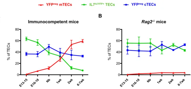
Figure 10 - IL7hi/YFP+ and YFPneg TECs exhibit distinct developmental kinetics under normal
and immunodeficient conditions ... 33 Figure 11 - Blockade of derived RANKL does not influence in the
thymocyte-mediated decline of IL7hi/YFP+ TECs ... 35
Figure 12 - Thymocyte-mediated decrease in IL7hi/YFP+ TECs is independent of mTEC
differentiation induced by RANK signaling ... 36
Figure 13 - DP thymocytes deficient in TCR expression do not profoundly affect IL7hi/YFP+
TECs ... 39
Figure 14 - Anti-CD3ε treatment improves TEC compartmentalization in Rag2-/- mice .... 40
Figure S2 - IL7hi/YFP+ TECs are markedly augmented in adult and old Rag2-/- thymi ... 65
Figure S3 - The decline in the proliferative status of TECs is independent of a normal programme of T cell development ... 65
Figure S4 - IL7hi/YFP+ TECs are unresponsive to RANK-derived signals independently of the
stage of thymic development. ... 66
Figure S5 - The cooperative effect between RANK and LTβR-derived signals does not
affect the homeostasis of IL7hi/YFP+ TECs ... 66
Figure S6 - Anti-CD3ε mAb treatment in Rag2-/- mice induces thymic expansion and
promotes the differentiation of DN thymocytes into the DP stage ... 67 Figure S7 - Molecular profile of the medullary thymic epithelium upon anti-CD3ε mAb
treatment in Rag2-/- mice ... 67
Figure S8 - The emergence of mTECs in Rag2-/- treated mice does not occur due to a
direct effect of anti-CD3ε mAb stimulation in the thymic epithelium ... 68 Figure S9 - Anti-CD3ε treatment does not affect the expression levels of thymocyte-derived RANKL and CD40L ... 68 Figure S10 - Thymic microenvironment from 2 weeks old immunocompetent mice ... 68 Figure S11 - The strength of TCR-MHC interactions during thymocyte selection modulates
Introduction
The Immune System
The immune system has evolved to protect the organism from a universe of external and internal threats. The environment contains a wide range of pathogenic microbes and allergenic substances that challenge the host through a large spectrum of mechanisms. To resolve these challenges, the immune system uses a complex myriad of cells and molecules that respond to foreign antigens whilst avoiding harm the host-tissues [1].
Immunologic defences are divided into two main branches distinguished by the speed and specificity of the response. The innate immunity, an ancient defence mechanism, initiates an immediate response against foreign pathogens that is largely non-specific and does not generate immunological memory. The innate immune system provides the first line of defence using chemical mediators and physical barriers, as well as cellular components, which become activated during an inflammatory response. The adaptive immunity, unlike the innate immune response, relies on the clonal expansion of antigen-specific B and T lymphocytes. A hallmark of this response is the ability to induce immunological memory, with the formation of long-lived cells that persist in a quiescent state and ensure a rapid response upon re-exposure to the same antigen. Nonetheless, despite the differences between innate and adaptive immune responses, the interplay between elements of both systems provides an effective defensive network [2].
All the components of the immune system are generated from a pool of multipotent hematopoietic stem cells (HSC), in a highly orchestrated process termed hematopoiesis. This event begins early during embryogenesis and has the fetal liver and bone marrow as the major niches of hematopoiesis. After expansion, some HSC migrates into distinct anatomical locations, such as the thymus, in order to commit and develop into a specific hematopoietic lineage [3].
Despite the benefits, a misguided immune response can not only compromise the ability of our system to respond against pathogens but also the immunological tolerogenicity to self-antigens, increasing the chance of developing allergic or autoimmune syndromes. Thus, the instruction and maintenance of an immunological self-tolerant state is ensured by the coordinated action of central and peripheral mechanisms [4]. Central tolerance plays an important role purging auto-reactive cells from the newly developing
lymphocyte repertoire (disclosed later in this thesis). However, this mechanism is not perfect and allows the escape of some auto-reactive lymphocytes into the periphery [5]. Therefore, peripheral tolerance provides a downstream control mechanism that include anergy and clonal deletion of auto-reactive lymphocytes as well their suppression by a specialized subset of T cells, the regulatory T cells (Treg) [5].
The Thymus as a T Lymphocyte School
The word “thymus” is thought to derive from a Greek word θυµοЅ, meaning “heart” or “soul”. The ancient Greeks were the first to originally describe this organ and they believe it was the place of the “soul” because its anatomical position lies above the heart. Along centuries the thymus was considered an enigmatic organ, surrounded by many questions regarding to its function. Although during the 1950s, the thymus was recognized as a lymphocyte producing-organ, the immunologists were sceptical about a possible immunological function of this organ and it was considered as a redundant “lymphocyte graveyard” [6]. However, the importance of the thymus was only documented in 1961 by Jacques Miller's studies on virus induced-leukaemia in young thymectomyzed mice. His observations lead to the recognition of the thymus as a non-redundant specialized organ of the immune system with a crucial role in the development of T cells [6,7].
Anatomically, the thymus is a bilobated organ that is located in the superior anterior mediastinum. Histologically, each lobe is composed by two distinct regions that includes an outer cortex and inner medulla [8]. The unique capacity of this organ to support T cell development, or thymopoiesis, relies on an organized tridimensional network of thymic stromal cells that provides the essential molecular cues for maturation, expansion and selection of T cell precursors, also known as thymocytes. The thymic stroma is heterogeneous and is composed of dendritic cells, macrophages, fibroblasts, endothelial cells and thymic epithelial cells (TECs) [9]. TECs are the major constituent of the thymic stroma and are categorized into cortical (cTECs) and medullary (mTECs), according to their spatial location, morphology and functional proprieties. These subsets are responsible for supporting distinct stages of T cell development.
Whereas cTECs support early stages of T cell development, mTECs guide later stages of T cell development [10–12].
Throughout life, the thymus contributes with a permanent output of naive T cells, reaching a peak of activity during the early adulthood in mice [13]. Nevertheless, the size and function of this organ starts progressively degenerating with age, concomitantly with the disruption of the normal cortical and medullary thymic architecture. Consequently, the capacity of the thymus to support thymopoiesis becomes gradually compromised, thereby contributing to a reduced immune surveillance. This phenomenon is known as thymic involution and is thought to be an important contributor for the immunosenescence-associated problems, by leading to an indirect increase in the susceptibility to infectious diseases, neoplastic and autoimmune disorders during elderly [14,15].
Thymic Organogenesis
Thymic organogenesis is a tightly coordinated process, that comprises a series of morphogenetic and differentiation events and relies on the interaction of cells from all three embryonic germ layers (endoderm-derived epithelium, ectoderm-derived neural crest (NC) mesenchyme, mesoderm-ectoderm-derived hematopoietic cells) [16].
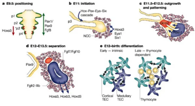
The murine thymus develops from the ventral region of the third pharyngeal pouches of the foregut endodermal tube. Although the signals that initiate the thymic development take place around embryonic day 9.5 of gestation (E9.5), the first morphological signs are only visible at E10.5 with the budding and outgrowth of the thymic anlage [16]. Between E11.5-E12, the first wave of lymphoid progenitor cells colonizes the thymic anlage, entering through the mesenchymal layer in response to chemoattractive factors [17]. The following stages of thymic organogenesis require detachment, patterning and differentiation of different thymic stromal cell subsets and migration of the thymus into its final anatomical position (Figure 1) [18,19].
During embryogenesis, the thymic primordium becomes delimited by a NC-derived mesenchyme, which contributes to the formation of the capsule and the thymic blood vasculature [8,20]. In addition, the surrounding NC mesenchymal cells influence the proliferation and homeostasis of TECs through the production of fibroblast growth factors (FGF), such as FGF7 and FGF10 [18,21,22], as well as
retinoic acid (RA) and insulin-like growth factor (IGF)-1 and -2 [23,24]. In addition, whereas the mesenchyme does not appear to be required for the initial thymic fate induction [8,25], the positioning of the organ and its separation from the pharynx relies on signals provided by NC-derived mesenchyme [26].
In the past, there were some conflicting interpretations about the precise contribution of the endodermal and ectodermal layers for the development of the thymic epithelium. Two different hypotheses had been proposed, one arguing in favour of a dual origin with participation of both endoderm and ectoderm layers, whereas the other one defends an exclusively endodermal origin for thymic epithelium. However, it is currently accepted that TECs originate solely from a unique germ layer, the endoderm [8,18,27].
The early organogenesis of the thymus is intimately connected with the formation of the parathyroid gland, which arises from the dorsal part of the third pharyngeal pouch, developing as a shared structure. Differentiation into parathyroid and thymus specific domains is driven by the spatial-specific expression of two transcription factors Glial cells missing homolog 2 (Gcm2) and Forkhead box N1 (Foxn1), respectively [28]. Foxn1, a gene encoded by the nude locus, exhibits a vital role during thymic organogenesis by governing the
Figure 1 - A schematic overview of thymic organogenesis: The thymic anlage is formed from an outgrowth of the 3rd
pharyngeal pouch. The surrounding NC-derived mesenchyme influences the thymic anlage growth and separation off the pharynx (Adapted from [18])
differentiation of TECs [29,30] in a cell-autonomous manner [31]. The expression of Foxn1 is first detectable in the ventral region of the third pharyngeal pouches as early as E11 [28], although the molecular mechanisms that initiate and control Foxn1 gene expression remain elusive. Some evidence suggests that Bone morphogenetic protein 4 (Bmp4) and Wingless-related MMTV integration (Wnt) signals might regulate Foxn1 expression in TECs in vitro [32,33], even though the role of these proteins in vivo remains to be proven.
Thymic Epithelial Cells - The Educators of Thymopoiesis
The partitioning of TECs into two specialized subpopulations, cTECs and mTECs, is essential to guide distinct stages of thymopoiesis. In recent years, TECs have been extensively studied with the purpose of gaining a better understanding about the precursor-progeny relationship and the cellular and molecular mechanisms behind the establishment of a functionally competent TEC microenvironment.
Despite the high level of heterogeneity observed among the thymic epithelium, the two TECs subtypes can be identified based on the phenotypically expression of specific markers [34,35]. Whereas cTECs are defined by the expression of cytokeratin-8 and -18 (K8+ and K18+), Ly51+ (also known as BP1) and CD205+ (also
known as DEC-205) [34–36], mTECs are commonly associated with the expression of cytokeratin-5 and -14 (K5+ and K14+), CD80+, mouse thymic stroma 10
(MTS10+), ERTR5+, the tight junction components claudin-3 and -4 (Cld3+ and
Cld4+) and the ability to bind lectin Ulex europaeus agglutinin 1 (UEA1+) (Figure 2)
[34,35,37]. Yet, both TEC lineages share common markers such as, the expression of Foxn1, Epithelial Cell Adhesion Molecule (EpCAM) and the Major Histocompatibility Complex Class II (MHCII) [35]. As aforementioned, TECs can diverge, not only in their phenotypic traits and spatial location but also in their functional properties to promote distinct stages of T cell development. cTECs mediate early stages of T cell development by providing an array of thymopoietic factors, including CCL25, IL-7, DLL4, β5t and TSSP, which are involved in the attraction, commitment, expansion and positive selection of immature thymocytes [12,34,35]. In contrast, mTECs are critical to regulate later stages of thymopoiesis, particularly negative selection, through the expression of Aire-dependent and -inAire-dependent tissue-restricted self-antigens (TRA) [38,39].
The concept that both TEC lineages arise from a common bipotent progenitor came with the identification of a particular TEC population co-expressing both cortical and medullary markers, namely K5+K8+ [40]. During fetal development,
K5+K8+ TECs are widely predominant within the thymic epithelium, whereas in
adulthood, this population represents a minor subpopulation located close to the cortico-medullary junction [40,41], emphasizing the idea of a common progenitor pool for K5+K8- mTECs and K5-K8+ cTECs. In line with the previous observations, a
subpopulation of embryonic TECs was identified based on the reactivity to the monoclonal antibody MTS24 [42,43]. MTS24+ TECs, which also co-express K5+K8+,
in contrast to MTS24- TECs, were shown to have the ability to generate a fully
competent thymic microenvironment, compartmentalized into cortex and medulla [42,43]. In addition, MTS24+ TECs also were shown to have the capacity to
support thymopoiesis when transplanted into the kidney capsule of nude mice, suggesting that MTS24+ TECs represent a putative TEC progenitor [42]. Yet, a
subsequent study showed that MTS24- TECs also had the potential to generate a
functional thymus, depending on the embryonic stage [44]. Whilst the evidence for the existence of a bipotent progenitor have been ascertained [45,46], the paucity of specific markers has significantly hampered the identification and localization of these cells in the embryonic and postnatal thymus.
The current model for TEC development suggests that cTEC and mTEC lineages originate from a bipotent precursor and develop through compartment-intermediate progenitors [10,12]. The first key evidence supporting this idea was based on the formation of multiple small clusters of mTECs derived from a single progenitor distinguished on the basis of different MHCII haplotypes in chimeric thymi [47]. Along this line, cTECs and mTECs committed progenitors have been identified during early ontogeny (E12). While mTEC progenitors are defined on the basis of the expression of tight junction components Cld3 and Cld4 [48], committed cTEC progenitors are established accordingly with the differential expression of CD205 and CD40 [49]. Moreover, the co-expression of a cTEC specific proteosomal subunit β5t along with CD205 was also associated with committed cTEC progenitors [50]. By E12, β5t+ TECs tend to be clustered in the
ventral and outer region of the thymic epithelium, in a distinct anatomical location from those containing mTEC progenitors expressing Cld3/4+ [48,50].
Thus, one can envisage the existence of landmarked spatial niches for cTEC/mTEC progenitors determined early during thymic organogenesis.
TEC differentiation can be temporally divided in two distinct phases, which occurs through a thymocyte-independent and -dependent mechanism [41]. The compartmentalization of distinct TEC microenvironments, particularly the expansion of thymic medullary regions beyond E15, is not a cell-autonomous process and depends on thymocyte-derived signals, a symbiotic relationship named "TEC-Thymocyte Crosstalk" [51]. In adult mice with a profound arrest in early T cell development, at the DN1-DN2 stage (Rag2-/-Il2rg-/- mice; human CD3ε
trangenic mice [Tge26] and Ikaros-/- mice), the thymic architecture is severely
compromised, with little organization and lack of epithelial maturation [35,41,52]. Phenotypically, the thymus from these immunodeficient mice resemble a fetal thymus at E12-E14, with an abundance of TECs co-expressing K5+K8+ [41], suggesting that in the absence of developing thymocytes, TECs
progenitors might be arrested in an immature stage. Still, the initial patterning of TECs seems to be a thymocyte-independent process [41,53], occurring presumably either in a cell-autonomous fashion or through interaction with non-hematopoietic cells during early embryonic development. Nevertheless, the ablation of perithymic mesenchyme from a fetal thymus during the E12 does not preclude the differentiation into cTEC and mTEC lineages [21,36], suggesting that beyond E12, the compartmentalization of TECs does not require sustained interactions with mesenchyme.
cTECs
CD205
+Ly51
+K8
+mTECs
MTS10
+UEA1
+CD80
+Aire
+Maturation
Figure 2 - A model for TEC differentiation: A common bipotent thymic epithelial progenitor cell (TEPC) has the potential to generate both cortical thymic epithelial cells (cTECs) and medullary TEC (mTECs), through compartment-specific intermediate progenitors, cortical TEC progenitor (cTEPC) and medullary TEC progenitor (mTEPC), respectively (Adapted from [22]).
How does the TEC Microenvironment Instruct T Cell
Development?
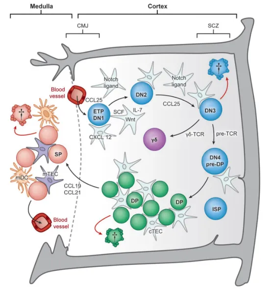
While most hematopoietic lineages differentiate within the bone marrow (BM), the generation of a diverse and self-restricted repertoire of T lymphocytes requires specialized niches exclusively found in the thymus. During their migration throughout distinct regions of the thymic microenvironment, developing thymocytes undergo a sequence of differentiation, proliferation and maturation events (Figure 3). In fact, thymopoiesis is a tightly regulated phenomenon during which several checkpoints purge the production of T cells bearing non-functional or auto-reactive T cell receptors (TCRs) [11,54,55]. As a result, only 1-3% of thymocytes are able to survive and migrate into the peripheral secondary lymphoid organs [54].
The earliest thymic progenitors that colonize the thymus display a limited self-renewal capacity. Therefore, in order to sustain T cell production, a continual input of BM progenitors is required [11]. Nevertheless, two recent reports demonstrated that, under particular conditions, neonatal thymi harbour thymocytes with a self-renewal capacity that are able to reconstitute the peripheral T-cell pool in the absence of intrathymic competition [56,57]. The entry of BM progenitors into the thymus does not seem to be a continuous process, but rather a temporally regulated event that is suggested to occur in discrete waves [58]. The initial seeding of hematopoietic precursors occurs prior to the vascularisation of the thymic primordium, through interaction with the surrounding mesenchymal layer [8,17]. However, in the postnatal thymus, once the vasculature is well developed, T cell progenitors enter across blood vessels located nearby the cortico-medullary junction (CMJ) [17,54,55]. The capacity of hematopoietic progenitors to migrate into the thymus is mediated by a chemotactic gradient, established by the expression of CC-chemokine ligand 21 (Ccl21), Ccl25 and CXC-chemokine ligand 12 (Cxcl12) within the thymus and the correspondent interaction with their receptors CC-chemokine receptor 7 (Ccr7), Ccr9 and CXC-chemokine receptor 4 (Cxcr4) expressed by hematopoietic progenitor cells. In addition, the expression of adhesion molecules by the thymic endothelium, such as P-selectin, also facilitates the colonization process [54,55,59].
Within the thymus, the earliest thymic seeding progenitors are uncommitted cells that retain both myeloid, NK, B and T cell lineage potential [60]. In order to be instructed into the T cell lineage, these cells undergo a developmental pathway that comprises different stages of maturation prior to the expression of CD4 or CD8. The most immature thymocytes are named double-negative (DN), due to the lack of expression of CD4 and CD8. While they migrate from the cortico-medullary region into the subcapsular zone, DN thymocytes sequentially progress across four distinct sub-stages, from DN1 to DN4, which can be monitored on the basis of the expression of CD25 and CD44. The migration of DN thymocytes occurs under the influence of a chemokine gradient, in particular to Cxcl12, Ccl19 and Ccl21. Moreover, during their migration to the subcapsular zone, the stromal production of stem cell factor (SCF), also known as Kit ligand, and Interleukin-7 (IL-7), are essential to support the survival and proliferation of these developing DN thymocytes [11,54,55].
The irreversible commitment towards the T cell lineage takes place at DN3 stage and is promoted by the Notch-signalling pathway, via Dll4-Notch1 interaction [61,62]. Simultaneously, the recombination activating gene (RAG) enzymes promote the rearrangement of the TCRγ, TCRδ and TCRβ locus. The productive rearrangement, assembly and signalling through the γδ-TCR allow the differentiation of DN3 thymocytes towards γδ T cell lineage [59]. With regard to αβ T cell differentiation, rearranged TCRβ chain assembles with an invariant pre-TCRα chain and CD3 signalling molecules on the cell surface, to form the pre-TCR complex. Nevertheless, only thymocytes bearing a functional pre-TCR are allowed to further develop into double positive (DP) thymocytes (CD4+CD8+), a process
known as β-selection. After passing β-selection, thymocytes initiate the TCRα locus rearrangement, which leads to the expression of a fully assembled αβ-TCR [11,54,55] and progress through a transient state as immature CD8 single positive (ISP) thymocytes (Figure 3) [11]. Subsequently, the newly generated DP thymocytes are screened for the ability of their TCR to recognize self-peptide-MHC complexes (pself-peptide-MHC) with low avidity, a process known as positive selection [63]. Only positively selected thymocytes receive signals to survive, which allow them to differentiate further into CD4 or CD8 single positive (SP) thymocytes. cTECs are the master regulators of positive selection due to the generation of MHC-bound peptides presented through distinct pathways, which are mediated
by the expression of cortex-specific proteosome subunit β5t, thymus-specific serine protease (TSSP) and Cathepsin L [64–66].
Upon positive selection, thymocytes migrate to the medullary compartment in response to Ccr7-mediated chemotactic signals, namely Ccl19 and Ccl21, expressed by mTECs (Figure 3) [67]. Once within the medulla, SP thymocytes bearing auto-reactive TCRs against self-antigens are deleted, an essential event essential for the establishment of central tolerance [68]. This process is termed negative selection, and together with positive selection, corresponds to the most important checkpoints that fine-tune T cell development. Due to its unique property, mTECs are capable of expressing an array of tissue-restricted antigens
Figure 3 - A schematic overview of thymopoiesis: Once within the thymus, the hematopoietic progenitors undergo a differentiation pathway passing through distinct niches and checkpoints until they complete their maturation as SP thymocytes. However, T cell development is not a cell-autonomous process and relies on essential molecules that are provided by TECs (Adapted from [11])
partially dependent on the transcription factor AIRE [38]. In addition, thymic dendritic cells (DCs) also regulate negative selection ably cross-presenting several self-peptides to developing SP thymocytes [70]. Thus, the interplay between mTECs and DCs during negative selection contributes to shape a self-tolerant T-cell repertoire.
SP thymocytes spend almost one week in the medullary compartment before egressing from the thymus. This process is regulated by the sphingosine-1-phosphate receptor 1 (S1P1), a G-protein coupled receptor (GPCR) expressed on SP thymocytes [71]. Sphingosine-1-phosphate (S1P), the physiological ligand of S1P1, is produced by pericytes, a NC-derived population positioned nearby the CMJ. S1P is found in higher concentration in the circulation, which allows the establishment of a chemotactic gradient that regulates the egress of mature T lymphocytes from the thymus into the circulation [54,55]. Furthermore, it seems that the Ccr7/Ccl19 axis also contributes to thymocyte export at least in newborn mice [72], suggesting perhaps a coordinated timely regulation between Ccl19- and S1P-mediated emigration along development.
Interleukin-7: A Critical Thymopoietic Cytokine
Interleukin-7, previously known as "lymhopoietin 1" and "pre-B cell factor", was first discovered in 1988 as a pre-B cell growth factor [73]. Subsequently, IL-7 was recognized as a non-redundant and multi-faceted cytokine indispensable for B cell and T cell immunity [74]. IL-7 is not only an important growth factor to T cell precursors during thymopoiesis, but is also required for the homeostasis of naive and memory T cells in peripheral lymphoid organs [75,76]. Moreover, IL-7 is essential to lymph node (LN) and Peyer's patches (PP) development, promoting the survival and proliferation of lymphoid tissue inducer (Lti) cells [77].
The pleiotropic effects of IL-7 are mediated via its receptor, IL-7 receptor (IL-7R) [78]. The IL-7R is a heterodimeric complex composed of IL-7 receptor alpha (IL7-Rα) chain (also known as CD127) and the common cytokine receptor γ-chain (γc), also shared by other cytokines of the γc family (IL-2, IL-4, IL-9, IL-15, IL-21)
[78,79]. IL-7-derived signals are transduced through multiple signalling pathways, which include the phosphatidylinositol 3-kinase (PI3K), Janus kinase and signal transducer and activator of transcription pathway (Jak and STAT) and Src family tyrosine kinases [76]. Interestingly, there are some evidence suggesting that IL-7
signalling is an evolutionarily conserved pathway in lower and higher vertebrates [80]. Mutations in the γc, IL7-Rα chains and in components associated with IL-7
signalling pathway result in Severe Combined Immunodeficiency (SCID) [79,81], a syndrome characterized by a profound impairment in B and T cell development.
Under steady-state conditions, IL-7 appears to be constitutively produced and its bioavailability limits the size of the lymphocyte pool. Nonetheless, the responsiveness to IL-7 is tightly controlled by the differential expression of IL7-Rα [82]. During thymopoiesis, IL7-Rα is expressed at the DN stage, particularly by DN2 and DN3 thymocytes, absent at the DP stage and re-expressed at SP stage [78,82]. Along these stages of T cell development, IL-7 acts as a survival factor balancing pro- and anti-apoptotic B cell lymphoma 2 (Bcl-2) family members [76,78,81]. Yet, despite its role as a pro-survival factor, IL-7 tends influences the T cell lineage differentiation. IL-7 signalling commits DP thymocytes into the CD8 lineage by inducing the expression of the transcription factor Runx3 [83]. Moreover, γδ T-cell development relies on IL-7 signals, which regulate the rearrangement of the γ-chain locus by permitting the chromatin accessibility to the RAG enzymes [84]. Such dependency is well documented in IL7-/- and IL-7Rα
-/-mice [85,86], in which γδ T cells are absent.
Several reports have shown the expression of IL-7 in multiple lymphoid tissues, which include the thymus, bone marrow, lymph nodes, spleen, fetal and adult liver and intestinal epithelium [76,87]. Interestingly, very low levels of IL-7 have been detected in adult, which support the idea that under normal conditions the bioavailability of IL-7 is limited in the body [88]. Despite the knowledge about its biological effects, the identity of IL-7 producing cells, the anatomical niches in which IL-7 is active, and the mechanisms that regulate its expression remain largely unknown.
The lack of experimental tools to monitor cells expressing IL-7 has hampered our comprehension about how IL-7 is regulated in vivo. Recently, four independent studies provide new insights into this issue by generating different bacterial artificial chromosome (BAC) transgenic mice that monitor IL-7 expression in vivo [87]. In one of these studies from our laboratory, the IL-7 reporter activity indicated the thymus as the main source of IL-7 and identified TECs as bona fide IL-7-producing cells [89]. IL-7 expressing TECs (hereafter referred as IL7hi/YFP+ TECs) emerge early during fetal thymic organogenesis and
TECs display a widespread distribution in the fetal thymus, they are predominantly located at the CMJ in the adult thymus [89,90]. One can envisage a correlation between the strategically distribution of IL7hi/YFP+ TECs close the CMJ
with the requirement of IL-7 signalling for early and post-selected thymocyte development.
Nowadays, it is recognized that IL-7 expression by TECs occurs in a Foxn1-independent fashion albeit it remains undetermined whether its expression initiates in a cell autonomous manner or is controlled by an exogenous signal [91]. In addition, the same report has also suggested that signals delivered by developing thymocytes regulate the IL-7 expression during ontogeny [91]. In this regard, in alymphoid Rag2-/-Il2rg-/- mice, which display a profound block in early
stages of thymopoiesis, the frequency of IL7hi/YFP+ TECs is significantly augmented
when compared with age-matched immunocompetent mice [90]. However, upon reconstitution with bone marrow precursors from either TCRα-/- or WT mice, the
frequency of IL7hi/YFP+ TECs is reduced, mirroring a similar phenotype observed in
immunocompetent mice [90]. Such evidence unveils the existence of a feedback mechanism in which TEC-thymocyte crosstalk curtails the expression of IL-7. Yet, the biological significance and the molecular events underlying this feedback mechanism remain far from disclosed.
TEC-Thymocyte Crosstalk Governs the Epithelial
Development
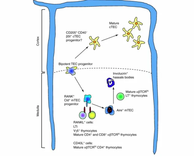
As mentioned above, the establishment of distinct cortical and medullary microenvironments is not a cell-autonomous process and is influenced by thymocyte-derived signals [51]. In recent years, although the cellular and molecular mechanisms that underlie the TEC-thymocyte crosstalk have been partially deciphered, the detailed nature of this bidirectional communication continues largely unclear.
Initial studies involving the characterization of mice deficient in signalling components of both canonical and non-canonical nuclear factor kappa B (NF-κB) pathway have contributed to elucidate the molecular basis behind mTEC development. Abrogation of downstream associated effectors NF-κB-inducing kinase (NIK), Tumor necrosis factor receptor-associated factor 6 (TRAF6)
related B (RelB), caused a disorganized mTEC architecture, concomitantly with a
marked reduction in Aire+ mTECs and consequent manifestations of
autoimmunity [92–94]. Research over the last years has recognized the tumour necrosis factor receptors super family (TNFSF) members as key activators of NF-kB signalling during mTEC development [37]. Lymphotoxin β receptor (LTβR), which plays a pivotal role in the organogenesis of most secondary lymphoid tissues, was the first TNFR member described to be involved in mTEC differentiation [95]. LTβR-mediated signals induce a diverse array of cellular processes that, albeit contributing to the development and proper organization of mTEC architecture, also guarantee an efficient negative selection process by regulating the expression of Aire-independent TRA and chemokine genes in mTECs [39,96]. Additional studies have described the individual and cooperative action of two other TNFSF members, receptor activator of NF-kB (RANK) and CD40, in the homeostasis of thymic medulla (Figure 4) [97,98].
RANK is a multifaceted receptor that regulate different physiological processes [99]. In the thymus, RANK-mediated signals enhance the growth and optimal development of mTECs, including Aire-expressing cells [100]. In fact, from all TNFSF members, RANK appears to have the strongest impact on the medullary epithelium. Compared with the lack of LTβR or CD40-signalling pathways, RANK-deficient mice exhibit the most severe phenotype characterized by a marked reduction in mTEC cellularity, particularly Aire+ mTECs, and impaired central
tolerance [97,98,100]. Different cellular populations appear to express RANK ligand (RANKL), the physiological ligand of RANK, within discrete developmental windows. While CD4+CD3- LTi and Vγ5+ γδ T cells are the major source of RANKL at
early phases of embryogenesis [101,102], RANKL is predominantly expressed by positively selected thymocytes during post-natal life [100]. Additionally, several studies have pointed to a participation of CD40-CD40L axis in the formation of thymic medulla after birth, simultaneously with the expression of CD40L by CD4+
SP thymocytes [97,103].
Despite their individual contributions, the establishment of a mature and functional medullary microenvironment relies on a stepwise interplay between LTβR, RANK and CD40. A recent report point to a synergistic interaction between LTβR and RANK at early stages, during which LTβR-signals enhances RANK expression on mTEC precursors, thereby making TECs more responsive to RANKL [98]. Nevertheless, this cooperation appears only to occur during early stages of
organogenesis, being then substituted during post-natal life by a collaborative action between RANK and CD40-mediated signals [97]. Moreover, an efficient delivery of RANKL and CD40L is likely to occur in the context of physical interactions between auto-reactive CD4+ SP thymocytes and mTECs displaying the
cognate self-antigen-MHCII complexes [104].
Although the cellular and molecular mediators that control mTEC maturation are partially understood, the underlying basis that regulates cTEC differentiation remain largely unanswered [35]. Unlike the medullary lineage, the paucity of cell surface markers has hampered the advances to map the developmental progression of cTECs. Only recently, a model for developmental checkpoints during cTEC differentiation has been proposed on the basis of the differential expression of CD205, CD40 and MHCII markers [49]. This model has proposed a
Figure 4 - A model of TEC-thymocyte crosstalk and its impact in the formation of distinct microenvironments: TECs derive from a bipotent precursor and develop through intermediate TEC-committed progenitors in distinct anatomical niches. While the initial development of TECs occur in the absence of thymocytes, its progression through different stages of development relies on thymocyte-derived signals (Adapted from [33])
stage-specific requirement of DN thymocyte-derived signals for the proper development of a cortical microenvironment [49]. However, the precise nature of such signals and how DN thymocytes shape cTEC maturation remains largely unknown.
Aims
The thymic epithelial microenvironment undergoes a series of morphological and architectural modifications throughout development, which are accompanied by a prominent reduction in the number of TECs, particularly in cTECs [105,106]. Interestingly, it has been demonstrated that cTECs have the potential to rejuvenate the cortical compartment upon thymic injury [107]. Yet, the cellular and molecular basis that governs the development and maintenance of the cortical microenvironment remain open questions.
In this thesis, we aim at defining novel checkpoints in the differentiation of the thymic cortical epithelium. Using state-of-the-art research mouse models (IL-7 reporter mice in immunocompetent and genetically altered background), together with in vitro thymic organotypic cultures and in vivo functional assays, we study (1) the lineage relationship between IL7hi/YFP+ TECs and other well-characterized
TEC subtypes and (2) the impact of distinct molecular interactions between thymocytes and TECs in the control of the homeostasis of the cortical epithelium.
The development of approaches to promote the functional rejuvenation of the thymus has been one of the biggest challenges in clinical immunology. Comprehending the mechanisms behind TEC development will aid in the elaboration of tailor-made therapeutic strategies aimed at repairing and improving thymic functioning in different pathophysiological conditions, including aged-associated thymic involution, immunodeficiencies and infection.
Material and Methods
Mice
These studies used transgenic mice (B6.Cg-Tg(Il7-EYFP)5Pas) carrying a bacterial artificial chromosome (BAC) encoding YFP under the control of the Il7 promoter (Figure 5), inserted by homologous recombination downstream of the ATG start codon located at the end of the exon 1 of Il7 locus. In these mice, the expression of YFP is purportedly controlled by the same molecular mechanism that drives the expression of endogenous Il7. IL-7 reporter mice were backcrossed onto C57BL/6 background wild type (WT), Rag2-/- mice [108] and
Marilyn Rag2-/- mice [109] (kindly provided by Jocelyne Demengeot, Instituto
Gulbenkian de Ciência, PT) for more than 9 generations.
Figure 5 – BAC sequence scheme comprising Il7 gene, its promoter and YFP insertion in the gene.
For fetal studies, the day of the vaginal plug detection was designated embryonic day E0.5.
Mice were housed under specific pathogen-free conditions and all animal experiments were conducted accordingly to the guidelines of the Portuguese National Authority for Animal Health (DGV) and European Union directive 2010/63/EU by FELASA-accredited researchers.
Genotyping
In the adopted breeding scheme, transgenic mice were crossed with immuno-competent and Rag2-/- C57BL/6 mice, resulting in a progeny of both transgenic
and WT littermates. Tails were digested in buffer (100 mM Tris pH 8.5; 5 mM EDTA; 0.2% SDS; 200 mM NaCl) with 0.4 mg/ml proteinase K (Eurobio) and the DNA was isolated using a standard mouse tail DNA isolation protocol. Transgenic mice were distinguished by standard PCR, using specific primers for the Il7-YFP junction sequence (Forward: 5’ tacacccacctcccgcagaccatggtgagcaagggcgag-gagctgttc 3’; Reverse: 5’ gcaccagagagcagcgcttaccatttacttgtacagctcgtccatgcc 3’). PCR reaction comprised 5 minutes at 95ºC for initial denaturation of DNA, and 35 cycles of 30 seconds at 95ºC and 1 minute at 72ºC for annealing and
amplifica-tion, followed by a final step of 5’ at 72ºC for extension. For Rag2-/- genotype, a
set of three primers was used to discriminate WT and KO alleles (Forward: 5’ gggaggacactcacttgccagta 3’; Reverse: 5’ agtcaggagtctccatctcactga 3’ and neo-Forward: 5’ cggccggagaacctgcgtgcaa 3’). PCR reaction comprised 4 minutes at 94ºC for initial denaturation of DNA, 35 cycles of 20 seconds at 94ºC, 30 seconds at 72ºC and 30 seconds at 74ºC, followed by an additional final step of 5 minutes at 74ºC for extension.
Isolation of Thymic Stromal Cells
Thymic lobes were dissected at indicated time points (for early developmental stages, by micro-dissection) and stromal cells were isolated as previously described [110]. Thymic fragments were digested for 30 minutes at 37ºC in trypsin (Sigma) supplemented with 1% collagenase (Sigma) and 0,02% DNaseI (Roche). Fragments were mechanically disrupted by ressuspension every 5 minutes with a syringe to obtain cell suspensions. For later time points (adult stages), thymic stromal cells were enriched by an additional selection procedure that depletes hemotopoietic cells using MACS CD45 MicroBeads (Miltenyi Biotec) and autoMACS separation columns according to the manufacturer’s instructions. Cell numbers were calculated using counting chamber (Hycor Biomedical).
Flow Cytometric Analysis
Cell suspensions were stained with CD8 (FITC); CD4, CD80, anti-CD40 and anti-Ly51 (PE); anti-CD205 (biotin) antibodies (Abs) and streptavidin (PE-Cy7) (Becton Dickinson); anti-I-A/I-E (Alexa780); anti-CD45.2 (PerCPCy5.5); anti-EpCAM and anti-CD8 (APC) Abs and anti-EpCAM (eFluor 450) Abs (Ebioscience). For cell cycle analysis, staining with propidium iodide (PI) solution (Sigma) was done after fixation with cold pure ethanol for 1 hour at 4ºC followed by over-night incubation at -20ºC. After PBS-washing, cells were incubated for 30 minutes at 37ºC with PI staining solution (50 µg/ml PI in PBS with 0.05% Triton X-100). Analysis was done using both FACSCantoII and FACSAria (BD Biosciences) and FlowJo software. Cell sorting was performed using FACSAria (BD Biosciences.
Gene Expression
For quantitative PCR (qPCR), total RNA from sorted cells was purified with an RNeasyMicroKit (Qiagen). The quantity of total RNA was assessed using ND-1000 spectrophotometer (Thermo Scientific). cDNA synthesis was performed by reverse transcription of the extracted RNA, using SuperScript III first-strand synthesis system for RT-PCR (Invitrogen) and Random Hexamers (Fermentas) according to the manufacturer’s instructions. qPCR were performed either using TaqMan Universal Master Mix (Applied Biosystems) and primers Hprt, 18s, Il7, Dll4, Ccl25,
Psmb11, Aire and Tnfrsf11a (RANK) (Applied Biosystems) or iQ™ SYBR® Green
Supermix (Bio-Rad) with primers specific for β-actin (forward: 5' cgtgaaaagatgacccagatca 3'; reverse: 5' tggtacgaccagaggcatacag 3'), Ctss
(Cathepsin S) (forward: 5' ccattgggatctctggaagaaaa 3'; reverse: 5'
tcatgcccacttggtaggtat 3'), Tnfsf11 (RANKL) (forward: 5' cacacctcaccatcaatgctgc 3'; reverse: 5 gaagggttggcacacctgaatgc 3') and Cd40lg (CD40L) (forward: 5' gtgaggagatgagaaggcaa 3'; reverse: 5 cactgtagaacggatgctgc 3') (Sigma). Il7 probes used were located between exons 1 and 2, in order to detect only endogenous Il7. All the samples were analysed as triplicates and the delta-delta-Ct method was used to calculate relative levels of target mRNA normalized to 18s/Hprt or β-actin. All procedures were performed according to the manufacturer’s protocols. Real-time PCR was performed on an iCycler iQ5 Real-Time PCR thermocycler (Bio-Rad). Data were analysed using iQ5 Optical System software (Bio-(Bio-Rad).
Fetal Thymic Organ Culture
E14 fetal thymi were dissected and thymic lobes were separated. Usually, three or four lobes were used per condition in order to normalize the intrathymic variations. A gel foam sponge was used to support thymic lobes on 0.8 µm Isopore membrane filter (Millipore), cultured in 6 well plates with 1ml DMEM medium (GIBCO) supplemented with 10% FCS, 1% L-Glutamine 200 mM (GIBCO) and 360 mg/L of 2-deoxyguanosine (Sigma) for 4 days. The agonistic anti-RANK (αRANK) (5 µg/ml; R&D Systems) or/and the agonistic anti-LTβR mAB AC.H6 (10 µg/ml; kindly provided by Jeff Browning, Biogenidec, US) was added on day 4. For the blocking experiment, the antagonistic anti-RANKL (Prepotech) was added to Fetal Thymic Organ Culture (FTOC) at a final concentration of 10 µg/ml on day 0
and 4. FTOCs were maintained for 8 days. TECs were isolated (as described above) and analysed by flow cytometry.
Immunohistological Analysis
Thymic lobes were fixed in 4% paraformaldehyde (Electron Microscopy Sciences) in PBS, washed twice with PBS and incubated in 50% sucrose-PBS solution before being embedded in OCT compound (Sakura) and frozen. 8 µm sections, obtained in the Ultramicrotome Leica Reichert SuperNova, were collected in Superfrost/Plus slides (Fisher Scientific) and stored at -20ºC. After re-hydration and blocking (10% BSA in PBS solution), samples were stained with rabbit anti-GFP, biotinylated CD205, rat anti-MHC Alexa647 as primary antibodies, Alexa Fluor 488 donkey anti-rabbit IgG, Alexa Fluor 647 goat anti-rat and streptavidin Alexa 555 as secondary antibodies (Invitrogen). Vectashield mounting medium with 4',6-diamidino-2-phenylindole (DAPI) (Vector Laboratories) was used to prepare the slides. Analysis was performed in an AxioImager Z1 (Carl Zeiss). Images were processed with Fiji Software.
In Vivo Anti-CD3ε Treatment
3-4 days old neonatal Rag2-/- mice (less than 3 days) were injected
intraperitoneally with purified anti-CD3ε monoclonal antibody (mAb) (10 µg/g body weight; Clone 1452C11; Kindly provided by Benedita Rocha, Hospital Necker, France). Mice thymi were collected 5 or 12 days after injections. For in
vitro studies, fetal thymic lobes from Rag2-/- fetus (day 14 of gestation) were used
to establish FTOC with or without 10 µg/ml anti-CD3ε mAb for 5 days. In both procedures, TECs were isolated, counted and analysed by flow cytometry according the procedures described above.
Statistical Analysis
Statistical analysis of the results was performed using the GraphPad Prism Software. The two-tailed Mann-Whitney test was used to analyse the differences between groups. A 95% confidence interval was applied in the calculations and samples with p values under 0.05 were considered significant.
Results
A Prelude on the ontogeny of IL7
hi/YFP+TECs
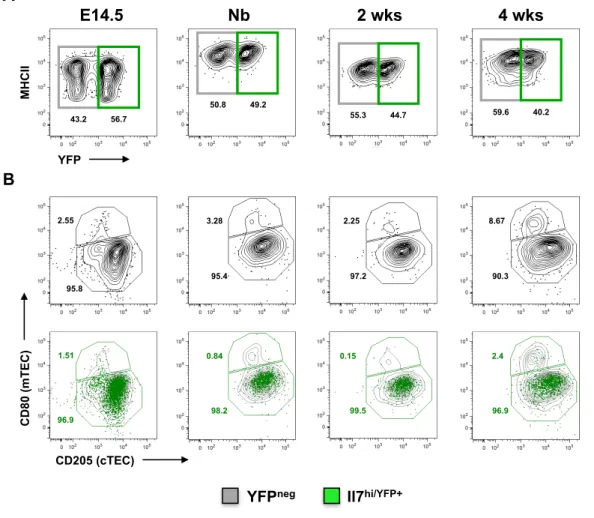
Previous studies using IL-7 reporter mice identified a particular population of TECs that expresses high levels of Il7 transcripts [89,90]. IL7hi/YFP+TECs appear
early during fetal development and gradually diminish along gestation concomitantly with hematopoietic colonization of the thymus. In addition, IL7hi/YFP+TECs segregate from mature CD80+ mTECs throughout development
indicating that they may constitute a specialized cortical subset [90]. To better define the identity of IL7hi/YFP+TECs within the cortical epithelium, we performed a
combinatory phenotypic-temporal analysis throughout thymic organogenesis employing two different cTEC-associated markers, CD205 and BP1 [35]. TECs were identified based on the expression of MHCII and EpCAM and the lack of CD45 markers accordingly to the gating strategy described in Figure S1. In line with previous results, IL7hi/YFP+TECs emerged early during fetal development, in
which they represented the majority of the thymic epithelium and gradually disappeared with age (Figure 6.A and Figure S2, top panel). The maturation of TECs could be monitored by the progressive expression of high levels of MHCII along thymic development (Figure 6.A). During early stages of thymic organogenesis, both IL7hi/YFP+ TECs and YFPneg TECs revealed a similar phenotypic
pattern, with the majority of cells of both subsets expressing cortical traits (CD205+BP1+). However, from E16.5 onwards, while IL7hi/YFP+TECs retained a
cortical-related phenotype, a subset of medullary epithelium (CD205-BP1-) started
to emerge within YFPneg TECs (Figure 6.Bi).
In addition, we analysed the epithelial compartment on the basis of the expression of the costimulatory molecule CD40, which was recently associated with TECs exhibiting a genetic profile linked to the mTEC lineage [49]. During thymic ontogeny, IL7hi/YFP+TECs progressively acquired the expression of CD40
while retaining the expression of CD205 (Figure 6.Bii). Noticeably, and in contrast to IL7hi/YFP+TECs, the increment in the expression of CD40 by YFPneg TECs
was accompanied by the expression of a medullary-associated marker CD80 (Figure 6.Biii), suggesting that YFPneg TECs might harbour a putative mTEC
Having established that IL7hi/YFP+ TECs retained a cTEC phenotype during thymic
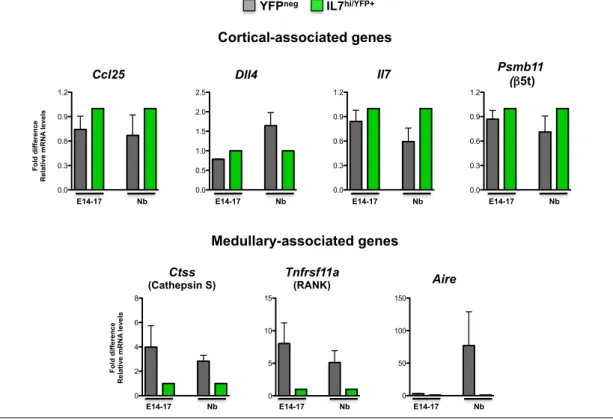
development, we aimed at investigating its molecular features relatively to cortical and medullary-associated genes. To this end, a molecular characterization of IL7hi/YFP+ and YFPneg TECs at different time points of thymic
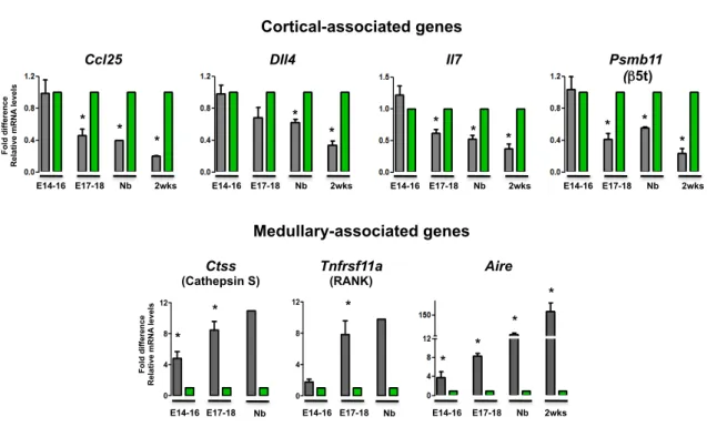
organogenesis were performed by quantitative PCR using a panel of TEC-associated genes: Ccl25, a chemokine responsible for the attraction the T cell progenitors to the thymus; Dll4, the Notch ligand involved in the commitment towards the T cell lineage; Il7, a cytokine that provides survival and proliferative signals for developing thymocytes; Psmb11 (β5t), a proteasome subunit involved in positive selection; Ctss (Cathepsin S), a lysosomal endoproteinase implicated in
Figure 6 - IL7hi/YFP+ TECs emerge during early thymic organogenesis and comprise an epithelial population that
displays cortical-associated traits. Thymi from IL-7 reporter mice (Tg) were analyzed at the indicated time points. A) Dynamics of IL7hi/YFP+ TECs (gated on CD45-/MHCII+/EpCAM+) (top panel). B) Phenotypic characterization of TECs
throughout fetal development using a panel of cTEC and mTEC-associated markers. i) BP1 and CD205. ii) CD40 and CD205 and iii) CD40 and CD80. The FACS plots in B represent an overlay of color-coded IL7hi/YFP+ (green dot plot) and
YFPneg (gray contour plot) TECs shown in A. A and B) Numbers indicate the percentage of gated cells. For B, numbers in
green represent the percentage of gated cells within IL7hi/YFP+ TECs.
E13.5 E15.5 Nb 2 wks 68 32 E16.5 80.6
18.8 MH C II YFP 66.8 33.2 A 39 39 61 49.1 50.9 B B P1 CD205 5.5 93.6 31.1 68.9 i 21.3 75.6 13.4 83.5 11.1 86.8 9.9 87.8 CD40 a" a" a" 9.9 8.9 83.1 CD205 ii YFPneg IL7hi/YFP+ CD80 CD40 79.9 6.9 9.0 30.3 0.7 67.1 57.1 1.5 38.1 78.3 1.4 16.8 iii
negative selection; Tnfrsf11a (RANK), a TNFSF member responsible for mTEC differentiation and Aire, the transcription factor that mediate the expression of TRA during negative selection [12,34,35].
In line with the phenotypic characterization (Figure 6), similar levels of cortical-associated transcripts (Ccl25, Dll4, Il7 and Psmb11) were found in both IL7hi/YFP+ and YFPneg TECs at early stages of thymic organogenesis (E14-16) (Figure
7, top panel). However, as the compartmentalization into distinct TEC microenvironments began, differences in the expression pattern between both subsets became more prominent, with IL7hi/YFP+ TECs retaining the expression of
cortical-associated genes (Figure 7, top panel). Regarding the medullary-associated genes, the expression of Ctss, Tnfrs11a and Aire was virtually undetected in IL7hi/YFP+ TEC fraction. Yet, these transcripts were already present in
YFPneg TECs at E14-16, particularly Ctss and Aire, and progressively increased
throughout thymic ontogeny (Figure 7, bottom panel). Taken together, our results suggest that IL7hi/YFP+ TECs are a determinant of the cortical lineage and
segregate from mature mTECs expressing Ctss, Tnfrs11a and Aire.
YFPneg IL7hi/YFP+
Fold d iffe re n ce R el ati ve mRNA levels Psmb11 (β5t) * * Dll4 * * Nb E17-18 2wks E14-16 Ccl25 * * Il7 * * * Nb E17-18 2wks
E14-16 E14-16 E17-18 Nb 2wks E14-16 E17-18 Nb 2wks
* * Cortical-associated genes 0 4 8 12 0 4 8 12 Fold d iffe re n ce R el ati ve mRNA levels 2wks Aire * * * Nb E17-18 E14-16 E17-18 E14-16 Ctss (Cathepsin S) E17-18 E14-16 Tnfrsf11a (RANK) * * * * Nb Nb Medullary-associated genes
Figure 7 - Molecular characterization of YFPneg and IL7hi/YFP+ TECs during thymic ontogeny. YFPneg (gray) and IL7hi/YFP+
TECs (green) purified at the indicated time points were analyzed for the expression of Ccl25, Dll4, Il7, Psmb11 (β5t),
Ctss (Cathepsin S), Tnfrsf11a (RANK) and Aire. Values were normalized to 18s, Hprt or ß-actin. IL7hi/YFP+ TECs were set
as 1, at each time point, and the fold difference in the relative mRNA expression was compared to YFPneg TECs. Average
values of 4-6 independent experiments are shown. (*) indicates that the difference between groups are statistically significant (p<0.05).