DNA markers for detection and infrasubspecific
discrimination of mastitis-causing Streptococcaceae
Dissertação de Mestrado em Genética Forense
Alexandre Miguel Santos Almeida
Faculdade de Ciências da Universidade do Porto
2012
discrimination of mastitis-causing Streptococcaceae
Dissertação submetida à Faculdade de Ciências da Universidade do Porto para
obtenção do grau de Mestre em Genética Forense.
Local de realização:
Departamento de Biologia
Faculdade de Ciências da Universidade do Porto
Orientadores:
Professor Doutor Fernando Tavares
Faculdade de Ciências da Universidade do Porto
Centro de Investigação em Biodiversidade e Recursos Genéticos
Doutor Ricardo Araújo
“A scientific truth does not triumph by convincing its opponents and making them see the light, but rather because its opponents eventually die and a new generation grows up that is familiar with it.”
Acknowledgements
Individual effort and skill can only take you so far. Without the help and dedication of colleagues, friends or family you’ll never accomplish much in life. Throughout this past year, several people in one way or another were essential for the outcome of this work.
First and foremost, I would like to thank my supervisor Prof. Fernando Tavares for all the dedication, cooperation and friendship granted during my thesis. In spite of being very busy with numerous responsibilities, he always managed to teach and guide me throughout this endeavour and make sure the work was successful. With his articulate speeches and guidance, we always managed to solve any challenge we faced.
I also have to issue a very special thanks to my laboratory partner Pedro Albuquerque. His tremendous kindness and willingness to always help and teach was one of the main driving forces for this work. He taught me all the techniques and protocols used throughout this year and even during his final months of his PhD he was always available to help and offer his expertise on the theoretical and technical aspects of the work. Without him this experience would have been extremely more difficult.
A special thanks to my co-supervisor Ricardo Araújo who, in spite of being institutionally apart, was always on par with the work, available to help and offer his interesting perspective and collaboration.
I would also like to thank Prof. Niza Ribeiro for offering his expertise as a more directly involved researcher in the subject of bovine mastitis. A special thanks as well to Paula Santos and João Sousa from SEGALAB for providing the bacterial isolates used during this study, which were essential for the experimental analyses.
Also, thank you to Prof. António Amorim, leader of the Population Genetics group of IPATIMUP, for giving me the opportunity to develop my thesis in the Sciences Faculty with Prof. Fernando Tavares.
Although these were the people directly involved in my dissertation, I would also like to thank my friends and colleagues from the Population Genetics group of IPATIMUP, Ana Carolina, Catarina Seabra, Catarina Xavier, Filipa Cabral, Inês Martins, Marisa Oliveira and Sofia Marques for their friendship and numerous fun times shared these
past couple of years. A special word to Ana Carolina who has a very special place in my heart for always supporting me during the good and the bad, and who has given me some of the best moments of my life.
A special thanks also to Maria João Fonseca and Eduarda Almeida from the Microbial Diversity and Evolution group of CIBIO, who welcomed me kindly to the group and were great companions in the laboratory during my work.
Last but not least, thank you to my parents for continuing to allow me to pursue my academic goals, and the rest of my friends and family who I may have forgotten during these busy times but that still hold a precious place in my life.
Abstract
Molecular methods have shown to be a fast and reliable approach for bacterial detection and identification. Among these, DNA-based methods encompass some of the most promising approaches, due to the development of several efficient techniques. Moreover, increasingly larger and more informative genome databases allow effective in silico analyses for the selection of taxa-specific DNA signatures. On the other hand, biochemical and phenotypic tests, frequently employed in routine diagnostic laboratories, present a limited accuracy and are inherently biased towards culturable organisms.
The applicability of these methods range from disease diagnosis and identification of biological agents to metagenomic studies and community profiling. In the field of veterinary medicine, bovine mastitis, an inflammatory disease in the mammary gland, is currently a major concern affecting dairy herds worldwide. Due to changes in milk quality and composition, this disease is responsible for significant financial losses in the dairy industry. Over 150 pathogenic agents have been identified, with particular prevalence of those belonging to the Streptococcaceae family. Thus, efficient detection and typing methods are required for disease prevention, source tracking, treatment assessment and control.
The aim of this work was to develop a rapid, reliable and inexpensive platform for the detection of prevalent bovine mastitis pathogens within the Streptococcaceae family and gain additional insight into the infrasubspecific diversity of this group to improve epidemiological characterisation.
Using Insignia and the Protein Family Database (Pfam), DNA signatures were selected for well-known bovine mastitis-causing taxonomic ranks: a broad-spectrum marker for the Streptococcaceae family, taxa-specific markers for the Lactococcus and Streptococcus genera, and specific markers for Streptococcus agalactiae and Streptococcus uberis, two particularly prevalent mastitis pathogens. Additionally, markers of functional traits associated with the virulence potential of bovine mastitis strains were used, based on the fructose and nisin operon. The virulence-associated genes hasC, gapC and oppF, frequently described in S. uberis strains, were also selected.
Experimental validation was carried out by PCR and dot blot hybridisation, and an image algorithm was used to allow an operator-independent interpretation of the results. A set of 44 reference strains and isolates, representative of the Streptococcaceae family, of closely related species and of organisms with matching hosts, was tested with the selected DNA markers. The isolates used were obtained from different mastitic milk samples of distinct locations within Portugal and previously identified by the automated identification system VITEK 2. Sequencing analysis of the 16S rRNA gene revealed an incorrect identification of some of these isolates, emphasising the increased reliability and accuracy of molecular methods.
Based on the results obtained, the broad-spectrum taxonomic marker was specific to the Streptococcus genus and the markers selected for Lactococcus, S. agalactiae and S. uberis were shown to be specific to the corresponding taxa. The functional markers allowed increased discrimination of strain-specific patterns of S. agalactiae and S. uberis: the fructose operon markers were specific to bovine isolates of S. agalactiae and the nisin operon markers were present in a particular cluster of strains with a common origin. Furthermore, dot blots using the virulence-associated markers revealed specific patterns that were able to discriminate additional species, such as Streptococcus bovis and Streptococcus parauberis, and detect other organisms closely related to the Streptococcaceae family.
This work suggests that the combined use of taxa-specific and functional markers presents a promising approach for the reliable, rapid and cost-effective detection and typing of bovine mastitis-causing pathogens, for the treatment and control of this disease.
Resumo
Os métodos moleculares têm vindo a adquirir um papel de destaque para a deteção e identificação bacteriana. As técnicas baseadas em DNA em particular englobam algumas das metodologias mais promissoras devido ao desenvolvimento de diversas técnicas eficazes. Além disso, a disponibilidade de bases de dados de genomas de microrganismos cada vez mais informativas permitem análises in silico fidedignas para a obtenção de assinaturas de DNA taxa-específicas. Por outro lado, testes bioquímicos e fenotípicos, frequentemente utilizados em laboratórios de rotina, apresentam uma precisão limitada e são enviesados inerentemente para organismos cultiváveis.
A aplicabilidade destes métodos vai desde o diagnóstico de doenças e agentes biológicos a estudos de metagenómica e caracterização de comunidades. A mastite bovina, uma doença inflamatória na glândula mamária, é atualmente uma das grandes preocupações para a medicina veterinária, afetando o gado bovino por todo o mundo. Isto leva a alterações significativas quer na composição, quer na qualidade do leite, resultando em grandes prejuízos económicos para a indústria. Mais de 150 agentes patogénicos já foram identificados, com especial prevalência de organismos pertencentes à família Streptococcaceae. Deste modo, métodos eficazes de deteção e tipagem são essenciais para prevenir, controlar e avaliar a eficácia do tratamento desta doença.
O objetivo deste trabalho foi desenvolver uma plataforma rápida, barata e fidedigna para a deteção de agentes patogénicos responsáveis por mastites bovinas pertencentes à família Streptococcaceae e inferir alguma diversidade infrasubespecífica para melhorar a caraterização epidemiológica.
Através das ferramentas Insignia e Protein Family Database (Pfam), foram selecionadas assinaturas de DNA para níveis taxonómicos frequentemente associados a organismos responsáveis por mastites bovinas: um marcador de largo espetro para a família Streptococcaecae, marcadores para os géneros Lactococcus e Streptococcus, e marcadores específicos para dois patogénicos com especial interesse, Streptococcus agalactiae e Streptococcus uberis. Além disso, foram utilizados marcadores funcionais de caraterísticas fenotípicas associadas à virulência de estirpes de mastites, baseados no operão da frutose e da nisina. Foram
selecionados também três genes de virulência descritos frequentemente em S. uberis: hasC, gapC e oppF.
A validação experimental foi realizada por PCR e hibridação em dot blot, complementada com um programa de análise automática de imagem para uma interpretação objetiva dos resultados obtidos. Os marcadores selecionados foram testados com um conjunto de 44 isolados e estirpes de coleção, representativo da família Streptococcaceae, de organismos filogeneticamente próximos e de outros que partilham um habitat comum. Os isolados foram obtidos a partir de amostras de leite contaminadas provenientes de diferentes localidades de Portugal e previamente identificados pelo sistema de identificação bacteriano VITEK 2. A sequenciação do gene 16S rRNA revelou que alguns isolados tinham sido mal identificados, o que realça o facto de os métodos moleculares serem alternativas mais exatas e precisas.
Com base nos resultados obtidos, o marcador taxonómico de largo espetro foi específico para o género Streptococcus e os marcadores selecionados para Lactococcus, S. agalactiae e S. uberis mostraram ser específicos para os grupos pretendidos. Os marcadores funcionais permitiram inferir alguns padrões específicos de linhagens de S. agalactiae e S. uberis: os marcadores do operão da frutose demonstraram ser específicos para isolados bovinos de S. agalactiae, enquanto os marcadores do operão da nisina foram detetados num conjunto de isolados de S. agalactiae com uma origem comum. Além disso, os ensaios com os marcadores de genes de virulência permitiram discriminar outras espécies, incluindo Streptococcus bovis, Streptococcus parauberis e outros organismos filogeneticamente próximos da família Streptococcaceae.
Este trabalho demonstra que o uso simultâneo de marcadores taxonómicos e funcionais é uma estratégia promissora para a deteção e tipagem eficazes de patogénicos responsáveis por mastites bovinas, podendo assim contribuir para um melhor tratamento e controlo desta doença.
List of tables
Table 1...34 Table 2...35 Table 3...36 Table 4...36 Table 5...38 Table 6...38 Table 7...40 Table 8...41 Table 9...44 Table 10...48 Table 11...48 Table 12...49 Table 13...50 Table 14...52 Table 15...54 Table 16...55 Table 17...56List of figures
Figure 1...27 Figure 2...27 Figure 3...44 Figure 4...45 Figure 5...45 Figure 6...47 Figure 7...47 Figure 8...51 Figure 9...52 Figure 10...55 Figure 11...62 Figure S1...73 Figure S2...74 Figure S3...74 Figure S4...76 Figure S5...77 Figure S6...77 Figure S7...77 Figure S8...77 Figure S9...78 Figure S10...78 Figure S11...78 Figure S12...78 Figure S13...78 Figure S14...79 Figure S15...79 Figure S16...79 Figure S17...79 Figure S18...80 Figure S19...80 Figure S20...80 Figure S21...81 Figure S22...82 Figure S23...83Figure S24...84 Figure S25...85
List of abbreviations
API - Analytical Profile Index
ATCC - American Type Culture Collection
BHI - Brain Heart Infusion Medium
BLAST - Basic Local Alignment Search Tool
bp - Base pairCAI - Codon Adaptation Index CDS - Coding DNA Sequence CMT - California Mastitis Test
CUPID - Core and Unique Protein Identification DGGE - Denaturing gradient gel electrophoresis DNA - Deoxyribonucleic acid
dNTPs - Deoxyribonucleotide triphosphates EC - Electrical conductivity
eCAI - Expected Codon Adaptation Index ELISA - Enzyme Linked Immunosorbent Assay FISH - Fluorescence in situ hybridisation
GAPDH - Glyceraldehyde-3-phosphate dehydrogenase GC content - Percentage of guanine or cytosine bases MRS - de Man, Rogosa and Sharpe Medium
MLST - Multilocus sequence typing
MLVA - Multilocus variable number of tandem repeats analysis ORF - Open Reading Frame
PCR - Polymerase Chain Reaction Pfam - Protein Family Database PFGE - Pulse field gel electrophoresis PTS - Phosphotransferase system
RAPD - Randomly amplified polymorphic DNA rRNA - Ribosomal ribonucleic acid
RFLP - Restriction fragment length polymorphism RNA - Ribonucleic acid
Table of contents
Acknowledgements ... 7 Abstract ... 9 Resumo ... 11 List of tables... 13 List of abbreviations ... 17 Introduction ... 211. Bacterial identification, detection and typing ... 21
1.1. Molecular methods ... 22
1.1.1. Identification and detection techniques ... 22
1.1.2. Typing techniques ... 23 1.1.3. DNA signatures ... 24 1.1.4. Bioinformatics tools ... 24 2. Bovine mastitis ... 25 2.1. Streptococcaceae ... 27 2.1.1. Lactococcus ... 28 2.1.2. Streptococcus ... 28 2.2. Diagnostic techniques ... 30 2.3. Epidemiological studies... 31 3. Objectives ... 31
Materials and methods ... 33
1. Selection of DNA signatures and in silico analyses ... 33
2. DNA markers design ... 36
3. Bacterial strains, culture conditions and DNA extraction ... 39
4. PCR ... 39
5. Dot blot validation and automatic image analysis ... 41
Results... 43
1. Selection of DNA signatures and in silico analyses ... 43
2. DNA markers design ... 46
3. Dot blot validation ... 49
3.1. Taxa-specific markers ... 50
3.2. Functional markers ... 50
3.3. Automatic image analysis ... 51
3.4. Further experimental validation with S. uberis ... 55
1. Current diagnostic methodologies ... 57
2. Selection of discriminatory DNA markers ... 57
3. Experimental validation by dot blot hybridisation ... 59
4. Developing a detection and genotyping platform ... 61
Future perspectives ... 63
References ... 65
Supplementary data ... 73
Introduction
1. Bacterial identification, detection and typing
The germ theory of disease, proposed and validated between the 17th and 19th century, marks the beginning of clinical microbiology and its relevance for modern medicine. The work of Anton van Leeuwenhoek, Louis Pasteur, Robert Koch and other notable figures contributed to the discovery that the presence and action of microorganisms in the human body are the cause of many diseases. Thus, bacterial identification and detection became essential throughout the following years to fields such as medicine, forensics, biotechnology and agriculture (Nakanishi et al., 2009; Trevors and Masson, 2010; Shome et al., 2011). Bacterial species are responsible for numerous diseases, including pneumonia, meningitis and tuberculosis (Hernandez-Pando et al., 2000; Aguilar et al., 2010; Zancolli et al., 2011), and their diagnosis and treatment is dependent on the accurate identification of the causative agent. In addition, community profiling can be used as a complementary tool for forensic identification (Fierer et al., 2010), whereas the advent of bioterrorist acts also highlights the need for simple and rapid methods of bacterial identification, detection and typing (Dance, 2008). Currently, the available tools for the identification and detection of bacterial species are based on phenotypic assays, serological methods or molecular biology techniques.
Phenotypic methods of bacterial identification are based on the ability to distinguish metabolic and morphological features of known bacteria. Culture in selective or differential media, biochemical tests, such as API 20, and automated identification systems like VITEK are some of the main phenotypic approaches currently used, primarily due to their accessible cost (Torsvik et al., 1990; O'Hara et al., 2000). However, culture-based methodologies are generally time-consuming, technically demanding and less reliable (Fortin et al., 2003; Saini et al., 2012).
Serological techniques, such as the Enzyme Linked Immunosorbent Assay (ELISA), allow the identification and detection of bacterial species based on the antibody-antigen interaction (Engvall and Perlmann, 1972). Nevertheless, these methods cannot be used in immunosuppressed individuals for the diagnosis of several diseases, are less specific to particular organisms and can only be applied to known bacteria (Jacobs, 1993; Daleine and Lagrange, 1995).
Therefore, culture-independent tools may stand out as more reliable, specific and cost-effective techniques for bacterial identification and detection.
1.1. Molecular methods
1.1.1. Identification and detection techniques
DNA-based methods are now the leading technology used for identification and detection purposes. PCR-based techniques specifically amplify a part of the DNA sequence to be subsequently analysed and compared. With its high specificity and sensitivity it can accurately detect a selected target organism even in dead or growth-inhibited bacterial cells(Forsman et al., 1997).
Furthermore, real-time PCR can also be used for quantification of bacterial cells, in which the amount of DNA specifically amplified is quantified throughout the reaction (Taponen et al., 2009).
In spite of these many advantages, PCR technology has its limitations. Due to its high sensitivity, contamination from non-template DNA present in the work environment can lead to deceptive results, requiring several precautions to minimise this problem. Moreover, sequencing analyses of PCR amplicons are hampered by conventional polymerase errors that can occur, due to the enzyme’s inability to correct misincorporated nucleotides. PCR-based technology is also dependent on primer specificity, especially at the 3’ end. In addition, false negative results can occur due to PCR inhibitors, or nonspecific amplification due to less restrictive PCR conditions (Wilson, 1997).
Hybridisation-based techniques are a viable alternative for detection purposes. Unlike PCR technology, a DNA probe is specifically hybridised against the DNA sequence of the target organism, under high-stringency conditions. Therefore, the hybridisation signals detected are highly specific. Nevertheless, these methods are, in general, more laborious, time-consuming and costly, usually requiring a previous PCR amplification to obtain the DNA probe.
DNA microarrays, fluorescence in situ hybridisation (FISH), southern blot and dot blot techniques have shown reliable and promising results as detection platforms (Amann et al., 2001; Volokhov et al., 2006; Vieira et al., 2007; Zhang et al., 2007).
Microarray assays consist of hybridisation between hundreds of DNA targets immobilized in a microarray slide, and the DNA probe conjugated with a fluorescent or chemiluminescent dye. However, this particular method is technically demanding and based on the use of a high number of reduced sized markers (less than 100 bp), which poses added costs to routine laboratories (Perez et al., 2004).
Dot blot hybridisation, a more viable and inexpensive alternative, has been successfully used for the detection of a number of bacterial species (Wirawan et al., 2006; Vieira et al., 2007; Albuquerque et al., 2011). Instead of a microarray slide, a nylon membrane is used as support for either the DNA of the targeted organisms (traditional dot blot) or multiple DNA markers (inverted dot blot), enabling the simultaneous detection of selected markers with increased length.
1.1.2. Typing techniques
Identification and detection techniques are essential for determining the pathogenic agent of interest. On the other hand, genotyping techniques, capable of discriminating organisms to the infrasubspecific level, allow identification of different strains, source tracking and identification of transmission routes (Zadoks and Schukken, 2006).
A number of established methods have been used as genotyping techniques. Pulse field gel electrophoresis (PFGE) consists in separating DNA band fragments, after enzymatic digestion, by electrophoresis of increased resolution. Multilocus sequence typing (MLST) is based on the comparative sequence analysis of housekeeping genes, which allows improved investigation of population structure and evolution (Maiden et al., 1998; Rato et al., 2008). Multilocus variable number tandem repeat analysis (MLVA) is another viable typing technique, discriminating different polymorphisms based on the number of repeats of specific loci. Other techniques, like restriction fragment length polymorphism (RFLP) and randomly amplified polymorphic DNA (RAPD) are also reliable alternatives, but usually require the previous species identification. These methods coupled with reliable diagnostic approaches can be used as efficient tools for the identification, detection and typing of bacterial species in complex environments.
Alternatively, for community profiling, denaturing gradient gel electrophoresis (DGGE) can be used, discriminating small differences in DNA fragments due to their relative migration in denaturant conditions.
The development of effective and reliable methods for bacterial identification, detection and genotyping will help in the study of microbial populations in the areas of medical science, food microbiology and forensics (Gunzburg et al., 1995; Ercolini, 2004; Kuang et al., 2009).
1.1.3. DNA signatures
The concept of DNA signatures was first proposed by Phillipy (Phillippy et al., 2007), as a sequence of nucleotides present in a particular organism or group of organisms and absent from all other species. However, the selection of DNA signatures for bacteria discrimination has been made mostly within phylogenetic or functional genes associated with bacteria virulence (Gunzburg et al., 1995; McDonald et al., 2005). Phylogenetic markers, such as the 16S rRNA gene, can present high sequence similarities in closely related species and considerable intragenomic heterogeneity, leading to a low discriminatory potential (Michon et al., 2010). Virulence-related functional markers, on the other hand, require a comprehensive knowledge of the bacterial metabolism, a particularly difficult endeavour in unculturable or poorly characterised bacteria, and are usually within highly dynamic and variable DNA regions. Nevertheless, in spite of being less specific, functional markers can help gain insight into strain-specific patterns and traits of particularly virulent organisms (Ote et al., 2011; Reinoso et al., 2011).
The increasingly larger and more reliable genomic databases allow accurate and efficient in silico analyses for the selection of discriminatory taxa-specific markers within the entire genome. Nevertheless, these bioinformatics tools are inherently biased towards fully sequenced organisms, requiring additional validation by reliable molecular methods.
1.1.4. Bioinformatics tools
Insignia (Phillippy et al., 2007) is an online utility that calculates target-specific DNA regions based on user-defined organisms. Previous studies have shown the reliability of this database as a preliminary tool for the selection of specific DNA signatures (Albuquerque et al., 2012). Most of the information is already calculated, so results can be quickly obtained. One of the main advantages of this application is the amount of flexibility it delivers for inputting experimental constraints: signature length, melting temperature and GC content can all be tweaked for a specific assay.
The Protein Family Database (Pfam) (Finn et al., 2006) is an online application suited for genomic and proteomic analysis, consisting of a large collection of protein families associated with sequenced taxonomic groups. This enables the user to search for taxa-specific domains of selected organisms (Vieira et al., 2007).
CUPID is a freely available database of taxa-specific proteins. Genus, species or strain levels’ specificity of available Open Reading Frames (ORF) are calculated by identifying the most closely related organism (Mazumder et al., 2005).
Complementing these tools for signature selection is the BLAST (Basic Local Alignment Search Tool) (Altschul et al., 1990) application. This utility uses a specific algorithm to assess the specificity of a selected sequence against the NCBI database, the largest available nucleotide databank. Nevertheless, it can also present some limitations in finding sequence similarities, due to algorithm simplifications for faster outputs (Nordberg, 2005).
Other bioinformatics tools have also been successfully used as preliminary in silico diagnostic methods (Albuquerque et al., 2009), but their reliability and efficiency is dependent on the available information in each database. Therefore, in silico analyses require further experimental validation through accurate and reliable molecular techniques.
The selection and validation of taxa-specific DNA signatures can help solve numerous challenges in clinical microbiology, veterinary medicine, microbial ecology and other biological sciences.
2. Bovine mastitis
Derived from the Greek word mastos (breast), mastitis refers to an inflammatory disease in the mammary gland, affecting dairy herds worldwide. Based on the severity of disease, mastitis is divided into clinical (symptomatic) and subclinical (asymptomatic) mastitis (McDonald, 1979; Jones and Bailey, 2009).
Causes of inflammation range from physical trauma to chemical irritants, but the most common cause of disease is pathogenic microorganisms. The teat skin cells act as a first line of defence against these infectious agents by producing keratin, a fibrous protein combined with lipid components that have bacteriostatic effects. However,
during inadequate milking procedures, lesions can occur and the teat canal becomes highly vulnerable. Furthermore, after milking, the teat canal remains dilated for 1-2 hours, increasing the likelihood of bacterial infection (Jones and Bailey, 2009). When pathogens enter the teat canal, they multiply and release toxins, enzymes and surface proteins which are responsible for adherence to the host’s extracellular matrix. Altogether these induce an inflammatory response from the host, increasing the number of polymorphonuclear neutrophils, phagocytes and other leukocytes (Fig. 1) (Jones and Bailey, 2009; Ote et al., 2011). The immune response can vary greatly, depending on the causative agent, lactation stage, age and health status of the cow (Harmon, 1994). Due to this somatic cell increase, milk quality and composition is significantly altered, reducing its economic value. Thus, mastitis is considered one of the costliest diseases of the dairy industry (Kitchen, 1981), causing, for instance, an annual financial loss of 1.7-2 billion dollars in the U.S (Jones and Bailey, 2009).
A significant number of microbial organisms have been isolated from the bovine mammary gland, indicating that mastitis infections can be caused by over 150 different species, belonging mostly to three major groups of organisms: Staphylococcus, Streptococcus and coliforms. Other mastitis-causing agents have been identified, albeit less frequently, including Enterococcus, Mycoplasma, pseudomonads, algae and yeasts (Hale et al., 1962; Watts, 1988; Zaror et al., 2011).
Pathogenic agents can be found either in the udder (contagious pathogens) or in the cow’s surroundings (environmental pathogens) and this distinction is correlated to their behaviour in dairy herds. Longer and more prevalent infections are caused by contagious organisms that spread during the milking process, whereas environmental agents typically cause a more clinically severe case of mastitis. The most common contagious pathogens are Staphylococcus aureus, Streptococcus agalactiae and Streptococcus dysgalactiae, whereas the most prominent environmental agents are Streptococcus uberis, Escherichia coli and Klebsiella pneumonia (McDonald, 1979).
Recently, with the development of more precise diagnostic techniques, the classical distinction between contagious and environmental agents is in question. Studies indicate that some bacterial strains within a species can display a contagious transmission pattern while others present an environmental origin (Munoz et al., 2007; Zadoks et al., 2011). This suggests that a more comprehensive diagnosis and assessment is required to classify a mastitis-causing organism with a contagious or environmental etiology.
The Streptococcaceae family plays a significant role in bovine mastitis with a high number of identified species from this taxon directly responsible for disease, including Lactococcus lactis, S. agalactiae and S. uberis (Kuang et al., 2009). Further research will help gain an insight into the importance of other organisms belonging to this family for the study of bovine infections.
Figure 1. Mechanism of the bovine mastitis disease.
2.1. Streptococcaceae
This family of gram-positive bacteria, placed within the order of Lactobacillales consists essentially of two main genera: Lactococcus and Streptococcus. Based on previous studies, both groups have been associated with mastitis-causing agents (Kuang et al., 2009). An additional genus, Lactovum, has been identified belonging to the Streptococcaceae family (Fig. 2), without known etiological relation to bovine mastitis.
Figure 2. Phylogenetic tree, based on small-subunit rRNA gene sequences, of the Streptococcaceae family and closely
2.1.1. Lactococcus
Lactococci, a group of gram-positive microaerophilic bacteria, have been involved in the dairy industry throughout the years. They can be identified as spherical or ovoid cells with 0.5-1 µm diameter (Teuber and Geis, 2006). As the first bacteria to be purely cultured, they have particular significance for microbiology (Lister, 1873). Initially identified as Streptococcus lactis, lactococci were later separated into a new genus (Schleifer et al., 1985). Currently, they are used in industrial fermentations as manufacturers of dairy products.
Animal environments are the main habitats of lactococci. Originally present only in the dairy cow and raw milk, colonisation and expansion to other animal species, due to evolutionary changes, has been suggested (Teuber and Geis, 2006).
Lactococci are nutritionally fastidious organisms, requiring complex media for optimal growth. Isolation from dairy products is even more troublesome due to the abundance of solid or semi-solid fat containing material, but there are a number of available and published isolation methods (Endo et al., 2011; Pavlidou et al., 2011; Yu et al., 2011).
In regards to bovine mastitis, L. lactis and Lactococcus garviae stand out as the most prevalent agents (Teuber and Geis, 2006; Kuang et al., 2009; Wyder et al., 2011). L. lactis, a mastitis causing agent, has also been studied for its production of bacteriocins with antimicrobial effects against more significant mastitis pathogens (Lee et al., 2011).
2.1.2. Streptococcus
This genus of gram-positive cocci comprises a diverse group of species, normally colonising mammalian skin membranes as commensal organisms. Although Streptococcus abundantly inhabit the mucosa and skin surface of mammals, they are also a cause of disease and some are even considered primary pathogens (Cleary and Cheng, 2006).
Regarding bovine mastitis, S. agalactiae, Streptococcus bovis, S. dysgalactiae, S. uberis and other streptococci have been studied and identified as causative agents, with S. agalactiae and S. uberis standing out as two particularly significant mastitis pathogens (McDonald et al., 2005; Jones and Bailey, 2009; Kuang et al., 2009; Unnerstad et al., 2009; Wyder et al., 2011).
The species S. agalactiae, causing mostly subclinical infections, is one of the most prevalent contagious pathogens. Since a single strain is able to infect multiple animals in a herd, particular importance is given to the study of the epidemiology of these pathogens (Jones et al., 2003; Zadoks et al., 2011). The specific microenvironment of the udder is necessary for the growth of this species, and differences in pathogenicity between strains is linked to factors determining their ability to adhere to the mammary epithelium. Infections caused by S. agalactiae are generally low-grade and persistent, but can be readily eliminated with intramammary therapy (Keefe, 1997). Recent studies of S. agalactiae have identified distinct populations between human and bovine mastitis isolates (Zadoks et al., 2011). These differences have been correlated to the bovine strains’ acquisition of genes through interspecies horizontal transfer, resulting in environmental adaptation and a competitive advantage of these strains during infection of bovine hosts (Richards et al., 2011).
The pathogen S. bovis, commonly identified as a mastitis agent, has been described as a genotypically heterogeneous group and is found primarily in the intestinal tract of bovines (Wyckoff et al., 1997).
In regards to S. dysgalactiae, it has been frequently associated with both clinical and subclinical bovine mastitis (Rato et al., 2011). Several virulence factors have been identified, associated with surface proteins that specifically interact with the host’s extracellular matrix, or plasma proteins (Mamo et al., 1987; Leigh et al., 1998). Other virulence-associated genes coding for mastitis-causing proteins, such as alpha-2-macroglobulin, immunoglobulin G- or immunoglobulin A-binding proteins, have also been described (Valentinweigand et al., 1990).
In S. uberis, mostly an environmental agent, a number of virulence factors related to the pathogenesis of bovine mastitis have been identified (Reinoso et al., 2011). Some of the most prevalent genes are responsible for promoting invasion of the host tissue, survival in the host environment, evasion of the host immune response and internalization in the mammary gland cells, suggesting a particular importance of this virulence pattern. Furthermore, recent studies have identified, within S. uberis strains, a nisin U operon with close similarity to S. agalactiae, which has been suggested to provide these bacteria with a competitive advantage during mastitis infection (Wirawan et al., 2006; Richards et al., 2011).
2.2. Diagnostic techniques
Etiological agents and microbiological profiles vary greatly between different geographic regions and also between types of mastitis (clinical and subclinical), therefore requiring fast and efficient detection methods (Petrovski et al., 2011; Sharma et al., 2012)
Bovine mastitis’ monitoring and diagnosis can be based solely on clinical signs, by visual observation of abnormal changes, or indirect measurements, like somatic cell count using the California Mastitis Test (CMT) (Dohoo and Leslie, 1991) and electrical conductivity (EC) measurement of the milk using a hand-held meter (Hillerton and Semmens, 1999). However, these techniques do not identify the causative agent, which is essential for the prevention, treatment and control of this disease.
To circumvent this limitation, bacteriological culturing methods are implemented in the routine identification of mastitis agents, using selective growth medium for prevalent known pathogens (Sears and McCarthy, 2003) or automated bacterial identification systems like VITEK 2. These phenotypic tests are based on particular differences in bacteria metabolism. Nevertheless, these methods also carry some limitations, namely the fact that they can only detect culturable organisms and are inherently biased towards those that grow more rapidly (Amann et al., 1995). The accuracy of these tests is also a major disadvantage, ranging from 50% to 70% in some cases (Ieven et al., 1995; Bal et al., 2010). In fact, some bacteria cannot be efficiently differentiated by biochemical tests, including the mastitis-causing S. uberis, which cannot be distinguished from Streptococcus parauberis (Facklam, 2002).
Culture-independent molecular methods are now becoming increasingly important as mastitis detection techniques, providing a more accurate and reliable approach. A number of studies have already been published using PCR-based techniques for identification of mastitis pathogens (Lee et al., 1998; Hassan et al., 2001). A multiplex PCR study has been developed for the simultaneous detection of 10 prevalent mastitis pathogens (Shome et al., 2011), while another was specifically developed towards streptococcal species using the cpn60 gene (Dmitriev et al., 2006).
Hybridisation-based techniques have also been used in the detection of bovine mastitis-causing pathogens. Microarray technology and dot bot hybridisation studies
have been published for detection of S. uberis, allowing to infer genome diversity and plasticity (Lang et al., 2009).
2.3. Epidemiological studies
Diagnostic techniques play a significant role in determining mastitis etiology and a number of detection platforms have already been developed for well-known pathogens. Furthermore, genotyping techniques are also an essential element to decide on treatment and control. This is especially significant considering that bovine mastitis can either be caused by contagious or environmental pathogens. Epidemiological research can help understand the population structure, diversity and behaviour of important species such as S. agalactiae and S. uberis, the most prevalent contagious and environmental agents, respectively (Keefe, 1997; Rato et al., 2008).
In regards to mastitis epidemiology, DGGE and RFLP studies of 16S rRNA genes have been described (McDonald et al., 2005; Kuang et al., 2009). However, these techniques have not been successfully used for strain identification, showing limited discriminatory resolution. On the other hand, PFGE and MLST studies have been successfully used, describing infrasubspecific diversity (Mork et al., 2005). MLST platforms, based on polymorphisms of housekeeping genes, have been published for important species identified in the bovine environment, including S. agalactiae and S. uberis (Jones et al., 2003; Zadoks et al., 2005; Rato et al., 2008). More recently, MLVA has also been used as a reliable typing technique for the epidemiological characterisation of S. agalactiae (Radtke et al., 2012).
3. Objectives
Streptococcaceae is one of the major taxa responsible for bovine mastitis. In the present work the general goal was to develop a fast and effective detection and typing platform for mastitis-causing bacteria within this family. Therefore, the specific objectives of this work were the following:
1) To find molecular markers with specificity to the family, genus and species of mastitis-causing pathogens from the Streptococcaceae family, using bioinformatics tools.
2) To find additional markers of strain-specific genes, capable of discriminating particular traits and the overall diversity of the most relevant bovine mastitis-causing pathogens.
3) To validate the selected markers using PCR-based techniques and dot blot hybridisation assays.
Materials and methods
1. Selection of DNA signatures and in silico analyses
Identification of taxa-specific DNA signatures was carried out using the Insignia online application (Phillippy et al., 2007), taking into account only chromosomal data. For each target taxon, the 10 largest signatures found were analysed for their specificity using the BLAST (blastn) utility (Altschul et al., 1990), and the most promising regions were selected for further analyses. One broad range signature for the Streptococcaceae family was selected (Ins1), in addition to specific regions for Lactococcus (Ins2), Streptococcus (Ins3), S. agalactiae (Ins4) and S. uberis (Ins5).
For the selection of the Streptococcaceae specific region (Ins1), a total of 65 sequenced genomes were compared and analysed in Insignia (Table 1). To retrieve a Lactococcus-specific region (Ins2), the two available genomes were used: Lactococcus lactis subsp. cremoris SK11 and Lactococcus lactis subsp. cremoris MG1363. For the Streptococcus signature (Ins3), several genus members’ full genome sequences were analysed (Table 2). The S. agalactiae signature (Ins4) was chosen based on the eight specific available genomes (Table 3). The S. uberis-specific region (Ins5) was selected using S. uberis 0140J, the only representative with a sequenced genome.
One additional signature (Pf1) specific to the Streptococcus genus was selected using the Protein Family Database (Pfam) (Finn et al., 2006). Using the “Taxonomy search” function of the database, the protein domains exclusive to Streptococcus were filtered. The corresponding DNA sequences were obtained and specificity was verified using the BLAST (blastn) utility (Altschul et al., 1990).
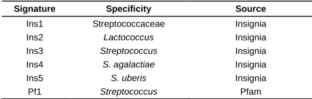
Overall, the bioinformatics analyses carried out, using Insignia and Pfam, allowed the selection of six DNA signatures specific to the target pathogens (Table 4).
Table 1. Sequenced genomes used in Insignia for the selection of a Streptococcaceae-specific region (Ins1).
Sequenced genomes
Streptococcus gordonii str. Challis substr. CH1 Challis Streptococcus pneumoniae CDC3059-06
Streptococcus salivarius SK126 Streptococcus pneumoniae MLV-016
Streptococcus sanguinis SK36 Streptococcus pneumoniae Taiwan19F-14
Streptococcus suis 89/1591 Streptococcus pneumoniae Hungary19A-6
Streptococcus suis 05ZYH33 Streptococcus pneumoniae 70585
Streptococcus suis 98HAH33 Streptococcus pneumoniae JJA
Streptococcus thermophilus LMG 18311 Streptococcus pneumoniae P1031
Streptococcus thermophilus CNRZ1066 Streptococcus pneumoniae G54
Streptococcus thermophilus LMD-9 Streptococcus pneumoniae CGSP14
Streptococcus mutans UA159 Streptococcus pneumoniae TCH8431/19A
Streptococcus agalactiae 2603V/R Streptococcus pneumoniae ATCC 700669
Streptococcus agalactiae NEM316 Streptococcus pneumoniae CCRI 1974M2
Streptococcus agalactiae 18RS21 Streptococcus pyogenes M1 GAS SF370
Streptococcus agalactiae 515 Streptococcus pyogenes MGAS5005
Streptococcus agalactiae H36B Streptococcus pyogenes SSI-1
Streptococcus agalactiae COH1 Streptococcus pyogenes MGAS315
Streptococcus agalactiae CJB111 Streptococcus pyogenes str. Manfredo
Streptococcus agalactiae A909 Streptococcus pyogenes MGAS10394
Streptococcus pneumoniae TIGR4 Streptococcus pyogenes MGAS8232
Streptococcus pneumoniae R6 Streptococcus pyogenes M49 591
Streptococcus pneumoniae D39 Streptococcus pyogenes ATCC BAA-1633
Streptococcus pneumoniae SP3-BS71 Streptococcus pyogenes MGAS6180
Streptococcus pneumoniae SP6-BS73 Streptococcus pyogenes MGAS9429
Streptococcus pneumoniae SP9-BS68 Streptococcus pyogenes MGAS2096
Streptococcus pneumoniae SP11-BS70 Streptococcus pyogenes MGAS10270
Streptococcus pneumoniae SP14-BS69 Streptococcus pyogenes MGAS10750
Streptococcus pneumoniae SP18-BS74 Streptococcus uberis 0140J
Streptococcus pneumoniae SP19-BS75 Streptococcus infantarius subsp. infantarius ATCC BAA-102
Streptococcus pneumoniae SP23-BS72 Streptococcus equi subsp. zooepidemicus ATCC BAA1716
Streptococcus pneumoniae CDC1087-00 Streptococcus equi subsp. equi 4047
Streptococcus pneumoniae CDC1873-00 Lactococcus lactis subsp. cremoris SK11
Streptococcus pneumoniae SP195 Lactococcus lactis subsp. cremoris MG1363
Table 2. Sequenced genomes used in Insignia for the selection of a Streptococcus-specific signature (Ins3).
Sequenced genomes
Streptococcus gordonii str. Challis substr. CH1 Challis Streptococcus pneumoniae CDC0288-04
Streptococcus salivarius SK126 Streptococcus pneumoniae CDC3059-06
Streptococcus sanguinis SK36 Streptococcus pneumoniae MLV-016
Streptococcus suis 89/1591 Streptococcus pneumoniae Taiwan19F-14
Streptococcus suis 05ZYH33 Streptococcus pneumoniae Hungary19A-6
Streptococcus suis 98HAH33 Streptococcus pneumoniae 70585
Streptococcus thermophilus LMG 18311 Streptococcus pneumoniae JJA
Streptococcus thermophilus CNRZ1066 Streptococcus pneumoniae P1031
Streptococcus thermophilus LMD-9 Streptococcus pneumoniae G54
Streptococcus mutans UA159 Streptococcus pneumoniae CGSP14
Streptococcus agalactiae 2603V/R Streptococcus pneumoniae TCH8431/19A
Streptococcus agalactiae NEM316 Streptococcus pneumoniae ATCC 700669
Streptococcus agalactiae 18RS21 Streptococcus pneumoniae CCRI 1974M2
Streptococcus agalactiae 515 Streptococcus pyogenes M1 GAS SF370
Streptococcus agalactiae H36B Streptococcus pyogenes MGAS5005
Streptococcus agalactiae COH1 Streptococcus pyogenes SSI-1
Streptococcus agalactiae CJB111 Streptococcus pyogenes MGAS315
Streptococcus agalactiae A909 Streptococcus pyogenes str. Manfredo
Streptococcus pneumoniae TIGR4 Streptococcus pyogenes MGAS10394
Streptococcus pneumoniae R6 Streptococcus pyogenes MGAS8232
Streptococcus pneumoniae D39 Streptococcus pyogenes M49 591
Streptococcus pneumoniae SP3-BS71 Streptococcus pyogenes ATCC BAA-1633
Streptococcus pneumoniae SP6-BS73 Streptococcus pyogenes MGAS6180
Streptococcus pneumoniae SP9-BS68 Streptococcus pyogenes MGAS9429
Streptococcus pneumoniae SP11-BS70 Streptococcus pyogenes MGAS2096
Streptococcus pneumoniae SP14-BS69 Streptococcus pyogenes MGAS10270
Streptococcus pneumoniae SP18-BS74 Streptococcus pyogenes MGAS10750
Streptococcus pneumoniae SP19-BS75 Streptococcus uberis 0140J
Streptococcus pneumoniae SP23-BS72 Streptococcus infantarius subsp. infantarius ATCC BAA-102
Streptococcus pneumoniae CDC1087-00 Streptococcus equi subsp. zooepidemicus ATCC BAA1716
Streptococcus pneumoniae CDC1873-00 Streptococcus equi subsp. equi 4047
Table 3. Sequenced genomes used in the Insignia bioinformatics tool for the selection of a S. agalactiae specific
signature (Ins4).
Sequenced genomes
Streptococcus agalactiae 2603V/R Streptococcus agalactiae H36B
Streptococcus agalactiae NEM316 Streptococcus agalactiae COH1
Streptococcus agalactiae 18RS21 Streptococcus agalactiae CJB111
Streptococcus agalactiae 515 Streptococcus agalactiae A909
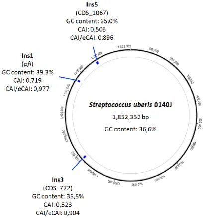
Further in silico analyses were performed on the obtained DNA signatures (Table 4). The circular chromosome map was visualised using Geneious Pro (Drummond et al., 2012), and the position of each marker was determined in Streptococcus uberis 0140J for signatures Ins1, Ins3 and Ins5; in Lactococcus lactis subsp. lactis CV56 for Ins2; and in Streptococcus agalactiae 2603V/R for the Ins4 region. The Codon Adaptation Index (CAI), the expected CAI and GC content were calculated using the CAIcal server (Puigbo et al., 2008).
Table 4. Selected DNA signatures, using Insignia and Pfam.
Signature Specificity Source
Ins1 Streptococcaceae Insignia
Ins2 Lactococcus Insignia
Ins3 Streptococcus Insignia
Ins4 S. agalactiae Insignia
Ins5 S. uberis Insignia
Pf1 Streptococcus Pfam
2. DNA markers design
The selection of DNA markers was based on the previously obtained signatures and carried out using the AlignX utility from the Vector NTI software (Invitrogen, Carlsbad, CA). Primer pairs (Table 5) were designed for each of the six selected regions, using the Vector NTI software (Invitrogen, Carlsbad, CA). All primer pairs were chosen with a predicted amplicon size of 150 to 500 bp and a calculated optimal annealing temperature greater than 50 ºC.
One marker specific to the Streptococcaceae family was selected taking into account the alignment of 15 target sequences (F1). Two markers from the Streptococcus-specific signature (Ins3) were obtained, based on nine available genomes (ST1 and ST2). One Lactococcus-specific marker (LC2) was selected using the alignment of six available sequences. One marker exclusive to S. uberis (SU) was chosen based solely on the Ins3 signature sequence of S. uberis 0140J. Two markers were obtained from a
S. agalactiae-specific signature (Ins4), using five target genomes (A1 and A2). One additional marker (ST3) was selected based on the Streptococcus signature obtained with Pfam (Pf1), using seven target sequences.
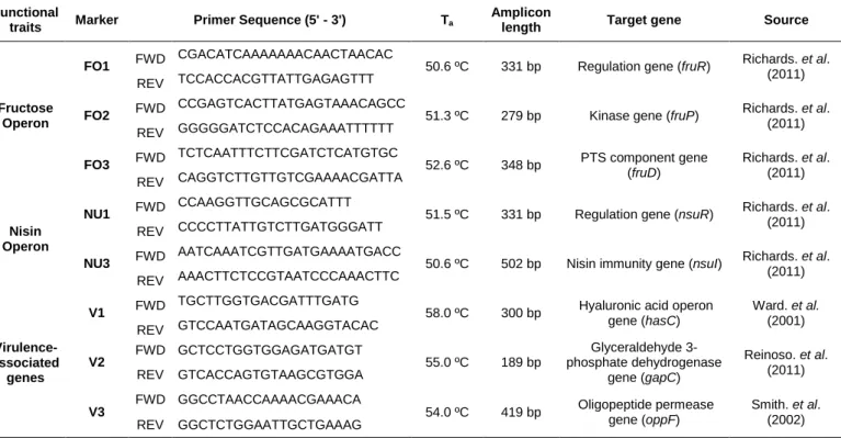
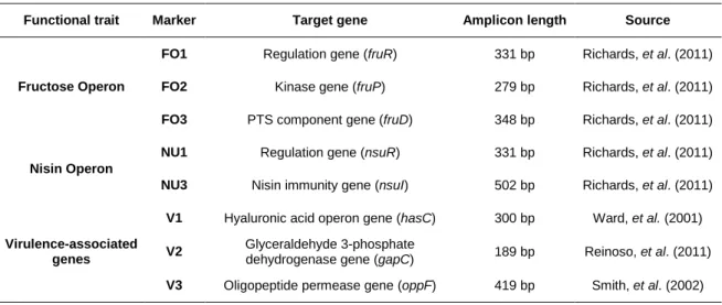
In addition, three primer pairs were designed for genes associated with the fructose operon of S. agalactiae: one for a transcriptional regulator gene (FO1), one for a fructose-1-phosphate kinase (FO2) and another for a phosphotransferase system (PTS) fructose-specific component (FO3). Two primer pairs were also designed for the nisin U operon of S. uberis: one for the gene responsible for the operon regulation (NU1), and another for the gene responsible for immunity to nisin U (NU3).
Primer pairs were designed having as template the sequence of Streptococcus uberis 0140J for primers F1 FWD/REV, ST1 FWD/REV, ST2 FWD/REV and SU FWD/REV; of Lactococcus lactis subsp. lactis CV56 for primers LC2 FWD/REV; of Streptococcus agalactiae 2603V/R for primers ST3 FWD/REV, A1 FWD/REV, A2 FWD/REV; of Streptococcus agalactiae FSL S3-026 for FO1 FWD/REV, FO2 FWD/REV and FO3 FWD/REV; and of Streptococcus uberis strain 42 for primers NU1 FWD/REV and NU3 FWD/REV. Primers were designed to anneal to the sites of each marker that showed higher specificity to the selected targets.
Three additional primer pairs were selected based on virulence-associated genes described in S. uberis (Ward et al., 2001; Smith et al., 2002; Reinoso et al., 2011): the hyaluronic acid operon gene hasC (V1), the glyceraldehyde 3-phosphate dehydrogenase gene gapC (V2) and the oligopeptide permease gene oppF (V3).
A total of 16 taxa-specific and functional markers were obtained for experimental validation (Tables 5 and 6).
Table 5. Selected taxa-specific markers with primer sequence, annealing temperature (Ta), amplicon length and marker
specificity.
Signature Marker Primer Sequence (5' - 3') Ta
Amplicon
length Specificity Source Ins1 F1 FWD TTATGCTCGTCTTGCTCTTTACGG 54.6 ºC 281 bp Streptococcaceae Insignia
REV GCACACGTCCAAGTGATGTAGCTG
Ins2 LC2 FWD TTTATGATTCAAAATTTAACCGCT 51.8 ºC 251 bp Lactococcus Insignia REV TGAATGCCGTATGCTCTTCC
Ins3
ST1 FWD TCCAGTTATGGTGACGCAATATGAT 53.4 ºC 333 bp Streptococcus Insignia REV GCTAAACTAGTATTCGGATGGGCTG
ST2 FWD CATTGGGAAAAGAGTCAGTGTTAG 51.1 ºC 194 bp Streptococcus Insignia REV TGATTCTGGCAATTTCTGTATAAG
Pf1 ST3 FWD GTTATGGATGGCTCCTGGAT 50.7 ºC 265 bp Streptococcus Pfam REV TCCCTAGTCTTAGATAGAACCGTTA
Ins4
A1 FWD ATGTAGCTGCTGATTCTGTCATAA 52.6 ºC 314 bp S. agalactiae Insignia REV AATAGCTGGTGTAGATTTGACTGC
A2 FWD ATGAACACAAAACAGCGTTTTTCA 50.8 ºC 192 bp S. agalactiae and
S. dysgalactiae Insignia
REV AGTAGGTGTCTCATTTGCTATGCT
Ins5 SU FWD TCGTTTGTATACGCTTGATGCT 50.6 ºC 229 bp S. uberis Insignia REV CACGTCTCTATAAAAGGAATTCCC
Table 6. Selected functional markers with primer sequence, annealing temperature (Ta), amplicon length and target
gene.
Functional
traits Marker Primer Sequence (5' - 3') Ta
Amplicon
length Target gene Source
Fructose Operon
FO1 FWD CGACATCAAAAAAACAACTAACAC 50.6 ºC 331 bp Regulation gene (fruR) Richards. et al. (2011) REV TCCACCACGTTATTGAGAGTTT
FO2 FWD CCGAGTCACTTATGAGTAAACAGCC 51.3 ºC 279 bp Kinase gene (fruP) Richards. et al. (2011) REV GGGGGATCTCCACAGAAATTTTTT
FO3 FWD TCTCAATTTCTTCGATCTCATGTGC 52.6 ºC 348 bp PTS component gene (fruD) Richards. et al. (2011) REV CAGGTCTTGTTGTCGAAAACGATTA Nisin Operon
NU1 FWD CCAAGGTTGCAGCGCATTT 51.5 ºC 331 bp Regulation gene (nsuR) Richards. et al. (2011) REV CCCCTTATTGTCTTGATGGGATT
NU3 FWD AATCAAATCGTTGATGAAAATGACC 50.6 ºC 502 bp Nisin immunity gene (nsuI) Richards. et al. (2011) REV AAACTTCTCCGTAATCCCAAACTTC
Virulence-associated
genes
V1 FWD TGCTTGGTGACGATTTGATG 58.0 ºC 300 bp Hyaluronic acid operon gene (hasC) Ward. et al. (2001) REV GTCCAATGATAGCAAGGTACAC V2 FWD GCTCCTGGTGGAGATGATGT 55.0 ºC 189 bp Glyceraldehyde 3-phosphate dehydrogenase gene (gapC) Reinoso. et al. (2011) REV GTCACCAGTGTAAGCGTGGA V3 FWD GGCCTAACCAAAACGAAACA 54.0 ºC 419 bp Oligopeptide permease gene (oppF) Smith. et al. (2002) REV GGCTCTGGAATTGCTGAAAG
3. Bacterial strains, culture conditions and DNA extraction
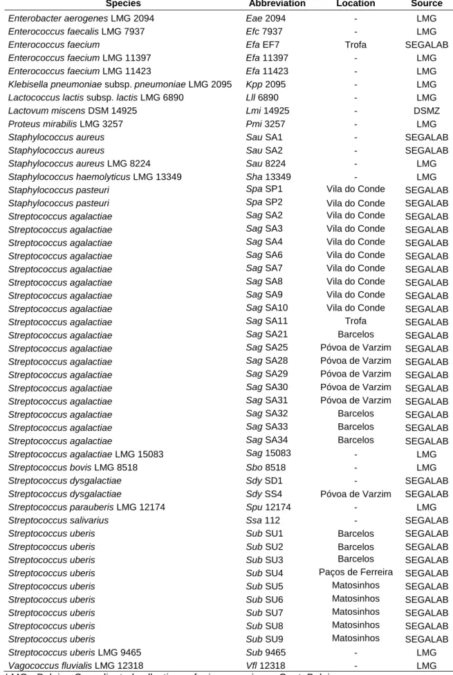
A total of 50 bacterial strains were used in this work, corresponding to 15 reference strains and 35 isolates representative of the Streptococcaceae family, of closely related species and of organisms with common habitats (Table 7). The bacterial isolates, obtained from different mastitic milk samples within Portugal, were provided by SEGALAB (Laboratório de Sanidade Animal e Segurança Alimentar, S.A.). Species identification was done using the VITEK 2 system (bioMérieux, Durham, NC).
All strains were cultured in Brain Heart Infusion (BHI) medium (Oxoid, Hampshire, England) at 37 oC, with the exception of Lactovum miscens DSM 14925, which was cultured in MRS broth medium (Oxoid, Hampshire, England) pre-reduced with cysteine 0.05% and supplemented with N-acetylglucosamine 2 mM at 25 oC, in anaerobic conditions, using the CampyGen Atmosphere Generation System (Oxoid, Hampshire, England).
DNA was extracted from pure bacterial cultures using the EaZy Nucleic Acid (E.Z.N.A.) bacterial DNA purification kit (Omega Bio-Tek, Norcross, GA), following the manufacturer’s instructions. The quality of the extracted DNA was assessed by electrophoresis in 1% agarose gel and each DNA sample was quantified using the Qubit 2.0 Fluorometer HS Assay (Invitrogen, Carlsbad, CA), according to the manufacturer’s instructions.
4. PCR
The PCR mastermix was prepared with 1x DreamTaq buffer, containing 1.5 mM of MgCl2 (Fermentas, Ontario, Canada), 0.2 mM of each deoxynucleoside triphosphate
(dNTP; Fermentas), 0.2 µM of each primer, 1 U of DreamTaq DNA polymerase (Fermentas) and ≈ 25 ng of genomic DNA as template for a total reaction volume of 20 µl. The PCR conditions were as follows: an initial denaturation step of 5 min at 95 °C, followed by 35 cycles of 30 s at 95 °C, 45 s at 55 °C, and 45 s at 72 °C, with a final extension step of 10 min at 72 °C.
Amplification of the 16S rRNA gene (primers listed in Table 8) was performed as mentioned above with 35 cycles of 30 s denaturing at 95 ºC, 30 s annealing at 55 ºC and 90 s extension at 72 ºC .
Table 7. Bacterial strains used in this study.
Species Abbreviation Location Source
Enterobacter aerogenes LMG 2094 Eae 2094 - LMG
Enterococcus faecalis LMG 7937 Efc 7937 - LMG
Enterococcus faecium Efa EF7 Trofa SEGALAB
Enterococcus faecium LMG 11397 Efa 11397 - LMG
Enterococcus faecium LMG 11423 Efa 11423 - LMG
Klebisella pneumoniae subsp. pneumoniae LMG 2095 Kpp 2095 - LMG
Lactococcus lactis subsp. lactis LMG 6890 Lll 6890 - LMG
Lactovum miscens DSM 14925 Lmi 14925 - DSMZ
Proteus mirabilis LMG 3257 Pmi 3257 - LMG
Staphylococcus aureus Sau SA1 - SEGALAB
Staphylococcus aureus Sau SA2 - SEGALAB
Staphylococcus aureus LMG 8224 Sau 8224 - LMG
Staphylococcus haemolyticus LMG 13349 Sha 13349 - LMG
Staphylococcus pasteuri Spa SP1 Vila do Conde SEGALAB
Staphylococcus pasteuri Spa SP2 Vila do Conde SEGALAB
Streptococcus agalactiae Sag SA2 Vila do Conde SEGALAB
Streptococcus agalactiae Sag SA3 Vila do Conde SEGALAB
Streptococcus agalactiae Sag SA4 Vila do Conde SEGALAB
Streptococcus agalactiae Sag SA6 Vila do Conde SEGALAB
Streptococcus agalactiae Sag SA7 Vila do Conde SEGALAB
Streptococcus agalactiae Sag SA8 Vila do Conde SEGALAB
Streptococcus agalactiae Sag SA9 Vila do Conde SEGALAB
Streptococcus agalactiae Sag SA10 Vila do Conde SEGALAB
Streptococcus agalactiae Sag SA11 Trofa SEGALAB
Streptococcus agalactiae Sag SA21 Barcelos SEGALAB
Streptococcus agalactiae Sag SA25 Póvoa de Varzim SEGALAB
Streptococcus agalactiae Sag SA28 Póvoa de Varzim SEGALAB
Streptococcus agalactiae Sag SA29 Póvoa de Varzim SEGALAB
Streptococcus agalactiae Sag SA30 Póvoa de Varzim SEGALAB
Streptococcus agalactiae Sag SA31 Póvoa de Varzim SEGALAB
Streptococcus agalactiae Sag SA32 Barcelos SEGALAB
Streptococcus agalactiae Sag SA33 Barcelos SEGALAB
Streptococcus agalactiae Sag SA34 Barcelos SEGALAB
Streptococcus agalactiae LMG 15083 Sag 15083 - LMG
Streptococcus bovis LMG 8518 Sbo 8518 - LMG
Streptococcus dysgalactiae Sdy SD1 - SEGALAB
Streptococcus dysgalactiae Sdy SS4 Póvoa de Varzim SEGALAB
Streptococcus parauberis LMG 12174 Spu 12174 - LMG
Streptococcus salivarius Ssa 112 - SEGALAB
Streptococcus uberis Sub SU1 Barcelos SEGALAB
Streptococcus uberis Sub SU2 Barcelos SEGALAB
Streptococcus uberis Sub SU3 Barcelos SEGALAB
Streptococcus uberis Sub SU4 Paços de Ferreira SEGALAB
Streptococcus uberis Sub SU5 Matosinhos SEGALAB
Streptococcus uberis Sub SU6 Matosinhos SEGALAB
Streptococcus uberis Sub SU7 Matosinhos SEGALAB
Streptococcus uberis Sub SU8 Matosinhos SEGALAB
Streptococcus uberis Sub SU9 Matosinhos SEGALAB
Streptococcus uberis LMG 9465 Sub 9465 - LMG
Vagococcus fluvialis LMG 12318 Vfl 12318 - LMG
LMG - Belgian Co-ordinated collections of microorganisms, Gent, Belgium.
Table 8. Primers used for the amplification of the 16S rRNA gene. M = A or C; Y = C or T.
Gene Primer Sequence (5' - 3') Source
16S rRNA FWD AGAGTTTGATCMTGGCTCAG Weisburg, et al. (1991)
REV TACGGYTACCTTGTTACGACTT
5. Dot blot validation and automatic image analysis
DNA probes were obtained from purified PCR amplicons, using the GFX PCR DNA and gel band purification kit (GE Healthcare, Buckinghamshire, United Kingdom). The identity of these amplicons was confirmed by sequencing (STAB Vida, Portugal).
Markers F1, ST1, ST2, SU, V1, V2 and V3 were obtained from Streptococcus uberis LMG 9465 (Sub 9465); marker LC2 was obtained from Lactococcus lactis subsp. lactis LMG 6890 (Lll 6890); markers ST3, A1 and A2 were obtained from Streptococcus agalactiae LMG 15083 (Sag 15083); markers FO1, FO2 and FO3 were obtained from Streptococcus agalactiae (Sag SA11) and markers NU1 and NU3 were obtained from Streptococcus uberis (Sub SU3).
Identification of Efa EF7, Spa SP1, Spa SP2 and of the isolates used as marker template (Sag SA11 and Sub SU3) was confirmed by sequencing of the 16S rRNA gene (Fig. S1-S4 of Supplementary data). The sequences obtained of each isolate were analysed by BLAST (Altschul et al., 1990) and on the Ribosomal Database Project (Cole et al., 2009).
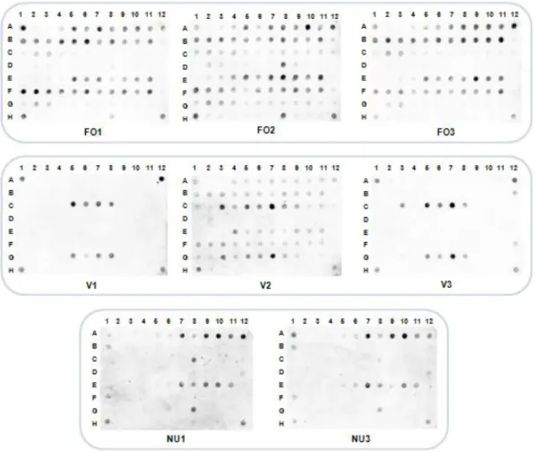
DNA probes were labelled with digoxigenin (DIG), using the DIG-High Prime labelling kit (Roche, Basel, Switzerland) according to the manufacturer’s instructions. For dot blot assays, 100 ng of heat-denatured DNA from each bacterial strain were spotted into a nylon membrane using a Bio-Dot apparatus (Bio-Rad). Hybridisation was carried out overnight at 68 °C, with a final probe concentration of 100 ng/ml. Washing and detection steps were performed according to the DIG system recommendations (Roche). DIG-labelled nucleic acids were detected by chemiluminescence using X-ray films (GE Healthcare) and a Molecular Imager Chemi-Doc system (Bio-Rad).
The analysis of the hybridisation data was complemented with an algorithm developed to automatically process and interpret the dot blot images. This software adjusts each image to a user-defined grid and outputs a probability value of each dot being a
positive signal, using as references the positive and negative controls present in each membrane (Marçal et al., 2009; Caridade et al., 2010).
Results
1. Selection of DNA signatures and in silico analyses
Numerous bacterial species belonging to Streptococcaceae have been associated to bovine mastitis (Wyder et al., 2011). In this work, six DNA signatures specific to different genera and species within this family were obtained.
Five taxonomic regions were selected using Insignia (Phillippy et al., 2007). This utility calculates 20 mer DNA signatures specific to the selected target organisms. The 10 largest signatures obtained for each taxon were analysed for specificity using BLAST (Altschul et al., 1990) and one region was selected for further experimental validation, taking into account the specificity results (i.e. the signature with the lowest E value).
For the selection of one Streptococcaceae-specific signature, a total of 3711 signatures were obtained and ordered by sequence length. The largest signature specific to Streptococcaceae was selected, corresponding to a 300 bp region (Ins1). Other regions exclusive to Lactococcus and Streptococcus were retrieved: a 282 bp signature was selected for Lactococcus (Ins2), the largest and most specific region out of 20288 computed sequences, while a specific 840 bp signature (Ins3) was filtered from 2775 sequences, found particularly unique to mastitis-causing streptococci. Furthermore, two additional signatures were obtained for prevalent mastitis pathogens. A 444 bp signature specific to S. agalactiae (Ins4) and a 400 bp S. uberis-specific sequence (Ins5), the largest out of 71208 and out of 108723 signatures obtained, respectively.
The Protein Family Database was used to obtain one additional Streptococcus-specific signature (Finn et al., 2006). Three protein domains exclusive to Streptococcus were obtained. The primary structure of the proteins was analysed and two multiple copy domains were filtered out. The remaining region of 195 bp (Pf1) was subjected to a specificity analysis using BLAST, which confirmed its specificity towards the Streptococcus genus, particularly S. agalactiae.
Therefore, six DNA signatures specific to taxonomic groups within Streptococcaceae were selected (Table 9).