UNIVERSIDADE DE LISBOA
Faculdade de Medicina Veterinária
Structure and Function of
Novel Carbohydrate-Active Enzymes (CAZymes)
and
Carbohydrate-Binding Modules (CBMs) involved in
Plant Cell Wall degradation
Immacolata Venditto
TESE DE DOUTORAMENTO EM CIÊNCIAS VETERINÁRIAS
ESPECIALIDADE DE CIÊNCIAS BIOLÓGICAS E BIOMÉDICAS
CONSTITUIÇÃO DO JURI ORIENTADOR
PRESIDENTE DO JURI: Doutor Carlos Mendes Godinho de Andrade Fontes
Reitor da Universidade de Lisboa CO-ORIENTADOR
VOGAIS: Doutor Shabir Najmudin
Doutor Pedro Maldonado Coutinho Doutor Marco Moracci
Doutor Luís Manuel dos Anjos Ferreira Doutor José António Mestre Prates
Doutor Carlos Mendes Godinho de Andrade Fontes Doutor Shabir Najmudin
2015 LISBOA
III
‘There is nothing more wonderful than being a scientist,
nowhere I would rather be than in my lab,
staining up my clothes and getting paid to play’
(Marie Skłodowska-Curie)
Ai miei genitori che sono stati un costante sostegno A mio fratello Angelo per i suoi consigli
A Federica Maria per il suo sorriso A Filipe per aver condiviso con me questa esperienza A te Signore che mi sei sempre accanto e mi dai la forza
IV
This work was supported by
the European Union Seventh Framework Programme (FP7 2007‐2013)
under the WallTraC project (Grant Agreement number 263916).
This thesis reflects the author’s views only.
The European Community is not liable for any use
that may be made of the information contained herein.
V
ACKNOWLEDGEMENTS
I would like to thank my supervisor Professor Carlos Fontes for giving me the opportunity to do this PhD, for his guidance throughout my years of PhD. I want to thank Dr. Shabir Najmudin for the X-ray crystallography and I am grateful for his patience and support. I would like to thank Professor Luís Ferreira and Professor Victor Alves for their help in my work.
I am also grateful for collaborations and I want to thank Professor William Willats for my secondment in Copenhagen and Professor Harry Gilbert for my secondment in Newcastle, for their help and valuable contributions to my work. They were very welcoming and I had a great experience in their lab.
I would like to thank Dr. Bernard Henrissat and Professor Pedro Coutinho for their contributions in the project.
I would like to thank all members of WallTraC project for their friendship. I want to thank Alexandre Thebaud for the help, support and patience.
I would like to thank all the people I have worked with in nutrition lab in FMV: Helena Santos, Joana Brás, Ana Luís, Monica Costa, Kate Cameron, Pedro Bule, Teresa Ribeiro, Vânia Fernandes, Virgínia Pires and Professor Maria Centeno.
I would like also to thank D. Maria Paula Silva and Dr. Anabela Gomes for their help and generosity during my years of PhD.
VI
RESUMO
Estrutura e Função de novas glucosil hidrolases (CAZymes) e de Módulos de Ligação a Hidratos de Carbono (CMBs) envolvidos na degradação da Parede Celular Vegetal.
Os polissacarídeos da parede celular vegetal são uma fonte de energia abundante, eficientemente utilizada por um vasto número de microrganismos, os quais desempenham um papel central na recilagem do carbono. As enzimas secretadas pelos microrganismos aeróbicos, que promovem a hidrólise de hidratos de Carbono (CAZymes), funcionam de froma individualizada, ao passo que as bactérias anaeróbicas organizam essas enzimas num complexo multi-enzimático designado por Celulossoma, o qual efetua uma degradação mais eficiente da parede celular vegetal. As CAZymes são enzimas modulares que contêm, além de domínios catalíticos, módulos de ligação a hidratos de Carbono (CBMs) com função não catalítica. Os CBMs direcionam as enzimas a eles ligadas para os substratos-alvo, potenciando assim a catálise. Neste trabalho mostra-se que os CBMs associado à endoglucanase 5A (EcCel5A) da Eubacterium cellulosolvens designados por CBM65A e CBM65B, possuem uma significativa preferência por xiloglucano. A estrutura tridimensional do CBM65B, em complexo com um derivado oligossacárido do xiloglucano e os estudos de mutagenese realizados no CBM65A, revelaram que o mecanismo de preferência destas proteínas pelo xiloglucano se deve ao estabelecimento de interações hidrofóbicas com as cadeias laterais (xilose) deste substrato (capítulo 2). O genoma da bactéria celulolítica do rúmen Ruminococcus flavifaciens, estirpe FD1, codifica um vasto número de putativas proteínas celulosomais, ainda não estudadas. Neste estudo, os genes que codificam os módulos celulosomais de funções desconhecidas foram clonados e as proteínas por eles codificadas foram expressas em níveis elevados em Escherichia coli. Técnicas complementares, combinando eletroforese em gel nativo, uma plataforma de matriz de alta densidade (microarray) e calorimetria de titulação isotérmica, foram usados para identificar novos CBMs em módulos celulosomais de função desconhecida. Esta estratégia permitiu a identificação de 8 novas famílias de CBMs. Foram determinadas as estruturas tridimensionais representativas de duas destas famílias (CBM-A e CBM-B1), e efectuada a sua caracterização funcional detalhada. O CBM-A e o CBM-B1 apresentam um enrolamento em sanduiche β. O CBM-A liga-se ao β-1,4-glucano ramificado através de uma fenda superficial, revelando preferência por xiloglucano. Em contraste, o CBM-B1 revela uma superfície plana complementar a uma fenda aberta que permite a ligação a uma série de glucanos de tipo β, incluindo o reconhecimento de celulose insolúvel (capítulo 3). Por último, a estrutura do CBM46 derivado de uma endoglucanase do Bacillus halodurans designada por BhCel5B, foi determinada. A BhCel5B é uma enzima multi-modular composta por um domínio catalítico da família GH5_4 no terminal N, seguida por um módulo interno do tipo da imunoglobulina (lg) e o CBM46 no terminal C. O BhCBM46 não se liga a polissacarídeos solúveis ou insolúveis. Porém, a estrutura tridimensional da BhCel5B revelou que o CBM46 é parte integrante da fenda onde se alojam os resíduos responsáveis pela catálise da enzima GH5_4 e, por conseguinte, desempenha um papel importante no reconhecimento do substrato (capítulo 4).
Palavras-chave: Enzimas Ativas em Hidratos de Carbono, Módulos de ligação a hidratos de carbono,
VII
ABSTR ACT
Structure and Function of novel Carbohydrate-Active Enzymes (CAZymes) and Carbohydrate Binding Modules (CBMs) involved in Plant Cell Wall degradation.
Plant cell wall polysaccharides offer an abundant energy source efficiently utilized by a large repertoire of micro-organisms, which thus play a central role in carbon re-cycling. Aerobic micro-organisms secrete Carbohydrate-Active Enzymes (CAZymes) as free-standing proteins, whereas anaerobic bacteria organize a diverse enzyme consortium in a multi-component complex, the cellulosome, which performs a more efficient deconstruction of this composite structure. CAZymes are modular enzymes containing, in addition to catalytic domains, non-catalytic Carbohydrate-Binding Modules (CBMs). CBMs direct the appended enzymes to their target substrates thus potentiating catalysis. Here we show that the CBMs of Eubacterium cellulosolvens endoglucanase 5A (EcCel5A), designated as CBM65A and CBM65B, display a significant preference for xyloglucan. The crystal structure of CBM65B in complex with a xyloglucan-derived oligosaccharide, in combination with mutagenesis studies on CBM65A, revealed the mechanism by which these proteins display a preference for xyloglucan by establishing hydrophobic interactions with xyloglucan xylose side chains (Chapter 2). The genome of the ruminal cellulolytic bacterium Ruminococcus flavefaciens strain FD-1 encodes a large number of putative novel cellulosomal proteins. Here, genes encoding cellulosomal modules of unknown function were cloned and their corresponding proteins expressed at high levels in
Escherichia coli. Complementary techniques combining affinity gel electrophoresis, a microarray
platform and isothermal titration calorimetry were used to identify novel CBMs in cellulosomal-modules of unknown function. This strategy allowed the identification of 8 novel CBM families. The structures of representative members of two of these families (CBM-A and CBM-B1) have been solved and detailed functional characterization of these CBMs was performed. CBM-A and CBM-B1 comprise β-sandwich folds. CBM-A binds decorated β-1,4-glucans at a shallow binding cleft and displays preference for xyloglucan. In contrast, CBM-B1 displays a flat surface complementary to an open cleft that allows binding to a range of β-glucans including insoluble cellulose recognition (Chapter 3). Finally, the structure of CBM46 derived from BhCel5B, a Bacillus halodurans endoglucanase, was solved.
BhCel5B is a multi-modular enzyme composed of a GH5_4 N-terminal catalytic domain, followed by
an internal immunoglobulin-like module (Ig) and a C-terminal CBM46. BhCBM46 does not bind soluble or insoluble polysaccharides. However, the crystal structure of BhCel5B revealed that CBM46 is integral to the GH5_4 enzyme catalytic cleft and thus plays an important role in substrate recognition (Chapter 4).
Key-words: Carbohydrate-Active Enzymes, Carbohydrate-binding module, Glycoside hydrolase,
VIII
INTERN ATION AL PEER -REVIEWED P APERS
This thesis was based on the following manuscripts:
Venditto I., Baslé A., Luís A. S., Temple M. J., Ferreira L. M. A., Fontes C. M. G. A., Gilbert
H. J. and Najmudin S. (2013) Overproduction, purification, crystallization and preliminary X-ray characterization of the C-terminal family 65 carbohydrate-binding module (CBM65B) of
endoglucanase Cel5A from Eubacterium cellulosolvens. Acta Cryst. F Structural Biology
Crystallization Communication 69, 191-194.
Luís A. S*., Venditto I*., Temple M. J*., Rogowski A., BasléA., XueJ., Knox P. J., PratesJ. A. M, Ferreira L. M. A., Fontes C. M. G. A., Najmudin S. and Gilbert H. J. (2013) Understanding how non-catalytic carbohydrate binding modules can display specificity for xyloglucan. The Journal of Biological Chemistry 288:4799-4809.
VendittoI., CentenoM. S. J, FerreiraL. M. A, FontesC. M. G. A. and NajmudinS. (2014) Expression, purification and crystallization of a novel carbohydrate-binding module from Ruminococcus flavefaciens Cellulosome. Acta Cryst. F Structural Biology Crystallization Communication, accepted for publication.
VendittoI., GoyalA., Thompson A., FerreiraL. M. A, FontesC. M. G. A. and NajmudinS. (2015) Crystallization and preliminary crystallographic studies of a novel, noncatalytic carbohydrate-binding module from Ruminococcus flavefaciens Cellulosome. Acta Cryst. F Structural Biology Crystallization Communication, accepted for publication.
VendittoI., FernandesV. O, RydahlM. G, BuleP., GoyalA., CentenoM. S. J, FerreiraL. M. A, WillatsW. G, CoutinhoP., HenrissatB., GilbertH. J., NajmudinS. and FontesC. M. G. A. Mining Ruminococcus flavefaciens cellulosome for the discovery of novel families of Carbohydrate-Binding Modules (CBMs). (2014) Work in progress.
.
Venditto I., Santos H., Ferreira L. M. A., Sakka K., Fontes C. M. G. A. and Najmudin S.
(2014) Overproduction, purification, crystallization and preliminary X-ray characterization of
the family 46 carbohydrate-binding module (CBM46) of endo-β-1,4-glucanase B (CelB) from
Bacillus halodurans. Acta Cryst. F Structural Biology Crystallization Communication 70, 754–
757.
Venditto I., Santos H., Sandy J., Sanchez-Weatherby J., Ferreira L. M. A., Sakka K., Fontes
tri-IX
modular endo-β-1,4-glucanase (Cel5B) from Bacillus halodurans. Acta Cryst. F Structural Biology Crystallization Communication, accepted for publication.
VendittoI., NajmudinS., Luís A. S., FerreiraL. M. A, SakkaK., GilbertH. J. and FontesC. M. G. A. Family 46 Carbohydrate-Binding Modules extend the capacity of xyloglucan specific sub-family 5_4 Glycoside Hydrolases to cleave mixed linked glucans. (2014) Work in progress.
X
INDEX
LIST OF FIGURES ... XV LIST OF TABLES ... XVIII LIST OF ABBREVIATIONS AND SYMBOLS ... XIX
1. BIBLIOGRAPHIC REVIEW AND OBJECTIVES ... 1
1.1. Introduction ... 1
1.2. The plant cell wall ... 2
1.2.1. Structure ... 2
1.2.2. Cellulose ... 4
1.2.3. Hemicellulose ... 5
1.2.3.1. Xyloglucan ... 5
1.2.3.2. β-1,3, β-1,4 mixed linked glucans ... 7
1.2.4. Pectin ... 7
1.2.5. Lignin ... 8
1.3. Plant Cell Wall Models ... 8
1.4. Hydrolysis of Plant Cell Wall Polysaccharides ... 9
1.4.1. Carbohydrate-Active enZymes (CAZymes) ... 10
1.4.1.1. Glycoside Hydrolases (GH) ... 11
1.4.1.1.1. Classification and nomenclature ... 11
1.4.1.2. GlycosylTransferases (GTs) ... 15
1.4.1.3. Polysaccharide Lyases (PLs) ... 15
1.4.1.4. Carbohydrate Esterases (CEs) ... 16
1.4.1.5. Cellulases and related enzymes in biotechnology ... 16
1.4.2. Carbohydrate-Binding Modules (CBMs) ... 17
1.4.2.1. CBM Classification ... 18
1.4.2.1.1. Type A CBMs – surface binding. ... 20
1.4.2.1.2. Type B CBMs – endo-type. ... 20
1.4.2.1.3. Type C CBMs – exo-type. ... 21
1.4.2.2. Functional Roles of CBMs ... 22
1.4.2.2.1. Aromatic amino acid side chains ... 23
1.4.2.2.2. Hydrogen bonding and Calcium ... 23
1.4.2.3. Multivalency ... 24
1.4.2.4. Biotechnological applications for CBMs ... 25
1.4.2.5. Using CBMs as molecular probes ... 26
XI
1.5.1. The Cohesin-Dockerin Interaction ... 30
1.5.2. The complexity of Ruminococcus flavefaciens strain FD-1 cellulosome ... 30
1.5.3. Applications of Cellulosomes ... 35
1.6. Objectives ... 36
2. XYLOGLUCAN RECOGNITION BY FAMILY 65 CBMs ... 37
2.1. Overproduction, purification, crystallization and preliminary X-ray characterization of the C-terminal family 65 carbohydrate-binding module (CBM65B) of endoglucanase Cel5A from Eubacterium cellulosolvens ... 37
2.1.1. Introduction ... 37
2.1.2. Material and Methods ... 39
2.1.2.1. Protein Production and Purification ... 39
2.1.2.2. Protein crystallization ... 40
2.1.2.3. Crystallization ... 42
2.1.2.4. Data collection and processing ... 43
2.2. Understanding how noncatalytic carbohydrate binding modules can display specificity for xyloglucan. ... 46
2.2.1. Introduction ... 47
2.2.2. Material and Methods ... 48
2.2.2.1. Protein Production and Purification ... 48
2.2.2.2. Site-directed Mutagenesis ... 49
2.2.2.3. Source of Sugars Used ... 49
2.2.2.4. Affinity Gel Electrophoresis ... 49
2.2.2.5. Isothermal Titration Calorimetry (ITC) ... 50
2.2.2.6. Immunofluorescence Cell Wall Imaging ... 50
2.2.2.7. Crystallization and Data Collection ... 50
2.2.2.8. Model Building and Refinement ... 51
2.2.3. Results ... 53
2.2.3.1. Quantitative Evaluation of the Binding of CBM65A to Its Ligands ... 53
2.2.3.2. Structure of CBM65A ... 59
2.2.3.3. The Ligand Binding Site in CBM65 ... 61
2.2.3.4. Site-directed Mutagenesis of CBM65A ... 62
2.2.3.5. Structural Similarity of CBM65 to Other Proteins ... 65
2.2.4. Discussion ... 65
3. DISCOVERING NOVEL CARBOHYDRATE-BINDING MODULES IN CELLULOSOMES ... 68
3.1. Expression, purification and crystallization of a novel carbohydrate-binding module from Ruminococcus flavefaciens Cellulosome ... 68
XII
3.1.1. Introduction ... 68
3.1.2. Material and Methods ... 69
3.1.2.1. Macromolecule production ... 69
3.1.2.2. Crystallization ... 70
3.1.2.3. Data collection and processing ... 72
3.1.3. Results and discussion ... 74
3.2. Crystallization and preliminary crystallographic studies of a novel, non-catalytic carbohydrate-binding module from Ruminococcus flavefaciens cellulosome. ... 75
3.2.1. Introduction ... 75
3.2.2. Materials and methods ... 76
3.2.2.1. Macromolecule production ... 76
3.2.2.2. Crystallization ... 77
3.2.2.3. Data collection and processing ... 79
3.2.3. Results and discussion ... 80
3.3. Mining Ruminococcus flavefaciens cellulosome for the discovery of novel families of Carbohydrate-Binding Modules (CBMs) ... 82
3.3.1. Introduction ... 83
3.3.2. Material and Methods ... 85
3.3.2.1. CBMs, Polysaccharides and Oligosaccharides ... 85
3.3.2.2. Cloning, Expression, and Purification of cellulosomal proteins of unknown function... 85
3.3.2.3. Site-Directed Mutagenesis ... 87
3.3.2.4. Affinity Gel Electrophoresis (AGE) ... 87
3.3.2.5. Binding to Insoluble Polysaccharide... 87
3.3.2.6. Isothermal titration calorimetry (ITC) ... 88
3.3.2.7. Microarray technology ... 88
3.3.2.7.1. Carbohydrate microarray platform ... 90
3.3.2.8. Crystallization and Data Collection ... 91
3.3.2.9. Structure Determination and Refinement ... 92
3.3.3. Results and discussion ... 95
3.3.3.1. Identification of modules of unknown function in cellulosomal proteins of R. flavefaciens FD-1 ... 95
3.3.3.2. Discovery of novel CBMs within R. flavefaciens FD-1 cellulosome ... 95
3.3.3.3. Crystal Structures of CBM-A and CBM-B1 ... 101
3.3.3.4. Probing the location of the ligand binding sites in CBM-A and CBM-B1 . 105 3.3.3.5. Properties of CBM-A and CBM-B families ... 113
XIII
4. STRUCTURE AND FUNCTION STUDIES ON FAMILY 5 ENDO-β-1,4-GLUCANASE B
(CEL5B) FROM BACILLUS HALODURANS ... 116
4.1. Overproduction, purification, crystallization and preliminary X-ray characterization of the family 46 carbohydrate-binding module (CBM46) of endo-β-1,4-glucanase B (Cel5B) from Bacillus halodurans ... 116
4.1.1. Introduction ... 116
4.1.2. Material and Methods ... 118
4.1.2.1. Protein Production and Purification ... 118
4.1.2.2. Crystallization ... 119
4.1.2.3. Data collection and processing ... 120
4.2. Crystallization and preliminary x-ray diffraction analysis of a tri-modular endo-β-1,4-glucanase (Cel5B) from Bacillus halodurans ... 122
4.2.1. Introduction ... 122
4.2.2. Materials and methods ... 123
4.2.2.1. Macromolecule production ... 123
4.2.2.2. Crystallization ... 124
4.2.2.3. Data collection and processing ... 125
4.2.3. Results and discussion ... 126
4.3. Family 46 Carbohydrate-Binding Modules extend the capacity of xyloglucan specific sub-family 5_4 Glycoside Hydrolases to cleave mixed linked glucans ... 127
4.3.1. Introduction ... 128
4.3.2. Material And Methods ... 130
4.3.2.1. Carbohydrates ... 130
4.3.2.2. Cloning, Expression and Purification ... 130
4.3.2.3. Site-Directed mutagenesis ... 132
4.3.2.4. Affinity-Gel Electrophoresis (AGE) ... 132
4.3.2.5. Isothermal Titration Calorimetry (ITC) ... 132
4.3.2.6. Interaction with insoluble polysaccharides ... 132
4.3.2.7. Enzyme Assays ... 133
4.3.2.8. Thin Layer Chromatography (TLC) ... 133
4.3.2.9. Crystallization and Data Collection ... 133
4.3.2.10. Structure Determination and Refinement ... 134
4.3.3. Results and Discussion ... 136
4.3.3.1. Expression and Purification of BhCel5B and its derivatives ... 136
4.3.3.2. Crystal structure of BhCBM46 ... 136
4.3.3.3. The mechanism by which BhCBM46 binds carbohydrates ... 138
XIV
4.3.3.5. The mechanism by which BhCBM46 modulates the catalytic activity of
GH5_4... 147
4.3.3.6. CBM46 is a monospecific family associated with GH5_4 ... 149
4.3.4. Conclusions ... 153
5. GENERAL DISCUSSION AND FUTURE PERSPECTIVES ... 154
BIBLIOGRAPHIC REFERENCES ... 160 ANNEXES... A
XV
LIST OF FIGURES
Figure 1.1| Schematic representation of the plant cell wall structure. ... 3
Figure 1.2| Structure of a primary cell wall. ... 4
Figure 1.3| Structure of cellulose and schematic representation of cellulose microfibrils. ... 4
Figure 1.4| Schematic representation of xyloglucan. ... 6
Figure 1.5| Representative structure of XXXG- and XXGG-type XYGS. ... 6
Figure 1.6| Structure of mixed linkage glucans. ... 7
Figure 1.7| Schematic structure of pectin. ... 7
Figure 1.8| Schematic structure of a primary cell wall. ... 9
Figure 1.9| Representation of the modular structure of a trypical CAZyme. ... 11
Figure 1.10| Glycoside hydrolases. ... 11
Figure 1.11| Representation of the main fold of catalytic domains of various glycoside hydrolase families. ... 13
Figure 1.12| Enzymatic degradation of polysaccharides... 14
Figure 1.13| The three types of active sites found in glycoside hydrolases. ... 15
Figure 1.14| Representative structure of a Type A CBM. ... 20
Figure 1.15| Representative structure of a Type B CBM. ... 21
Figure 1.16| Representative structure of a Type C CBM. ... 21
Figure 1.17| The three types of binding-site ‘platforms’ formed by aromatic amino acid residues. ... 23
Figure 1.18| The structural features of CBMs that contribute to their carbohydrate specificity... ... 24
Figure 1.19| Schematic representation of cellulosomes bound to cellulose and the cell surface. ... 27
Figure 1.20| Molecular basis for the organization of cellulosomes. ... 29
Figure 1.21| The scaffoldin gene cluster in R. flavefaciens FD-1 and 17. ... 31
Figure 1.22| The complexity of Ruminococcus flavefaciens strain FD-1 cellulosome. ... 31
Figure 1.23| Schematic representation of the proposed cellulosome architecture in R. flavefaciens FD-1 versus strain 17. ... 32
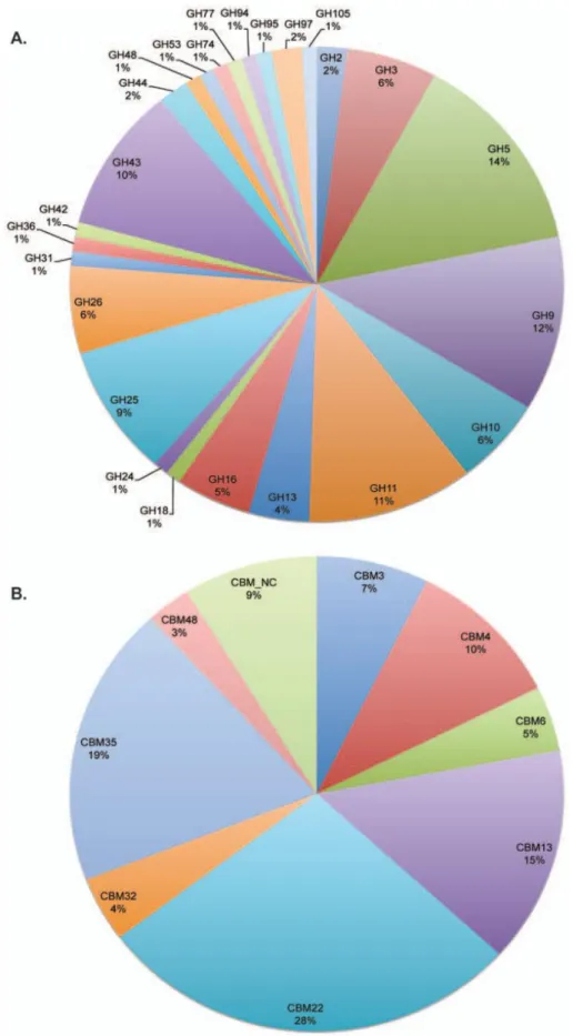
Figure 1.24| Glycoside hydrolase modules and carbohydrate-binding modules detected in R. flavefaciens FD-1. ... 34
Figure 2.1.1| Sequence comparison of CBM65 family members. ... 38
Figure 2.1.2| A coomassie brilliant blue-stained 14% page gel evaluation of protein purity. . 39
Figure 2.1.3| Assembly of crystals. ... 40
Figure 2.1.4| Phase diagram applying to crystal growth. ... 41
XVI
Figure 2.2.1| Schematic of EcCel5A. ... 53
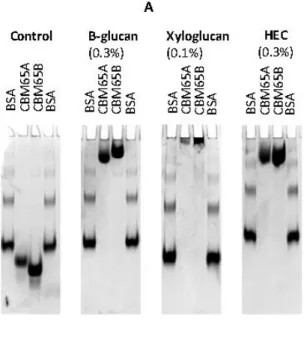
Figure 2.2.2| Examples of affinity gel electrophoresis of CBM65A and CBM65B against soluble polysaccharides. ... 55
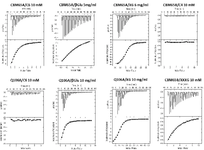
Figure 2.2.3| Representative ITC data of CBM65s binding to soluble ligands. ... 56
Figure 2.2.4| Structure of CBM65A. ... 60
Figure 2.2.5| Immunofluorescence analysis of CBM65a binding to cell walls in situ. ... 64
Figure 3.1.1| Schematic showing the modular architecture of the full-length Ruminococcus flavefaciens glycoside hydrolase family 5 containing protein. ... 69
Figure 3.1.2| A coomassie brilliant blue-stained 16% page gel evaluation of protein purity. . 70
Figure 3.1.3| Crystals of native CBM-Rf1 obtained by both sitting-drop and hanging-drop vapour-diffusion methods. ... 71
Figure 3.2.1| Schematic showing the modular architecture of the full-length Ruminococcus flavefaciens FD-1 RfCel9A. ... 76
Figure 3.2.2| A coomassie brilliant blue-stained 16% PAGE gel evaluation of protein purity. 77 Figure 3.2.3| Crystals of CBM-Rf6A obtained by sitting-drop vapour diffusion method in the crystallisation conditions. ... 78
Figure 3.3.1| Detection of CBMs in glycan microarrays. ... 90
Figure 3.3.2| Affinity gel electrophoresis of R.flavefaciens proteins of unknown function against soluble ligands ... 96
Figure 3.3.3| Affinity of modules of unknown function from R.flavefaciens cellulosome for carbohydrate ligands as detected by microarray analysis. ... 98
Figure 3.3.4| Representative ITC data of CBM-H to soluble ligands. ... 101
Figure 3.3.5| 3D structures of CBM-A (panel A) and CBM-B1 (panel B). ... 104
Figure 3.3.6| Representative ITC and AGE data of CBM-A binding to soluble ligands. ... 106
Figure 3.3.7| Representative ITC and AGE data of CBM-B1 binding to soluble ligands. .... 110
Figure 3.3.8| Binding of CBM-A and CBM-B1 to insoluble cellulose as probed by pull down assays and ITC . ... 111
Figure 3.3.9| Alignments of CBM-A (panel A) and CBM-B1 (panel B) with other family members. ... 114
Figure 4.1.1| Schematic showing the modular architecture of full-length B. halodurans endo-β-1,4-glucanase (Cel5B). ... 117
Figure 4.1.2| A coomassie brilliant blue-stained 16% page gel evaluation of protein purity... ... 118
Figure 4.1.3| Crystals of BhCBM46 (with 10 mm 1,4-β-d-cellohexaose) and SeMet-BhCBM46 obtained by both sitting-drop and hanging-drop vapour-diffusion methods. ... 119
Figure 4.2.1| SDS–page [14%(w/v)] showing overexpression and purification of BhCel5B... ... 124
XVII
Figure 4.3.1| The architectural arrangement of BhCel5B and truncated derivatives produced in this work. ... 130 Figure 4.3.2| 3D structure of BhCBM46. ... 137 Figure 4.3.3| Examples of affinity gel electrophoresis of BhCBM46, BhCel5B_E296A and other mutant derivatives against xyloglucan and β-glucan. ... 138 Figure 4.3.4| Representative ITC data of BhCBM46, BhCel5B_E296A and other derivatives binding to soluble ligands. ... 141 Figure 4.3.5| Binding studies of BhCBM46 against insoluble forms of cellulose. ... 141 Figure 4.3.6| Examples of affinity gel electrophoresis of BhCel5B_E296A and BhCel5B_W501A_F504A_F507A_Y509A_R531A_E296A. ... 143 Figure 4.3.7| 3D structure of BhCel5B... 146 Figure 4.3.8| TLC of BhCel5B and BhGH5-Ig with xyloglucan, barley β-glucan, HEC and CMC.. ... 148 Figure 4.3.9| pH and temperature profile of BhCel5B (panel A) and thermostability of BhCel5B and BhGH5-Ig (panel B). ... 149 Figure 4.3.10| BLAST search of the endo-β-1,4-glucanase B (BhCel5B). ... 150 Figure 4.3.11| Alignment of BhCBM46 with all 45 representatives members of CBM46. .... 151 Figure 4.3.12| Alignment of BhCel5B with 4 proteins displaying an identical molecular architecture. ... 152
XVIII
LIST OF TABLE
Table 1.1| GH clans of related families.
... 12
Table 1.2| Acronyms for genes and encoded enzymes. ... 14
Table 1.3| CBM fold families. ... 18
Table 2.1.1| Data collection statistics. ... 44
Table 2.2.1| Data collection and structure refinement statistics. ... 52
Table 2.2.2| Affinity gel electrophoresis of CBM65A and CBM65B. ... 54
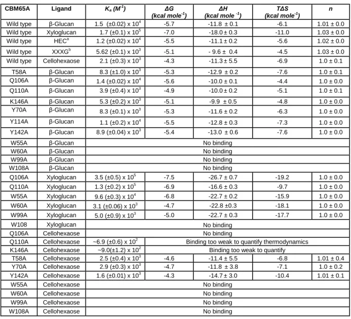
Table 2.2.3| Affinity and thermodynamic parameters of the binding of CBM65A and its variants to polysaccharide and oligosaccharide ligands. ... 58
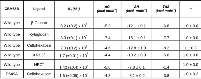
Table 2.2.4| Affinity and thermodynamic parameters of the binding of CBM65B and its variant D649A to polysaccharide and oligosaccharide ligands. ... 59
Table 3.1.1| Data collection and processing ... 73
Table 3.2.1| Data collection statistics. ... 80
Table 3.3.1| Primers used to clone the genes encoding CBM-A and CBM-B1 and to generate their mutant derivatives. ... 86
Table 3.3.2A| Data collection and structure refinement statistics for CBM-A. ... 93
Table 3.3.2B| Data collection and structure refinement statistics for CBM-B1. ... 94
Table 3.3.3| Molecular architecture of enzymes containing novel CBMs and initial biochemical characterization of 9 novel CBMs identified in R. flavefaciens Cellulosome. ... 100
Table 3.3.4| Thermodynamic parameters of the binding of CBM-H to polysaccharide ligands as determined by ITC... ... 101
Table 3.3.5| Thermodynamic parameters of the binding of CBM-A and its derivatives to polysaccharide ligands. ... 107
Table 3.3.6| Thermodynamic parameters of the binding of CBM-B1 and its derivatives to polysaccharide ligands. ... 109
Table 3.3.7| Thermodynamic parameters of the binding of CBM-A and CBM-B1 to regenerated cellulose. ... 113
Table 4.1.1| Data collection statistics. ... 121
Table 4.2.1| Data collection and processing. ... 125
Table 4.3.1| Primers used to clone the genes in the present study. ... 131
Table 4.3.2| Structures statistics. ... 135
Table 4.3.3| Affinity gel electrophoresis of Bh-CBM46, BhCel5B_E296A and other mutant derivatives against soluble polysaccharides. ... 139
Table 4.3.4| Affinity and thermodynamic parameters of the binding of BhCBM46, BhCel5B_E296A and its derivatives to polysaccharide ligands. ... 140
Table 4.3.5| Enzyme kinetics of BhCel5B and BhGH5-Ig against xyloglucan and barley β-glucan.. ... 147
XIX
LIST OF ABBREVIATIONS AND SYMBOLS
% Percentage
Å Angstrom
A600 Absorbance at 600 nanometers
AGE Affinity gel electrophoresis
Ala Alanine (A)
Arg Arginine (R)
Asn Asparagine (N)
Asp Aspartic acid (D)
BhCBM46 Carbohydrate binding module family 46 from Bacillus halodurans
BSA Bovine serum albumin
C. acetobutylicum Clostridium acetobutylicum C. cellulolyticum Clostridium cellulolyticum C. cellulovorans Clostridium cellulovorans
C. josui Clostridium josui
C. thermocellum Clostridium thermocellum
C6 Cellohexaose
CaCl2 Calcium Chloride
Cal Calorie
CAZymes Carbohydrate-active enzymes
CBD Cellulose-binding domain
CBM Carbohydrate-binding module
CBM3a Cellulose binding module
CBM6 Carbohydrate binding module from family 6
CBM62 Carbohydrate binding module from family 62
CBM65 Carbohydrate binding module from family 65
CBM9 Carbohydrate binding module from family 9
CCP4 Collaborative Computational Project Number 4
CE Carbohydrate esterase
Ce3B-Doc Dockerin of the family 3 carbohydrate esterase
Cel44A-doc Family 44 enzyme-borne dockerins
Cel5B Endo-β-1,4-glucanase B
CfCBM4B Family 4 Carbohydrate binding module from Cellulomonas fimi
CipA C. thermocellum Cellulosome integrating protein
CjCBM10 Family 10 Carbohydrate binding module from Cellvibrio japonicus
CjXyn11A GH11 xylanase from Cellvibrio japonicus
CMC Carboxymethyl cellulose
XX
CmLic5A Lichenase 5A from Cellvibrio mixtus
Coh Cohesin
CoMPP Comprehensive Microarray Polymer Profiling
CttA Cellulose-binding protein
Cys Cysteine (C)
Da Dalton
DE Degree of esterification
DNSA 3,5-dinitrosalicylic acid
Doc Dockerin
DTT Dithiothreitol
E. coli Escherichia coli
EC Enzyme Commission number
EcCel5A Endoglucanase from Eubacterium cellulosolvens
EDTA Ethylenediaminetetraacetic acid
ESFR European Synchrotron Radiation Facility
g Gram
GH Glycoside hydrolase
GH43 Glycoside hydrolase from family 43
GH44 Glycoside hydrolase from family 44
GH5 Glycoside hydrolase from family 5
GH9 Glycoside hydrolase from family 9
Gln Glutamine (Q)
Glu Glutamic acid (E)
Gly Glycine (G) GT GlycosylTransferase h Hour H2O Water molecule HCl Hydrochloric Acid HEC Hydroxyethylcellulose
HEPES Hydroxyethyl piperazineethanesulfonic acid
His Histidine (H)
His6-tag Six Histidines tag
HTP High-through put
Ig Immunoglobulin-like module
Ile Isoleucine (I)
IMAC Immobilized Metal Affinity Chromatography
IPTG Isopropyl β-D-1-thiogalactopyranoside
XXI K Kelvin Ka Association constant Km Michaelis constant L Litre LB Luria Bertani LNK Linker Lys Lysine (K) M Molar
mAbs Monoclonal antibodies
MES 2-(N-morpholino)ethanesulfonic acid
Met Methionine (M)
mg Milligram
min Minute
mL Milliliter
mM milliMolar
MPPBS Milk powder dissolved in Phosphate-buffered saline
MR Molecular replacement
n Stoichiometry of binding
NaCl Sodium Chloride
NaHCO3 Sodium bicarbonate
nm nanometer
ºC Celcius degree
PBS Phosphate-buffered saline
PCR Polymerase chain reaction
PD-10 Gel filtration collumns GE Healthcare
PDB Protein data bank
PeCBM29B Family 29 Carbohydrate binding module from Piromyces equi
PEG Polyethylene glycol
pH Negative decimal logarithm of the hydrogen ion activity in a solution
Phe Phenylalanine (F)
PL Polysaccharide Lyase
R Universal gas constant
R. flavefaciens Ruminococcus flavefaciens
RC Regenerated cellulose
SAD Single wavelenght Anomalous Dispersion
ScaA Anchoring scaffoldin
ScaB Anchoring scaffoldin
XXII
ScaE Anchoring scaffoldin
SDS-PAGE Sodium Dodecyl Sulfate-Polyacrylamide Gel Electrophoresis
Se-Met Selenomethionine
Ser Serine (S)
SP Signal peptide
T Absolute temperature
T. reesei Trichoderma reesei
Thr Threonine (T)
TLC Thin layer chromatography
Tris 2-Amino-2-hydroxymethyl-propane-1,3-diol
Tyr Tyrosine (Y)
UNK Domain of unknown function
Val Valine (V)
w/v Weight per volume
XG Xyloglucan
XXXG Xyloglucan heptasaccharide
β-Glu Barley β-glucan
ΔG Gibbs Energy
ΔH Change in Enthalpy of a system
1
1.
BIBLIOGRAPHIC REVIEW AND OBJECTIVES
1.1.
Introduction
Society today faces the challenging problem of finding alternative and renewable energy sources to the conventional and still widely used fossil fuels. Plant cell wall polysaccharides offer an extraordinary source of carbon and energy that can be utilized by various microorganisms, which thus play a central role in the carbon cycle (Bayer et al., 2004). The main components of plant cell walls are cellulose, hemicellulose and lignin. These components form complex structures that provide the plant with physical strength (Somerville et al., 2004). A large repertoire of microorganisms has evolved the capacity to use the energy stored in plant cell wall polysaccharides. These microbes occupy a broad range of habitats: some are free living and rid the environment of such polysaccharides by converting them to the simple sugars that are subsequently assimilated; others are linked closely with cellulolytic animals colonising the digestive tracts of ruminants and other grazers or the guts of wood-degrading termites and worms (Doyle, 1992). In contrast to aerobic microorganisms, which secrete numerous Carbohydrate Active enZYmes (CAZymes) that act individually but in synergy during plant cell wall hydrolysis, a subset of anaerobic bacteria organize cellulases and hemicellulases in multi-enzyme complexes termed cellulosomes. Cellulosomes are highly elaborate nanomachines that degrade cellulose and hemicellulose very efficiently (Fontes & Gilbert, 2010). Construction of multiprotein complexes is one of the key emerging fields in nanotechnology and modern chemistry and cellulosomes represent the blueprint for the construction of recombinant protein complexes that might benefit from enzyme proximity. Notwithstanding the importance of organizing CAZymes in multi-protein complexes to favour carbohydrate re-cycling it is now well established that these enzymes also have a modular architecture. Thus, cellulases and hemicellulases generally contain one or more catalytic domains connected, via linker sequences, to usually more than one non-catalytic Carbohydrate-Binding Modules (CBMs). The practical use of CBMs has been proposed in different fields of biotechnology and the number of published articles and patents reporting novel applications for CBMs is steadily rising. Considering the complexity of plant cell walls, it is becoming apparent that the number of CBM ligand specificities and CAZymes that remain to be discovered may be remarkably large. Recently the genome sequences of several cellulosome producing bacteria have been elucidated. These data reveal the presence of an unprecedented large number of cellulosomal catalytic sub-units, the great majority of those being of unknown function. Since cellulosomes play a key role in plant cell wall deconstruction it is believed that they comprise an extremely interesting source for the discovery of new CAZYmes and CBMs. This work aims to develop novel strategies to discover novel cellulases and hemicellulases in cellulosomes. This introduction begins with a
2
general review of plant cell wall structure. Subsequently, attention is focussed on hydrolysis of plant cell wall polysaccharides. The following subchapters deal with the role of CAZymes (in particular for Glycoside Hydrolases) and CBMs in plant cell wall degradation. Cellulosome complexity and functionality will be analysed, with particular attention to the cellulosome of Ruminococcus flavefaciens, an anaerobic, cellulolytic bacterium that plays an important role in the ruminal digestion of plant cell walls. Finally, this chapter finishes with the identification of the specific objectives of this thesis. Chapters 2, 3 and 4, including the respective subchapters, are organized in papers based on scientific manuscripts, already published or submitted to international journals. Each chapter is composed by an abstract, introduction, experimental procedures, results, discussion and conclusions. Finally, chapter 5 aims to provide a general discussion combining the most insightful findings reported in the experimental chapters.
1.2.
The plant cell wall
The deposition and modification of cell walls play an essential role during plant growth and development. Carbon is incorporated into cell wall polymers, making plant cell walls the most abundant source of terrestrial biomass and renewable energy. Cell wall material is also of great practical importance for human and animal nutrition and as a source of natural fibers for the production of textiles and paper-based products. For these reasons, the study of cell wall synthesis is of considerable interest (Reiter, 2002).
1.2.1. Structure
The plant cell wall is a complex, macromolecular, extracellular matrix that is presented at the surface of the plasma membrane. Plant cell walls consist of multiple layers. The first and most external layer is the middle lamella which is deposited just after cell division. The primary cell wall is formed secondly, over the middle lamella, and is sufficiently dynamic to accommodate both cell growth and development. When cells differentiate or cease growing they may deposit a secondary cell wall, which is formed between the plasma membrane and the primary cell wall. The secondary cell wall is thus the most internal plant cell wall layer and can be deposited in distinct layers, usually termed S1, S2 and S3 (Figure 1.1).
3
Figure 1.1| Schematic representation of the plant cell wall structure.
Adapted from (http://www.ccrc.uga.edu/)
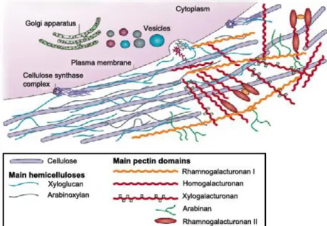
The plant cell wall is composed predominantly of the polysaccharides cellulose, hemicellulose and pectin. In contrast, secondary walls are often rigidified by the impregnation of lignin, a non-glycoside heterogeneous aromatic polymer. Plant cell walls also contain many proteins and glycoproteins, including various enzymes and structural proteins. For example, arabinogalactan proteins are structurally complex molecules found on the plasma membrane and in the cell wall matrix; these enzymes are thought to play important roles in cell recognition and signalling (Ellis et al., 2010).The plant cell wall, on average, contains roughly 40% cellulose, 30% hemicelluloses and 20% lignin (Carpita & Gibeaut, 1993). The exact composition of polysaccharides in cell wall of an individual type of plant varies greatly. Cellulose microfibrils are synthesized by large hexameric complexes in the plasma membrane, whereas hemicelluloses and pectins, which compose the matrix polysaccharides, are synthesized in the Golgi apparatus and are deposited on the wall surface by vesicles. In most plant species the main hemicellulose is xyloglucan, while hemicelluloses such as arabinoxylans and mannans are found in lesser amounts. The main pectin polysaccharides include rhamnogalacturonan I and homogalacturonan, with smaller amounts of xylogalacturonan, arabinan, arabinogalactan I and rhamnogalacturonan II. Pectin domains are believed to be covalently linked together and to bind to xyloglucan by covalent and non-covalent bonds (Figure 1.2) (Cosgrove, 2005).
4 Figure 1.2| Structure of a primary cell wall.
The hemicellulose–cellulose network is shown on the left part of the cell wall without pectins, which are emphasized on the right part of the figure. Adapted from (Cosgrove, 2005).
1.2.2. Cellulose
Cellulose is the major constituent of plant matter and represents the most abundant organic polymer on Earth. Cellulose is a remarkably stable polymer, consisting of a linear polysaccharide of 100 to over 10,000 β-1,4 linked glucose units. Chemically, the repeating unit is simply glucose, but structurally the repeating unit is the disaccharide cellobiose, that is 4-O-(β-D-glucopyranosyl)-D-glucopyranose, since each glucose residue is rotated 180° relative to its neighbour (Fig. 1.3).
Figure 1.3| Structure of cellulose and schematic representation of cellulose microfibrils.
Three parallel chains are shown and a glucose moiety and repeating cellobiose unit are indicated. The arrow indicatesparallel cellulose chains aggregate into crystalline structures called microfibrils. Adapted from (Horn et
5
Hydrogen bonding between different molecules of cellulose allows the assembly of the so called microfibrils of cellulose that generally display a crystalline structure. Crystallinity of the cellulose microfibrils renders this macromolecule non-soluble and thus recalcitrant to enzymatic attack (Horn et al., 2012). Interspersed in the well ordered crystalline regions, cellulose also contains amorphous regions. A measure of the weight fraction of the crystalline regions is one of the most important measurable properties of cellulose that influences its enzymatic digestibility. Many studies have shown that completely disordered or amorphous cellulose is hydrolyzed at a much faster rate than partially crystalline cellulose (Fan et al., 1980). Four different crystalline allomorphs of cellulose (cellulose I, II, III and IV) have been identified. Cellulose I is the most abundant form found in nature. It is known that the crystalline structure of cellulose I is found as parallel chains in the two forms Iα (triclinic) and Iβ (monoclinic) (Atalla & Vanderhart, 1984). Cellulose Iα is the predominant form found in bacteria and algae, whereas the cellulose in higher plants is mostly Iβ. Cellulose II can be prepared by two distinct routes, mercerization (alkali treatment) and regeneration (solubilization and subsequent recrystallization). Cellulose III1 and III2 can be formed from
cellulose I and II,respectively, by treatment with liquid ammonia. Cellulose IV1 and IV2 can be
obtained by heating cellulose III1 and III2 respectively (Mittal et al., 2011).
1.2.3. Hemicellulose
Hemicelluloses are polysaccharides in plant cell walls that have β-(1→4)-linked backbones, including xyloglucans, xylans, mannans, glucomannans and β-(1→3,1→4)-glucans, which may be decorated with a diverse range of carbohydrate side-chains. These types of hemicelluloses are present in the cell walls of all terrestrial plants, except for β-(1→3,1→4)-glucans, which are restricted to Poales and a few other groups (Scheller & Ulvskov, 2010). The main backbone of hemicellulose is usually made of one or two sugars, which determines their classification. For example the predominant hemicellulose in monocots is xylan whose backbone is composed of 1,4-linked-β-D-xylopyranose units. The backbone of galactoglucomannans is made of linear 1,4-linked glucopyranose and β-D-mannopyranose units which may be decorated with α-1,6-linked galactose residues.
1.2.3.1. Xyloglucan
Xyloglucan is the quantitatively predominant hemicellulosic polysaccharide in the primary walls of dicots and non-graminaceous monocots. Xyloglucan may account for up to 20% of the dry weight of the primary wall. Xyloglucans have a main β-D-(1-4)-glucan backbone (denoted as G) generally branched with α(1-6)-linked D-xylopyranosyl (denoted as X) or β-D-galactopyranosyl (1-2)-D-xylopyranosyl residues (denoted as L) and a terminal fucosyl
α-L-6
(1-2) units linked to branching β-D-galactosyl residues (denoted as F) (Del Bem & Vincentz, 2010) (Figure 1.4).
Figure 1.4| Schematic representation of xyloglucan.
Xyloglucan [β-D-Glcp-(1 4)]n backbone substituted with side chains as seen in pea and arabidopsis. Adapted from (Scheller & Ulvskov, 2010).
Xyloglucans are classified as XXXG-type and XXGG-type oligosaccharides considering the type of decorations. XXXG-type has three consecutive backbone residues bearing an α-D-Xylp substituent at O6 and a fourth, unbranched backbone residue. In XXGG-type xyloglucans have two consecutive backbone residues bear an α-D-Xylp substituent at O6, the third and fourth backbone residues are not branched (Figure 1.5).
Figure 1.5| Representative structure of XXXG- and XXGG-type XYGS.
a. XXXG-type XyGs, comprising a Glc4Xyl3 repeating motif with variable branch extensions (bold residues). Tamarind seed XyG and primary cell wall XyGs (for example, from lettuce leaves) are distinguished by the absence of fucose in the former. b. XXGG-type XyGs, comprising a Glc4Xyl2 repeating motif. These XyGs are common to solanaceous species (for example, tomato) and are typified by branches extended with arabinofuranosyl residues. Standard single-letter abbreviations for designating backbone decorations are shown. Adapted from (Larsbrink et al., 2014). c. The Protein Data Bank (PDB) is a key resource for the three-dimensional structural data. Pdb accession number 2YPJ is shown. Glucose residues are blue and the xylose side chains as green.
7 1.2.3.2. β-1,3, β-1,4 mixed linked glucans
β-(1→4)-linked glucans with interspersed single β-(1→3)-linkages are well known in grasses. Mixed linkage glucans are dominated by cellotriosyl and cellotetrasyl units linked by β-(1→3) linkages. The β-(1→3,1→4)-glucans play a role in cell expansion in primary walls and have not been found in dicots but are found throughout Poales (Scheller & Ulvskov, 2010) (Figure 1.6).
Figure 1.6| Structure of mixed linkage glucans.
Linear chain with mixed linkage of β-(1,3) and β-(1,4).
1.2.4. Pectin
The primary roles of cell walls are to give physical strength to the plant and to provide a barrier against the outside environment. The main role of pectin is to participate in these two functions together with the other polymers. Various pectic polysaccharides can be detected in the cell wall, including homogalacturonan (HG), xylogalacturonan (XGA), rhamnogalacturonan I (RGI), and rhamnogalacturonan II (RGII) (Figure 1.7) (Harholt et al., 2010).
Figure 1.7| Schematic structure of pectin.
Pectin consists of four different types of polysaccharides and their structures are shown. HG and RGI are much more abundant than the other components. Adapted from (Harholt et al., 2010).
8
Rhamnogalacturan I consists of alternative residues of α-1,4 D-galacturonic acid and α-1,2 L-rhamnose, and has side branches that contain other pectin domains (primarily arabinan and galactan side chains). Little is known about the function of RGI, it has been suggested that ramnogalacturan I functions as a scaffold to which other pectins, such as ramnogalacturan II and homogalacturonan are covalently attached as side chains (Somerville et al., 2004). Homogalacturonan comprises a linear chain of α-1,4 D-galacturonic acid residues, whereas xylogalacturonan are often methyl esterified and are modified by the addition of xylose branches. Xylogalacturonan has α-1,4 D-galacturonic acid residues that is substituted with β-1,3 xylose. Xylogalacturonans are found in plant cell walls but little is known about the function of the polysaccharide.
Rhamnogalacturonan II is the most complex polysaccharide. RGII is a complex pectin domain that contains 11 different sugar residues and forms dimers through borate (B) esters. It has α-1,4 galacturonic acid backbone, the same as HG. The neutral arabinans and arabinogalactans are also linked to the acidic pectins and it has been proposed that they promote wall flexibility and that they bind to the surface of cellulose (Cosgrove, 2005).
1.2.5. Lignin
Lignin is a heterogeneous, racemic, polydisperse, high-molecular-weight hydrophobic polymer, which consists of no repeating aromatic monomers connected via phenoxylinkages (Lewis & Yamamoto, 1990). Because of its recalcitrant chemical structure and its close association with cellulose and hemicellulose, lignin is an important factor in impeding the biodegradation of these plant polysaccharides. The degradation of lignin is limited to filamentous prokaryotes and fungi under aerobic, oxidative conditions.
1.3.
Plant Cell Wall Models
Over the years, several models have been proposed to explain the organization of plant cell wall components (Keegstra et al., 1973; Carpita & Gibeaut, 1993; Somerville et al., 2004). Most of the models have focused on understanding the organization of components in primary cell walls that would allow regulated reorganization of wall components during cell growth and differentiation. Keegstra et al. in 1973 proposed that polymers from the matrix (xylan, xyloglucan, pectic polysaccharides and structural proteins) were covalently linked and formed a very large macromolecular network. Later a tether model was proposed, where xyloglucan molecules are hydrogen bonded to and cross-link cellulose microfibrils, the pectin polysaccharides and structural proteins occupy the space between xyloglucan chains (Hayashi, 1989). Although this is presently the most popular model, there are two other models proposed: the multicoat model and stratified model. In the multicoat model each
9
cellulose microfibril is coated by a series of less-tighly bound polysaccharides layers (Talbott & Ray, 1992). In the stratified model the cellulose-xyloglucan lamellae are separated by strata of pectic polysaccharides (Ha et al., 1997).
A simplified model of the primary cell wall is represented in Figure 1.8 based on the initial model proposed by Keegstra et al. in 1973.
Figure 1.8| Schematic structure of a primary cell wall.
The orthogonally arranged layers of cellulose microfibrils (green) are tied into a network by the cross-linking glycans (red) that form hydrogen bonds with the microfibrils. The network of cellulose and cross-linking glycans provides tensile strength, while the pectin network resists compression. Cellulose, cross-linking glycans and pectin are typically present in roughly equal amounts in a primary cell wall. Adapted from (Scheller & Ulvskov, 2010).
1.4.
Hydrolysis of Plant Cell Wall Polysaccharides
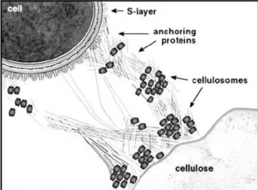
The microbial degradation of the plant cell wall is a fundamental biological process that is of considerable industrial importance. Cell wall polysaccharides, primarily cellulose and hemicellulose, are a major reservoir of carbon and energy. However, only a restricted number of microorganisms have acquired the capacity to deconstruct these structural carbohydrates (Fontes & Gilbert, 2010). The requirement for a consortium of enzymes to achieve total or partial degradation of plant cell wall polysaccharides reflects the physical association of carbohydrates within the plant cell wall, which demands that the catalytic entities act in synergy to degrade this composite structure (Gilbert, 2007). Carbohydrate substrates are often insoluble and microorganisms use extracellular enzymes, free or in complex, to convert the polysaccharides into soluble products that are transportable into the cells (Wilson, 2008).The extracellular organization of the plant cell wall degrading apparatus
10
of aerobic and anaerobic microorganisms is quite different. Aerobic microorganisms produce free enzymes that physically do not associate (Tomme et al., 1995; Warren, 1996). For example Bacillus halodurans is a rod-shaped, Gram-positive bacterium found in soil and water. The bacterium produces many industrially useful alkaliphilic enzymes such as proteases (protein degrading enzyme), cellulases (cellulose degrading enzyme) and amylases (starch degrading enzyme) (Horikoshi, 1999). In most anaerobic microorganisms, the plant cell wall degrading enzymes associate in a supramolecular complex, termed the ‘cellulosome’ (Bayer et al., 1998; Gilbert, 2007; Fontes & Gilbert, 2010). In several organisms, Clostridium thermocellum, Clostridium cellulovorans, Ruminococcus flavefaciens, Acetivibrio cellulolyticus, Clostridium cellulovorans, the cellulosome can be attached to the cell surface. Anaerobic bacteria and fungi in therumen have developed a wide array of multi-modular cellulases and hemicellulases that act individually and as organized cellulosomes for the hydrolysis of plant cell-wall polysaccharides to soluble sugars (Bayer et al., 2004;
Fontes & Gilbert, 2010). For example the rumen anaerobic cellulolytic microbe Eubacterium
cellulosolvens produces a large consortia of cellulases and hemicellulases responsible for plant cellwall degradation (Flint et al., 2008).
1.4.1. Carbohydrate-Active enZymes (CAZymes)
The diversity of complex carbohydrates found in nature is processed by a range of enzymes involved in their assembly (glycosyltransferases) and their breakdown (glycoside hydrolases, polysaccharide lyases, carbohydrate esterases), collectively designated as Carbohydrate-Active enZymes (CAZymes). CAZymes have been classified in sequence-based families for more than 20 years (Lombard et al., 2014). CAZyme families are accessible through the CAZy database (http://www.cazy.org/) that is constantly updated with genomic, proteomic and bibliographic data.
The first defining feature of CAZyme classification is that families are defined based on significant amino acid sequence similarity (usually over 30%) with at least one biochemically characterized founding member (Henrissat, 1991). A second defining feature is that the classification is made module by module. CAZymes are frequently modular proteins containing a catalytic module connected to a variable number of other discrete modules, which can be either catalytic or not (Figure 1.9). The most prevalent non-catalytic modules appended to CAZYmes are Carbohydrate-Binding Modules (CBMs) which bind enzymes to carbohydrates (Figure 1.9). Thus a modular CAZyme can be assigned to several families if its constitutive modules belong to separate families. The third important feature is that the analysis of protein sequences is released daily in GenBank (Lombard et al., 2014). Additionally Henrissat in 1991 noted that the sequence-based families of glycoside hydrolases grouped together enzymes of different substrate specificities (i.e. enzymes with ‘different’ EC numbers) suggesting that acquisition of novel specificities from a common
11
ancestral has been a common occurrence during evolution. The classes of enzymes activities currently covered in CAZy database (www.cazy.org) are: Glycoside Hydrolases (GHs), GlycosylTransferases (GTs), Polysaccharide Lyases (PLs) and Carbohydrate Esterases (CEs).
Figure 1.9| Representation of the modular structure of a typical CAZyme.
CAZymes are modular enzymes, which contain one or more catalytic domains connected, via linker sequences, to usually one or more non-catalytic CBMs.
1.4.1.1. Glycoside Hydrolases (GH)
Glycoside hydrolases (EC 3.2.1.-) are a widespread group of enzymes which hydrolyse the glycosidic bond in di-, oligo and poly-saccharides and are found in all living organisms. Glycoside hydrolases are also referred to as glycosidases, and sometimes also as glycosyl hydrolases (Figure 1.10).
Figure 1.10| Glycoside hydrolases.
Glycoside hydrolases can catalyze the hydrolysis of O-, N- and S-linked glycosides.
Glycoside Hydrolases (GHs) proceed with catalysis via two different mechanisms. Retaining enzymes perform catalysis through a double displacement mechanism, which leads to either transglycosylation or hydrolysis reactions with retention of configuration at the anomeric center. In contrast, inverting enzymes perform catalysis through a single displacement mechanism and do not catalyse transglycosylation reactions but exclusively hydrolysis with the inversion of configuration at the anomeric center (McCarter & Withers, 1994).
1.4.1.1.1. Classification and nomenclature
The International Union of Biochemistry and Molecular Biology enzyme nomenclature (IUB-MB; 1984) is based on the enzymes substrate specificity and occasionally on their molecular mechanism; such a classification does not reflect the structural features of these enzymes. Classification of CAZymes in families is based on amino acid sequence similarity and allows
12
for the integration of both structural and mechanistic features of these enzymes (Henrissat, 1991). Because there is a direct relationship between the amino acid sequence and the folding of an enzyme, this classification reflects the structural features of these enzymes better than substrate specificity alone, helps to reveal the evolutionary relationships between these enzymes and provides a convenient tool to derive mechanistic information from the protein sequence data.
The CAZy database (www.cazy.org) provides a continuously updated list of the GH families. Because the fold of proteins is better conserved than their sequences, some of the families can be grouped in ‘clans’ when new sequences are found to be related to more than one family, when the sensitivity of sequence comparison methods is increased or when structural determinations demonstrate the resemblance between members of different families (Henrissat & Bairoch, 1996) (Table 1.1) (Figure 1.11).
Table 1.1| GH clans of related families.
Clans of Related Families Protein fold Glycoside Hydrolase Families
GH-A (β/α)8 1 2 5 10 17 26 30 35 39 42 50 51 53 59 72 79 86 113 128
GH-B β-jelly roll 7 16
GH-C β-jelly roll 11 12 GH-D (β/α)8 27 31 36
GH-E 6-fold β-propeller 33 34 83 93 GH-F 5-fold β-propeller 43 62 GH-G ( α/α )6 37 63 GH-H (β/α)8 13 70 77 GH-I α+β 24 46 80 GH-J 5-fold β-propeller 32 68 GH-K (β/α)8 18 20 85 GH-L ( α/α )6 15 65 125 GH-M ( α/α )6 8 48 GH-N β-helix 28 49
The GHs catalytic modules are currently classified into 133 different families based on amino acid sequence similarities (March 2014). Adapted from (http://www.cazy.org).
13
Figure 1.11| Representation of the main fold of catalytic domains of various glycoside hydrolase families.
Ribbon representation of the main folds GHs, β-strands are shown in cyan and α-helices in red. Adapted from (Davies & Henrissat, 1995).
In accordance with standard practice in bacterial genetics, genes and their products are designated by three letters. A similar strategy was proposed for the nomenclature of CAZYmes. For example an enzyme from family 5 of glycoside hydrolases will be Cel5 or Man5, depending on its preferred substrate, cellulose or mannan respectively (Table 1.2). If an organism produces multiple enzymes from a family, these will be designated Cel5A, Cel5B, etc., with the letters after the family number corresponding to the order in which the enzymes were reported. If an enzyme contains more than two catalytic domains, the designation would include all of them. For example, endoglucanase CelA from Caldocellulosiruptor saccharolyticum is composed of two cellulases, one from family 9 and the other from family 48. The enzyme will be CsCel9A-Cel48A, written in the conventional sense from the amino- to the carboxyl-terminus (Henrissat et al., 1998).
14 Table 1.2| Acronyms for genes and encoded enzymes.
Enzyme Gene Protein EC designations
Cellulase cel Cel
EC 3.2.1.4; EC 3.2.1.91
Xylanase xyn Xyn EC 3.2.1.8
Mannanase man Man EC 3.2.1.78
Lichenase lic Lic
EC 3.2.1.73; EC 3.2.1.58
Laminarinase lam Lam EC 3.2.1.39
Adapted from (Henrissat et al., 1998)
Cellulases and hemicellulases are family members of the broad group of glycoside hydrolases which in general catalyze the hydrolysis of oligosaccharides and polysaccharides (Warren, 1996). GHs can cleave their substrates in the middle of the chain (endo-acting enzymes) or at the chain ends (exo-acting enzymes) (Davies & Henrissat, 1995; Flint et al., 2008). Distinctions between endo- and exo-acting enzymes, etc. are not absolute; rather, an enzyme has a predominantly exo- or endo-glycolytic mode of action. Particular enzymes may be referred to as endoglucanase Cel5A, cellobiohydrolase Cel6A, cellodextrinase Cel3, and so on (Henrissat et al., 1998) (Figure 1.12).
Figure 1.12| Enzymatic degradation of polysaccharides.
GHs can cleave in the middle of the chain (endo-acting enzymes) or at the chain ends (exo-acting enzymes).
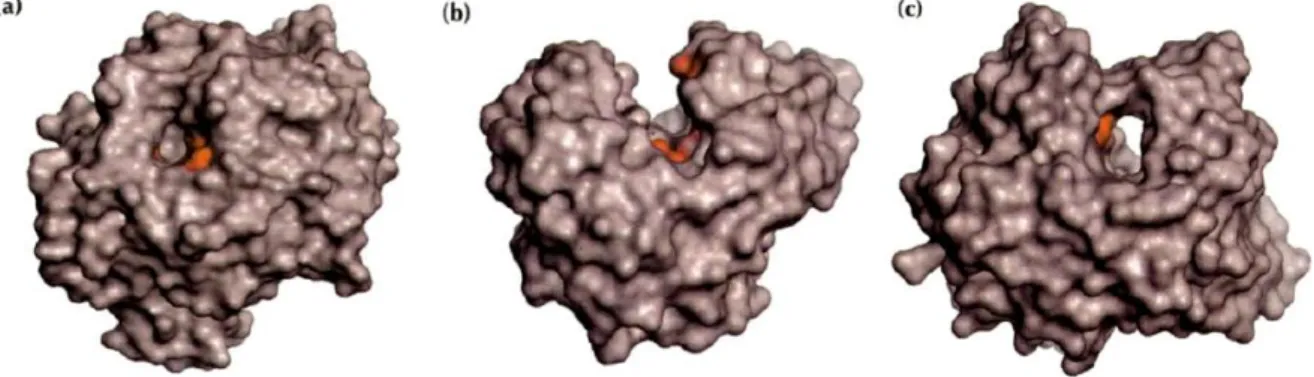
Interestingly, the distinction between endo- and exo-acting enzymes is also reflected by the architecture of the respective class of active site. The overall topologies of the active sites fall into just three general classes. These three topologies (Figure 1.13) can, in principle, be built on the same fold, with the same catalytic residues (Davies & Henrissat, 1995). Endoglucanases, for example, are commonly characterized by the presence of a groove or cleft into which any part of a cellulose chain can fit. Enzymes displaying this topology are mostly endo-acting and cleave randomly along polysaccharide chains producing different sized fragments depending on the composition of the polysaccharide (Davies & Henrissat, 1995). On the other hand, exoglucanases bear tunnel-like active sites, which can only accept a substrate chain via its terminus. The exo-acting enzyme would seem to thread the cellulose chain through the tunnel, where in successive units (e.g., cellobiose) the polysaccharide would be cleaved in a sequential manner. The sequential hydrolysis of a cellulose chain is a
15
notion of growing importance, which has earned the term “processive enzymes” (Davies & Henrissat, 1995). The pocket topography is displayed by exo-acting enzymes which are non-processive. Enzymes displaying this topology usually cleave on side chains of polysaccharides backbone, providing greater access for the endo-acting enzymes (Davies & Henrissat, 1995).
Figure 1.13| The three types of active sites found in glycoside hydrolases.
The catalytic residues are showed in red.a) The pocket or crater found in non processive exo-acting enzymes (glucoamylase from Aspergillus awamori); b) The cleft or groove found in endo-acting enzymes (endoglucanase E2 from Thermononospora fusca); c) The tunnel found in processive exo-acting enzymes (cellobiohydrolase II from Trichoderma reesei).Adapted from (Davies & Henrissat, 1995).
1.4.1.2. GlycosylTransferases (GTs)
Biosynthesis of disaccharides, oligosaccharides and polysaccharides involves the action of different glycosyltransferases (GTs) (EC 2.4.x.y). These enzymes catalyse the transfer of sugar moieties from activated donor molecules to specific acceptor molecules, forming novel glycosidic bonds. Glycosyltransferases can be classified as either retaining or inverting enzymes. Presently 95 families of GT are described in the CAZy database (March 2014).
1.4.1.3. Polysaccharide Lyases (PLs)
Polysaccharide lyases (EC 4.2.2.-) are a group of enzymes that cleave the glycosidic bonds of uronic acid-containing polysaccharide chains via a β-elimination mechanism to generate an unsaturated hexenuronic acid residue and a new reducing end. Cazy database presents a classification of these enzymes in families and subfamilies based on amino acid sequence similarities. These enzymes show a large variety of fold types (or classes), suggesting that PLs have been invented more than once during evolution from totally different scaffolds (Lombard et al., 2010). Currently 23 families of PL are described in the CAZy database (March 2014).