FACULDADE DE FARMÁCIA
POLYMERASE BASIC PROTEIN 1 (PB1) AS A MOLECULAR
DETERMINANT OF FITNESS AND ADAPTATION IN INFLUENZA A VIRUS
Marta Tiago Gíria
Orientadores: Professora Doutora Helena Rebelo de Andrade
Professor Doutor José Moniz Pereira
Tese especialmente elaborada para a obtenção do grau de Doutor em Farmácia,
especialidade Microbiologia.
FACULDADE DE FARMÁCIA
POLYMERASE BASIC PROTEIN 1 (PB1) AS A MOLECULAR DETERMINANT OF FITNESS AND ADAPTATION IN INFLUENZA A VIRUS
Marta Tiago Gíria
Orientadores: Professora Doutora Helena Rebelo de Andrade Professor Doutor José Moniz Pereira
Tese especialmente elaborada para a obtenção do grau de Doutor em Farmácia, especialidade Microbiologia.
Júri:
Presidente: Professora Doutora Matilde da Luz Santos Duque da Fonseca e Castro Vogais: Professor Doutor Paulo Jorge Pereira Cruz Paixão
Professora Doutora Perpétua da Conceição Rodrigues Gomes Cavaco Silva Professor Doutor Miguel Agostinho Sousa Pinto Torres Fevereiro
Professor Doutor José Miguel Azevedo Pereira
Professora Doutora Helena Paula Lopes Henriques Rebelo de Andrade
Marta Gíria teve apoio financeiro da Fundação para a Ciência e Tecnologia através de uma bolsa de Doutoramento (SFHR/BD/65211/2009).
Todas as afirmações efetuadas no presente documento são da exclusiva responsabilidade do seu autor, não cabendo qualquer responsabilidade à Faculdade de Farmácia da Universidade de Lisboa pelos conteúdos nela apresentados.
1
Acknowledgments
I would like to thank everyone that has contributed to making this project possible.
I begin by thanking Prof. Doutora Helena Rebelo de Andrade for being my mentor for the past 16 years, with the most respect and admiration for her scientific knowledge, her perseverance and her impressive ability to resolve and coordinate in extreme and emergency situations. Thank you for your supervision, for your constant guidance and for continuously pushing me forward, but most of all thank you for providing me such different work experiences which have contributed so much to my professional training and to my personal development.
I would like to thank Prof. Doutor José Moniz Pereira for permitting the development of this research project at the Departamento de Microbiologia e Imunologia da Faculdade de Farmácia da Universidade de Lisboa and for co-supervising the work developed.
I would also like to thank Prof. Doutor José Miguel Azevedo Pereira for integrating me in his lab but above all for his constant availability and for being so attentive and considerate.
To Doutora Madalena Almeida Santos from the Hospital de Curry Cabral, Lisboa, I would like to thank the essential contribution to this work by providing the clinical specimens that were a basis of our experimental approach.
Additionally, I would like to thank Dr. Ruben Donis from the Influenza Division of the National Center for Immunization & Respiratory Diseases of the Centers for Disease Control and Prevention, Atlanta, for providing my internship in reverse genetics that has proven determinant for the accomplishment of this research, and Dr. Li-Mei Chen and Dr. Guaniri Mateu-Petit for the attentive supervision.
Also, I acknowledge the Fundação para a Ciência e Tecnologia for the financial support that has permitted the development of this research.
To my colleagues and friends Luis Santos, Vanessa Correia and João Louro, I thank you for your contribution to the experimental aspects of this work. To you and to Anabela Coelho, Paulo Gonçalves and Sónia Pedro, I thank for always accompanying me.
To my colleagues and friends Rita Calado, Marta Calado, Inês Bártolo, João Perdigão, Pedro Borrego, Cheila Rocha, Joana Duarte, Andreia Martins, Francisco Martin, Carla Silva, Ana Rita Dinis, Inês Figueiredo, Inês Moranguinho, Jaciara Guimarães and Claudia Palladino, I particularly thank the extremely relevant and irrelevant conversations that have contributed so much to my support in these past years. João Perdigão, thank you for being so patiently helpful.
To my family…thank you so much. To my parents, for the high standards set and for being so caring. To my sisters Joana, Rita e Sara for being such a strong brotherhood and a reference. Pedro, thank you for having changed my life and for always being with me, my love. Frederica, Vicente and baby, you are my life.
Preface
The research described in this thesis was performed from Abril 2010 to October 2016, under the supervision of Prof. Doutora Helena Rebelo de Andrade and the co-supervision of Prof. Doutor José Moniz Pereira.
All experimental work was performed at the Host-pathogen Interaction Unit, Research Institute for Medicines (iMed.ULisboa), Faculty of Pharmacy, Universidade de Lisboa and the major findings were communicated in the form of research papers published in peer review international journals, oral communications and poster communications. The research on influenza and epidemiology of influenza infections has contributed to strengthen our knowledge and background on emerging infections and public health, which ultimately resulted in the additional authorship of a scientific book chapter.
Papers published in peer review international journals:
Gíria M, Santos L, Louro J, Rebelo de Andrade H. Reverse genetics vaccine seeds for influenza: Proof of concept in the source of PB1 as a determinant factor in viral growth and antigen yield. Virology. 2016; 486:21-27. doi: 10.1016/j.virol.2016.05.015. Epub 2016 May 27.
Gíria M, Rebelo de Andrade H. Genetic evolution of PB1 in the zoonotic transmission of influenza A(H1) virus. Infect Genet Evol. 2014; 27: 234-243. doi: 10.1016/j.meegid.2014.07.024. Epub 2014 Aug 1.
Gíria MT, Rebelo de Andrade H, Santos LA, Correia VM, Pedro SV, Santos MA. Genomic signatures and antiviral drug susceptibility profile of A(H1N1)pdm09. J Clin Virol. 2012 Feb;53(2):140-4. doi: 10.1016/j.jcv.2011.11.002. Epub 2011 Dec 15.
Manuscript in preparation:
Gíria M, Louro J, Rebelo de Andrade H. Compatibility between influenza PB1 and HA: phenotypic evaluation of putative residues for genomic interaction and their relevance in the overall viral fitness. Manuscript in preparation.
The present thesis is based on the previous papers published in peer review international journals and manuscript in preparation.
Oral communications in international conferences:
Gíria M, Rebelo de Andrade H. Reverse genetic vaccine seeds for Influenza, presented at the Influenza Vaccines for the World - Fifth International Conference, 2015, Albufeira, Portugal.
Oral communications in national conferences:
Gíria M, Rebelo de Andrade H. Vírus Influenza do tipo A : Emergência na população humana, presented at the VIII Congresso Científico AEFFUP Doenças Infecciosas Emergentes, 2013, Porto, Portugal.
Gíria M, Rebelo de Andrade H. Compatibilidade funcional do complexo de replicação como determinante de virulência em vírus Influenza A. Presented at Seminários CPM-URIA, Faculdade de Farmácia de Universidade de Lisboa, 2012, Lisboa, Portugal.
Book chapter:
Gíria M, Rebelo de Andrade H, Valadas E. 2014. Doenças infecciosas emergentes. In: Barroso H, Melo Cristino J, Taveira N. Microbiologia Médica. Lidel – edições técnicas, 1.ª Edição, (ISBN:978-972-757-576-3). capítulo 51, pag.541-551.
Poster presentation:
Gíria M, Santos L, Louro J, Rebelo-de-Andrade H. Functional compatibility between PB1 and antigenic proteins as a determinant of viral fitness and adaptation in the A(H1N1)pdm09, presented at OPTIONS IX for the control of influenza, 2014, Chicago, USA.
Other communications:
Papers published in peer review international journals:
Correia V, Santos LA, Gíria M, Almeida-Santos MM, Rebelo-de-Andrade H. Influenza A(H1N1)pdm09 resistance and cross-decreased susceptibility to oseltamivir and zanamivir antiviral drugs. J Med Virol. 2015; 87: 45-56. doi: 10.1002/jmv.23986. Epub 2014 Jul 21.
Santos, LA, Correia V, Gíria M, Pedro S, Santos MM, Silvestre MJ, Rebelo-de-Andrade H. Genetic and Antiviral Drug Susceptibility Profiles of Pandemic A(H1N1)v Influenza Virus Circulating in Portugal. Influenza and Other Respiratory Viruses. 2011; 5 (Suppl. 1), 294?300. doi: 10.1111/j.1750-2659.2011.00221.x
Oral communications in international conferences:
L Santos, V Correia, M Gíria, S Pedro, M Santos, M Silvestre, H Rebelo-de-Andrade. Genetic and Antiviral Drug Susceptibility Profiles of Pandemic A(H1N1)v Influenza Virus Circulating in Portugal, presented at the Options for the Control of Influenza VII, 2010, Hong Kong SAR, China.
Resumo
A Organização Mundial da Saúde e o National Institute of Allergy and Infectious Diseases reportaram deficiências no crescimento de vírus vacinais do subtipo A(H1N1)pdm09, produzidos por genética reversa, que comprometeram a imunização eficiente e atempada durante a pandemia de 2009 e que acentuaram a necessidade de melhorar o processo de produção de vacinas. Esta situação tinha também sido anteriormente detetada em investigação com vacinas pré-pandémicas para o subtipo A(H5N1) e atribuída a uma possível interação menos eficiente entre as proteínas virais. A dinâmica da evolução genética dos vírus influenza A sugere que poderá existir um padrão de cosegregação entre a subunidade Básica da Polimerase 1 (PB1) e as proteínas antigénicas Hemaglutinina (HA) e Neuraminidase (NA). Nos episódios de rearranjo genómico que resultaram na emergência dos vírus pandémicos de 1957 e 1968, os vírus sazonais adquiriram segmentos PB1 juntamente com proteínas antigénicas com origem em vírus aviários. Em 1947 foi identificado um padrão semelhante, em que um episódio de rearranjo entre vírus sazonais envolvendo o segmento PB1 e proteínas antigénicas resultou num aumento da dispersão geográfica face ao que carateriza as epidemias sazonais. Nestas situações, a presença do segmento PB1 homólogo às proteínas antigénicas parece ter favorecido o fitness viral. Em estudos retrospetivos acerca da composição genómica de vírus vacinais de elevado rendimento, produzidos por rearranjo clássico, foi frequentemente detetada a incorporação do segmento PB1 juntamente com os das proteínas antigénicas, sugerindo também que a sua interação poderá ter impacto no fitness viral.
Neste contexto, propusemos avaliar se o segmento PB1 é um determinante molecular do fitness viral, se especificamente a sua compatibilidade com as proteínas antigénicas condiciona o fitness
e a adaptação dos vírus influenza A e, também, se este conceito será passível de ser explorado para melhorar a produção de vírus vacinais. O vírus A(H1N1)pdm09 foi utilizado como modelo nesta investigação por ser um produto de rearranjo com uma combinação única de segmentos com origem aviária, suína e humana. Adicionalmente, o vírus vacinal pandémico apresentou deficiências no crescimento e, uma vez que o A(H1N1)pdm09 se mantém em circulação com um perfil sazonal, permanece a necessidade de produzir vacinas anualmente para fazer face aos frequentes drifts antigénicos.
Os objetivos desta investigação foram definidos como 1) avaliar a evolução genética do segmento PB1 na transmissão zoonótica de vírus suínos e inferir a sua possível contribuição para o fitness viral e a adaptação e 2) determinar se a compatibilidade funcional ou estrutural entre o segmento PB1 e as proteínas antigénicas é um determinante do fitness viral, em protótipo de vírus vacinal A(H1N1)pdm09 utilizado como modelo, produzido por genética reversa. Para a concretização do objetivo 1, foi selecionada uma amostra de sequências nucleotídicas de segmentos PB1 de vírus suínos que infetaram o homem e efetuada uma analise filogenética e uma avaliação de mutações. Os resultados mais relevantes incluíram a confirmação de que é possível identificar a história evolutiva do segmento PB1 no que diz respeito à distinção entre linhagens e hospedeiros. Ainda, foram detetados possíveis marcadores de adaptação a hospedeiros mamíferos no segmento PB1, como os 336I, 361R, 468K e 584Q, e a novas composições genómicas, o que ocorre provavelmente na sequência de episódios de rearranjo, como os 638D e 618D. Os resíduos 298I, 386K e 517V na PB1 foram identificados como possivelmente associados a uma maior compatibilidade entre PB1 e HA do subtipo H1, em hospedeiros mamíferos. O possível papel destes marcadores na adaptação viral foi inferido com base na epidemiologia molecular dos vírus, na localização genómica dos marcadores e nas propriedades dos aminoácidos envolvidos. O impacto fenotípico da aquisição das mutações L298I, R386K e I517V pelo A(H1N1)pdm09 durante a sua história evolutiva, foi também avaliado in vitro através da construção de vírus recombinantes por genética reversa. Os resultados obtidos permitiram-nos sugerir que a aquisição das mutações poderá ter resultado em alterações na conformação da proteína PB1, mas também no aumento do número de nucleótidos complementares aos do segmento HA e que poderão estar envolvidos na interação entre os dois segmentos ao nível do RNA. Por outro lado, verificámos que a aquisição destas mutações teve
um impacto negativo na cinética de crescimento viral in vitro, o que sugere que a interação entre os segmentos genómicos ao nível do RNA pode ser um fator determinante para a cosegregação mas que os mecanismos que lhe estão associados provavelmente não serão dependentes de uma vantagem replicativa. Esta situação seria concordante com um modelo de empacotamento seletivo das partículas virais proposto por outros autores.
Para a concretização do objetivo 2) determinar se a compatibilidade funcional ou estrutural entre o segmento PB1 e as proteínas antigénicas é um determinante do fitness viral em protótipo de vírus vacinal A(H1N1)pdm09 produzido por genética reversa, foi selecionada uma estirpe imunogénica A(H1N1)pdm09 protótipo. Com base nesta estirpe foram construídos protótipos de vírus vacinais com a composição genómica do vírus vacinal, embora contendo o segmento PB1 homólogo ou heterólogo às proteínas antigénicas, e avaliados parâmetros da sua cinética de crescimento e do rendimento em antigénio. Para a seleção de uma estripe imunogénica protótipo foi avaliado o perfil genético de uma amostra de estirpes de A(H1N1)pdm09, que circularam em Portugal durante o período pandémico, e selecionada uma estirpe semelhante ao consenso. Os protótipos de vírus vacinais foram construídos por genética reversa num esqueleto de A/PuertoRico/08/34, com a composição genómica 6:2 do protótipo vacinal clássico (PR8:HA,NA A(H1N1)pdm09) e a composição 5:3 na qual o segmento PB1 incorporado é o da estirpe imunogénica, juntamente com as proteínas antigénicas HA e NA (PR8:HA,NA,PB1 A(H1N1)pdm09). Esta abordagem permitiu identificar que a presença do segmento PB1 homólogo às proteínas antigénicas resultou num aumento significativo na cinética de crescimento viral, capacidade hemaglutinante e atividade da Neuraminidase. Com base nestes resultados, consideramos que poderá ser possível obter uma melhoria significativa do crescimento viral e rendimento em antigénio em protótipos vacinais PR8:A(H1N1)pdm09 produzidos por genética reversa, em comparação com o protótipo vacinal clássico, através da introdução do segmento PB1 da estirpe imunogénica.
Consideramos ainda que, adicionalmente ao papel da subunidade PB1 da polimerase na replicação viral, o segmento genómico PB1 poderá ser um determinante molecular do fitness e um fator determinante na epidemiologia molecular dos vírus influenza, através de interações que estabelece com outros segmentos genómicos ao nível do RNA e da sua aparente capacidade de acumular alterações genéticas adaptativas que são o maior fator impulsionador do fitness
viral. É necessária mais investigação para clarificar os mecanismos de empacotamento viral, o papel das interações ao nível do RNA em estabelecer padrões de cosegregação e as especificidades destas interações de acordo com o subtipo viral. No entanto, a compatibilidade funcional ou estrutural entre proteínas ou segmentos genómicos parece poder ser explorada para aumentar o fitness viral e melhorar a produção de vírus vacinais de outros subtipos. Ainda, uma vez que se reconhece que a composição genómica dos vírus influenza pode ter um impacto significativo no fitness viral, e consequentemente constituir um determinante de virulência, consideramos que incluir a sua análise na avaliação de risco de novas estirpes seria extremamente relevante no contexto sazonal e de ameaça pandémica.
Abstract
The World Health Organization and the National Institute of Allergy and Infectious Diseases reported growth deficits of influenza A(H1N1)pdm09 reverse genetic pandemic vaccine virus seeds. These have compromised the effective and timely distribution of vaccines in the 2009 pandemics and accentuated the need to improve the process of vaccine production. In pre-pandemic A(H5N1) research, seed viruses produced by reverse genetics have also been reported to present growth deficits. These deficits have been attributed to a putative sub-optimal protein interaction. The dynamics of the genetic evolution of influenza A viruses appears to suggest a gene segregation pattern between the Polymerase Basic protein 1 (PB1) and antigenic proteins Hemagglutinin (HA) and Neuraminidase (NA). In the reassortment events that lead to the emergence of the 1957 e 1968 pandemic viruses, the contemporary seasonal viruses acquired PB1 genomic segment together with antigenic glycoproteins originating from avian viruses. A similar pattern was identified in 1947, where a reassortment event between seasonal viruses, involving PB1 and antigenic proteins, has altered the epidemiology of the infection to a near-pandemic geographic dispersion. In both situations, viral fitness appears to have benefitted from acquiring a PB1 genomic segment homologous to antigenic proteins. Also, in retrospective studies on the genomic composition of high yield seasonal vaccine seeds produced by classical reassortment, PB1 is frequently co-incorporated with antigenic proteins HA and NA, further suggesting that the interaction between these proteins could have an impact in viral fitness. In this context, we proposed to address the question of PB1 genomic segment being a molecular determinant of fitness and adaptation in influenza A virus and, particularly, of the functional compatibility between PB1 and antigenic proteins being a driver of the overall viral fitness and
putatively exploitable to improve seed virus production. The A(H1N1)pdm09 virus was used a model for this research because it is a product of viral reassortment with an unprecedented genomic composition of segments originating from avian, swine and human seasonal viruses. Additionally, the 2009 pandemic vaccine virus presented severe growth deficits and, since the A(H1N1)pdm09 persists in circulation with a seasonal epidemiologic profile, the demand for high yield A(H1N1)pdm09 vaccine seeds will be continuous and the need to adequate the immunogenic strain to the circulating viruses will be recurrent because of antigenic drifts. The objectives of this research were defined as 1) to evaluate the genetic evolution of PB1 in the zoonotic transmission of swine influenza virus and infer its putative contribution towards viral fitness and adaptation, and 2) to determine if the functional or structural compatibility between PB1 and antigenic proteins is a molecular determinant of the overall virus fitness in the reverse genetics A(H1N1)pdm09 vaccine seed model.
The approach followed to accomplish objective 1 was to select a study sample of PB1 nucleotide sequences from swine virus that have infected the human host, to analyze phylogeny and mutation trends and to search for putative markers for viral adaptation on the basis of viral molecular epidemiology, genomic location of the polymorphisms and amino-acid properties. Our major findings were that the evolutionary history of PB1 is traceable in terms of lineage and host origin. Specific genomic markers in PB1 appear to putatively relate to the viral adaptation to mammalian hosts, 336I, 361R, 468K and 584Q, and to the viral adaptation to new genomic backgrounds possibly in the sequence of reassortment events, such as 638D and 618D. Residues 298I, 386K and 517V have been found to putatively relate to an enhanced compatibility between PB1 and HA of the H1 subtype, in the mammalian host. A subsequent in vitro investigation of the phenotypic impact of mutations L298I, R386K and I517V acquired by the A(H1N1)pdm09 during its evolutionary history, was performed by generating an A(H1N1)pdm09 recombinant virus and an A(H1N1)pdm09 reassortant in which the specific mutations have been reverted, by reverse genetics. This approach has resulted in two major findings. Acquiring these mutations has been found to putatively promote conformational changes in PB1 and enhance the span of complementary nucleotides possibly involved in PB1 interaction with HA at the RNA level and, on the other hand, has proven detrimental to viral growth kinetics in vitro. These findings have lead us to suggest that the interaction between genomic segments at the RNA level could be a
determinant of co-segregation, concordant with a selective packaging model proposed by other authors, but that the mechanisms that drive this process are probably not dependent on a replicative advantage.
Our approach to accomplishing objective 2) to determine if the functional or structural compatibility between PB1 and antigenic proteins is a molecular determinant of the overall virus fitness in the reverse genetic A(H1N1)pdm09 vaccine seed model, was to determine the genetic profile of A(H1N1)pdm09 strains circulating in Portugal during the pandemic period and select a prototype immunogenic strain, to generate reassortant viruses with the genomic composition of A(H1N1)pdm09 seed viruses prototypes bearing PB1 homologous and heterologous to antigenic proteins, and to evaluate viral growth and antigen yield in vitro. A sample of specimens collected from the pandemic period in Portugal were evaluated for genetic and phenotypic features and a strain similar to the consensus was selected as a prototype strain. Vaccine seed prototypes of the selected A(H1N1)pdm09 strain in an A/PuertoRico/08/34 backbone were generated by reverse genetics to present the genomic compositions of the 6:2 classical vaccine seed (PR8:HA,NA A(H1N1)pdm09) and a 5:3 seed prototype in which the PB1 segment from the immunogenic strain is co-incorporated with the antigenic proteins (PR8:HA,NA,PB1 A(H1N1)pdm09). Our major findings were that the presence of PB1 homologous to antigenic protein significantly increased viral replication, hemagglutination capacity and Neuraminidase activity. We have establishing proof of concept that, in the PR8:A(H1N1)pdm09 seed virus model, viral growth and antigen yield can be significantly improved by the inclusion of PB1 from the immunogenic strain when compared to the classical seed virus prototype.
We consider that, additionally to the role of PB1 protein in viral replication, PB1 genomic segment may be a molecular determinant of the overall virus fitness and a determinant factor in the molecular epidemiology of the viruses by establishing interactions with other segments at the RNA level and by, apparently, being able to genetically change and adapt to improve these interactions. Further research is necessary to clarify the mechanisms of viral genome packaging, the role of interactions at the RNA level in establishing the co-segregation patterns and the specificities of this interactions at the subtype level. However, it becomes clear that the functional compatibility between PB1 and antigenic proteins is a driver of the overall viral fitness in the A(H1N1)pdm09 and is putatively exploitable to improve seed virus production.
We also consider that exploring the concept of the compatibility between gene segments or proteins being a determinant factor in the overall viral fitness, can result in major improvements in the production of reverse genetics seed viruses of different influenza subtypes. Also, being aware of the fact that the genomic composition of influenza viruses can have a major phenotypic impact, and that consequently is a determinant of virulence even though the mechanisms that drive the selective packaging remain unclear, we consider that its inclusion in the risk assessment of influenza strains would be extremely relevant for seasonal and pandemic preparedness.
Abbreviations
µl Microliters
µg Micrograms
A Alanine
aa Amino acid
AIV Avian Influenza Virus
BLAST Basic Local Alignment Search Tool
b bp
Nucleotide base Base pair
C Cysteine
CDC Centers for Disease Control and Prevention
cDNA Complementary Deoxyribonucleic Acid
D Aspartic Acid
dN Non-synonymous substitution
DNA Deoxyribonucleic Acid
dS Synonymous substitution
E Glutamic Acid
ECDC European Centers for Disease Control and Prevention EMA
FDA G
European Medicines Agency Food and Drug Administration Glycine
H Histidine
HA Hemagglutinin
HRBC Human Red Blood Cells
I Isoleucine ILI K Influenza-Like illness Lysine L Leucine LRT M
Lower Respiratory Tract Matrix protein
M Methionine
MDCK Madin Darby Canine Kidney Cells
ml Milliliters
MOI Multiplicity of Infection
mRNA Messenger Ribonucleic Acid
MTS Mitochondrial Targeting Sequence
N Asparagine
NA Neuraminidase
NAI Neuraminidase Inhibitor
NEP NIAID
Nuclear Export protein
National Institute of Allergy and Infectious Diseases
NLS Nuclear Localization Signal
NP Nucleoprotein
NS Non-Structural protein
nt Nucleotide
ORF Open Reading Frame
P Proline
PA Polymerase Acidic protein
PB1 Polymerase Basic protein 1
PB2 Polymerase Basic protein 2
PR8 A/PuertoRico/08/34
Q Glutamine
R Arginine
rfu Relative Fluorescence Units
RNA Ribonucleic Acid
RNP Ribonucleoprotein
RT Reverse Transcription
RT-PCR Reverse Transcription Polymerase Chain Reaction
rtRT-PCR Real Time Reverse Transcription Polymerase Chain Reaction
S Serine
SIV Swine Influenza Virus
SOIV Swine Origin Influenza Virus
T Threonine
TCID Tissue Culture Infectious Dose
TRIG Triple Reassortment Internal Genes
TR-SOIV Triple Reassortant Swine Origin Influenza Virus URT
V
Upper Respiratory Tract Valine
vRNA Viral Ribonucleic Acid
W Tryptophan
WHO World Health Organization
wt Y
Wild-Type Tyrosine
Table of contents
Acknowledgments ... i Preface ... iii Resumo ... vii Abstract ... xi Abbreviations ... xvTable index ... xxiii
1 Chapter 1: General introduction ... 1 1.1 Biology of influenza A viruses and human disease ... 3 1.1.1 Classification ... 3 1.1.2 General organization of the genome and viral proteins ... 3 1.1.3 Life cycle and replication ... 5 1.1.4 Human disease, prevention and treatment ... 6 1.2 Ecology and epidemiology of influenza A viruses ... 9 1.2.1 Reservoir, intermediate hosts and the human-animal interface ... 9
1.2.2 Historical perspective of the emergence of influenza A viruses in the human host and current seasonal epidemiology ... 12 1.2.3 Molecular determinants of virulence and adaptation ... 15 1.2.4 Risk assessment ... 17 1.3 Viral fitness and evolution ... 20 1.3.1 Fitness ... 20 1.3.2 Population diversity of RNA viruses... 21 1.3.3 Generation of diversity in Influenza A viruses ... 22 1.3.4 Gene segregation patterns in influenza A viruses ... 23 1.4 Research context and objectives ... 24
2 Chapter 2: Genetic Evolution of PB1 in the zoonotic transmission of Influenza A(H1) virus ... 29 2.1 Abstract ... 31 2.2 Introduction ... 32 2.3 Methods ... 34 2.4 Results and discussion ... 35 2.5 Conclusions ... 49 2.6 Acknowledgments ... 50 2.7 Statement of author’s contribution ... 50 2.8 Supplementary data ... 51
3 Chapter 3: Compatibility between influenza PB1 and HA: putative residues for vRNA and vRNP interaction and their relevance in the overall viral fitness ... 53 3.1 Abstract ... 55 3.2 Short communication ... 56 3.3 Conflict of interest statement ... 64 3.4 Acknowledgments ... 64
4 Chapter 4: Genomic signatures and antiviral drug susceptibility profile of A(H1N1)pdm09 ... 65 4.1 Abstract ... 67 4.2 Background ... 68 4.3 Objectives ... 68 4.4 Study design ... 69 4.5 Results ... 69 4.6 Discussion ... 73 4.7 Funding ... 76 4.8 Competing interest ... 76 4.9 Ethical approval ... 76 4.10 Acknowledgments ... 76
5 Chapter 5: Reverse genetics vaccine seeds for influenza: proof of concept in the source of PB1 as a determinant factor in virus growth and antigen yield ... 77 5.1 Abstract ... 79 5.2 Introduction ... 80 5.3 Materials and methods ... 81 5.4 Results ... 85 5.5 Discussion ... 91 5.6 Conflict of interest statement ... 93 5.7 Acknowledgments ... 93
6 Chapter 6: General discussion and future research perspectives ... 95 6.1 Limitations of the research ... 97 6.2 Major discussion concepts and future research perspectives ... 99
2
Table index
Table 2.1: Amino acid residues in PB1 protein of swine-origin influenza viruses isolates from the human host ... 39
Table 2.2: Amino acid residues in PB1-F2 protein of swine-origin influenza viruses isolates from the human host ... 40
Table S2.1: Accession numbers of the PB1 nucleotide sequences. ... 51 3
4
Figure index
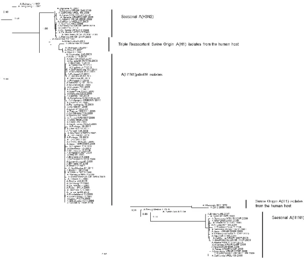
Figure 2.1: Phylogenetic tree of the PB1 coding region of swine-origin influenza viruses isolates from the human host ... 36
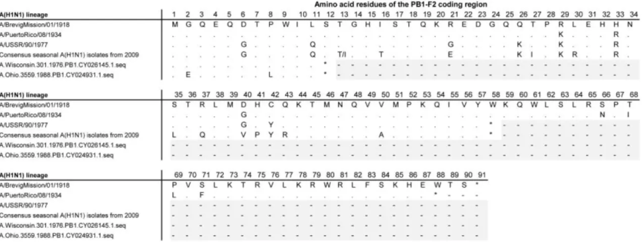
Figure 2.2: Amino acid alignment of PB1-F2 from A(H1N1) lineage viruses ... 37
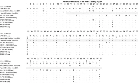
Figure 2.3: Amino acid alignment of PB1-F2 from A(H3N2) lineage viruses ... 38
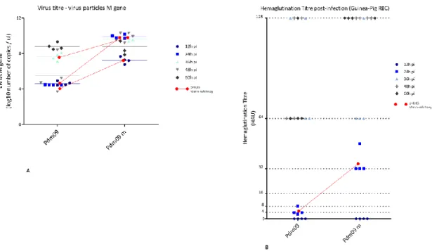
Figure 3.1: Evaluation of the viral growth and the hemagglutination capacity of a A(H1N1)pdm09 wild-type recombinant (pdm09) and a reverse genetic reassortant bearing a PB1 gene to which mutations K386R, V517I and I298L have been induced (pdm09 m) ... 60
Figure 3.2: Evaluation of Neuraminidase activity and Infectious virus titer of an A(H1N1)pdm09 wild-type recombinant (pdm09) and a reverse genetic reassortant bearing a PB1 gene to which mutations K386R, V517I and I298L have been induced (pdm09 m) ... 61
Figure 4.1: Phylogenetic analysis of the amino acids sequences of HA1 subunit of hemagglutinin (A) and neuraminidase (B) genes of Influenza A(H1N1)pdm09 strains circulating in Portugal ... 70
Figure 4.2: Phenotypic evaluation of NAIs susceptibility. IC50 values obtained by fluorescence assay for influenza A(H1N1)2009 ... 71
Figure 5.1: Hemagglutination titer of reverse genetic reassortants PR8, A(H1N1)pdm09 (pdm09), the classical 6:2 seed prototype PR8:HA,NApdm09 and the 5:3 reassortant PR8:HA,NA,PB1pdm09 ... 86
Figure 5.2: Evaluation of viral growth of reverse genetic reassortants PR8, A(H1N1)pdm09 (pdm09), the classical 6:2 seed prototype PR8:HA,NApdm09 and the 5:3 reassortant PR8:HA,NA,PB1pdm09 ... 87
Figure 5.3: Evaluation of antigen yield of the reassortants PR8, A(H1N1)pdm09 (pdm09), the classical 6:2 seed prototype PR8:HA,NApdm09 and the 5:3 reassortant PR8:HA,NA,PB1pdm09 ... 89
Figure 5.4: Evaluation of cell death and apoptosis of the reassortants PR8, A(H1N1)pdm09 (pdm09), the classical 6:2 seed prototype PR8:HA,NApdm09 and the 5:3 reassortant PR8:HA,NA,PB1pdm09 ... 91
1 Chapter 1: General introduction
Chapter 1
1.1 Biology of influenza A viruses and human disease
1.1.1 Classification
Influenza viruses belong to the Orthomyxoviridae family, which includes types A through D distinguished by host range, variability of the antigenic surface glycoproteins Hemagglutinin (HA) and Neuraminidase (NA) and genome organization[1-4]. They are enveloped, pleomorphic viruses with segmented single strand negative RNA (ss(-)RNA)) genomes[1, 2].
Influenza A viruses infect a wide range of mammalian and avian species. They are characterized by the subtype of the antigenic surface glycoproteins and further distinguished into strains. There are 18 subtypes of HA and 11 of NA identified to date[4]. Avian influenza viruses (AIV) are further classified as highly pathogenic (Highly Pathogenic Avian Influenza, HPAI) or low pathogenic (Low Pathogenic Avian Influenza, HPAI), according to its pathogeny and mortality rate in chickens in laboratory setting[4]. Because of the genetic and antigenic diversity, host-range and ability to reassort, influenza A viruses have the potential to cause pandemics[4, 5].
Influenza type B predominantly infects the human host. Type B viruses are classified into lineages and further divided into strains[4]. The genetic diversity is lower when compared to influenza A viruses and because of this and the more restricted host range, influenza B viruses may cause seasonal epidemics but do not cause pandemics[4]. Influenza type C viruses also predominantly infect the human host but are not known to cause epidemics. Type D viruses infects cattle and are not known to infect the human host[4].
1.1.2 General organization of the genome and viral proteins
Influenza A genome is composed of 8 segments that code for up to 15 proteins identified to date, although for some of these proteins the levels of expression in different viral subtypes or different hosts may vary and the exact role has not been clarified[1, 6-8]. Segments range from 890 to 2341 bases (b) long, in a total genome size of 13,5Kb. Each viral RNA (vRNA) segment is composed of a central coding region in antisense orientation. Flanking this region are 3’ and 5’ untranslated short sequences (up to 58nt long) that include a 12nt long and a 13nt long sequence,
respectively, highly conserved between all vRNAs[6]. The vRNA segments are associated with the polymerase complex and the Nucleoprotein (NP) forming viral ribonucleoproteins vRNPs[6]. The segments PB2, PB1 and PA code for the polymerase complex, composed by the Polymerase Basic protein 2 (PB2), Polymerase Basic protein 1 (PB1) and Polymerase Acidic protein (PA), responsible for replication and transcription. PB2 and PB1 genomic segments are 2341b long each. PB1 segment, additionally to PB1 protein, codes for PB1-F2 and N40 proteins. PB1-F2 is only found in infected cells. It is coded from a different open reading frame (ORF), ORF(+1), and is primarily associated with the induction of cellular apoptosis in a late stage of infection. N40 is a truncated form of PB1 at the N-terminal with no ability to bind to PA. It is thought to have a role in the regulation of transcription, but it remains unclear to what extent[1, 2]. PA genomic segment is 2233b long and, additionally to the PA protein, codes for PA-X, PA-155 and PA-182. PA-X is a truncated form of PA which appears to be involved in the modulation of the host response to infection by repressing cellular expression, although further research is necessary to clarify its role[7]. PA-155 and PA-182 are N-truncated forms of the PA protein whose function is not clarified[8].
The HA and NA segments, 1778 and 1413b long respectively, encode the Hemagglutinin and Neuraminidase antigenic transmembrane glycoproteins[1, 2].
The NP segment is 1565b long and encodes the Nucleoprotein, which has the primary function of encapsulating the virus genome for the purposes of RNA transcription, replication and packaging[1, 2].
The M segment is 1027b long and codes for M1 and M2 proteins. M1 is the Matrix protein, the fundamental structural component of the virus particle. It binds the membrane, interacts with both viral RNA and NP of the RNP complex and interacts with components of the host cells mostly for regulatory functions. M2 is an ionic-channel with the main function of regulating pH across the viral membrane, which is important in both the early and late stage of the replication cycle[1, 2]. The NS segment is 890b long and codes for Non-Structural proteins 1 and 2 (NS1 and NS2) which are present in the cell but are not incorporated in the infectious virus particle. NS1 protein is mostly associated with the activation of anti-apoptotic mechanisms in the early stage of cell infection. NS2, also designated as the Nuclear Export protein (NEP), mediates the export of RNP complexes from the nucleus[1, 2].
1.1.3 Life cycle and replication
Transmission of influenza A viruses occurs via aerosols and droplets containing viruses expelled by infected individuals, either by direct or indirect contact and airborne[9].
The virus HA binds to sialic acid (N-acetyl-neuraminic acid) cellular receptors and enters the cell by receptor-mediated endocytoses in an endosome[9, 10]. The acidic pH of the endosome leads to a conformational change in HA exposing the HA2 subunit, which is a fusion peptide that drives the fusion between the endosomal and viral membranes[9, 10]. The acidic pH of the endosome also leads to the opening of the M2 ionic channel, the acidification of the virion and the consequent release of the vRNPs from M1 protein and entry into the cells cytoplasm[9, 10]. The proteins composing the vRNP, PB2, PB1, PA and NP have Nuclear Localization Signals (NLS) which are recognized by the cellular nuclear import mechanisms[9, 10].
The influenza A virus replication process is multigenic. In the nucleus, the negative single strands of RNA are converted into positive sense RNA, designated as cRNA, which is the template for vRNA replication. The viral RNA-dependent RNA polymerase transcribes vRNA to mRNA[9, 10]. The mRNA is transported to the cytoplasm for translation into viral proteins using the cellular mechanisms[9, 10]. Still in the nucleus, the viral RNA-dependent RNA polymerase replicates vRNA which is assembled into vRNP together with the new PB2, PB1, PA and NP proteins, which enter the nucleus after being translated in the cytoplasm. The vRNPs are transported to the cytoplasm and used in the assembly of new virus particles[9, 10]. RNP export from the nucleus occurs via NS2/NEP. M1 protein association with NEP is essential for the export signaling of RNP from the nucleus to the cytoplasm, for protein synthesis, and to prevent reuptake of RNP to the nucleus. The new virus particles are formed using the cellular membrane, where HA, NA and M2 are incorporated[9, 10].
The same gene segments coding for the proteins involved in the replication process, code for additional proteins that are associated with the control of cellular apoptosis during the viral infection. PB1-F2 and NS1 initiate antagonist mechanisms of cell apoptosis in different stages of viral infection. NS1 is associated with the activation of anti-apoptotic mechanisms on the early stage of cell infection. These mechanisms will inhibit programmed cell death induced by the viral
infection, permitting replication to occur[11, 12]. PB1-F2 has a known pro-apoptotic function in the host cells in a later stage of infection, promoting the release of newly formed infectious virus particles. Also, this protein has been associated with an increase of the polymerase activity by binding to the viral polymerase PB1 subunit[13]. The expression of PB1-F2 is a known determinant of virulence, although the mechanisms behind the increased virulence are not fully clarified.
The cassette of internal genes of influenza A virus, coding for non-membrane non-antigenic proteins, therefore controls the major processes by which viral fitness is determined: replication and induction of apoptosis.
1.1.4 Human disease, prevention and treatment
Influenza A viruses infect the human respiratory epithelium and causes a respiratory illness mostly associated with fever, cough, sore throat and fatigue. Influenza can lead to severe complications and death, predominantly in risk groups including children, elderly, pregnant women and individuals with medical conditions such as weakened immune systems, extreme obesity or chronic diseases[14]. It is highly contagious, with an estimated reproduction number (R) of 1,28 secondary cases generated for typical infectious case in seasonal epidemics (seasonal influenza addressed in section 1.2 Ecology and epidemiology of influenza A viruses of the General Introduction chapter)[15]. Pandemic influenza viruses are associated with the introduction of new antigenic proteins in the human host and the absence of protective immunity can lead to an increase in the number of cases. This has occurred in the past pandemics, where R has ranged from 1,46 to 1,8 secondary cases generated for typical infectious case (pandemic influenza is reviewed in section 1.2 Ecology and epidemiology of influenza A viruses, of the General Introduction chapter)[15].
Vaccination against influenza is recommended by the World Health Organization (WHO) as the most effective measure to prevent infection and severe outcomes in risk groups with increased risk of exposure, such as the health care workers, and in risk groups for developing severe complications, identified before[16]. Influenza vaccines have to be reformulated in a regular basis, given the genetic evolution and the molecular epidemiology of the viruses (the genetic
evolution and epidemiology of influenza A viruses is reviewed in sections 1.3.3 Generation of diversity in Influenza A viruses and 1.2.2 Historical perspective of influenza A emergence in the human host and current seasonal epidemiology, of the General Introduction chapter). Particularly, in seasonal vaccination, the selection of strains that compose the vaccine needs to be addressed annually because of viral antigenic drifts, to avoid mismatches and to maximize immune protection for every season, for both southern and northern hemispheres[16]. Also, new antigenic glycoproteins are recurrently introduced in the human host by zoonotic transmission and novel vaccines may have to be developed for the prevention or mitigation of the infection by these emergent immunogenic strains[16].
Currently, Inactivated Influenza Vaccines (IIV), such as whole virus, split and subunit vaccines, and Live Attenuated Influenza Vaccines (LAIV) are licensed and available[16, 17]. IIV consist of influenza vaccine viruses propagated in embryonated eggs or cell cultures and subsequently inactivated[18, 19]. These constitute whole virus vaccines, which can also be disrupted and used as subvirion preparations designated as split vaccines, retaining the immunogenic characteristics but being less reactive when compared to whole virus[18]. Split vaccines have been the most extensively used in seasonal influenza[20]. For subunit vaccines, the antigenic proteins HA and NA are isolated and further purified, also retaining immunogenicity and reduced reactivity[19]. All IIV are recommended for intramuscular administration[18]. LAIV consist of vaccine viruses attenuated to cold adaptation and temperature sensitive features, which permits its replication in the nasopharynx but limits it in the lower respiratory tract (LRT)[19, 21]. LAIV are administered intranasally and can also induce mucosal immunity in addition to antibody response[20].
IIV and LAIV are intended to induce the production of antibodies directed towards the HA protein, which contribute primarily to prevent the illness, and the NA protein, contributing to reduce the severity[18, 20]. Because these are highly variable proteins, the effectiveness of the vaccines may be reduced due to mismatches with the current circulating strains, as referred above[20]. Given the restriction in the use of LAIV in risk groups for developing severe complications or their care givers because of virus shedding, these vaccines have not been recommended by the Advisory Committee on Immunization Practices (ACIP) for the forthcoming 2016-2017 season[20, 22].
Most seasonal influenza vaccines include two influenza A strains, of the A(H1N1) and the A(H3N2) subtypes, and a type B strain, which are selected based on prediction of strains likely to circulate in the subsequent influenza season (current seasonal epidemiology of influenza A viruses reviewed in the 1.2.2 Historical perspective of influenza A emergence in the human host and current seasonal epidemiology section). Quadrivalent vaccines including 2 influenza type B strains of the major lineages circulating have also been licensed in the Unites States of America in 2012 and in Europe in 2013[16, 17, 23]. In Europe, and particularly at the national level, vaccination with IIV is recommended to risk groups of developing serious complications, such as chronic patients, individuals >65 years and pregnant women, and to risk groups for transmitting the infections such as the health professionals and other care givers[24, 25].
Antivirals are available for treatment, but the recommendation for their use differs worldwide, from a more generalized use to reduce the probability of developing illness, as in the United States, to a more restricted use in situations where the vaccine may fail or is not recommended, as occurs in Europe. Specifically, at the national level, the use of antivirals is recommended by the General Directorate of Health to prevent severe complications or to prevent transmission by health professionals to risk groups[26-28].
The two major classes of antivirals are Neuraminidase Inhibitors (NAI) and M2 Inhibitors. Currently, the European Medicines Agency (EMA) approved influenza antiviral drugs are NAI Oseltamivir and Zanamivir, whose mechanisms of action is to block NA function preventing the release and spread of infectious virus particles. The Food and Drug Administration (FDA) has licensed a third NAI, Peramivir[26, 29, 30]. Although some strains with reduced inhibition for Oseltamivir have been detected worldwide, most viruses analyzed from the 2015/2016 influenza season were susceptible to Oseltamivir and all were susceptible to Zanamivir[30, 31]. Because of structural differences, mutations associated with a reduced inhibition are not necessarily the same for these drugs and may not have the same effect in all influenza subtypes[29]. Influenza virus circulating in the 2015/2016 season continued to be susceptible to Neuraminidase Inhibitors and their use is recommended for the forthcoming season of 2016/2017[26, 32].
The mechanisms of action of M2 Inhibitors is to specifically block the ion channel function of the M2 protein, which consequently interferers with the steps of the life cycle that involve this protein.
These have been the first class of antivirals developed but its effectiveness has been limited by the rapid spread of drug resistant mutations. All viruses analyzed from the 2015/2016 influenza season were resistant to the two FDA and EMA approved M2 Inhibitors Amantadine and Rimantadine, and their use is not recommended for the forthcoming season of 2016/2017[26, 30, 32].
1.2 Ecology and epidemiology of influenza A viruses
1.2.1 Reservoir, intermediate hosts and the human-animal interface
Wild aquatic birds are the primary natural reservoir of influenza viruses. All influenza A subtypes circulate in this reservoir, with the exception of H17N10 and H18N11 which have only been identified in bats. Consequently, this species may also be acting as a reservoir[33]. In the avian reservoir, infection by most subtypes is asymptomatic or causes mild disease but HPAI subtypes such as H5 or H7 can cause severe symptoms and death. AIV spread worldwide because of migrations and transmit to domestic poultry where they become closer to human and other animal hosts. AIV are considered species-specific but interspecies transmission occasionally occurs and the avian reservoir is thought to be the source of influenza A virus in other animal species[34, 35]. Additionally to the human host, a broad range of different animal hosts have been infected with influenza A viruses such as swine, poultry, canine, equine, marine mammals and small number of other incidental hosts[33, 36].
The major risk associated with the zoonotic transmission of AIV is the possibility of these viruses being able to adapt and effectively transmit within the human host. This may have been the origin of the 1918 influenza pandemics, described in section 1.2.2 Historical perspective of the emergence of influenza A viruses in the human host and current seasonal epidemiology, although aspects of the interspecies transmission profile between the avian reservoir and the human and swine host populations remain unclear.
AIV subtypes H5, H6, H7, H9 and H10 have intermittently infected the human host in 1996, 1997, 2003-5, 2007, 2012-4 and 2016[37-45]. Most infections have been associated with the A(H5N1) subtype, over 800 from 2003 to 2016 with an approximate 50% fatality rate, and the A(H7N9)
subtype, which has caused over 570 human infections since 2013, with a fatality rate of approximately 40%[46-48].
The zoonotic transmission of AIV was thought to require an intermediate host, such as the swine, to permit the adaptation of the virus to effectively replicate and transmit within the human host [49, 50]. However, in 1997, during an outbreak of avian A(H5N1) in China, direct transmission from poultry to the human host was recognized for the first time and the intermediate infection of swine may now be considered a facilitating factor other than a requirement for human infection with an AIV[18, 19].
The pathogenicity in poultry is not necessarily reflected in the human host. HPAI A(H5N1) is highly pathogenic in humans but the LPAI A(H7N9), for example, has also been found to cause severe human disease and death, and both LPAI and HPAI A(H7) strains have been associated with conjunctivitis and influenza-like-illness (ILI) [46, 51, 52]. Also, there is insufficient data to clarify if the human cases of infection are proportional to the level of circulation of these viruses in the reservoir or if some subtypes infect the human host more easily[51]. AIV A(H10N7) and A(H10N8), for example, have caused human infections but its circulation in the avian reservoir appears to be very limited[53-55].
Most of the reported cases of zoonotic transmission of AIV occurred in China and the Middle East, where the viruses circulate among poultry, subsequently to close exposure to infected poultry or contaminated environments, and this remains the primary risk factor for human host infection[56]. The situations where human-to-human transmission have been detected were occasional, associated with very close, unprotected and prolonged contact between an infected individual and a caregiver, and eventually self-exhausted[56, 57].
In parallel to the threat of direct human infection with an AIV, and its adaptation to replicate and transmit within the human host, and of the infection of an intermediate mammalian host where this adaptation can occur, AIVs pose an additional threat. Their reassortment with a human, or other mammal adapted influenza virus, may result in the emergence of a new virus with an already established ability to effectively transmit within the human host. This has been the origin of the 1957 and 1968 pandemics, described in section 1.2.2 Historical perspective of the emergence of
influenza A viruses in the human host and current seasonal epidemiology of the General Introduction chapter.
The swine population, additionally to possibly acting as an intermediate host to human infection with AIV and facilitate the adaption of the virus to a mammalian host, has also been considered a possible missing vessel, where avian and human or swine viruses can reassort. Swine can be naturally infected with avian and human viruses, because of the sialic acid receptors expressed, and co-infection can potentiate reassortment. However, the human and swine epithelia has been proposed to have similar receptor distribution, which raises the possibility of human host also acting a mixing vessel[58, 59].
Swine influenza virus (SIV) subtypes A(H1N1) and A(H3N2) circulate and cause outbreaks in the swine population, mostly reported in the United States. SIV do not generally infect the human host but sporadic infection with swine subtypes A(H1N1), A(H3N2) and with viral reassortants and genetic variants of these subtypes, have occurred[60-62]. Because it is clinically similar to infection with seasonal viruses, the majority of cases are presumed to remain undiagnosed. There were over 50 cases reported from 1970 to 2000 by SIV A(H1N1) and 11 notifications of human infection by a Triple-Reassortant SIV A(H3N2) since 2005. In 2009 the emergence of a SIV A(H1N1) caused a pandemic in the human host, designated as A(H1N1)pdm09, as described in section 1.2.2 Historical perspective of the emergence of influenza A viruses in the human host and current seasonal epidemiology. After this period, since 2011, approximately 300 cases of human infection with a genetic variant of SIV A(H3N2) which has acquired the M gene from the A(H1N1)pdm09 virus, A(H3N2)v, have been detected[51, 63-66]. In 2015 and 2016, 7 cases of human infection with SIV A(H1N1) and A(H3N2)v were reported[45, 67]. The rise in the number of cases of human infection with SIV is mostly attributed to an enhancement in the surveillance system. Mostly all cases of zoonotic transmission have occurred by close contact with infected swine, in the United States, and, with the exception of the 2009 pandemics, only limited human-to-human transmission has been detected[62, 68].
Infection of the swine host with avian A(H9N12) and A(H4N6) viruses have also been reported, but the viruses do not seem to be enzootic in pigs and no zoonotic transmissions to the human host has been detected[69, 70].
The transmission of influenza virus from the human to the swine host has also been shown to occur[71]. Since 1990, at least 20 introductions of human seasonal viruses in the swine population have been detected in the United States and, between 2009 and 2011, approximately 50 introductions of the A(H1N1)pdm09 pandemic virus from the human host to the swine population were reported globally[72].
Other animal hosts include horses and dogs. Influenza A subtype A(H3N8) circulate in horses causing outbreaks and has been transmitted to the canine host where it now co-circulates with an A(H3N2) subtype[73]. In 2011, an outbreak of infection with A(H3N8) was also detected in seals. A(H3N8) is thought to have been transmitted directly from avian species, because of the genetic homology to the avian virus and because similar mammal adaptive mutations were found in these hosts[74].
The biodiversity of the reservoir and animal hosts is a determinant source of emergent subtypes. On the other hand, the genetic diversity and evolutionary dynamics of influenza viruses are crucial factors that may permit the efficient zoonotic transmission and the sustained transmission in the new host species. Nevertheless, the human activity remains a critical factor driving the emergence of influenza subtypes in the human host because it extends the interface by creating exposure. The interspecies transmission of influenza virus is a continual threat to public health and understanding the determinants of viral fitness and the mechanisms of adaptation is crucial for the risk assessment of emerging subtypes or variants.
1.2.2 Historical perspective of the emergence of influenza A viruses in the human host
and current seasonal epidemiology
In 1918, an AIV A(H1N1) emerged in the human and swine populations. The interspecies transmission profile, from the avian reservoir to the human and swine host populations, remains unclear[64, 75]. The virus caused a pandemic in the human host, designated as the "Spanish Flu"
and is estimated to have resulted in 20–50 million deaths. Since the post-pandemic period, the virus has acquired a seasonal epidemiologic profile and its subsequent genetic evolution appears to have been characterized by mutation and intra-subtype reassortment events[76]. These have resulted in the co-circulation of different clades which have caused epidemic outbreaks of increased severity in 1928-1929, 1932-1933, 1936-1937 and in 1943-1944, in Europe and the United States of America[76].
In 1947, a seasonal A(H1N1) strain from a minor clade, not dominant and undetected at the time, acquired PB1, NA and NP genomic segments from a strain belonging to the dominant A(H1N1) clade by a reassortment event. The emerging virus designated as A-Prime has caused a total vaccine failure and has spread with a near pandemic geographical dispersion although reduced mortality[64, 76]. Again, in 1951, a seasonal A(H1N1) intra-subtype reassortment event has caused a strain to acquire PB2 and HA segments from a second strain, contemporary but phylogenetically more similar to the ones that circulated around 1940. The severity of the disease was enhanced when compared to a common influenza season, but the HA had little antigenic change and virulence was attributed to a putative increase in replication capacity[64, 76]. Intra-subtype reassortment between seasonal viruses is an important factor in the dynamics of influenza A evolution and these events demonstrate a putatively relevant epidemiologic consequence in terms of severity and geographic dispersion.
The circulation of the seasonal HA and NA antigenic proteins of the A(H1N1) subtype has been eliminated from human host in 1957, in an inter-subtype reassortment event where the seasonal virus acquired new HA, NA and PB1 genomic segments from an A(H2N2) avian origin. The emerging reassortant caused a pandemic, designated as the "Asian Flu" and estimated to have caused 1–4 million deaths, after which has acquired a seasonal epidemiologic profile[64, 76]. In 1968, the seasonal A(H2N2) virus acquired HA and PB1 from an avian subtype A(H3) origin, by reassortment, and the emerging A(H3N2) caused the third documented pandemic in the human host designated the "Hong Kong Flu" and also estimated to have caused approximately 1–4 million deaths. The pandemic virus has subsequently acquired a seasonal epidemiologic profile in the human population[64, 76].
Twenty years past the elimination of the A(H1N1) subtype, in 1977, a putative accidental laboratory release causes the emergence of an A(H1N1) strain phylogenetically similar to the seasonal strains that circulated in 1950, but distinct from the ones isolated in 1947 and 1957[77]. Its detection occurred primarily in the former Soviet Union, and then also in Thailand and northeast China affecting mostly children who were immunologic naive[64]. The emerging A(H1N1) subtype assumed a seasonal epidemiologic profile but did not cause the elimination of the previous seasonal A(H3N2) virus[64]. Unprecedented, these viruses co-circulated with a seasonal epidemiologic profile, alternating in dominant circulation until the emergence of the 2009 pandemic virus. The 2009 pandemic virus was a swine origin A(H1N1) subtype, product of a reassortment in which the TR-SIV acquired NA and M genes from a swine virus from the Eurasian lineage[78]. The 2009 pandemic is estimated to have caused over 200 000 deaths in the first 12 months, after which the A(H1N1)pdm09 assumed a seasonal epidemiologic profile that is maintained to date, and eventually eliminated the previous seasonal A(H1N1) subtype but not the seasonal A(H3N2)[79, 80].
Seasonal epidemics occur every year in temperate climates and in tropical regions more irregular outbreaks occur throughout the year[1, 52]. The estimated annual attack rate for annual epidemics and outbreaks ranges from 5%–10% in adults and 20%–30% in children and are estimated to result in about 3 to 5 million cases of severe illness and about 250 000 to 500 000 deaths [81]. Additionally to the direct costs associated with care providing, indirect costs of productivity losses and premature death and disability also contribute to the economic burden[82]. Influenza seasons differ in duration and severity, depending on the prevailing virus and the immunity of the population, and generally peak between December and March in the northern hemisphere[67]. Since the 2009 pandemics, A(H1N1)pdm09 and A(H3N2) co-circulate with influenza B virus alternating dominance and in the 2015/2016 season A(H1N1)pdm09 was the prevailing virus in circulation in Europe[67].
1.2.3 Molecular determinants of virulence and adaptation
Influenza A viruses differ in the presence of genetic features that translate into virulence phenotypes[35, 83]. Some of these features are characteristic of AIV and others are product of viral adaptation to mammalian hosts, acquired in the interspecies transmission.
The HA cleavage site is a known virulence determinant. The HA protein is synthetized as a precursor HA0 protein that is cleaved in HA1 and HA2 subunits, exposing the antigenic head and the transmembrane stalk, respectively, and permitting the virus to be infectious[35, 83]. In LPAI and in human and other mammalian influenza viruses, the cleavage site is monobasic and recognized by trypsin-like proteases present in the human and respiratory tract and in the avian respiratory and intestinal tracts[35, 83]. In HPAI, the cleavage site is multibasic, recognized by proteases with a broad distribution, and systemic infection can occur[35, 83]. The removal of the multibasic cleavage site is an attenuation factor in HPAI A(H5), although, in mammalian hosts, tissue tropism for viral replication is also determined by other factors and the introduction of a multibasic cleavage site may not solely promote systemic replication and enhance virulence[35, 83].
The receptor binding affinity of influenza A viruses is also a known virulence determinant. Human influenza viruses preferably bind α2,6-linked sialic acid receptors which are located in the upper respiratory tract (URT), as described in section 1.1.2 Life cycle and replication. AIVs preferably bind α2,3-linked sialic acid receptors which are present in the intestinal tract of avian species. For the A(H1), A(H2) and A(H3) subtypes, several mutations have been identified to promote a receptor switch use from α2,3 to α2,6, specifically E190D and D225G for A(H1) and Q226L and G228S for A(H2) and A(H3), which means that the host range and cell tropism are changed from avian to human[35].
In the A(H5N1), the switch from avian to human receptor usage was accomplished in vitro, proving that it is possible for the virus to naturally acquire the capacity to infect the human URT[84]. An additional threat posed by non-human influenza virus is that some subtypes have been proven to acquire dual use receptor capacity, meaning that they can use both α2,3 to α2,6-linked sialic acid receptors, such as the 1918 A(H1N1) pandemic virus, the avian A(H5N1) and A(H7N9) and some strains of the 2009 pandemic virus, A(H1N1)pdm09. The infection of the URT of the human host can evolve to the LRT where α2,3-linked sialic acid receptors are found and result in a more
severe infection[85-87]. The removal of glycosylation sites in the HA of A(H1N1)pdm09 and A(H5N1), such as in position 158-160, have also been demonstrated to result in changes in virulence in animal models, because they play an important role in antigenicity and in receptor binding[88, 89].
In parallel to the binding of HA to sialic acid containing cell receptors, the cleavage of the sialic acids from the cell receptors by the NA may also contribute to virulence changes[90]. Specific mutations in the NA of AIV A(H7N7) have been shown to possibly increase the severity of the disease in mammalian hosts, by enhancing NA activity and consequently promoting the release of infectious virus particles and prevent their agglomeration[91, 92]. Deletions in the NA stalk have also been associated with the transmission of AIV in poultry, resulting in an enhanced virulence, although the mechanisms are not fully understood[93, 94].
The PB2 subunit of the viral polymerase protein is a known essential factor in the adaptation of avian virus to mammalian hosts. The translocation of vRNP to the nucleus requires PB2 and NP to bind to an adaptor protein, importin-α. There are several isoforms of importin-α and the interspecies transmission of AIV to the human host requires a switch from the preferential binding of PB2 to importin-α3 to importin-α7, which can be acquired by the accumulation of specific mutations in PB2, K627E and D701N or compensatory mutations G590S and Q591R[83, 95]. This is thought to be related to overcoming the constraint of viral replication at a body temperature of 33ºC in the human URT, for a virus originally adapted to replicate at a temperature of 41ºC in the intestinal tract of avian species[83]. The affinity of an avian PB2 binding to importin-α7 has a significant impact in the efficiency of the viral replication in the human host and therefore is a determinant factor in pathogenicity and virulence[35, 95].
The induction of cellular apoptosis by the PB1-F2 protein is known to increase virulence in influenza viruses and, additionally to the full length coding of the protein, mutation N66S has been found to further increase pathogenicity in mice by reducing the production of interferon which is part of the innate immune response[96, 97]. PB1-F2 protein has also been found to promote
inflammatory response of the lungs and more severe secondary bacterial infections, which is thought to be associated with the 1918 pandemic virulence[98-100].
The antagonizing of the antiviral response by limiting the production of interferon is a major determinant of virulence and is mostly accomplished by NS1 protein, by limiting IFN production, blocking IFN-induced gene expression or suppression of the effector molecule involved in the IFN signaling pathway[11, 101, 102]. NS1 is highly variable and several polymorphisms have been detected, but the core functions appear to be conserved in influenza subtypes[101]. A D92E substitution has been shown to particularly increase virulence in the A(H5N1) by enhancing viral replication in the presence of interferon, but the mechanisms remain unclear and further research is necessary the clarify the specific role of polymorphisms in the virulence phenotype[102, 103].
1.2.4 Risk assessment
The risk assessment for influenza is focused in determining the probability and the possible impact in public health of a specific event, such as a genetic or antigenic change, a reassortment event or the zoonotic transmission of animal non-human viruses, and is intended to adequate the prevention and control measures to minimize the impact. The main factors currently considered are the characteristics of the particular viral threat, the level of exposure and the context where it has occurred or will putatively occur. The viral threat is evaluated on the basis of its biology and epidemiology, the evaluation of the human exposure to the threat contemplates the population groups exposed or in risk of exposure and, particularly, their level of immunity to the threat, and the analyses of the context will mainly reflect the social, economic and political environment that could be relevant to the implementation and effectiveness of the prevention and control measures.
Seasonal influenza risk assessment is specifically intended to provide a description of the epidemiologic profile of the disease from the beginning of the season, to identify the populations affected and infer the impact in health services and to evaluate the vaccine effectiveness and viral susceptibility to antiviral drugs. The basic clinical, epidemiologic and virologic information is generally collected at a national level through the surveillance systems, when existent.