UNIVERSIDADE DE LISBOA
Faculdade de Medicina de Lisboa
Validation of an automated equipment for depression induction in
a rodent model
Cátia Sofia Carvalheiro Alves
Supervisor:
Prof
aDoutora Luísa Alexandra Meireles Pinto
Co-supervisor:
Prof
aDoutora Luísa Maria Vaqueiro Lopes
Dissertação especialmente elaborada para obtenção do grau de Mestre em
Neurociências
Funding Institution: Bn’ML – Behavior and Molecular Lab
(project developed in a business context)
A impressão desta dissertação foi aprovada pelo Conselho Científico da
Faculdade de Medicina de Lisboa na reunião de 21 de Fevereiro de 2017
ix
ACKNOWLEDGMENTS/ AGRADECIMENTOS
Agradeço profundamente à Doutora Luísa Pinto por toda a orientação, todo o conhecimento que me transmitiu, toda a humildade e simpatia. Agradeço a inspiração pela investigadora dedicada, pela líder irrepreensível e pelo caracter que sempre demonstrou. Sem dúvida um exemplo enquanto pessoa e enquanto profissional que me acolheu e me integrou na sua equipa.
À doutora Luísa Lopes agradeço a aceitação da coorientação deste trabalho. Uma investigadora com um percurso notável e inspirador.
À Doutora Patrícia Patrício agradeço todo o conhecimento partilhado, toda a dedicação, todo o profissionalismo e rigor que sempre manifestou. Agradeço a disponibilidade e ajuda prestada bem como o pensamento crítico e honestidade que tão bem a caracterizam.
Ao Doutor João Oliveira agradeço todo o apoio e preocupação. Agradeço todo a persistência, entusiasmo e motivação que sempre demonstrou.
Ao Mestre Joaquim Cerqueira, agradeço imenso toda a disponibilidade e dedicação que tanto me ajudaram a atingir os objetivos e metas propostas.
Ao Doutor António Pinheiro agradeço toda a visão e lucidez que revelou. O seu querer e ambição contagiam facilmente os que estão em seu redor.
Ao Doutor Nuno Sousa agradeço a sua frontalidade, a sua opinião coerente e o seu carisma. Agradeço os conselhos que me deu e que me permitiram delinear uma estratégia e encontrar o meu caminho.
Ao Doutor João Bessa, agradeço as oportunidades que me proporcionou bem como toda a preocupação e simpatia.
À Joana Correia, Sónia Gomes e Jorge Silva agradeço a todos eles pelo seu trabalho árduo e disponibilidade. O seu auxílio, seja no esclarecimento de dúvida, no trabalho de laboratório, no biotério ou na análise de dados foi indispensável.
À Francisca Bravo, Cláudia Antunes, Luísa Santa Marinha, Mónica Morais, Vanessa Sardinha e a todos os meus colegas de laboratório agradeço a sua amizade, os seus conselhos e toda a boa disposição que alegrou os meus dias.
A todos os meus familiares e amigos um grande obrigado por me apoiarem e me motivarem para este projeto.
xi
RESUMO
A depressão é uma doença psiquiátrica multidimensional que afeta cerca de 350 milhões de pessoas em todo o mundo. O uso de modelos animais é muito importante no contexto da investigação e desenvolvimento de novos medicamentos para tratar a depressão, na fase de ensaios pré-clínicos. No entanto, a criação de modelos animais de depressão válidos e eficazes tem sido um desafio para vários investigadores deste campo.
Os modelos mais usados são baseados nos seguintes critérios de validação: semelhança (face validity), homologia (construct validity) e predição (predictive validity), validados por Willner. O critério de semelhança diz respeito à capacidade do modelo de mimetizar os principais sintomas da doença. O critério de homologia tem em consideração a fundamentação teórica por detrás das características observadas e o critério de predição avalia a correlação com a eficácia do tratamento na clínica.
Há vários modelos animais de depressão, uns são baseados na exposição ao stress, outros em manipulações (bio)químicas, outros baseados em genética e há mesmo alguns derivados de lesões. Para estudos pré-clínicos os modelos baseados na exposição ao stress são os mais fidedignos porque permitem uma avaliação do fármaco num organismo, enquanto sistema integrado. Para além disso, o stress crónico moderado e imprevisível (uCMS) é um modelo amplamente utilizado pela sua similaridade etiológica com a doença humana e pelo preenchimento de todos os critérios de validação referidos. A principal fraqueza do modelo é a sua pouca reprodutibilidade entre laboratórios, muito provavelmente derivada da falta de padronização. Adicionalmente, trata-se de um modelo moroso, exigente do ponto de vista operacional e suscetível a variabilidade na execução do protocolo.
Para ultrapassar estes problemas, a empresa onde desenvolvi esta tese de mestrado,
Bn’ML – Behavior and Molecular Lab, desenvolveu um equipamento capaz de executar o
protocolo de uCMS de uma forma padronizada e automatizada. Esta empresa é uma start-up da Universidade do Minho que trabalha na avaliação de efeitos comportamentais e moleculares de compostos terapêuticos, em modelos animais de doenças psiquiátricas.
Este trabalho, desenvolvido no laboratório ICVS/3B’s - onde está incubada a Bn’ML, teve 2 objetivos principais: o primeiro foi o acompanhamento e supervisão da construção do protótipo assim como o teste e melhoramento individual dos stressores adaptados; o segundo foi a validação do protocolo de uCMS adaptado num estudo piloto utilizando um pequeno número de animais.
xii No âmbito do primeiro objetivo concluímos a construção do protótipo (semelhante a uma rack) e conectámos este equipamento a um computador, o que permitiu a robotização de um processo que era integralmente executado pelo operador. De seguida reformulámos algun s dos stressores que fazem parte do protocolo de uCMS, para isso dividimo-los em três categorias principais: stressores totalmente automáticos, stressores parcialmente automáticos e stressores não automáticos. Os stressores totalmente automáticos foram introduzidos nas funções do equipamento - estes stressores são executados sem qualquer manipulação por parte do operador. Stressores parcialmente automáticos não são controlados pela rack mas a sua forma de implementação foi modificada de forma a ser adaptada a uma estrutura de mais fácil uso para o operador. Finalmente, stressores não automáticos refere-se aos que permaneceram iguais e portanto a sua execução foi exatamente igual à do protocolo original. Após provar que estes stressores adaptados desenvolviam a uma resposta ao stress similar aquela que ocorre em stressores originais (através do seu teste individual com animais), estes stressores foram integrados no equipamento para possibilitar o desenvolvimento de um protocolo completo, no formato de um estudo piloto.
Para o segundo objetivo, integrámos todas as adaptações ao protocolo original (uCMS) num estudo piloto, o que nos possibilitou atingir resultados úteis e originais no que diz respeito às abordagens técnicas e metodológicas. Os principais resultados obtidos foram a redução do espaço necessário para executar este protocolo, a poupança de tempo, a diminuição da intensidade de trabalho, uma exposição mais uniforme das caixas dos animais ao protocolo (menos variabilidade), assim como a redução da manipulação dos animais. Estas alterações podem contribuir para ultrapassar as conhecidas limitações do protocolo de uCMS.
Os resultados do estudo piloto foram avaliados para perceber se os critérios de validação propostos foram preenchidos. Para isso, um grupo de animais foi exposto ao protocolo automatizado (auCMS) e comparado com o grupo exposto ao protocolo manual (uCMS) e com o grupo controlo (CT). Alguns animais do grupo de auCMS foram tratados com um antidepressivo, a fluoxetina, para verificar a sua capacidade de reverter o fenótipo induzido. Os critérios de validação foram avaliados através de análises moleculares e celulares (critério de homologia), testes comportamentais (critério de semelhança) e eficácia do tratamento (critério de predição).
A nível molecular, os níveis de corticosterona mostraram uma disrupção do padrão de secreção desta molécula nos animais expostos ao auCMS, tal como foi observado com o grupo uCMS. Relativamente à morfologia dos neurónios do giro dentado do hipocampo, amb os os grupos stressados (uCMS and auCMS) apresentaram resultados inconclusivos no que diz respeito ao comprimento das dendrites e à complexidade dos neurónios do giro denteado dorsal. Apesar
xiii da validação da componente molecular, o critério de homologia foi apenas parcialmente validado devido à falta de dados robustos na componente celular.
O critério de semelhança (face validity) foi avaliado através de testes comportamentais para as 3 dimensões (cognição, ansiedade e humor), conhecidas por estar afetadas nesta doença. Resultados da cognição e ansiedade apresentaram diferenças significativas entre o grupo controlo e o grupo auCMS, com pior desempenho deste último grupo. A dimensão do humor foi avaliada através de 3 testes diferentes: SPT e SDT (testes anedónicos) que apenas mostraram diferenças significativas entre o grupo CT e o grupo uCMS; e pelo FST (teste de desamparo aprendido) que mostrou diferenças significativas entre o grupo controlo e o grupo auCMS. Estes resultados, pouco claros, podem estar relacionados com as dificuldades metodológicas deste tipo de testes. Apesar destas inconsistências, é possível alegar uma validade deste critério, visto que todos os testes comportamentais apresentaram piores desempenhos do grupo auCMS quando comparado com o grupo CT, sendo que naqueles onde não há estatística significativa existe uma tendência nesse sentido.
Por último, uma parte dos animais do grupo auCMS foram tratados com fluoxetina e expostos aos mesmos testes comportamentais. Apesar de nem todos atingirem a validade estatística, os resultados mostram uma melhoria do grupo tratado em todos os testes, o que poderá ser considerado como um preenchimento do critério de predição.
Para além destes critérios, Belzung introduziu outro conceito que pretende validar as estirpes usadas. O nosso estudo também teve em conta este parâmetro, uma vez que a nossa escolha recaiu nos Wistar Han – uma estirpe consensualmente aceite para estudar a depressão. No geral, tendo em conta os critérios de validação analisados os resultados revelaram-se promissores visto que a maioria dos resultados do protocolo automatizado revelaram uma resposta similar ao manual e divergente dos controlos.
É importante não esquecer que este estudo piloto produziu resultados preliminares baseados num pequeno número de animais, o que levanta a necessidade de repetir a experiência para confirmar os resultados observados. Outra limitação está relacionada com a natureza em si de um estudo piloto, como foi o primeiro estudo desenvolvido com o equipamento algumas necessidades de aperfeiçoamento foram identificadas. Tal como esperado, ocorreram algumas falhas do equipamento assim como erros no protocolo programado; estas questões foram prontamente resolvidas mas ainda assim estes problemas podem ter influenciado a indução de stress nos animais e pode ter sido a causa para algumas alterações na aquisição do fenótipo.
Apesar de este trabalho ter sido desenvolvido para estudar a depressão através de um protocolo de exposição a stress crónico, o equipamento apresentado pode ser usado para outras doenças uma vez que este é capaz de desenvolver outro tipo de protocolos.
xiv Nós acreditamos que esta inovação permitirá obter modelos animais mais robustos e fidedignos. De facto, modelos animais viáveis são cruciais para a investigação em ciências da saúde, particularmente para a melhoria das abordagens pré-clínicas atuais. Considero assim, que se deu um passo importante para o progresso no campo da investigação pré-clínica.
xv
ABSTRACT
Depression is a multidimensional psychiatric disorder that affects around 350 million people worldwide. In order to study new treatment approaches for this disease it is of major importance to use animal models. However, the generation of valid and effective animal models of depression has been a challenging task for many researchers that work in this field.
The mostly used models are based on the following validation criteria: face, construct and predictive, validated by Willner. Face validity is whether the model mimics the core symptoms of the disease, construct validity takes into account the theoretical rational behind the features observed and predictive validity evaluates its correlation with treatment efficiency in the clinics.
There are several animal models of depression, some are based on stress exposure, others on (bio)chemical manipulations, others are models based on genetics or even derived from lesions. For pre-clinical studies the models based on stress exposure are the most reliable because they allow to evaluate the effects of a drug on an organism, as an integrated system. Furthermore, the Unpredictable chronic mild stress (uCMS) is a widely used model, based on stress, mainly due to its etiological similarity with the human disease and also for the fulfilment of all the validation criteria mentioned before. The main weakness of this model is its reduced reproducibility between laboratories, most likely to derive from the lack of standardization. Additionally, this model is very time-consuming, demanding from the operational point of view and susceptible to variability in protocol execution.
In order to overcome these issues, the company where I performed this master thesis, Bn’ML – Behavior and Molecular Lab, developed an equipment capable of performing the uCMS protocol in a standardized and automated manner. This company is a start-up from the University of Minho that works in the evaluation of behavioural and molecular effects of therapeutic compounds, in animal models of psychiatric diseases.
This work was developed at the ICVS/3B’s laboratory, where B’nML is incubated and had two main goals: the first was the accompaniment and supervision of the prototype construction as well as to test and improve the adapted stressors, individually; the second was to validate this protocol adaptation in a pilot study with a small cohort of animals.
Within the scope of the first objective, we completed the prototype construction (similar to a rack) and connected this equipment to a computer, which allowed the robotization of a process that was fully executed by the experimenter. Then, we reformulated some of the actual
xvi stressors that are part of the uCMS protocol, for that we divided them into three main categories: total automated stressors, partial automated stressors and non-automated ones.
Total automated stressors were introduced in the equipment functions – these stressors are performed without any manipulation of the experimenter. Partial stressors are not controlled by the rack but their way of being implemented was modified in order to adapt to a more user-friendly structure. Finally, non-automated stressors refers to the ones that remained unchanged and so their execution was exactly the same as in the original protocol. After proving that these adapted stressors could lead to a similar stress response when compared to the original stressors (through their individual test with animals), these adapted ones were integrated into the equipment to allow the performance of a complete protocol, in a pilot study format.
For the second objective, we integrate all the adaptations to the original procotol (uCMS) in a pilot-study, which enable us to reach useful and original outcomes with regard to technical and methodological approaches. The main outcomes obtained were a space-demanding reduction, saving time, a decrease in labour-intensity work, a more uniform exposure of the cages to the protocol (less variability) as well as a reduction of experimenter manipulation of the animals. These alterations may contribute to overcome some known limitations of the uCMS protocol.
The results from the pilot study were evaluated to understand if the proposed validation criteria were fulfilled. For that, a group of animals was exposed to the automated protocol (auCMS) and compared to a group exposed to the manual protocol (uCMS) and to the control group (CT). Some of the animals from the auCMS group were treated with an antidepressant, fluoxetine, to verify its ability to revert the induced phenotype. The validation criteria were assessed through molecular and cellular insights (construct validity), behavioural tests (face validity) and treatment efficacy (predictive validity). Molecular findings from corticosterone levels showed a disruption of the circadian regulation of animals exposed to auCMS, like it was observed with the uCMS group. Regarding neuronal morphology in the hippocampal dentate gyrus (DG), both stressed groups (uCMS and auCMS) showed unclear results in dendritic length and in complexity of dorsal DG neurons. Despite the validation of the molecular component, construct validity can only be partially validated due to the lack of robust results from cellular findings.
Face validity was evaluated through behavioural tests for the 3 dimensions (cognition, anxiety and humor), known to be affected in this disease. Results from cognition and anxiety show significant differences between control group and auCMS group, with a worse performance of this last group. Mood dimension was assessed through 3 different tests: SPT and SDT (anehonic tests), in which we only observed statistical differences between the CT and uCMS groups; and
xvii the FST (learn-helplessness test) that showed statistical differences between the CT and auCMS groups. These unclear results can be related to methodological difficulties of this type of tests.
Despite this inconsistence it is possible to claim a validation of this criteria since all the behavioural tests showed a worse performance in the auCMS group when compared to CT ones, and in those where there is not a statistical significance it exists a tendency in that way.
Lastly, some animals from the auCMS group were treated with fluoxetine and allowed to perform the same behavioural tests. Although not all animals reached statistical differences, results showed an improvement of the treated group in all the tests, which may be considered a fulfilment of the predictive validity.
Apart from these criteria, Belzung introduced another concept that intends to validate the strains used. Our study also took this parameter into account since our choice relapsed on Wistar Han – a strain that is consensually accepted to study depression.
Overall the validation criteria evaluated showed promising results since the majority of them indicate a similar response to the manual protocol and divergent from the control ones.
Importantly, this pilot study produced preliminary results based on a small number of animals raising the need to repeat the experiments in order to confirm the results observed. Another limitation is related to the nature of a pilot study itself, as it was the first study developed with the equipment and some improvement needs were identified. As it was expected, some failures of the equipment occurred as well as errors in the programmed protocol; these issues were promptly resolved, however, these problems may have influenced the animals stress induction and could have been a cause for some alterations in the phenotype acquisition.
Although this work has been developed to study depression through a protocol of chronic stress exposure, the stated equipment can be used for other diseases since it is capable of performing other types of protocols. We believe that this innovation will allow developing more robust and reliable animal models. In fact, viable animal models are crucial to health sciences research, particularly to the improvement of the actual pre-clinical approaches. I consider that an important step toward progress in the field of preclinical research has been made.
xviii
ABBREVIATIONS LIST
ACTH – Corticotropic hormone AD – Antidepressant
AMPAR – α-amino-3-hydroxy-5-methyl-4-isoxazolepropionic acid receptor auCMS – Automated Unpredictable Chronic Mild Stress
BDNF – Brain-derived neurotrophic factor BrdU – Bromodeoxyuridine
CNS – Central Nervous System CORT – Corticosterone
CRF – Corticotropin-Releasing Factor CRH – Corticotropin-releasing hormone CT – Control
CUS – Chronic Unpredictable Stress DCX – Doublecortin
DNA – deoxyribonucleic acid) DG – Dentate Gyrus
DSM – Diagnostic and Statistical Manual of Mental Disorders FLX – Fluxoxetine
FST – Forced Swimming Test GABA – Gamma-AminoButyric Acid GAD – Generalized anxiety disorder GC – Glucocorticoid
GFAP - Glial fibrillary acidic protein GR – Glucocorticoid receptor
HPA – Hypothalamic-Pituitary-Adrenal KO – Knock-Out
LTM – Long Term Memory
MAOI – Monoamine Oxidase Inhibitor MD – Major Depression
MDD – Major depressive disorder MDE – Major depressive episode MR – Mineralocorticoid receptor NA – Non-Automated
xix NAc – Nucleus Accumbens
NMDAR – N-metil D-Aspartat receptor NOR – Novel object recognition NPC – Neural Progenitor Cells NSF – Novelty supressed feeding
OCT – Optimal cutting temperature compound PA – Partial Automated
PBS – Phosphate-buffered saline PFA – Paraformaldehyde
PFC – Prefrontal cortex
PNS – Peripheral Nervous System SEM – Standard Error of Mean SDT – Sweet Drive Test
SPT – Sucrose Preference Test SGZ – Subgranular Zone
SNRI – serotonin-norepinephrine reuptake inhibitor SSRI – selective serotonin reuptake inhibitor
STM – Short Term Memory
STAR*D – Sequenced Treatment Alternatives to Relieve Depression SGZ – Subgranular zone
SVZ – Subventricular zone TA – Total Automated
TCA – Tricyclic antidepressant
uCMS - Unpredictable Chronic Mild Stress VTA – Ventral Tegmental Area
xx
FIGURE INDEX
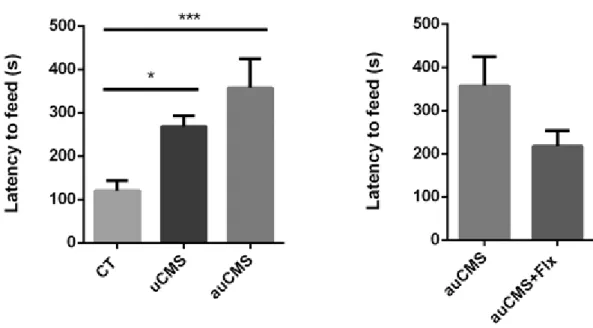
Figure 1. The Hypothalamic-Pituitary-Adrenal axis in Depression (25) ...4 Figure 2. Timeline of the procedures: The induction of chronic stress was performed manually or automatically during 6 weeks and in the last two weeks of this protocol fluoxetine was administered. After, depression induction protocols several analysis were performed. ... 20 Figure 3. Representative scheme of the automated prototype for uCMS... 29 Figure 4. Demonstration of the operation mode of our space restrictor. ... 37 Figure 5. Scheme of two versions of partial automated stressor – space restrictor, from the oldest (left) to the new (right)... 40 Figure 6. Corticosterone levels measured in the serum of rats exposed to a manual confinement procedure and to an automated confinement procedure. Abbreviations: CORT, corticosterone; Automated, exposure to an automated stressor; Manual, exposure to a manual stressor. ... 41 Figure 7. Assessment of anhedonic behaviour through the SDT at the 6th week of the uCMS protocol. The groups exposed to this anhedonic behavioural test were control animals, animals exposed to the uCMS and auCMS protocols as well as auCMS animals treated with fluoxetine. Abbreviations: CT, control; uCMS, manual stressed animals injected with vehicle; auCMS, automated stressed animals injected with vehicle; auCMS+FlX, automated stressed animals treated with fluoxetine. Data is presented as mean ± SEM. * p<0.05; ** p<0.01; **** p<0.0001. n= 4-10 animals per group... 43 Figure 8. Assessment of anhedonic behaviour through the SPT.. The groups exposed to this anhedonic behavioural test were control animals, animals exposed to the uCMS and auCMS protocols as well as auCMS animals treated with fluoxetine. Abbreviations: CT, control; uCMS, manual stressed animals injected with vehicle; auCMS, automated stressed animals injected with vehicle; auCMS+FlX, automated stressed animals treated with fluoxetine. Data is presented as mean ± SEM. * p<0.05; ** p<0.01; **** p<0.0001. n= 5-10 animals per group... 44 Figure 9 . Assessment of mood dimension through the FST. The groups exposed to learned helplessness were control animals, animals exposed to the uCMS and auCMS protocols as well as auCMS animals treated with fluoxetine. Abbreviations: CT, control; uCMS, manual stressed animals injected with vehicle; auCMS, automated stressed animals injected with vehicle; auCMS+FlX, automated stressed animals treated with fluoxetine. Data is presented as mean ± SEM. * p<0.05; ** p<0.01; **** p<0.0001. n= 6-10 animals per group... 45 Figure 10. Assessment of short-term (ST) memory. The groups subjected to the NOR test were control animals, animals exposed to the uCMS and auCMS protocols as well as auCMS animals treated with fluoxetine. Abbreviations: CT, control; uCMS, manual stressed animals; auCMS,
xxi automated stressed animals injected with vehicle; auCMS+Flx, automated stressed animals treated with fluoxetine. Data is presented as mean ± SEM. *p<0.05; ** p<0.01. n= 4-10 animals per group. ... 46 Figure 11. Assessment of long-term (LT) memory. The groups subjected to the NOR test were control animals, animals exposed to the uCMS and auCMS protocols as well as auCMS animals treated with fluoxetine. Abbreviations: CT, control; uCMS, manual stressed animals; au CMS, automated stressed animals injected with vehicle; auCMS+Flx, automated stressed animals treated with fluoxetine. Data is presented as mean ± SEM. * meaning p<0.05; ** meaning p<0.01. n= 4-10 animals per group... 46 Figure 12. Assessment of anxiety-like behaviour. The groups subjected to the NSF test were control animals, animals exposed to the uCMS and auCMS protocols as well as auCMS animals treated with fluoxetine. Abbreviations: CT, control; uCMS, manual stressed animals; auCMS, automated stressed animals injected with vehicle; auCMS+FLX, automated stressed animals treated with fluoxetine. Data is presented as mean ± SEM. *P<0.05; ***P<0.001. n= 6-10 animals per group. ... 47 Figure 13. Corticosterone levels measured in the serum of rats between 8:00 and 9:00 am (basal levels; nadir, N) and between 8:00 pm and 9:00 pm (peak levels; zenith, Z). This figure shows corticosterone levels of manual-stressed animals, automated-stressed animals, non-stressed animals as well as fluoxetine-treated animals. Abbreviations: CT, control; uCMS, manual stressed animals; auCMS, automated stressed animals injected with vehicle; auCMS+FlX, automated stressed animals treated with fluoxetine. Data is presented as mean ± SEM. *p<0.05; **p<0.01. n= 6-10 animals per group... 48 Figure 16. Number of proliferating cells in the dorsal dentate gyrus of auCMS-exposed animals untreated and treated with fluoxetine. Abbreviations: auCMS, automated stressed animals injected with vehicle; auCMS+FlX, automated stressed animals treated with fluoxetine. Data is presented as mean ± SEM. *p<0.05. n=3 animals per group, 3-11 neurons per animal. ... 49 Figure 14. Morphological analysis (total dendritic length) of neurons from the dorsal hippocampal dentate gyrus of control non-stressed, manual-stressed, automated-stressed and fluoxetine-treated animals. A representative image of neurons from the different groups are shown above the graphs. Abbreviations: CT, control; uCMS, manual stressed animals; auCMS, automated stressed animals injected with vehicle; auCMS+FlX, automated stressed animals treated with fluoxetine. Data is presented as mean ± SEM. n= 3-10 animals per group. ... 50 Figure 15. Morphological analysis – Sholl analysis of neurons from the dorsal dentate gyrus of control non-stressed, manual-stressed, automated-stressed and fluoxetine-treated animals. Both treated and non-treated groups were analyzed. Abbreviations: CT, control; uCMS, manual
xxii stressed animals; auCMS, automated stressed animals injected with vehicle; auCMS+FlX, automated stressed animals treated with fluoxetine. Data is presented as mean ± SEM. n=3-5 animals per group, 6-12 neurons per animal. ... 51
xxiv
PHOTO INDEX
Photo 1. Control computer linked to the equipment. ... 30
Photo 2. Mechanical feature of the equipment – opening three cages at once. ... 30
Photo 3. Mechanical feature of the equipment – tilted cage stressor ... 32
Photo 4. Mechanical detail – metallic ring for the shaking movement correction ... 32
Photo 5. Water/air supply system – wet bed or cold water stressor ... 33
Photo 6. Detail of the water tubs – clamps to seal the system ... 34
Photo 7. Detail of the water tubs – mini taps to control water volume ... 34
Photo 8. Software that controls the equipment... 35
Photo 9. Accessories linked to the equipment – strobe lights stressor... 35
Photo 10. Accessories linked to the equipment – startle noise stressor ... 36
Photo 11. Accessories for partial automated stressors – space restrictor in metal (left) and in acrylic (right) ... 40
Photo 12 . Accessories for partial automated stressors – food restrictor from the oldest to the new version (left to right). ... 41
xxv
TABLE INDEX
xxvi
INDEX
ACKNOWLEDGMENTS/ AGRADECIMENTOS...ix RESUMO ...xi ABSTRACT ... xv ABBREVIATIONS LIST ... xviii FIGURE INDEX ... xx PHOTO INDEX ... xxiv TABLE INDEX... xxv 1. INTRODUCTION ...1 1.1 Depression ...1 1.1.1 Global impact ...1 1.1.2 Diagnosis...1 1.1.3 Treatment ...2 1.1.4 Aetiology...2 1.1.5 Pathophysiology of Depression ...3 1.2 Validation criteria of animal models... 11 1.3 Animal models of depression ... 13 1.3.1 Animal models based on (bio)chemical manipulations ... 13 1.3.2 Genetic-related models ... 13 1.3.3 Lesion model ... 14 1.3.4 Animal models based on stress exposure ... 14 1.4 Research Objectives ... 18 2. MATERIALS AND METHODS... 19 2.1 Animals ... 19 2.1.1 Optimization phase ... 19 2.1.2 Pilot-study ... 20 2.2 Procedures ... 21 2.2.1 Manual unpredictable chronic mild stress ... 21 2.2.2 Prototype ... 22 2.2.3 Automated unpredictable chronic mild stress... 22 2.3 Drugs administration ... 22 2.4 Behavioural Tests ... 23 2.4.1 Sucrose preference test ... 23
xxvii 2.4.2 Sweet drive test ... 23 2.4.3. Novel object recognition ... 24 2.4.4 Novelty supressed feeding ... 25 2.4.5 Forced Swimming Test ... 25 2.5 Plasma corticosterone level ... 25 2.6 BrdU Immunostaining... 25 2.7 Morphological analysis ... 26 2.8 Statistical analysis ... 27 3. RESULTS ... 29 3.1 Rack development and optimization ... 29 3.1.1 General features ... 29 3.1.2 Mechanical details ... 30 3.1.3 Stressors categories ... 31 3.1.4 Totally Automated stressors ... 31 3.1.5 Partially automated stressors ... 36 3.1.6 Non-automated stressors ... 37 3.2 Optimization of individual stressors with animals... 38 3.2.1 Total Automated stressors ... 38 3.2.2 Partial automated stressors (accessories) ... 39 3.3 Pilot study: Testing an uCMS protocol with animals in the automated rack ... 42 3.3.1 Behaviour dimensions ... 42 3.3.2 Corticosterone levels ... 47 3.3.3 Cellular alterations - proliferation ... 48 3.3.4 Cellular alterations – Neuronal morphology in the hippocampal DG ... 49 4. DISCUSSION ... 53 4.1 Equipment development and optimization... 53 4.2 Pilot study phase ... 57 4.3 Main limitations ... 61 5. REFERENCES... 63
Chapter 1. Introduction
1
1. INTRODUCTION
1.1 Depression
1.1.1 Global impact
Major depressive disorder (MDD) is a highly prevalent mood disorder, known to affect around 350 million people worldwide(1). According to the World Health Organization, MDD will be the second leading cause of disability worldwide by 2020. This disorder affects more women than men and it often appears at a young age(2,3). Moreover, this disease affects various functioning areas like social, professional or occupational, posing a tremendous burden on society.
1.1.2 Diagnosis
According to Diagnostic and Statistical Manual of Mental Disorders (DSM)-V, MDD is defined by, at least, one major depressive episode (MDE) as well as the absence of mania and hypomania(4). Nine symptoms can be present in an MDD patient: loss of interest or pleasure in usually pleasurable situations or activities (anhedonia), depressed mood, change in appetite and weight, loss of energy, less concentration, changes in sleep patterns, guilty feelings or worthlessness, psychomotor retardation or agitation and suicidal ideation. To be considered a MDE, five of these nine symptoms must be present during the same 2 -week period, being depressed mood or anhedonia one of them. During this 2-week period the required frequency can vary by symptom but most of the times it needs to be present almost every day (4).
MDD can be considered a multi-dimensional psychiatric disorder because it often covers impairments in different behavioural domains, including cognition, anxiety and mood(5). Indeed, generalized anxiety disorder (GAD) seems to appear more nosologically related to MDD than previously thought due to high comorbidity between mood and anxiety disorders (6). Cognitive impairments have also been commonly associated with MDD, particularly executive dysfunction(7).
Chapter 1. Introduction
2
1.1.3 Treatment
Antidepressants are the first line treatment for depression. Still, many patients do not benefit from currently available antidepressants. According to START*D (Sequenced Treatment Alternatives to Relieve Depression) trial, which was an interesting study that gathered data about the effectiveness of antidepressant drugs treatment in MDD patients, only 28-33% of the patients remitted after the first antidepressant treatment(8). This trial was the largest and longest study ever conducted to evaluate depression treatment(9). This rate of remission is quite low for a disease with such prevalence, reflecting the need for the development of new antidepressants.
Antidepressants may be divided in several classes. First-line antidepressants to treat MDD are selective serotonin reuptake inhibitors (SSRIs) and serotonin-norepinephrine reuptake inhibitors (SNRIs) because they are associated with a better safety profile and tolerability. SS RIs include fluoxetine, sertraline, paroxetine, fluvoxamine, citalopram and escitalopram whereas SNRIs encompass venlafaxine and duloxetine.
Tricyclic antidepressants (TCAs) are other class of antidepressants which inhibit the reuptake of both serotonin and norepinephrine by blocking each transporter.However they are not specific and they also block other receptor sites (like histaminic, cholinergic, and α1-adrenergic). These are recommended as second-line antidepressants while monoamine oxidase
inhibitors (MAOIs) are third-line antidepressants due to their increased number of side effects (10,11).
In persistent and severe cases, where antidepressants fail or produce insufficient response, combination of antidepressants are a frequently used strategy or, even its augmentation by the use of other drug classes like antipsychotics.
In particular cases, non-medication treatments are also chosen, including deep brain stimulation, electroconvulsive therapy and vagus nerve stimulation (12).
1.1.4 Aetiology
Depression is thought to result from the interplay between genetic and environmental factors(13,14). Genetic contributions are relevant to the onset of depression, however, heritability is only moderate (40% to 50%) and as such, depression is not simply considered a genetic disorder(15). Taking into account that around 60% of the factors involved in depression aetiology are not explained by genetic variability, environmental features were shown to play a crucial role in depression(16).
Stress is a widely known environmental precipitating factor for this disease(17–19). However, not all stress types are maladaptive (20). Many factors are used to categorize stress;
Chapter 1. Introduction
3 the effects of stress depend on the neurodevelopment stage (for example childhood adversity is a major risk factor for depression), the intensity and duration, the nature, the predictability and controllability (21). As such, stress within specific contexts or exceeding a certain intensity and/or duration can affect physiological and behavioural homeostasis, leading to maladaptive responses(22). Additionally, individual susceptibility, i.e. the way different individuals cope with stress is highly variable, depending on (epi)genetic elements. Understanding the mechanisms and the conditions for these variations is crucial to improve knowledge on the etiopathogenesis of neuropsychiatric disorders (21,23).
Among all the stress types, chronic stress has a leading position within environmental precipitating factors contributing to the development of MDD. As I will explain next, hypothalamic-pituitary-adrenal axis (HPA-axis) deregulation is one of the links that I will approach to explain this relationship between stress and depression.
1.1.5 Pathophysiology of Depression
The Central Nervous System (CNS) is responsible for the processing of both external and internal inputs and for adjusting responses according to possible changes or stimuli. Neuroimaging and postmortem studies in depressed patients have revealed changes in several brain regions. Structural and functional alterations in the prefrontal cortex (PFC) and hippocampus can explain the cognitive alterations usually observed in patients, like memory impairments, hopelessness or suicidal ideation. Amygdala, as well as related parts of the striatum (mainly ventral tegmental area and nucleus accumbens), are involved in the reward responses and in mediating aversive stimulus; these structures are known to be affected in depression since hedonic deficits, anxiety and decreased motivation is often seen in depressed patients(24).
In summary, neural circuitry pathways involved in emotion, reward response and executive function are impaired in this disease(12). In depression, it is possible to observe the disruption of a wide variety of systems that will be next briefly described:
Chapter 1. Introduction
4
Figure 1. The Hypothalamic-Pituitary-Adrenal axis in Depression (25)
1.1.5.1 Neuroendocrine system
The Hypothalamic-Pituitary-Adrenal (HPA) axis controls glucocorticoids (GCs) release, a system deeply involved in stress response and in depression.
Stress is perceived in the cortex and this sensory information is transmitted to the hypothalamus where corticotropin-releasing factor (CRF) is released. This results in the secretion of the corticotropic hormone (CRH) from the anterior pituitary which will, in turn, stimulate the release of GCs from the adrenal cortex (cortisol in humans and corticosterone in rodents)(12). In fact, around 50% of depressed patients show elevated cortisol levels in the plasma and CRH in the cerebrospinal fluid(25). These GCs will activate the HPA axis through the binding to high-affinity MR (mineralocorticoid) and low-high-affinity glucocorticoid receptors (GR) – highly expressed in the hippocampus. The physiological regulation of this axis occurs by negative feedback triggered when GCs bind to their receptors.
In depressed patients, there is an hyperactivity of the HPA axis manifested by the increase of CRF and reduced feedback inhibition of the axis(12). This loss of the negative feedback loop is explained by a decrease of corticosteroid receptors in the hippocampus and PFC, also responsible
Chapter 1. Introduction
5 for the negative regulation of the HPA axis. As a consequence, this will lead to a persistent elevation of GCs secretion(22) (Fig 1).
Importantly, genetic studies also found a correlation between the genes encoding proteins involved in the regulation of the HPA axis and some variables related to the severity of the disease and its response to antidepressants(26).
1.1.5.2 Neuroimmune system
The neuroimmune system is associated with neuroinflammation and the neuroendocrine system, known to be affected in depression(27). Indeed, patients of infectious and autoimmune diseases often show depressive symptoms that are reverted with antidepressants(25). The other way around seems also to be true as MD patients have increased levels of the proinflammatory cytokines, tumor necrosis factor-α and interleukin-6(28), which were shown to affect the HPA axis and monoamines expression(25).
Similarly, chronic mild stress in rodents triggers the production of inflammatory cyto kines with an increase of interleukin-1𝛽, tumor necrosis factor-α, interleukin-6 and interleukin-4 expression(29). Cytokine changes may be secondary to the stress associated with the illness and may not be related to the mood disturbance per se. In fact, cytokine elevations are most predominant in severe depression(30). Some explanations are beginning to emerge, suggesting that this elevation in cytokine levels may contribute to some aspects of the atypical symptomatology, including decrease in sex drive, increased sleep and muscle fatigue, which are well documented effects of proinflammatory cytokines.
1.1.5.3 Neurotransmission
A decrease monoaminergic neurotransmission was proposed as a model to explain the pathophysiology of depression. This proposal was based on the knowledge that mono amine-based agents are potent antidepressants – strong predictive validity(31). Indeed, norepinephrine and serotonin have critical roles in the mechanisms of action of several pharmacological treatments. However, serotonin and norepinephrine depletion does not induce depressive symptoms in healthy individuals(32).
The cause of depression is far from being a simple lack of central monoamines. Some results suggest that pre-synaptic monoaminergic receptors (that modulate monoamine release) have a reduced sensitivity with depression. Also, second-messengers of the signalling cascade of serotonergic and noradrenergic systems were shown to have a reduced functioning, which may impair neurotransmitter activity even without changing monoamine levels or receptor numbers.
Chapter 1. Introduction
6 As a result, caution must be taken when associating depression with a direct reduction in monoamine neurotransmitters(25,31).
Apart from monoamines, other neurotransmitters seem to be altered in depression, specifically glutamate which is the major excitatory neurotransmitter in the brain. A post-mortem study in brain tissue showed an increase in glutamate levels in the frontal cortex of MDD individuals and a decrease of plasma glutamate levels after antidepressants treatment(33). Contrary, other studies show increased levels of this neurotransmitter in the occipital cortex(25) and decreased levels in the prefrontal cortex of depressed patients(34), these inconsistent results need to be clarified in future studies.
Additionally, some studies show reduce expression of excitatory aminoacid transporters (EAAT1 and EAAT2) as well as glutamine synthetase (convert glutamate to glutamine) within glia in several brain regions of MDD patients. In line with this, a role of glia was reported with impairments glutamate uptake and metabolism(22,33).
Animal models support this findings, acute stress exposure induced an increase of extracellular glutamate in the hippocampus, amygdala and PFC. Repeated restraint stress lead to a reduction in AMPA-R and NMDA-R mediated synaptic currents in the PFC(22,33). Although, most of the animal findings support the idea that GC induces the enhancement of excitatory transmission there are some gaps between stress paradigms outcomes and their relationship with this NT that must be clarified (33).
Supporting the importance of this NT in depression, ketamine has appear as a putative innovative antidepressant. Actually, drugs that target the glutamate system must be deeply studied because they could bypass the typical delay of action of monoaminergic drugs (33). Ketamine, one of the most common NMDA antagonists, this drug is also an activator of AMPA receptors and an agonist of the D2 receptor of dopamine is an example of that(35). It was shown that a single subanesthetic dose of this drug was able to induce rapid and sustained antidepressant efficacy in depressed and treatment-resistant patients. In chronic stress rodent models, ketamine showed antidepressant-like properties, promoting an increase in synaptic connectivity and reversing the neuronal atrophy and behavioural deficits(36). Recent studies also suggest that antidepressant-like effects of NMDA antagonists depend on the enhancement of AMPA-R activation, which increases expression of BDNF and stimulates neurogenesis(33).
Another neurotransmitter that has been under study in depression is GABA, the main inhibitory neurotransmitter of the brain. Alterations of the GABAergic system in depression are not well understood yet(36,37). Some studies in MD patients reported reduced levels of GABA in the plasma, occipital cortex, PFC and cerebrospinal fluid. Unmediated depressed individuals also showed decreased protein and mRNA levels. This was also reported for PFC as well as decreased
Chapter 1. Introduction
7 protein and mRNA levels of glutamic acid decarboxylase (GAD) 67 (a GABA synthesizing enzyme) (25,36,37), which was not evident in treated patients. Additionally, remission from depression of patients exposed to SSRIs or transcranial magnetic stimulation was shown to be linked with the normalization of GABA levels.
Studies with animal models (GABAA mutant mice) showed that GABAA reduced receptor binding leads to an anhedonic phenotype. Several animal models of chronic stress report a decrease of expression of GABAA receptor in frontal cortex and other brain regions. Another approach to study this NT was through the administration of GABA directly in the hippocampus of rats which protected them from developing learned helplessness(37).
Although some studies have been made in this topic further analysis need to be made in order to ensure robust outcomes.
1.1.5.4 Neurotrophic factors
Neurotrophic factors are key molecules for growth, survival and differentiation of neural cells; in particular neurotrophins are proteins that act in neurons (38)(39).
Brain-derived neurotrophic factor (BDNF) is an important neurotrophin shown to be involved in depression pathophysiology. Clinical studies demonstrated that BDNF levels in the serum were decreased in drug-free MDD patients when compared to healthy participants; other studies reported increased BDNF levels after antidepressant treatment (more prominent in responders rather than non-responders). However, no clear relationship was shown between BDNF levels and depression severity(40).
Animal models of stress also show a downregulation of BDNF in several hippocampal sub-regions (dentate gyrus, CA3 and CA1), which has a negate impact in neuronal plasticity. Also, an experiment where exogenous corticosterone was administrated to rats showed that BDNF expression was reduced in hippocampus. According to that, a single infusion of BDNF into the hippocampus of animals produced a massive and long-lasting antidepressant effect(39). Actually, the majority of the current available antidepressants increase BDNF expression (in regions as hippocampus and prefrontal cortex of animals exposed to stress). Moreover, the efficacy of ADs is very reduced when BDNF expression or TrkB signalling are disrupted(41).
Pre-clinical studies show that an impaired BDNF expression does not lead to depressive-like behaviour but does affect ADs efficacy, apparently it seems that most of the BDNF role is involved with therapeutic action(41).
Not in line with the beneficial effects of BDNF is the finding that inflammation in the brain and some neurotoxins increase brain BDNF levels. In the same way, blockade of BDNF activity in the Ventral Tegmental Area - Nucleus Accumbens (VTA-NAc) pathway exerts an
antidepressant-Chapter 1. Introduction
8 like effect in rodent models of stress(42). Other findings show that chronic stress cause an upregulation of BDNF in the basolateral amygdala(43).
It is clear that BDNF actions are not always beneficial, so a region-specific BDNF signalling is being studied as well as its impact through epigenetic modifications (e.g maternal separation early in life is capable of influencing this mechanism)(25,42). Epigenetic processes are very dynamic and tissue specific. Some groups have reported a hypermethylation of the BDNF gene promoter in MDD patients, a post-mortem analysis of patients that commit suicide have reported a lower BDNF expression, in Wernicke’s area, associated with an increase of DNA methylation of four CpG sites located at BDNF promotor 4.
Studies are not limited to BDNF, for example nerve growth factor (NGF) has also been shown to be decreased in the hippocampus of suicide victims as well as in animals exposed to stress(44). Additionally, NT-3 was also reduced in the hippocampus after stress exposure(45). Moreover, an infusion of this neurotrophic factor in the hippocampus produced an antidepressant response in the forced swimming test (FST)(44).
Another neurotrophic factor implicated in depression is the glial cell-line derived neurotrophic factor (GDNF). Some studies showed a decreased expression of this protein in the peripheral blood of depressed patients and reduced levels in the hippocampus were observed in a rodent model of chronic unpredictable stress(46).
1.1.5.5.Neural plasticity
Neural plasticity is a process that include re-organization of dendrites and synapses (synaptic alterations, re-orientation of dendrites and axons and modifications in branching structure) as well as the generation of new neuronal and glial cells, a process called neurogenesis and gliogenesis, respectively. Additionally, long-term potentiation and long term depression are known as functional neuroplastic changes. These physiological neuroplastic changes occur as a response to environmental stimuli, yielding functional alterations and gene expression alterations in order to achieve adaptation and further homeostasis(47,48).
Neurogenesis is the process by which neural progenitors divide mitotically to generate new neurons. This neuroplastic process happens at least in two regions of the adult mammalian brain: the subgranular zone (SGZ) of the dentate gyrus (DG) in the hippocampus(49) and the subventricular zone (SVZ) lining the lateral ventricles(50).
Particularly, the SGZ contains radial glial-like stem cells that express several markers, including GFAP (fibrillary acidic protein) and the intermediate filament nestin. These cells divide asymmetrically and produce Type-2 daughter cells, called transient amplifying progenitor cells or fast proliferating cells, which also express nestin and are much more proliferative than type-1
Chapter 1. Introduction
9 cells. Type-2 cells give raise to neuroblasts – type 3 cells. Type-3 cells are negative to GFAP and nestin but positive to doublecortin (DCX). This last stage correspond to transition between a slow proliferation neuroblast (which is exiting the cell cycle) to a postmitotic immature neuron that will migrate into the granule cell layer. These new cells will then became maturate into granule neurons with their axons being growth toward CA3 area of the hippocampus. After 2/3 weeks the cells express calbindin a marker of mature granule cells and after 4-8 weeks they are fully integrated in the pre-existing neuronal network(45,51,52).
Several neuroplastic changes have been found in human samples. Hippocampal atrophy is a clear feature seen in meta-analyses studies of depressed patients(53). Additionally, postmortem studies of depressed patients showed reduced glial cell density in the prefrontal cortex, hippocampal DG and anterior cingulate cortex(46,54,55) as well as a decrease in neuronal size observed in the dorsal PFC and anterior cingulate cortex(55,56).
Insights from animal models of depression have produced relevant findings regarding neural plastic changes induced by depression. Dendritic atrophy and spine loss was observed in neurons from the PFC and hippocampus of animal models of chronic stress (57). Also, a reduction in hippocampal neurogenesis was reported in animal models of stress exposure(58). Indeed, hippocampal neurogenesis was shown to be an important contributor for a sustained remission from depressive-like behaviour(59). The relevance of neurogenesis for depression is reinforced by the fact that antidepressant drugs take 3 to 4 weeks to exert their beneficial effects in patients, which corresponds to the same period for the maturation of adult born neurogenesis(45). Not only neuronal cells are affected in animal models of stress exposure. Indeed, chronic stress is known to induce a decrease in the proliferation of glial progenitor cells and reduce the number of GFAP-positive cells in the PFC and hippocampal DG. Additionally, impairments in astroglial cell morphology, metabolism and function are also observed(22,57,60,61).
In fact, astrocytes have an active control on neuronal activity and synaptic plasticity ; this bidirectional communication between neurons and astrocytes is called ‘tripartite synapse’. In this process astrocytes are responsible for glutamate clearance from the synaptic cleft through transporters (EAAT1 and EAAT2); conversion of glutamate into glutamine (precursor of glutamate and GABA); release of trophic factors; metabolic support; intervention in the neuronal activity through variations of intracellular Ca2+, among other functions(33,62).
Changes in the number or astroglial cells remodelling may affect the glutamatergic tripartite synapse through a decrease of extracellular glutamate clearance and subsequent activation of extrasynaptic glutamatergic receptors that will result in excitoxicity(22).
To conclude, it was proved that both neuronal and glial neuroplastic alterations, could be reversible after ADs treatment(46,61,63). Treatment with SSRIs, MAOIs or TCA allows a fast
Chapter 1. Introduction
10 recovery of spine density as well as dendritic neuronal architecture which is associated with the remission from depressive-like behaviour(59,63,64). TCA and SSRIs are also capable of restoring neuro- and glio-genesis as well as BDNF levels(65)(51).
Together, these neuroplastic reestablishments, potentiated by antidepressants, are crucial for the long-term remission from depression(66).
Chapter 1. Introduction
11
1.2 Validation criteria of animal models
Nowadays, searching for better and accurate animal models is an effort of many researchers. In fact, a wide variety of animal models have been used to mimic the human depression. Many of the symptoms of depression (e.g. depressed mood, feelings of worthlessness, suicidal ideation) cannot be easily measured in laboratory animals because they involve higher cognitive abilities. However, by trying to improve the quality and the validity of the models it is possible to step-by-step get closer to the human disease in order to help finding new therapeutic targets as well as new insights about the pathology seen in the clinics(67,68). Due to this reason, animal models are fundamental and must be continuously improved. For that, several validation criteria must be fulfilled, aiming an intra- and inter-laboratory reproducibility - linked to the standardization power of the model.
In 1969, William McKinney was the first to establish the minimal requirements for animal models of depression. These criteria include: an analogy between the symptomatology of human depression and what is observed in the animals; assessment of clear and objective behavioural changes that should agree between independent observers; treatment effective modalities in humans should reverse the changes observed in the animals; and the existence of reproducibility between investigators(69).
In 1984, Paul Willner refined the validation criteria proposed by McKinney. Most of the researches, working in the field of animal models of depression, rely on this last proposal by Willner that suggests 3 main validation criteria: predictive, face and construct validity. He described predictive validity as the capacity of the model to identify antidepressant treatments through the coherence between animal and human (un)successful agents as well as the model correlation with the success level seen in the clinics(70). Face validity is the resemblance between phenomenological similarities between the model and the disease through a coexistence of several aspects that are specific to depression. In other words, this criterion describes if the model mimics the diagnostic criteria of depression (core symptoms), while not referring to etiological or biological basis(68,70). Construct validity is the capacity to resemble the features and behavioural changes seen in human depression with a solid theoretical rational with unambiguous and homologous interpretation.
Later, Willner updated these criteria for a concept that aligns the theoretical explanation of the human disease with the behaviour and biological dysfunctions seen in the animal model. Etiology is now included in this concept through the identification of the triggering factors that cause a depressive-like state, its characterization (unpredictability, chronic, etc) and its
Chapter 1. Introduction
12 correlation with biological processes involved in depression(12,68). Willner compared several animal models and stated that predictive validity seems to be the easiest criterion to achieve. Face validity and construct validity are the hardest criteria to get because their validation is filled in only a reduced number of models(70).
More recently, in 2011, Belzung and Lemoine made a reformulation of these classic validation criteria. First, they referred the importance of internal validity which concerns the consistency of the experimental design (reproducibility, inter-observer reliability, randomization, blind experimentation and others) as well as the external validity that refers to the applicability of the results of a study on a sample to its extrapolation to the target population (68). Regarding the validation criteria, they introduced a different perspective of the validity concepts. However, the global outcomes being assessed remained the same, with an exception to homological validity, which is a totally new validity criteria. This criterion refers to the adequate choice of the animal species and of a particular strain. For instance, species more prone to display depressive-like states at behavioural and biological levels are a better choice as an animal model than resilient species(68).
Chapter 1. Introduction
13
1.3 Animal models of depression~
1.3.1 Animal models based on (bio)chemical manipulations
1.3.1.1 Chronic GC administration
As the name implies, this animal model is developed by the chronic administration of GCs (known to be elevated in depression). These animals display some behavioural alterations similar to those observed in the clinics and also some molecular alterations including: anhedonia and learned helplessness phenotype(12); molecular and cellular changes are observed, like decreased BDNF expression in hippocampus(44), reduced neurogenesis, retraction of dendrites in CA3 pyramidal neurons(71), increased CRF synthesis and secretion(12). These effects are reversed by chronic administration of antidepressants, like amitriptyline (a tricyclic antidepressant)(72).
1.3.1.2 Stimulation of the immune system
Up to 50% of patients with autoimmune diseases, such as multiple sclerosis, experience clinically significant depression. As it was previously stated, the activation of the inflammatory system seems to be correlated with depression onset. Moreover, cytokine alterations seem to elicit central monoamine and CRH changes(72). Induction of endotoxins and cytokines are models that show alterations at different levels, including brain neurochemistry changes, neuroendocrine and neuroimmune function alterations and behavioural changes – coincident with MDD alterations in humans. Additionally, the exposure to endotoxins or cytokines induce anhedonia phenotype, increase stress hormones and decrease locomotor activity and body weight. This is a low cost and easy-to-implement model but with a poor etiological validity.
1.3.2 Genetic-related models
1.3.2.1 Selective breeding
As stated before, depression requires genetic and/or environmental vulnerability. Selective breeding is based on genetic individual differences found in animals, an example is the Wistar-Kyoto (WKY) rat strain(73). This tool can be very useful however more work is needed to establish a line for a reliable model(74).
1.3.2.2 Targeted overexpression or KO (knock-out) of specific candidate genes
These are not considered formal animal models of depression but are helpful in same cases. For instance, 5-HT transporter knockout is used because it is the target of many antidepressants and as such it can give some insights about the molecular mechanisms. Also, the
Chapter 1. Introduction
14 knockout of tackykinin (NK1) has been associated with stress and anxiety, s howing worse performances in the FST.
HPA transgenic animals are also used in this context. These genetic mutant animals express irregular levels of GR, disturbing the normal negative feedback of the HPA axis.(67).
Moreover, it is possible to test candidate-driven mutations, for example to alter a protein known to be implicated in depression and then characterize the resulting phenotype. This reverse genetics allows to start with a gene and study the backwards to identify its function(74).
1.3.3 Lesion model
Bilateral olfactory bulbectomy is a model of depression where the two lobes are ablated. This lesion leads to anosmia and most importantly to the loss of detection of pheromones which are crucial to reproductive behaviour, gender recognition, social dominance among other behavioural and physiological status of the animal(75).
The olfactory system is part of the rat limbic region, and consequently this lesion will lead to a dysfunction of the cortical-hippocampal-amygdala circuit. More so, this model shows changes in behaviour, like impaired food-motivated behaviour, alterations in cognitive tasks (like morris water maze performance) and increased exploratory behaviour/open field activity. Also increased activity of the HPA axis is observed and some evidences claim that there are alterations of the immune system response.
Importantly, this model shows predictive validity because chronic administration (not acute) of antidepressants can revert behavioural, endocrine, immune and neurotransmitter changes(75). However, this model lacks etiological validity because, in humans, the loss of olfaction does not produce self-rated depression(68).
1.3.4 Animal models based on stress exposure
Depression is a disease with a highly influence from environmental factors(16). Actually, models based on environmental stressors have great aetiological validity compared to the previously mentioned (brain lesions or biochemical manipulations)(12).
1.3.4.1 Acute stress models
These models are used as tools to rapidly screen putative antidepressant compounds. An example is the FST that consists of placing the animal in an inescapable cylinder tank filled with water. The animal starts struggling, swimming and climbing and eventually will stay immobile. It is measured the amount of time that the animal takes to stay immobile. One strength of the
Chapter 1. Introduction
15 model is the reduced errors in results measurement, however it lacks some validation criteria since the phenotype is reversed by acute administration of antidepressants(12,72).
Still within the learned helplessness category, another approach is to expose the animals to inescapable electric shocks that allow them to develop a “helplessness” state. This happens because when animals are re-exposed to shocks, with an easy way out, they display a big latency or fail completely to escape. The major weakness is the same as above, successful response to acute drug treatment. Regarding positive aspects of the model, it is relevant to mention that rats develop alterations in sleep patterns; alterations in HPA axis activity; decreased number of synapses in the hippocampus; elevated CRF and corticosterone levels which all correlate with the human disease (74)(72).
1.3.4.2 Chronic stress models
Psychosocial stress (defeat or social isolation)
This is a model with an interesting etiological validity. In social defeat the animal is repeatedly exposed to a dominant/aggressive animal. These animals show a reduced preference for sucrose in the sucrose preference test (SPT) and changes in the neuroendocrine system, which are reversed through chronic administration of antidepressants. Despite the fulfilment of the validation criteria, social behaviour is not well characterized in rodents. Also these models have poor reproducibility which reduced their application(72).
Maternal care (maternal separation or prenatal stress)
This is an early life stress model that produce neuroendocrine and behavioural changes. For example, it is seen that a bigger vulnerability to learned helplessness, persistent into adulthood, and hyperactive HPA axis. Their reproducibility is relatively good and successful results are shown in many different species from rodents to non-human primates. Antidepressants administration can reverse these abnormalities(72).
Chronic stress exposure in adulthood
These models are among the most valid ones(70). A crucial explanation for that is their naturalistic essence(74). As mentioned before, stress is one of the most consensual precipitating factors for depression.
There are two widely used models of chronic stress: unpredictable chronic mild stress (uCMS) and chronic unpredictable stress (CUS)(76,77). The first one refers to a permanent exposure during 6 weeks to mild stressors in an unpredictable manner(77), whereas CUS protocol
Chapter 1. Introduction
16 is an intermittent exposure (1h per day) of more aggressive stressors during a 4 week period (76). Chronic unpredictable stress models reveal increases in plasma corticosterone levels, augmented serotonergic activity in the hypothalamus(76), alterations in aggressiveness, sexual behaviour as well as grooming deficits. These phenotypes are reversed by chronic administration of antidepressants either during the stress or as a post-stress treatment(72).
Katz and colleagues were the first to develop a chronic mild stress procedure(78). Later, Willner developed a protocol based on this previous one but reduced the severity of the stressors to increase the resemblance with the human daily life stressors. Moreover, he considered reactivity towards a reward as the most relevant behavioural test to assess the phenotype onset, instead of locomotor activity previously used by Katz(79). This Willner procedure consists of a permanent exposure to a variety of mild stressors (e.g. overnight illumination; periods of food and/or water deprivation; cage tilt; change of cage mate, damp bedding), which must be changed every few hours(77) during a period of several weeks(80). The diversity, unpredictability and mild intensity of the stressors are crucial aspects to prevent habituation to the protocol.
Many applications are possible through the uCMS, this protocol can be used to discover new antidepressant drugs but also to provide insights about the pathophysiology of the disease (in both, cellular or molecular scale)(80).
A closer look at uCMS model allow us to consider it as one of the most valid for the 3 main criteria (predictive, face, construct and homological)(68,77), as it is explained below.
A fulfilment of the predictive validity is observed because both behavioural and molecular alterations can be reversed through chronic administration of antidepressants from different classes (MAOs, SSRIs, TCAs)(5,64).
Regarding face validity, it is possible to observe a decreased preference for a palatable sucrose solution(77,81), a diminished sexual behaviour, locomotor activity as well as alterations in sleep changes and self-care(77,79). Not only mood is impaired, cognition and anxiety are also affected in this model, resembling the multidimensional nature of depression in humans. Stressed animals show high anxiety levels and a worse performance in cognitive tests when compared to control animals(5). All together, these findings cover the main symptoms observed in humans and can be seen in this animal model.
Construct validity is the theoretical rationale behind the alterations reported. For that, molecular and cellular analyses are needed. Indeed, increased activity in the HPA axis, which includes adrenal hypertrophy and corticosterone hypersecretion are observed in this animal model. Downregulation of hippocampal 5-HT1A receptors and hippocampal GR as well as reductions in frontocortical and hippocampal BDNF protein are also observed(80). Moreover, it is possible to observe abnormalities in the immune system(77), altered neuroplasticity