Article
RhoD Inhibits RhoC-ROCK-Dependent Cell
Contraction via PAK6
Graphical Abstract
Highlights
d
Vaccinia F11 protein is required for virus-induced cell
contraction and blebbing
d
F11-induced cell contraction depends on RhoC, but not
RhoA, signaling to ROCK
d
RhoD recruits Pak6 to the plasma membrane to antagonize
RhoC signaling
d
F11 inhibits RhoD signaling to its downstream effector Pak6
Authors
Charlotte H. Durkin, Flavia Leite,
Joa˜o V. Cordeiro, Yutaka Handa,
Yoshiki Arakawa, Ferran Valderrama,
Michael Way
Correspondence
[email protected]
In Brief
Many different viral infections induce cell
contraction and blebbing. Durkin, Leite,
et al. show that during vaccinia infection,
RhoC and not RhoA regulates this
ROCK-mediated cytopathic effect of cell
contraction. They delineate a pathway for
RhoC regulation, in which the viral protein
F11 blocks RhoC antagonism by
RhoD-recruited Pak6.
Durkin et al., 2017, Developmental Cell41, 315–329
May 8, 2017ª 2017 The Francis Crick Institute. Published by Elsevier Inc. http://dx.doi.org/10.1016/j.devcel.2017.04.010
Developmental Cell
Article
RhoD Inhibits RhoC-ROCK-Dependent
Cell Contraction via PAK6
Charlotte H. Durkin,1,2,3Flavia Leite,1,2Joa˜o V. Cordeiro,1,4Yutaka Handa,1,5Yoshiki Arakawa,1,6Ferran Valderrama,1,7
and Michael Way1,8,*
1Cellular Signalling and Cytoskeletal Function Laboratory, The Francis Crick Institute, 1 Midland Road, London NW1 1AT, UK 2These authors contributed equally
3Present address: MRC Centre for Molecular Bacteriology and Infection, Imperial College London, Armstrong Road, London SW7 2AZ, UK 4Present address: Centro de Investigac¸a˜o em Sau´de Pu´blica (CISP), Escola Nacional de Sau´de Pu´blica, Universidade Nova de Lisboa,
Avenida Padre Cruz, 1600-560 Lisbon, Portugal
5Present address: AbbVie GK, 3-5-27 Mita, Minato-ku, Tokyo 108-6302, Japan
6Present address: Department of Neurosurgery, Kyoto University Graduate School of Medicine, 54 Kawahara-cho, Shogoin, Sakyo-ku,
Kyoto 606-8507, Japan
7Present address: Centre for Biomedical Education, St George’s, University of London, Cranmer Terrace, London SW17 0RE, UK 8Lead Contact
*Correspondence:[email protected] http://dx.doi.org/10.1016/j.devcel.2017.04.010
SUMMARY
RhoA-mediated regulation of myosin-II activity in the
actin cortex controls the ability of cells to contract
and bleb during a variety of cellular processes,
including cell migration and division. Cell contraction
and blebbing also frequently occur as part of the
cytopathic effect seen during many different viral
in-fections. We now demonstrate that the vaccinia virus
protein F11, which localizes to the plasma
mem-brane, is required for ROCK-mediated cell
contrac-tion from 2 hr post infeccontrac-tion. Curiously, F11-induced
cell contraction is dependent on RhoC and not
RhoA signaling to ROCK. Moreover, RhoC-driven
cell contraction depends on the upstream inhibition
of RhoD signaling by F11. This inhibition prevents
RhoD from regulating its downstream effector Pak6,
alleviating the suppression of RhoC by the kinase.
Our observations with vaccinia have now
demon-strated that RhoD recruits Pak6 to the plasma
mem-brane to antagonize RhoC signaling during cell
contraction and blebbing.
INTRODUCTION
The cortical actin cytoskeleton consists of a dense network of actin filaments, crosslinked by myosin-II and other actin-binding proteins, that is intrinsically linked to the cytoplasmic face of the plasma membrane (Biro et al., 2013; Charras et al., 2006; Clark et al., 2013; Fritzsche et al., 2013; Koster and Mayor, 2016; Mo-rone et al., 2006). The regulation of the density and organization of actin filaments in the cell cortex, as well as their myosin-II driven contraction, provides the cell with mechanical resilience (Bovellan et al., 2014; Clark et al., 2013; Fritzsche et al., 2013, 2016; Gauthier et al., 2012; Salbreux et al., 2012; Tinevez et al., 2009). It also enables the cortical actin cytoskeleton to regulate
the shape of the cell during a variety of cellular processes including migration, mitotic rounding, and cytokinesis (Friedl and Wolf, 2003; Matthews et al., 2012; Sedzinski et al., 2011; Za-tulovskiy et al., 2014). A frequent hallmark of myosin-II-driven contraction of the actin cortex during these cellular processes is the transient and rapid appearance of spherical blebs on the plasma membrane (Charras et al., 2008). These protrusions occur at regions where the plasma membrane separates from the un-derlying actin cortex because of increased hydrostatic pressure within the cell (Charras et al., 2008; Paluch et al., 2006; Tinevez et al., 2009).
Over the last decade, it has become clear that blebbing of the plasma membrane helps drive ameboid-based cell motility dur-ing development and tumor cell migration (Blaser et al., 2006; Charras and Paluch, 2008; Diz-Munoz et al., 2010; Fackler and Grosse, 2008; Friedl and Wolf, 2003; Kardash et al., 2010; Paluch and Raz, 2013; Sahai and Marshall, 2003; Sanz-Moreno et al., 2008; Tozluoglu et al., 2013). A key determinant for assembly and contraction of the actin cortex is the activation of myosin-II by ROCK-mediated phosphorylation of the myosin light chain (MLC) (Amano et al., 1997, 1996, 2010; Amin et al., 2013; Sahai and Marshall, 2003). ROCK also indirectly increases the activ-ity of myosin-II by inhibiting the MLC phosphatase through phosphorylation of the myosin phosphatase-targeting subunit 1 (MYPT1) (Amano et al., 2010; Amin et al., 2013; Kawano et al., 1999). The small guanosine triphosphatase (GTPase) RhoA is most widely implicated in activating ROCK to drive cell contraction (Charras et al., 2006; Costigliola et al., 2010; Gutjahr et al., 2005; Sahai and Marshall, 2003; Sanz-Moreno et al., 2008). However, ROCK can also interact with the closely related GTPase RhoC, which is known to promote ameboid-based motility, tumor invasion, and metastasis (Clark et al., 2000; Ha-kem et al., 2005; Kitzing et al., 2010; Leung et al., 1996; Ruth et al., 2006; Sahai and Marshall, 2002; Simpson et al., 2004).
Cell contraction and membrane blebbing are also frequently observed as part of the cytopathic effect induced by many different viruses during their replication cycles (Agol, 2012). For example, within a few hours of vaccinia virus infection, cells begin to contract and bleb, in a process that is independent of
Developmental Cell 41, 315–329, May 8, 2017ª 2017 The Francis Crick Institute. Published by Elsevier Inc. 315 This is an open access article under the CC BY license (http://creativecommons.org/licenses/by/4.0/).
B A
D C
E F
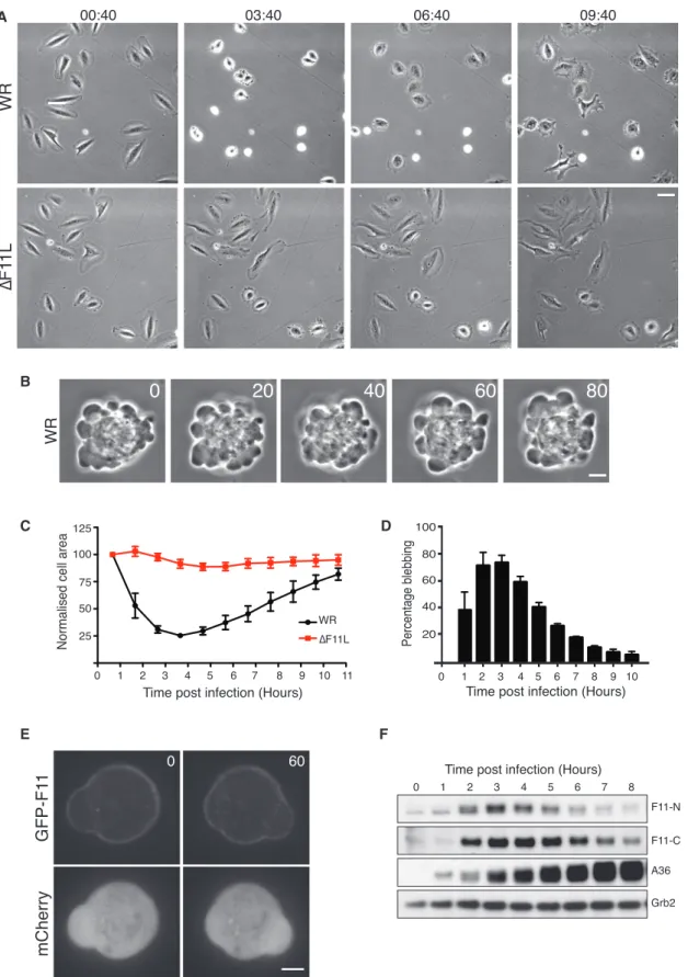
Figure 1. Vaccinia Stimulates F11-Dependent Cell Contraction
(A) Phase-contrast images showing the morphology of HeLa cells at the indicated time points (hours:minutes) after infection with WR or the DF11L virus (see
Movie S1). Scale bar, 30 mm.
(B) Phase-contrast images, 20 s apart, showing WR-induced bleb formation in HeLa cells 3 hr 40 min after infection. Scale bar, 5 mm.
(legend continued on next page)
apoptosis (Bablanian et al., 1978; Barry et al., 2015; Schepis et al., 2006; Schramm et al., 2006). Previous observations suggest the vaccinia protein F11, which is expressed early in infection, is involved in vaccinia-induced cell contraction and/or the loss of cell-cell adhesion (Cordeiro et al., 2009; Mo-rales et al., 2008; Valderrama et al., 2006). F11 downregulates RhoA signaling in the latter stages of the virus replication cycle (Arakawa et al., 2007b; Cordeiro et al., 2009; Valderrama et al., 2006). F11 binds directly to GTP-bound RhoA using a motif that is conserved in ROCK (Cordeiro et al., 2009; Valderrama et al., 2006). Once bound, RhoA is inactivated by the Rho GTPase-activating protein (RhoGAP) activity of myosin-9A, which binds to the central PDZ-like domain of F11 via its C-ter-minal PDZ-binding motif (Handa et al., 2013). Ultimately, F11-mediated inhibition of RhoA signaling late during viral replication promotes the spread of vaccinia infection by stimulating cell migration, increasing microtubule dynamics, and enhancing viral release by modulating the cortical actin beneath the plasma membrane (Arakawa et al., 2007a, 2007b; Cordeiro et al., 2009; Handa et al., 2013; Valderrama et al., 2006). Consistent with its role during vaccinia virus infection, ectopic expression of F11 can enhance the cell-to-cell spread and oncolytic potential of myxoma virus, which lacks F11 (Irwin and Evans, 2012; Irwin et al., 2013).
Previous studies on F11 have largely focused on its role in in-fected cells at 8–20 hr post infection (hpi) (Arakawa et al., 2007a, 2007b; Cordeiro et al., 2009; Handa et al., 2013; Valderrama et al., 2006). However, F11 expression correlates with the onset of virus-induced cell contraction (Cordeiro et al., 2009; Morales et al., 2008). With this in mind, we set out to investigate whether and how F11 plays a role in vaccinia-induced cell contraction and blebbing. We found that vaccinia stimulates cell contraction independently of RhoA by activating RhoC-mediated signaling to ROCK. Furthermore, the ability of RhoC to promote this effect depends on F11-mediated inhibition of RhoD signaling to Pak6.
RESULTS
F11 Induces Cell Contraction Early during Vaccinia Infection
In agreement with earlier studies, live-cell imaging demonstrates that within the first few hours of infection the Western Reserve (WR) strain of vaccinia induces contraction and blebbing of HeLa cells (Figures 1A and 1B;Movie S1). Quantification of the cell area reveals that maximum contraction and blebbing occurs approximately 3 hr 40 min post infection, after which time in-fected cells begin to respread (Figures 1C and 1D). Vaccinia-induced cell contraction is not specific to HeLa cells, as it is also seen in U-2 OS cells (Figure S1A). In contrast, infection with WR lacking the F11L gene (DF11L virus) does not induce
cells to contract or bleb (Figures 1A, 1C, andS1A). In agreement withSchepis et al. (2006), we also found that the highly attenu-ated virus strain, Modified Vaccinia Ankara (MVA), which does not express a functional F11, did not induce HeLa cell contrac-tion early during infeccontrac-tion (Figure S1B). Using a recombinant vi-rus expressing GFP-tagged F11 from its endogenous promoter, we found that F11 associates with the plasma membrane, consistent with a possible role in regulating the actin cortex ( Fig-ure 1E andMovie S2). Immunoblot analysis of WR-infected cells confirmed that the onset of cell contraction correlates with the expression of F11 (Figure 1F). Moreover, respreading correlates with a gradual loss of F11, suggesting that its presence promotes vaccinia-induced cell contraction. Consistent with this, pro-longed expression of F11 in U-2 OS cells suppresses their respreading at later time points (Figure S1A). A role for F11 in promoting cell contraction is unexpected, given previous obser-vations demonstrating that the viral protein inhibits, rather than activates, RhoA signaling.
Vaccinia-Induced Cell Contraction Depends on ROCK It is well established that RhoA signaling to ROCK promotes myosin-II-mediated cell contraction and blebbing by phosphor-ylating MLC and also inhibiting MYPT1 (Amin et al., 2013; Julian and Olson, 2014). To investigate whether ROCK is also required for vaccinia-induced cell contraction, we infected cells in the presence of one of three different ROCK inhibitors. In all cases, we found that there was a dramatic inhibition in virus-induced cell contraction, consistent with the reduction in MLC2 and MYPT1 phosphorylation seen in immunoblots (Figures 2A and S2A). Lack of cell contraction was not due to an inhibition of viral entry, as the drug treated cells were equally as well infected as control cells (Figure S2B). To extend our pharmacological anal-ysis, we examined the impact of small interfering RNA (siRNA)-mediated depletion of ROCK1 and ROCK2 on virus-induced cell contraction. Independent knockdown of either kinase did not significantly impair cell contraction (Figures 2B and S2C). When both proteins were depleted, early viral protein expression was not impaired but there was a substantial inhibition of cell contraction (Figures 2B andS2D). To explore whether additional signaling pathways also contribute to myosin-II activation, we treated cells with siRNA against myotonic dystrophy kinase-related Cdc42-binding kinases (MRCKa/b) and zipper-interact-ing protein kinase (ZIPK) (Nehru et al., 2013; Usui et al., 2014; Zhao and Manser, 2015). We found that WR-infected cells still contract in the absence of MRCKa/b and ZIPK (Figures 2C and 2D). In addition, we treated infected cells with ML7 or ML9 to inhibit MLC kinase (MLCK) to examine whether it also partici-pates in virus-driven cell contraction. Consistent with a role for myosin-II during viral entry (Mercer and Helenius, 2008), we found that inhibition of MLCK with ML7 or ML9 dramatically
(C) Quantification of the average area of HeLa cells infected with WR (black) or DF11L (red) over 11 hr. Each cell area is normalized to its initial value at the start of acquisition.
(D) Quantification of the percentage of WR-infected cells blebbing at the indicated times post infection.
(E) Images showing the association of GFP-F11 with the plasma membrane in cells infected with WR for 3 hr 40 min. The time is indicated in seconds and mCherry provides a volume marker (seeMovie S2). Scale bar, 5 mm.
(F) Immunoblot analysis with two different F11 antibodies reveals the level of endogenous F11 expression in HeLa cells at the indicated times after infection with WR. A36 and Grb2 represent viral and HeLa cell loading controls, respectively.
Error bars in graphs represent the SEM from three independent experiments, in which a total of 60 cells were analyzed. See alsoFigure S1.
A B C E D F
Figure 2. Vaccinia-Induced Cell Contraction Is Dependent on ROCK Signaling
(A) Images of HeLa cells infected with WR for 3 hr 40 min in the absence (DMSO) or presence of ROCK inhibitors (GSK49286A, H1152, and Y27632) together with quantification of cell area. Scale bar, 30 mm.
(B) Images showing the morphology of HeLa cells treated with siRNA against ROCK1 and ROCK2 at 3 hr 40 min post infection with WR together with quanti-fication of cell area. Scale bar, 30 mm.
(legend continued on next page)
reduced early viral protein expression (Figure 2E). To overcome this inhibition, we infected cells in the presence of cycloheximide to block viral uncoating (Mercer et al., 2012), before adding ML7 or ML9 (Figure 2F). Removal of the cycloheximide in the presence of ML7 or ML9 restarts the stalled replication cycle, resulting in early viral protein expression and cell contraction (Figure 2F). Taken together, our results indicate that ROCK-mediated regulation of myosin-II is required for vaccinia-induced cell contraction and that both isoforms are functionally redun-dant. The global levels of phospho-MLC did not appreciably
(C) The graphs show the percentage of remaining MRCKa, MRCKb, or ZipK mRNA following treatment with the indicated siRNA relative to the non-targeting AllStar control (NT). The Immunoblot shows the level of MRCKa and ZipK after siRNA treatment. A36 and MCM7 represent viral and HeLa cell loading controls, respectively.
(D) Quantification of the average area of HeLa cells infected with WR for 3 hr 40 min and treated with the indicated siRNA.
(E) Immunoblot analysis at 5 hpi reveals that in contrast to the ROCK inhibitors, ML7 and ML9 suppress viral entry based on reduced early viral protein expression (A36). MLC represents a cell loading control.
(F) Immunoblot analysis demonstrates that washout of cycloheximide at 2 hpi in the presence or absence of ML7 and ML9 does not inhibit viral entry based on expression of F11 and A36. Actin and Grb2 represent loading controls. The graph reveals that after cycloheximide washout WR-infected cells still contract in the presence of ML7 and ML9.
Error bars in graphs represent the SEM from three independent experiments in which a total of 60–100 cells were analyzed. ***p < 0.001; ns, not significant. See alsoFigure S2.
A
B
C
Figure 3. Vaccinia-Mediated Cell Contrac-tion Is Not Dependent on RhoA
(A) Phase-contrast images of HeLa cells infected with the F11-VK virus and quantification of their average area (blue line). The F11-VK infection was performed at the same time as WR (black line) and DF11L (red line) taken fromFigure 1A.
(B) Images of HeLa cells infected with WR treated with non-targeting (NT) control or RhoA siRNA, together with quantification of their average area. The immunoblot shows the efficiency of RhoA knockdown, and a-tubulin represents a loading control.
(C) Representative images of HeLa cells infected with WR in the presence or absence of the C3 Rho inhibitor, together with quantification of their average area.
All scale bars, 5 mm. Error bars represent the SEM from three independent experiments, in which a total of 60 (A), 80 (B), and 100 (C) cells were analyzed. See alsoFigure S3.
change during WR or DF11L virus infection, but the subcellular distribution of phospho-MLC was strikingly different (Figures S2A and S2E). Live-cell imag-ing also reveals that RFP-MLC2 is re-cruited to the blebbing cell cortex ( Fig-ure S2F andMovie S3), suggesting that localized ROCK activity drives the redis-tribution of myosin-II to promote cell contraction.
Vaccinia Promotes RhoC- but Not RhoA-Dependent Cell Contraction Given the involvement of ROCK1/2, we wondered whether an interaction of F11 with RhoA is also required for vi-rus-induced cell contraction. To address this question, we infected cells with a recombinant virus ex-pressing F11-VK, which is defective in RhoA binding (Cordeiro et al., 2009). We found that the F11-VK virus is as effective as the parental WR strain in inducing cell contraction (Figure 3A andMovie S1). Furthermore, siRNA-mediated depletion of RhoA did not impact on the ability of WR to induce cell contraction ( Fig-ure 3B). In contrast, the Clostridium botulinum toxin C3, a pan-RhoA, B, and C inhibitor (Aktories, 2011), blocked virus-induced cell contraction but not infection (Figures 3C andS3). This sug-gests that vaccinia-induced cell contraction is independent of
RhoA but reliant on the activation of ROCK by another RhoGTPase.
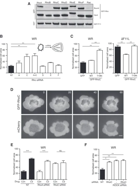
Previous studies have linked RhoB, RhoC, RhoD, RhoE, and RhoF (Rif) to ROCK signaling (Fan et al., 2010; Leung et al., 1996; Riento and Ridley, 2003; Tsubakimoto et al., 1999). Given this finding, we examined whether F11 also interacts with these RhoGTPases. Pull-down assays on lysates from infected HeLa cells demonstrate that glutathione S-transferase (GST)-F11 asso-ciates with GFP-tagged RhoC, D, E, and F but not RhoB or Rac (Figure 4A). To examine whether any of these RhoGTPases are required for vaccinia-induced cell contraction, we performed siRNA-mediated ablation of each protein (Figures S4A and S4B). We found that only depletion of RhoC had a significant impact on the ability of WR to induce cell contraction (Figure 4B). Consistent with the lack of involvement of RhoA, the combined loss of RhoA and RhoC did not result in a greater inhibition of WR-induced cell contraction than RhoC alone (Figures 4B andS4A). In addition, expression of GFP-tagged
dominant-nega-A
B C
D
E F
Figure 4. RhoC Is Required for Vaccinia-Induced Cell Contraction
(A) Immunoblot analysis of glutathione pull-downs on WR-infected HeLa cell lysates reveals that GST-F11 interacts with GFP-tagged RhoA, RhoC, RhoD, RhoE, and RhoF.
(B) Quantification of the area of HeLa cells treated with the indicated siRNA at 3 hr 40 min post infection with WR.
(C) Quantification of the area of HeLa cells ex-pressing GFP or GFP-tagged wild-type (WT) or T19N (dominant-negative) RhoC 3 hr 40 min after infection with WR (left) or DF11L (right). (D) Images showing the association of GFP-RhoC with the plasma membrane in cells infected with WR for 3 hr 40 min. The time is indicated in sec-onds and mCherry provides a volume marker (see
Movie S4). Scale bar, 5 mm.
(E) Quantification of WR-infected HeLa cell area at 3 hr 40 min post infection in the presence or absence of C3, 72 hr after treatment with the indicated siRNA.
(F) Normalized cell area of WR-infected HeLa cells at 3 hr 40 min post infection, 72 hr after treatment with the indicated siRNA.
Error bars in graphs represent the SEM from three independent experiments, in which a total of 90 (B), 60 (C), and 80 (E and F) cells were analyzed. *p < 0.05, **p < 0.01, ***p < 0.001; ns, not signifi-cant. See alsoFigure S4.
tive RhoC-T19N blocked WR-mediated cell contraction (Figure 4C). In contrast, expression of GFP-RhoC stimulated par-tial contraction of DF11L virus-infected cells (Figure 4C). In agreement with its role in promoting cell contraction, RhoC is associated with the plasma membrane of blebs (Figure 4D andMovie S4). The involvement of RhoC explains why C3 toxin blocks WR-induced cell contraction (Figure 3C). Moreover, C3 treatment did not increase the suppression of cell contraction induced by the loss of RhoC (Figure 4E). To confirm that RhoC acts via ROCK to promote contraction of infected cells, we depleted both RhoC and ROCK1/2. Depletion of all three proteins impaired WR-induced cell contraction to the same extent as the loss of ROCK1/2 or RhoC alone (Figure 4F). Our ob-servations demonstrate that vaccinia-induced cell contraction in-volves RhoC-mediated activation of ROCK.
F11-Mediated Inhibition of RhoD Promotes RhoC-Driven Cell Contraction
It is well established that RhoGTPases frequently regulate each other (Guilluy et al., 2011). Given their interaction with F11, it is possible that RhoD, RhoE, and RhoF may have an inhibitory role in promoting vaccinia-induced cell contraction. We found that loss of RhoD but not RhoE or RhoF results in contraction of DF11L virus-infected cells (Figures 5A andS5A). Moreover, this contraction has temporal dynamics similar to those of WR-induced cell contraction (Figure 5B). GFP-tagged RhoD is
A C E F G D B
Figure 5. RhoD Negatively Regulates Vaccinia-Stimulated Cell Contraction
(A) Quantification of the area of HeLa cells treated with the indicated siRNA for 72 hr at 3 hr 40 min post infection with the DF11L virus. (B) Quantification of the change HeLa cell area after treatment with RhoD siRNA and infection with the DF11L virus.
(legend continued on next page)
associated with the plasma membrane of vaccinia-induced blebs (Figure 5C andMovie S5) and can also interact with GST-F11-VK in lysates from infected cells (Figure S5B). Furthermore, in vitro pull-down assays with recombinant proteins demonstrate that F11 interacts directly with RhoD (Figure S5C). We further examined the impact of expressing siRNA-resistant GFP-tagged wild-type (WT), constitutively active (G26V), or dominant-negative (T31N) RhoD on DF11L-infected cells treated with RhoD siRNA. GFP-tagged RhoD or its constitutively active G26V mutant sup-pressed the contraction of DF11L-infected cells induced by the depletion of endogenous RhoD (Figure 5D). In contrast, GFP-RhoD-T31N did not inhibit cell contraction. Taken together, these data suggest that RhoD and RhoC have an antagonistic role during vaccinia-mediated cell contraction. While the presence of RhoC and activity of ROCK are required for F11-induced cell contraction, RhoD must be inactive or absent. Consistent with this, GFP-RhoD-G26V, but not the dominant-negative T31N mutant, suppressed contraction of WR-infected cells (Figure 5D). To determine whether RhoD and RhoC are working in the same or parallel pathways, we investigated whether pharmacological inhi-bition of ROCK, or C3-mediated loss of RhoC signaling, sup-presses contraction of DF11L-infected cells that lack RhoD. Indeed, both inhibitors blocked contraction of DF11L-infected cells treated with RhoD siRNA (Figure 5E). Furthermore, DF11L-infected cells did not contract in the absence of both RhoC and RhoD, following siRNA silencing of both proteins (Figure 5F). Curiously, loss of RhoA, which promotes the spreading of DF11L-infected cells, was partially able to suppress the impact of the loss of RhoD during DF11L infection (Figure S5D). Notwithstanding this, taken together our results demonstrate that F11 facilitates RhoC-mediated contraction of WR-infected cells by inhibiting RhoD signaling. Moreover, in non-infected cells expression of dominant-negative GFP-RhoD-T31N increases the level of GTP-bound RhoC, confirming that RhoD can inhibit RhoC activity outside the context of vaccinia infection (Figure 5G).
Our previous observations demonstrated that RhoA is inacti-vated by the RhoGAP activity of myosin-9A, which interacts with F11 (Handa et al., 2013). It is possible that myosin-9A bound to F11 also inhibits RhoD signaling. However, this does not appear to be the case, as WR still induces contraction of cells treated with myosin-9A siRNA (Figure S5E). Furthermore, unlike RhoD siRNA, loss of myosin-9A does not induce contraction of DF11L-infected cells (Figure S5E).
RhoD Inhibits RhoC Signaling via Pak6
There are many documented examples and different mecha-nisms regulating RhoGTPase crosstalk (Guilluy et al., 2011).
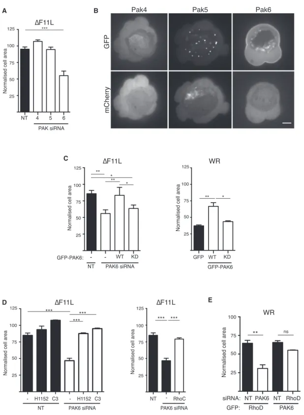
For example, Rac antagonizes RhoA signaling via the p21-acti-vated kinase family members, Pak1 and Pak4, which phosphor-ylate and inhibit a number of different Rho guanine nucleotide exchange factors (RhoGEFs) to prevent RhoA activation (Barac et al., 2004; Nimnual et al., 2003; Rosenfeldt et al., 2006). Pak5, a class II family member, is also one of a few known RhoD binding partners (Wu and Frost, 2006). Given this informa-tion, we decided to investigate whether Pak family kinases mediate the antagonistic crosstalk between RhoD and RhoC. We found that pharmacological inhibition of the class I family members Pak1–3 with IPA3 did not stimulate contraction of DF11L-infected cells (Figure S6A). In contrast, siRNA-mediated loss of Pak6 but not Pak4 or Pak5 results in cell contraction ( Fig-ures 6A, S6B, and S6C). Furthermore, GFP-tagged Pak6 but not Pak4 or Pak5 associates with the plasma membrane of blebs (Figure 6B andMovie S6). The DF11L-induced contraction of cells treated with Pak6 siRNA is specific, as it is inhibited by expression of siRNA-resistant GFP-PAK6 (Figure 6C). In contrast, the Pak6-K436A kinase-dead mutant did not suppress contraction of DF11L-infected cells treated with Pak6 siRNA (Figure 6C). In agreement with this, GFP-PAK6, but not its kinase-dead mutant, blocked WR-induced cell contraction (Figure 6C).
To confirm that Pak6 acts upstream of RhoC and ROCK, we examined the impact of inhibiting RhoC or ROCK with C3 and H115, respectively, in DF11L-infected cells depleted of Pak6. In both cases, DF11L-induced contraction of cells lacking Pak6 was suppressed (Figure 6D). Cells simultaneously depleted of Pak6 and RhoC also failed to contract (Figure 6D). To examine whether Pak6 and RhoD act in the same pathway to inhibit RhoC/ROCK signaling, we expressed GFP-tagged RhoD or Pak6 in cells depleted of endogenous Pak6 or RhoD, respec-tively. We found that the ability of GFP-RhoD to inhibit contrac-tion of WR-infected cells depends on the presence of Pak6 ( Fig-ure 6E). In contrast, GFP-Pak6 blocks WR-induced contraction in the presence or absence of RhoD (Figure 6E). Our data demonstrate that RhoD acts through Pak6 to inhibit RhoC-medi-ated cell contraction.
RhoD Interacts with Pak6 and Mediates Its Recruitment to the Plasma Membrane
It is thought that RhoGTPases regulate class II Pak kinases by controlling their subcellular location rather than directly stimu-lating their kinase activity (Ha et al., 2015). We therefore exam-ined whether RhoD is required to recruit Pak6 to the plasma membrane. We found that there was a dramatic reduction in the association of GFP-Pak6 with the plasma membrane of blebs
(C) Images showing the association of GFP-RhoD with the plasma membrane in cells infected with WR for 3 hr 40 min. The time is indicated in seconds and mCherry provides a volume marker (seeMovie S5). Scale bar, 5 mm.
(D) Left graph shows the quantification of the area of HeLa cells, treated with RhoD siRNA and expressing GFP-tagged wild-type (WT), G26V (constitutively active), or T31N (dominant-negative) RhoD and infected with DF11L at 3 hr 40 min post infection. Right graph shows the area of HeLa cells expressing GFP-tagged G26V (constitutively active) or T31N (dominant-negative) RhoD and infected with WR for 3 hr 40 min.
(E) The area of RhoD-depleted HeLa cells, with or without H1152 (top) or C3 (bottom) treatment, and infected with DF11L at 3 hr 40 min post infection. (F) The area of HeLa cells depleted of RhoD or RhoD and RhoC and infected with DF11L at 3 hr 40 min post infection. The immunoblot shows the efficiency of RhoD and RhoC knockdown, and actin represents a loading control.
(G) The graph shows the level of GTP-bound RhoC in non-infected U-2 OS cells expressing GFP, GFP-RhoD G26V, or T31N mutant. Error bars represent SEM from four independent experiments. The immunoblot illustrates a representative assay.
Error bars in cell-area graphs represent the SEM from three independent experiments, in which a total of 90 (A and B), 70 (E), and 60 (D and F) cells were analyzed. *p < 0.05, **p < 0.01, ***p < 0.001. See alsoFigure S5.
A B
C
D E
Figure 6. Pak6 Inhibits RhoC-Driven Cell Contraction
(A) Quantification of the area of HeLa cells treated for 72 hr with the indicated siRNA and infected with the DF11L virus for 3 hr 40 min.
(B) Images showing the subcellular localization of GFP-tagged Pak4, Pak5, and Pak6 during WR infection. Only GFP-Pak6 is associated with the plasma membrane, and mCherry provides a volume marker (seeMovie S6). Scale bar, 5 mm.
(C) Quantification of the area of WR- or DF11L-infected HeLa cells treated with Pak6 siRNA and expressing GFP-tagged WT or kinase-dead (KD) Pak6. (D) The area of Pak6 depleted HeLa cells infected with DF11L at 3 hr 40 min post infection and treated with H1152, C3, or RhoC siRNA.
(E) The area of WR-infected HeLa cells depleted of RhoD or Pak6 and expressing GFP-tagged Pak6 or RhoD, respectively.
Error bars in graphs represent the SEM from three independent experiments, in which a total of 90 (A and D) or 60 (C and E) cells were analyzed. *p < 0.05, **p < 0.01, ***p < 0.001; ns, not significant. See alsoFigure S6.
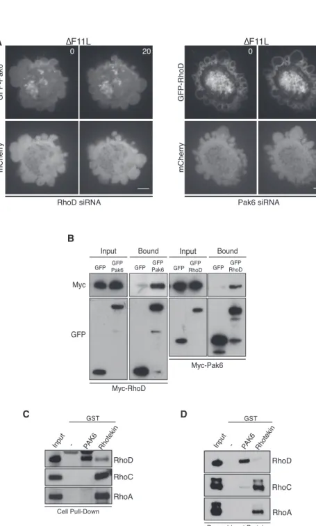
in DF11L-infected cells treated with RhoD siRNA (Figure 7A and Movie S7). In contrast, loss of Pak6 had no obvious impact on the recruitment of GFP-RhoD to the plasma membrane ( Fig-ure 7A andMovie S7). These data suggest that RhoD interacts with Pak6 to recruit the kinase to the plasma membrane. Consis-tent with this, reciprocal pull-downs on myc- and GFP-tagged RhoD and Pak6 demonstrate that the two proteins interact with each other in uninfected cells (Figure 7B). Recombinant GST-Pak6 can also retain RhoD but not RhoC or RhoA from cell lysates (Figures 7C andS7A). Moreover, pull-down assays with recombinant proteins demonstrate that the interaction be-tween RhoD and Pak6 is direct (Figures 7D andS7B). Our data clearly demonstrate that RhoD inhibits RhoC-ROCK-induced cell contraction via its downstream effector Pak6.
A
B
C D
Figure 7. RhoD Interacts Directly with Pak6
(A) Images showing that siRNA-mediated ablation of RhoD leads to a loss of GFP-Pak6 recruitment to the plasma membrane in cells infected with DF11L. In contrast, loss of Pak6 does not impact on recruitment of GFP-RhoD to the plasma membrane (seeMovie S7). Scale bar, 5 mm. (B) Immunoblot analysis with the indicated anti-bodies of a GFP-Trap pull-down on cell lysates from uninfected HeLa expressing Myc-RhoD and GFP-Pak6 or GFP-RhoD and Myc-Pak6. (C) Immunoblot of glutathione-Sepharose pull-downs on HeLa cell lysates demonstrates that GST-Pak6 interacts with RhoD, while Rhotekin preferentially associates with RhoA and RhoC. (D) Immunoblot of glutathione-Sepharose pull-downs with recombinant proteins demonstrates that GST-Pak6 interacts with RhoD but not RhoA or RhoC.
See alsoFigure S7.
DISCUSSION
The organization and contraction of the cortical actin cytoskeleton plays an essential role in controlling cell shape and movement during development and tumor cell invasion (Blaser et al., 2006; Charras and Paluch, 2008; Fackler and Grosse, 2008; Friedl and Wolf, 2003; Pal-uch and Raz, 2013; Sahai and Marshall, 2003; Sanz-Moreno et al., 2008; Tozluoglu et al., 2013; Zatulovskiy et al., 2014). While it is well established that myosin-II drives contraction of the actin cortex, we still lack a complete understanding of the or-ganization and composition of the cortical actin cytoskeleton, as well as the signaling networks controlling its form and function (Barry et al., 2015; Biro et al., 2013; Bovel-lan et al., 2014; Fritzsche et al., 2013, 2016). One of the hallmarks of myosin-II-driven contraction of the actin cortex is the formation of blebs at the plasma mem-brane (Charras et al., 2008). Cell blebbing, which occurs during ameboid-based cell motility, mitotic round-ing, and cytokinesis (Friedl and Wolf, 2003; Matthews et al., 2012; Sedzinski et al., 2011), is also frequently seen as part of the so-called cytopathic effect during many different viral infec-tions (Agol, 2012). In the case of vaccinia virus, cells start to con-tract and bleb within a few hours of infection (Bablanian et al., 1978; Barry et al., 2015; Schepis et al., 2006; Schramm et al., 2006). We have now demonstrated that vaccinia-induced cell contraction and blebbing is dependent on the viral protein F11. The ability of F11 to induce ROCK-dependent cell contraction early during viral replication contrasts with its ability to inhibit RhoA signaling in the latter stages of infection (Arakawa et al., 2007b; Cordeiro et al., 2009; Handa et al., 2013; Valderrama et al., 2006). F11-mediated inhibition of RhoA signaling at 8 hpi
stimulates infected cell migration and also facilitates the release of new viral progeny by increasing microtubule growth toward the plasma membrane and modulating the cortical actin cytoskel-eton (Arakawa et al., 2007a, 2007b; Cordeiro et al., 2009; Handa et al., 2013; Morales et al., 2008; Valderrama et al., 2006).
Considering the ability of RhoA to bind F11, it was surprising that F11-induced cell contraction depends on RhoC and not RhoA, as both GTPases have been shown to bind and activate ROCK to induce actin stress fiber formation and myosin-II contraction (Leung et al., 1996; Ridley, 2013). Historically it has been difficult to assign individual effects or cellular responses to RhoA and RhoC, in part because, until relatively recently, most studies tended to focus only on RhoA. Furthermore, expression of constitutively active or dominant-negative RhoA mutants impacts on the activity of both GTPases, as does the inhibitory effect of C3 toxin (Aktories, 2011). More recently, the use of RNAi-based approaches has begun to uncover distinct functions for RhoA and RhoC (Bravo-Cordero et al., 2011, 2013; Korkina et al., 2013; Sahai and Marshall, 2003; Vega et al., 2011). However, it is striking that the evidence for RhoA driving cell contraction and blebbing is largely based on the use of reagents that do not fully discriminate between RhoA and RhoC (Charras et al., 2006; Costigliola et al., 2010; Gutjahr et al., 2005; Sahai and Marshall, 2003; Sanz-Moreno et al., 2008). Our observations demonstrate that RhoC plays the greater role in stimulating ROCK-mediated myosin-II contraction of the actin cortex during vaccinia infection. This conclusion has important implications for a variety of cellular processes involving cell contraction and blebbing that have previously only been assigned to RhoA. It is also striking that there is more evidence suggesting that RhoC is more important than RhoA in promoting tumor cell invasion and metastasis (Clark et al., 2000; Dietrich et al., 2009; Hakem et al., 2005; Kitzing et al., 2010; Leung et al., 1996; Ridley, 2013; Ruth et al., 2006; Sahai and Marshall, 2002; Simpson et al., 2004).
F11 interacts directly with RhoA using a motif that is similar to that of the Rho-binding site in ROCK (Cordeiro et al., 2009; Valder-rama et al., 2006). It is likely that F11 will also bind directly to RhoC, as its effector binding switchI/II regions are essentially identical to those in RhoA (Ridley, 2013; Wheeler and Ridley, 2004). Our in vitro pull-down assays with recombinant protein reveal that RhoC interacts much less efficiently with F11 than with RhoA (Figure S5C). The reason for this dramatic difference is not immediately obvious. However, we found that F11 regulates RhoC-mediated cell contraction by inhibiting RhoD signaling. RhoD is emerging as a RhoGTPase family member with diverse functions including regulation of centrosome duplication, cell-cy-cle progression, actin structures, and membrane trafficking ( Gas-man et al., 2003; Koizumi et al., 2012; Kyrkou et al., 2013; Nehru et al., 2013). Previous analysis demonstrates that overexpression of constitutively active RhoD disrupts focal adhesions and stress fibers, and also suppresses the ability of active RhoA to promote actin assembly (Tsubakimoto et al., 1999). These overexpression data led the authors to suggest that RhoD antagonizes RhoA signaling (Tsubakimoto et al., 1999). Our data now clearly demon-strate that RhoD antagonizes the function of RhoC, which repre-sents another example of RhoGTPase crosstalk, whereby the action of one GTPase regulates the activity of another. GTPase crosstalk can regulate RhoGEFs and RhoGAPs, GTPase stability,
and downstream signaling (reviewed inGuilluy et al., 2011). One well-documented example of GTPase crosstalk is the inhibition of RhoA by Rac. Following Rac activation, the downstream effec-tors Pak1 and Pak4 phosphorylate p115-RhoGEF, GEF-H1, PDZ-RhoGEF, and Net1 (Alberts et al., 2005; Barac et al., 2004; Rosenfeldt et al., 2006; Zenke et al., 2004). Phosphorylation of these RhoGEFs inhibits their exchange activity or alters their stability and/or localization, leading to a loss of downstream RhoA activation. There remains the possibility that Pak1 and Pak4 can also control RhoC-mediated cell contraction, as some of these RhoGEFs also regulate the activity of RhoC (Jaiswal et al., 2011; Ren et al., 1998). This possibility, together with the observation that RhoD interacts with Pak5 (Wu and Frost, 2006), led us to investigate whether Pak kinases were involved in vaccinia-induced cell contraction. We found that only deple-tion of Pak6 stimulated RhoC/ROCK-dependent contracdeple-tion of DF11L virus-infected cells. Conversely, Pak6 overexpression suppressed contraction of WR-infected cells. Furthermore, RhoD suppressed WR-induced cell contraction through Pak6, suggesting that Pak6 is a RhoD effector. Consistent with this notion, we found that RhoD can interact directly with Pak6 and is responsible for recruitment of the kinase to the plasma membrane.
Our data are in agreement with previous observations sug-gesting that RhoGTPases principally regulate class II Pak family members by controlling their subcellular location rather than directly stimulating their kinase activity (Ha et al., 2015). Never-theless, our results clearly show that the kinase activity of Pak6 is required to inhibit RhoC-induced cell contraction. It is possible that Pak6 may directly phosphorylate RhoC to regulate its activ-ity. Seven of the 22 RhoGTPases, including RhoA, have a serine residue between their phospholipid-binding polybasic region and the CAAX motif that can be phosphorylated (Wheeler and Ridley, 2004). Phosphorylation of this serine weakens the asso-ciation of the RhoGTPase with the membrane and also increases its affinity for Rho GDP-dissociation inhibitor (RhoGDI), which extracts the GTPase from membrane (Boulter et al., 2010; Eller-broek et al., 2003; Garcia-Mata et al., 2011). However, RhoC does not have an equivalent serine residue between its polybasic region and CAAX motif, so is unlikely to be regulated by this mechanism. Furthermore, our pull-down assays demonstrate that Pak6 does not interact with RhoC. Given this finding it may be that, as with Rac-mediated inhibition of RhoA, Pak6 suppresses the activity of RhoC by phosphorylating one or more RhoGEF. This raises the question of how specificity of a RhoGEF(s) for RhoC over RhoA is achieved. The RhoGEF bind-ing switch I regions of RhoA, B, and C are almost identical, with only a single change for RhoB (Q29E) or RhoC (V43I) (Wheeler and Ridley, 2004). Structural analysis of RhoA in complex with the LARG, PDZ-RhoGEF, or p63RhoGEF reveals that Val43 par-ticipates in van der Waals interactions at the RhoGTPase:GEF interface (Chen et al., 2010; Kristelly et al., 2004; Lutz et al., 2007; Oleksy et al., 2006). A bulkier isoleucine residue at this point could sterically block the interaction between the RhoGTPase and its GEF. Indeed, the GEF XPLN interacts with RhoA and RhoB but not RhoC, the distinguishing feature being the isoleucine residue at position 43 in RhoC (Arthur et al., 2002; Sloan et al., 2012). Conversely, it is also conceivable that RhoD/Pak6 can activate a GAP capable of downregulating
RhoC signaling. We previously found that RhoA is inactivated by the GAP activity of myosin-9A, in an F11-dependent manner in late stages of infection (Handa et al., 2013). However, we now found that in early stages of infection there is no role for myos9A, as WR still induces cell contraction and those in-fected with DF11L remain spread when myosin-9A is depleted (Figure S5E). These data also suggest that myosin-9A has no GAP activity toward RhoD.
From our studies it is clear that early during vaccinia infection there must be an additional stimulus activating RhoC, as DF11L virus-infected cells depleted of RhoD or Pak6 contract in an RhoC-dependent fashion. This is consistent with our hypothesis that in DF11L-infected cells RhoD recruits Pak6 to the plasma membrane where it can locally antagonize RhoC signaling to ROCK to inhibit cell contraction. Moreover, the activation of RhoC by an additional viral protein within the first few hours of infection might explain why F11 promotes very different pheno-types at early and late time points during infection. It is also possible that the selectivity of RhoC during vaccinia-induced contraction is actually dependent on this unknown stimulus, leaving the possibility open that Pak6 suppresses both RhoA and RhoC signaling. The task ahead is to understand how vaccinia activates RhoC and to use vaccinia-induced cell contraction as an assay to uncover the basis for Pak6-mediated inhibition of RhoC signaling.
STAR+METHODS
Detailed methods are provided in the online version of this paper and include the following:
d KEY RESOURCES TABLE
d CONTACT FOR REAGENT AND RESOURCE SHARING
d EXPERIMENTAL MODEL AND SUBJECT DETAILS
B Cell Culture
B Viruses and Infections
d METHOD DETAILS B Plasmids
B Drug Treatments
B siRNA and DNA Transfections
B Reverse Transcription Quantitative PCR (RT-qPCR)
B Purification of Recombinant Proteins
B Pull Downs Assays
B Immunoblotting
B Live Cell Imaging
B Confocal Microscopy
d QUANTIFICATION AND STATISTICAL ANALYSIS SUPPLEMENTAL INFORMATION
Supplemental Information includes seven figures, one table, and seven movies and can be found with this article online athttp://dx.doi.org/10.1016/j.devcel. 2017.04.010.
AUTHOR CONTRIBUTIONS
C.H.D., F.L., and M.W. designed the study and wrote the manuscript. C.H.D., F.L., and J.V.C. performed and analyzed the experiments. Y.A. and F.V. performed initial experiments leading to this study. Y.H. generated the recombinant GFP-F11 virus. All authors commented on the manu-script text.
ACKNOWLEDGMENTS
We would like to thank members of the M.W. laboratory, Erik Sahai (The Francis Crick Institute, London), and Helen Walden (University of Dundee) for comments on the manuscript. We would like to thank Harry Mellor (Bristol University, UK) and Erik Sahai (The Francis Crick Institute, London) for providing Rho clones as well as Prof. Geoffrey Smith (Department of Pathol-ogy, Cambridge University, UK) for the DF12L virus. This work was supported by the Francis Crick Institute, which receives its core funding from Cancer Research UK (FC001209), the UK Medical Research Council (FC001209), and the Wellcome Trust (FC001209). F.V. and Y.H. were additionally supported by European Community Marie Curie Fellowship (HPMF-CT-2000-01021) (F.V.) and EMBO Long-Term Fellowship ALTF 813-2009 (Y.H.), and The Japan Society for the Promotion of Science (JSPS) Postdoctoral Fellowship for Research Abroad (Y.H.), respectively.
Received: January 30, 2014 Revised: January 5, 2017 Accepted: April 12, 2017 Published: May 8, 2017
REFERENCES
Agol, V.I. (2012). Cytopathic effects: virus-modulated manifestations of innate immunity? Trends Microbiol. 20, 570–576.
Aktories, K. (2011). Bacterial protein toxins that modify host regulatory GTPases. Nat. Rev. Microbiol. 9, 487–498.
Alberts, A.S., Qin, H., Carr, H.S., and Frost, J.A. (2005). PAK1 negatively regulates the activity of the Rho exchange factor NET1. J. Biol. Chem. 280, 12152–12161.
Amano, M., Ito, M., Kimura, K., Fukata, Y., Chihara, K., Nakano, T., Matsuura, Y., and Kaibuchi, K. (1996). Phosphorylation and activation of myosin by Rho-associated kinase (Rho-kinase). J. Biol. Chem. 271, 20246–20249.
Amano, M., Chihara, K., Kimura, K., Fukata, Y., Nakamura, N., Matsuura, Y., and Kaibuchi, K. (1997). Formation of actin stress fibers and focal adhesions enhanced by Rho-kinase. Science 275, 1308–1311.
Amano, M., Nakayama, M., and Kaibuchi, K. (2010). Rho-kinase/ROCK: a key regulator of the cytoskeleton and cell polarity. Cytoskeleton (Hoboken) 67, 545–554.
Amin, E., Dubey, B.N., Zhang, S.C., Gremer, L., Dvorsky, R., Moll, J.M., Taha, M.S., Nagel-Steger, L., Piekorz, R.P., Somlyo, A.V., et al. (2013). Rho-kinase: regulation, (dys)function, and inhibition. Biol. Chem. 394, 1399–1410.
Arakawa, Y., Cordeiro, J.V., Schleich, S., Newsome, T.P., and Way, M. (2007a). The release of vaccinia virus from infected cells requires RhoA-mDia modulation of cortical actin. Cell Host Microbe 1, 227–240.
Arakawa, Y., Cordeiro, J.V., and Way, M. (2007b). F11L-mediated inhibition of RhoA-mDia signaling stimulates microtubule dynamics during vaccinia virus infection. Cell Host Microbe 1, 213–226.
Arthur, W.T., Ellerbroek, S.M., Der, C.J., Burridge, K., and Wennerberg, K. (2002). XPLN, a guanine nucleotide exchange factor for RhoA and RhoB, but not RhoC. J. Biol. Chem. 277, 42964–42972.
Bablanian, R., Baxt, B., Sonnabend, J.A., and Esteban, M. (1978). Studies on the mechanisms of vaccinia virus cytopathic effects. II. Early cell rounding is associated with virus polypeptide synthesis. J. Gen. Virol. 39, 403–413.
Barac, A., Basile, J., Vazquez-Prado, J., Gao, Y., Zheng, Y., and Gutkind, J.S. (2004). Direct interaction of p21-activated kinase 4 with PDZ-RhoGEF, a G protein-linked Rho guanine exchange factor. J. Biol. Chem. 279, 6182–6189.
Barry, D.J., Durkin, C.H., Abella, J.V., and Way, M. (2015). Open source soft-ware for quantification of cell migration, protrusions, and fluorescence inten-sities. J. Cell Biol. 209, 163–180.
Biro, M., Romeo, Y., Kroschwald, S., Bovellan, M., Boden, A., Tcherkezian, J., Roux, P.P., Charras, G., and Paluch, E.K. (2013). Cell cortex composition and homeostasis resolved by integrating proteomics and quantitative imaging. Cytoskeleton (Hoboken) 70, 741–754.
Blaser, H., Reichman-Fried, M., Castanon, I., Dumstrei, K., Marlow, F.L., Kawakami, K., Solnica-Krezel, L., Heisenberg, C.P., and Raz, E. (2006). Migration of zebrafish primordial germ cells: a role for myosin contraction and cytoplasmic flow. Dev. Cell 11, 613–627.
Boeda, B., Briggs, D.C., Higgins, T., Garvalov, B.K., Fadden, A.J., McDonald, N.Q., and Way, M. (2007). Tes, a specific Mena interacting partner, breaks the rules for EVH1 binding. Mol. Cell 28, 1071–1082.
Boulter, E., Garcia-Mata, R., Guilluy, C., Dubash, A., Rossi, G., Brennwald, P.J., and Burridge, K. (2010). Regulation of Rho GTPase crosstalk, degrada-tion and activity by RhoGDI1. Nat. Cell Biol. 12, 477–483.
Bovellan, M., Romeo, Y., Biro, M., Boden, A., Chugh, P., Yonis, A., Vaghela, M., Fritzsche, M., Moulding, D., Thorogate, R., et al. (2014). Cellular control of cortical actin nucleation. Curr. Biol. 24, 1628–1635.
Bravo-Cordero, J.J., Oser, M., Chen, X., Eddy, R., Hodgson, L., and Condeelis, J. (2011). A novel spatiotemporal RhoC activation pathway locally regulates cofilin activity at invadopodia. Curr. Biol. 21, 635–644.
Bravo-Cordero, J.J., Sharma, V.P., Roh-Johnson, M., Chen, X., Eddy, R., Condeelis, J., and Hodgson, L. (2013). Spatial regulation of RhoC activity de-fines protrusion formation in migrating cells. J. Cell Sci. 126, 3356–3369.
Charras, G., and Paluch, E. (2008). Blebs lead the way: how to migrate without lamellipodia. Nat. Rev. Mol. Cell Biol. 9, 730–736.
Charras, G.T., Hu, C.K., Coughlin, M., and Mitchison, T.J. (2006). Reassembly of contractile actin cortex in cell blebs. J. Cell Biol. 175, 477–490.
Charras, G.T., Coughlin, M., Mitchison, T.J., and Mahadevan, L. (2008). Life and times of a cellular bleb. Biophys. J. 94, 1836–1853.
Chen, Z., Medina, F., Liu, M.Y., Thomas, C., Sprang, S.R., and Sternweis, P.C. (2010). Activated RhoA binds to the pleckstrin homology (PH) domain of PDZ-RhoGEF, a potential site for autoregulation. J. Biol. Chem. 285, 21070–21081.
Clark, E.A., Golub, T.R., Lander, E.S., and Hynes, R.O. (2000). Genomic analysis of metastasis reveals an essential role for RhoC. Nature 406, 532–535.
Clark, A.G., Dierkes, K., and Paluch, E.K. (2013). Monitoring actin cortex thick-ness in live cells. Biophys. J. 105, 570–580.
Cordeiro, J.V., Guerra, S., Arakawa, Y., Dodding, M.P., Esteban, M., and Way, M. (2009). F11-mediated inhibition of RhoA signalling enhances the spread of vaccinia virus in vitro and in vivo in an intranasal mouse model of infection. PLoS One 4, e8506.
Costigliola, N., Kapustina, M.T., Weinreb, G.E., Monteith, A., Rajfur, Z., Elston, T.C., and Jacobson, K. (2010). RhoA regulates calcium-independent periodic contractions of the cell cortex. Biophys. J. 99, 1053–1063.
Cudmore, S., Cossart, P., Griffiths, G., and Way, M. (1995). Actin-based motility of vaccinia virus. Nature 378, 636–638.
Dietrich, K.A., Schwarz, R., Liska, M., Grass, S., Menke, A., Meister, M., Kierschke, G., Langle, C., Genze, F., and Giehl, K. (2009). Specific induc-tion of migrainduc-tion and invasion of pancreatic carcinoma cells by RhoC, which differs from RhoA in its localisation and activity. Biol. Chem. 390, 1063–1077.
Diz-Munoz, A., Krieg, M., Bergert, M., Ibarlucea-Benitez, I., Muller, D.J., Paluch, E., and Heisenberg, C.P. (2010). Control of directed cell migration in vivo by membrane-to-cortex attachment. PLoS Biol. 8, e1000544.
Dodding, M.P., Newsome, T.P., Collinson, L.M., Edwards, C., and Way, M. (2009). An E2-F12 complex is required for intracellular enveloped virus morphogenesis during vaccinia infection. Cell. Microbiol. 11, 808–824.
Ellerbroek, S.M., Wennerberg, K., and Burridge, K. (2003). Serine phosphory-lation negatively regulates RhoA in vivo. J. Biol. Chem. 278, 19023–19031.
Fackler, O.T., and Grosse, R. (2008). Cell motility through plasma membrane blebbing. J. Cell Biol. 181, 879–884.
Fan, L., Pellegrin, S., Scott, A., and Mellor, H. (2010). The small GTPase Rif is an alternative trigger for the formation of actin stress fibers in epithelial cells. J. Cell Sci. 123, 1247–1252.
Fram, S., King, H., Sacks, D.B., and Wells, C.M. (2014). A PAK6-IQGAP1 com-plex promotes disassembly of cell-cell adhesions. Cell. Mol. Life Sci. 71, 2759–2773.
Friedl, P., and Wolf, K. (2003). Proteolytic and non-proteolytic migration of tumour cells and leucocytes. Biochem. Soc. Symp. 277–285.
Frischknecht, F., Moreau, V., Rottger, S., Gonfloni, S., Reckmann, I., Superti-Furga, G., and Way, M. (1999). Actin-based motility of vaccinia virus mimics receptor tyrosine kinase signalling. Nature 401, 926–929.
Fritzsche, M., Lewalle, A., Duke, T., Kruse, K., and Charras, G. (2013). Analysis of turnover dynamics of the submembranous actin cortex. Mol. Biol. Cell 24, 757–767.
Fritzsche, M., Erlenkamper, C., Moeendarbary, E., Charras, G., and Kruse, K. (2016). Actin kinetics shapes cortical network structure and mechanics. Sci. Adv. 2, e1501337.
Garcia-Mata, R., Boulter, E., and Burridge, K. (2011). The ’invisible hand’: regulation of RHO GTPases by RHOGDIs. Nat. Rev. Mol. Cell Biol. 12, 493–504.
Gasman, S., Kalaidzidis, Y., and Zerial, M. (2003). RhoD regulates endosome dynamics through Diaphanous-related Formin and Src tyrosine kinase. Nat. Cell Biol. 5, 195–204.
Gauthier, N.C., Masters, T.A., and Sheetz, M.P. (2012). Mechanical feedback between membrane tension and dynamics. Trends Cell Biol. 22, 527–535.
Guilluy, C., Garcia-Mata, R., and Burridge, K. (2011). Rho protein crosstalk: another social network? Trends Cell Biol. 21, 718–726.
Gutjahr, M.C., Rossy, J., and Niggli, V. (2005). Role of Rho, Rac, and Rho-ki-nase in phosphorylation of myosin light chain, development of polarity, and spontaneous migration of Walker 256 carcinosarcoma cells. Exp. Cell Res.
308, 422–438.
Ha, B.H., Morse, E.M., Turk, B.E., and Boggon, T.J. (2015). Signaling, regula-tion, and specificity of the type II p21-activated kinases. J. Biol. Chem. 290, 12975–12983.
Hakem, A., Sanchez-Sweatman, O., You-Ten, A., Duncan, G., Wakeham, A., Khokha, R., and Mak, T.W. (2005). RhoC is dispensable for embryo-genesis and tumor initiation but essential for metastasis. Genes Dev. 19, 1974–1979.
Handa, Y., Durkin, C.H., Dodding, M.P., and Way, M. (2013). Vaccinia virus F11 promotes viral spread by acting as a PDZ-containing scaffolding protein to bind myosin-9A and inhibit RhoA signaling. Cell Host Microbe
14, 51–62.
Irwin, C.R., and Evans, D.H. (2012). Modulation of the myxoma virus plaque phenotype by vaccinia virus protein F11. J. Virol. 86, 7167–7179.
Irwin, C.R., Favis, N.A., Agopsowicz, K.C., Hitt, M.M., and Evans, D.H. (2013). Myxoma virus oncolytic efficiency can be enhanced through chemical or ge-netic disruption of the actin cytoskeleton. PLoS One 8, e84134.
Jaiswal, M., Gremer, L., Dvorsky, R., Haeusler, L.C., Cirstea, I.C., Uhlenbrock, K., and Ahmadian, M.R. (2011). Mechanistic insights into specificity, activity, and regulatory elements of the regulator of G-protein signaling (RGS)-contain-ing Rho-specific guanine nucleotide exchange factors (GEFs) p115, PDZ-RhoGEF (PRG), and leukemia-associated PDZ-RhoGEF (LARG). J. Biol. Chem.
286, 18202–18212.
Julian, L., and Olson, M.F. (2014). Rho-associated coiled-coil containing ki-nases (ROCK): structure, regulation, and functions. Small GTPases 5, e29846.
Kardash, E., Reichman-Fried, M., Maitre, J.L., Boldajipour, B., Papusheva, E., Messerschmidt, E.M., Heisenberg, C.P., and Raz, E. (2010). A role for Rho GTPases and cell-cell adhesion in single-cell motility in vivo. Nat. Cell Biol.
12, 47–53, sup pp 1–11.
Kawano, Y., Fukata, Y., Oshiro, N., Amano, M., Nakamura, T., Ito, M., Matsumura, F., Inagaki, M., and Kaibuchi, K. (1999). Phosphorylation of myosin-binding subunit (MBS) of myosin phosphatase by Rho-kinase in vivo. J. Cell Biol. 147, 1023–1038.
Kitzing, T.M., Wang, Y., Pertz, O., Copeland, J.W., and Grosse, R. (2010). Formin-like 2 drives amoeboid invasive cell motility downstream of RhoC. Oncogene 29, 2441–2448.
Koizumi, K., Takano, K., Kaneyasu, A., Watanabe-Takano, H., Tokuda, E., Abe, T., Watanabe, N., Takenawa, T., and Endo, T. (2012). RhoD activated by fibroblast growth factor induces cytoneme-like cellular protrusions through mDia3C. Mol. Biol. Cell 23, 4647–4661.
Korkina, O., Dong, Z., Marullo, A., Warshaw, G., Symons, M., and Ruggieri, R. (2013). The MLK-related kinase (MRK) is a novel RhoC effector that mediates lysophosphatidic acid (LPA)-stimulated tumor cell invasion. J. Biol. Chem.
288, 5364–5373.
Koster, D.V., and Mayor, S. (2016). Cortical actin and the plasma membrane: inextricably intertwined. Curr. Opin. Cell Biol. 38, 81–89.
Kristelly, R., Gao, G., and Tesmer, J.J. (2004). Structural determinants of RhoA binding and nucleotide exchange in leukemia-associated Rho guanine-nucle-otide exchange factor. J. Biol. Chem. 279, 47352–47362.
Kyrkou, A., Soufi, M., Bahtz, R., Ferguson, C., Bai, M., Parton, R.G., Hoffmann, I., Zerial, M., Fotsis, T., and Murphy, C. (2013). RhoD participates in the regu-lation of cell-cycle progression and centrosome duplication. Oncogene 32, 1831–1842.
Leung, T., Chen, X.Q., Manser, E., and Lim, L. (1996). The p160 RhoA-binding kinase ROK alpha is a member of a kinase family and is involved in the reorga-nization of the cytoskeleton. Mol. Cell. Biol. 16, 5313–5327.
Lutz, S., Shankaranarayanan, A., Coco, C., Ridilla, M., Nance, M.R., Vettel, C., Baltus, D., Evelyn, C.R., Neubig, R.R., Wieland, T., et al. (2007). Structure of Galphaq-p63RhoGEF-RhoA complex reveals a pathway for the activation of RhoA by GPCRs. Science 318, 1923–1927.
Matthews, H.K., Delabre, U., Rohn, J.L., Guck, J., Kunda, P., and Baum, B. (2012). Changes in Ect2 localization couple actomyosin-dependent cell shape changes to mitotic progression. Dev. Cell 23, 371–383.
Mercer, J., and Helenius, A. (2008). Vaccinia virus uses macropinocytosis and apoptotic mimicry to enter host cells. Science 320, 531–535.
Mercer, J., Snijder, B., Sacher, R., Burkard, C., Bleck, C.K., Stahlberg, H., Pelkmans, L., and Helenius, A. (2012). RNAi screening reveals proteasome-and Cullin3-dependent stages in vaccinia virus infection. Cell Rep. 2, 1036–1047.
Morales, I., Carbajal, M.A., Bohn, S., Holzer, D., Kato, S.E., Greco, F.A., Moussatche, N., and Krijnse Locker, J. (2008). The vaccinia virus F11L gene product facilitates cell detachment and promotes migration. Traffic 9, 1283–1298.
Morone, N., Fujiwara, T., Murase, K., Kasai, R.S., Ike, H., Yuasa, S., Usukura, J., and Kusumi, A. (2006). Three-dimensional reconstruction of the membrane skeleton at the plasma membrane interface by electron tomography. J. Cell Biol. 174, 851–862.
Nehru, V., Almeida, F.N., and Aspenstrom, P. (2013). Interaction of RhoD and ZIP kinase modulates actin filament assembly and focal adhesion dynamics. Biochem. Biophys. Res. Commun. 433, 163–169.
Nimnual, A.S., Taylor, L.J., and Bar-Sagi, D. (2003). Redox-dependent down-regulation of Rho by rac. Nat. Cell Biol. 5, 236–241.
Oleksy, A., Opalinski, L., Derewenda, U., Derewenda, Z.S., and Otlewski, J. (2006). The molecular basis of RhoA specificity in the guanine nucleotide ex-change factor PDZ-RhoGEF. J. Biol. Chem. 281, 32891–32897.
Paluch, E.K., and Raz, E. (2013). The role and regulation of blebs in cell migra-tion. Curr. Opin. Cell Biol. 25, 582–590.
Paluch, E., Sykes, C., Prost, J., and Bornens, M. (2006). Dynamic modes of the cortical actomyosin gel during cell locomotion and division. Trends Cell Biol.
16, 5–10.
Pinner, S., and Sahai, E. (2008). PDK1 regulates cancer cell motility by antag-onising inhibition of ROCK1 by RhoE. Nat. Cell Biol. 10, 127–137.
Ren, Y., Li, R., Zheng, Y., and Busch, H. (1998). Cloning and characterization of GEF-H1, a microtubule-associated guanine nucleotide exchange factor for Rac and Rho GTPases. J. Biol. Chem. 273, 34954–34960.
Ridley, A.J. (2013). RhoA, RhoB and RhoC have different roles in cancer cell migration. J. Microsc. 251, 242–249.
Riento, K., and Ridley, A.J. (2003). Rocks: multifunctional kinases in cell behaviour. Nat. Rev. Mol. Cell Biol. 4, 446–456.
Rietdorf, J., Ploubidou, A., Reckmann, I., Holmstro¨m, A., Frischknecht, F., Zettl, M., Zimmermann, T., and Way, M. (2001). Kinesin dependent movement on microtubules precedes actin based motility of vaccinia virus. Nat. Cell Biol.
3, 992–1000.
Rosenfeldt, H., Castellone, M.D., Randazzo, P.A., and Gutkind, J.S. (2006). Rac inhibits thrombin-induced Rho activation: evidence of a Pak-dependent GTPase crosstalk. J. Mol. Signal. 1, 8.
Ro¨ttger, S., Frischknecht, F., Reckmann, I., Smith, G.L., and Way, M. (1999). Interactions between vaccinia virus IEV membrane proteins and their roles in IEV assembly and actin tail formation. J. Virol. 73, 2863–2875.
Ruth, M.C., Xu, Y., Maxwell, I.H., Ahn, N.G., Norris, D.A., and Shellman, Y.G. (2006). RhoC promotes human melanoma invasion in a PI3K/Akt-dependent pathway. J. Invest. Dermatol. 126, 862–868.
Sahai, E., and Marshall, C.J. (2002). ROCK and Dia have opposing effects on adherens junctions downstream of Rho. Nat. Cell Biol. 4, 408–415.
Sahai, E., and Marshall, C.J. (2003). Differing modes of tumour cell invasion have distinct requirements for Rho/ROCK signalling and extracellular proteol-ysis. Nat. Cell Biol. 5, 711–719.
Salbreux, G., Charras, G., and Paluch, E. (2012). Actin cortex mechanics and cellular morphogenesis. Trends Cell Biol. 22, 536–545.
Sanz-Moreno, V., Gadea, G., Ahn, J., Paterson, H., Marra, P., Pinner, S., Sahai, E., and Marshall, C.J. (2008). Rac activation and inactivation control plasticity of tumor cell movement. Cell 135, 510–523.
Schepis, A., Schramm, B., de Haan, C.A., and Locker, J.K. (2006). Vaccinia virus-induced microtubule-dependent cellular rearrangements. Traffic 7, 308–323.
Schramm, B., de Haan, C.A., Young, J., Doglio, L., Schleich, S., Reese, C., Popov, A.V., Steffen, W., Schroer, T., and Locker, J.K. (2006). Vaccinia-vi-rus-induced cellular contractility facilitates the subcellular localization of the viral replication sites. Traffic 7, 1352–1367.
Sedzinski, J., Biro, M., Oswald, A., Tinevez, J.Y., Salbreux, G., and Paluch, E. (2011). Polar actomyosin contractility destabilizes the position of the cytoki-netic furrow. Nature 476, 462–466.
Simpson, K.J., Dugan, A.S., and Mercurio, A.M. (2004). Functional analysis of the contribution of RhoA and RhoC GTPases to invasive breast carcinoma. Cancer Res. 64, 8694–8701.
Sloan, C.M., Quinn, C.V., Peters, J.P., Farley, J., Goetzinger, C., Wernli, M., DeMali, K.A., and Ellerbroek, S.M. (2012). Divergence of Rho residue 43 im-pacts GEF activity. Small GTPases 3, 15–22.
Tinevez, J.Y., Schulze, U., Salbreux, G., Roensch, J., Joanny, J.F., and Paluch, E. (2009). Role of cortical tension in bleb growth. Proc. Natl. Acad. Sci. USA
106, 18581–18586.
Tozluoglu, M., Tournier, A.L., Jenkins, R.P., Hooper, S., Bates, P.A., and Sahai, E. (2013). Matrix geometry determines optimal cancer cell migration strategy and modulates response to interventions. Nat. Cell Biol. 15, 751–762.
Tsubakimoto, K., Matsumoto, K., Abe, H., Ishii, J., Amano, M., Kaibuchi, K., and Endo, T. (1999). Small GTPase RhoD suppresses cell migration and cyto-kinesis. Oncogene 18, 2431–2440.
Usui, T., Okada, M., and Yamawaki, H. (2014). Zipper interacting protein ki-nase (ZIPK): function and signaling. Apoptosis 19, 387–391.
Valderrama, F., Cordeiro, J.V., Schleich, S., Frischknecht, F., and Way, M. (2006). Vaccinia virus-induced cell motility requires F11L-mediated inhibition of RhoA signaling. Science 311, 377–381.
Vega, F.M., Fruhwirth, G., Ng, T., and Ridley, A.J. (2011). RhoA and RhoC have distinct roles in migration and invasion by acting through different targets. J. Cell Biol. 193, 655–665.
Wheeler, A.P., and Ridley, A.J. (2004). Why three Rho proteins? RhoA, RhoB, RhoC, and cell motility. Exp. Cell Res. 301, 43–49.
Wilkinson, S., Paterson, H.F., and Marshall, C.J. (2005). Cdc42-MRCK and Rho-ROCK signalling cooperate in myosin phosphorylation and cell invasion. Nat. Cell Biol. 7, 255–261.
Wu, X., and Frost, J.A. (2006). Multiple Rho proteins regulate the subcellular targeting of PAK5. Biochem. Biophys. Res. Commun. 351, 328–335.
Zatulovskiy, E., Tyson, R., Bretschneider, T., and Kay, R.R. (2014). Bleb-driven chemotaxis of Dictyostelium cells. J. Cell Biol. 204, 1027–1044.
Zenke, F.T., Krendel, M., DerMardirossian, C., King, C.C., Bohl, B.P., and Bokoch, G.M. (2004). p21-activated kinase 1 phosphorylates and regulates 14-3-3 binding to GEF-H1, a microtubule-localized Rho exchange factor. J. Biol. Chem. 279, 18392–18400.
Zhang, W.H., Wilcock, D., and Smith, G.L. (2000). Vaccinia virus F12L protein is required for actin tail formation, normal plaque size, and virulence. J. Virol.
74, 11654–11662.
Zhao, Z., and Manser, E. (2015). Myotonic dystrophy kinase-related Cdc42-binding kinases (MRCK), the ROCK-like effectors of Cdc42 and Rac1. Small GTPases 6, 81–88.
STAR+METHODS
KEY RESOURCES TABLEREAGENT or RESOURCE SOURCE IDENTIFIER
Antibodies
Mouse Monoclonal anti-b-Actin (AC-74) Sigma-Aldrich Cat#A2228; RRID: AB_476697 Rabbit Anti-Glutathione-S-Transferase (GST) antibody Sigma-Aldrich Cat#G7781; RRID: AB_259965 Mouse Monoclonal Anti-polyHistidine antibody
(clone HIS-1)
Sigma-Aldrich Cat#H1029; RRID: AB_260015
Mouse Monoclonal Anti-c-Myc antibody (clone 9E10) Cancer Research UK N/A
Mouse Monoclonal Anti-Mcm7 antibody (clone DCS-141) Sigma-Aldrich Cat#M7931; RRID: AB_260666 Mouse Monoclonal Anti-alpha-Tubulin Antibody
(clone B-5-1-2)
Sigma-Aldrich Cat#T6074; RRID: AB_477582
Rabbit Anti-Zip Kinase antibody Sigma-Aldrich Cat#Z0134; RRID: AB_261899 Rabbit monoclonal MYPT1 (D6C1) Cell Signaling Technology Cat#8574S; RRID: AB_10998518 Rabbit Phospho-MYPT1 (Thr696) Antibody Cell Signaling Technology Cat#5163S; RRID: AB_10691830) Rabbit Phospho-Myosin Light Chain 2 (Ser19) Antibody Cell Signaling Technology Cat#3671S; RRID: AB_330248 Rabbit monoclonal RhoA (67B9) antibody Cell Signaling Technology Cat#2117S; RRID: AB_10693922 Rabbit Monoclonal Anti-RhoC (Clone D40E4) Cell Signaling Technology Cat#3430S; RRID: AB_2179246 Rabbit Monoclonal ROCK1 (C8F7) Cell Signaling Technology Cat#4035S; RRID: AB_2238679 Rabbit Monoclonal ROCK2 (D1B1) Cell Signaling Technology Cat#9029S;RRID: AB_11127802 Rabbit Polyclonal Anti-Human PAK5 ProSci Cat#3075; RRID: AB_735027 Rabbit Polyclonal PAK6 antibody GeneTex Cat#GTX127915; RRID: N/A Rabbit polyclonal RHOD antibody Anbobio Cat#C18411; RRID: N/A Rabbit polyclonal RHOD antibody OriGene Cat#TA312722; RRID: N/A Mouse Monoclonal anti-GFP (clone 3E1) Cancer Research UK N/A
Mouse Monoclonal Anti-GRB2 Antibody (clone 81) BD Transduction Cat#610112, RRID: AB_397518 Rabbit anti-A36 Ro¨ttger et al., 1999 N/A
Rabbit anti-F11-N Handa et al., 2013 N/A
Rabbit anti-F11-C This paper N/A
Rabbit anti-MRCKa Wilkinson et al., 2005 N/A
Peroxidase-AffiniPure Goat Anti-Rabbit IgG antibody Jackson ImmunoResearch Labs Cat# 111-035-003; RRID: AB_2313567 Peroxidase-AffiniPure Goat Anti-Mouse IgG antibody Jackson ImmunoResearch Labs Cat#115-035-003; RRID: AB_10015289 FITC-Donkey anti-Rabbit IgG Jackson ImmunoResearch Labs Cat#711-095-152; RRID:AB_2315776 Texas Red-X Phalloidin Molecular Probes Cat# T7471
Bacterial and Virus Strains
Vaccinia virus Western Reserve Cudmore et al., 1995 N/A Vaccinia virus Western Reserve DF11L Cordeiro et al., 2009 N/A Vaccinia virus Western Reserve F11-VK Cordeiro et al., 2009 N/A Vaccinia virus Western Reserve GFP-F11 This paper N/A Escherichia coli BL21-CodonPlus (DE3)-RIL Agilent Technologies Cat# 230245 Chemicals, Peptides, and Recombinant Proteins
Rho inhibitor (C3) Cytoskeleton Inc. Cat#CT04 ROCK inhibitor Y27632 Sigma-Aldrich Cat#Y0503 ROCK inhibitor H1152 Tocris Bioscience Cat#2414 ROCK inhibitor GSK429286A Tocris Bioscience Cat#3726 Myosin Light Chain Kinase (MLCK) inhibitor ML7 Sigma-Aldrich Cat#I2764 Myosin Light Chain Kinase (MLCK) inhibitor ML9 Sigma-Aldrich Cat# C1172 Cyclohexamide (CHX) Sigma-Aldrich Cat#C7698 Group I p21-activated kinase (PAK) inhibitor IPA-3 Tocris Bioscience Cat#3622
(Continued on next page)
Continued
REAGENT or RESOURCE SOURCE IDENTIFIER
EDTA-free Protease Inhibitor Cocktail Roche Cat#05056489001 PhosSTOP Phosphatase inhibitor tablets Roche Cat#04906837001 PDIKLDAVLDRDGNFRPADC (F11 residues 101-120) Handa et al., 2013 N/A
CGGNFITKEIKNRDK (F11 residues 323-334) This paper N/A HiPerFect Transfection Reagent Qiagen Cat#301707 FuGENE 6 Transfection Reagent Promega Cat#E2691
GST-F11 Valderrama et al., 2006 N/A
GST-Pak6 This paper N/A
GST-Rhotekin (residues 7-89) Millipore Cat# 14-383
His-RhoA This paper N/A
His-RhoB This paper N/A
His-RhoC This paper N/A
His-RhoD This paper N/A
His-RhoE This paper N/A
His-RhoF This paper N/A
His-Rac1 This paper N/A
Critical Commercial Assays
GFP-Trap_A ChromoTek Cat#GTA-20
Rho Assay Reagent (Rhotekin RBD, agarose) Millipore Cat#14-383 Glutathione Sepharose 4B Amersham Cat#17-0756-01
Ni-NTA resin Qiagen Cat#30230
RNeasy Mini kit Qiagen Cat#74104
SuperScript II reverse transcriptase Invitrogen Cat#18064-014 ECL Western Blot detection reagent Amersham Cat#RPN2106 Experimental Models: Cell Lines
Human: HeLa cells ATCC Cat#CCL-2
Human: U-2 OS cells The Francis Crick Institute Cell Services
N/A
Oligonucleotides
siRNA sequences This paper, seeTable S1 N/A Recombinant DNA
pCB6-GFP Handa et al., 2013 N/A
pCB6-Myc Handa et al., 2013 N/A
pMW172-GST Boeda et al., 2007 N/A
pMW172-His Boeda et al., 2007 N/A
pEL-GFP Frischknecht et al., 1999 N/A
pEL-GST Frischknecht et al., 1999 N/A
pLVX MLC2-RFP Barry et al., 2015 N/A
pEL-GST-F11L Valderrama et al., 2006 N/A pMW-GST-F11L Valderrama et al., 2006 N/A pEL-GFP-RhoA Valderrama et al., 2006 N/A pMW-His-RhoA Valderrama et al., 2006 N/A
pEL-GFP-RhoB This paper N/A
pMW-His-RhoB This paper N/A
pEL-GFP-RhoC This paper N/A
pMW-His-RhoC This paper N/A
pCB6 GFP-RhoC WT This paper N/A
pCB6 GFP-RhoC T19N This paper N/A
pEL-GFP-RhoD This paper N/A
(Continued on next page)
CONTACT FOR REAGENT AND RESOURCE SHARING
Further information and requests for resources and reagents should be directed to and will be fulfilled by the Lead Contact, Michael Way ([email protected]).
EXPERIMENTAL MODEL AND SUBJECT DETAILS Cell Culture
HeLa cells were cultured in Modified Eagle Medium (MEM) supplemented with 10% SFB, penicillin (100u/ml) and streptomycin (100mg/ml). U-2 OS cells were cultured in Dulbecco’s Modified Eagle Medium (DMEM) supplemented with 10% SFB, penicillin (100u/ml) and streptomycin (100mg/ml). HeLa and U2-OS cells were authenticated by STR profiling and are both negative for mycoplasma.
Viruses and Infections
The wild-type virus used in this work is the Western Reserve strain (WR) (Cudmore et al., 1995). The recombinant viruses DF11L or F11-VK have been previously described (Cordeiro et al., 2009). The virus encoding GFP-F11, with a GGRGG linker between the GFP and N-terminus of F11 was generated by homologous recombination between a targeting vector encoding F12-GFP-F11 and the DF12L virus (Dodding et al., 2009; Zhang et al., 2000; Rietdorf et al., 2001). The F12-GFP-F11 targeting vector was transfected into HeLa cells 3 hours after infection with the DF12L virus, at a multiplicity of infection of 0.1 plaque-forming units. Two days later, cells were harvested and ruptured by freeze thawing. The cell lysate was then used to infect confluent BSC-1 cell monolayers, which were overlaid with 0.9% agarose 2 h after infection. Four days later, individual plaques were picked based on the rescue of the DF12L small plaque phenotype as well as expression of GFP. The virus in the isolated plaques was then amplified by re-infecting HeLa cells for 48 h. A further 4 rounds of plaque purification were performed until the recombinant GFP-F11 virus was clonal. The fidelity of the recombinant virus was confirmed by sequencing. HeLa or U-2 OS cells were infected with the WR strain of Vaccinia or its Continued
REAGENT or RESOURCE SOURCE IDENTIFIER
pMW-His-RhoD This paper N/A
pCB6 GFP-RhoD WT This paper N/A
pCB6 GFP-RhoD T31N This paper N/A
pCB6 GFP-RhoD G26V This paper N/A
pCB6 Myc-RhoD This paper N/A
pEL-GFP-RhoE This paper N/A
pMW-His-RhoE This paper N/A
pEL-GFP-RhoF This paper N/A
pMW-His-RhoF This paper N/A
pEL-GFP-Rac1 This paper N/A
pMW-His-Rac1 This paper N/A
pCB6-GFP-Pak4 This paper N/A
pCB6-GFP-Pak5 This paper N/A
pCB6-GFP-Pak6 This paper N/A
pCB6-GFP-Pak6 KD This paper N/A
pCB6-Myc-Pak6 This paper N/A
pLVX-GFP Barry et al., 2015 N/A
pLVX-mCherry Barry et al., 2015 N/A
Software and Algorithms
MetaMorph Microscopy Automation and Image Analysis Software
Molecular Devices https://www.moleculardevices.com/ systems/metamorph-research-imaging
ImageJ National Institutes of Health https://imagej.nih.gov/ij/
SlideBook Intelligent Imaging Innovations https://www.intelligent-imaging.com/ slidebook
Other
NuPAGE 4-12% Bis-Tris Protein Gels, 1.0 mm, 10-well Invitrogen Cat#NP0321BOX NuPAGE 3-8% Tris-Acetate Protein Gels, 1.0 mm, 10-well Invitrogen Cat#EA0375BOX