Bacterial diversity and antibiotic resistance from the
water source to the tap
Thesis submitted to the Universidade Católica Portuguesa to attain the degree of PhD in Biotechnology with specialization in
Microbiology
By
Ivone Cristina Vaz Moreira
Bacterial diversity and antibiotic resistance from the
water source to the tap
Thesis submitted to the Universidade Católica Portuguesa to attain the degree of PhD in Biotechnology - with specialization in
Microbiology
By
Ivone Cristina Vaz Moreira
Under the supervision of Professor Célia Maria Manaia Rodrigues Under the co-supervision of Professor Olga Cristina Pastor Nunes
‘‘Everything comes from water! And everything is kept alive by water!’’
J.W. von Goethe, Faust II, 1833
to all the persons who crossed my life and taught me something
i
ABSTRACT
Water is one of the most important habitats for bacteria in the environment. The continuous flux in the urban water cycle carries water through many places, dragging bacteria and numerous chemical contaminants. This makes of water one of the most important vehicles, not only for the dissemination of the chemical substances, but also for the dissemination of organisms and, consequently, the respective resistance genes in the environment. The main goal of this study was to investigate if drinking water production and distribution could represent a hotspot for the proliferation, selection or incoming of antibiotic resistant bacteria, and the likelihood of these organisms to reach the final consumer, via tap water. In order to meet this objective, the study was planned aiming the
tracking of bacterial communities and individual isolates from the source to the tap.
Firstly, the abundance and diversity of bacteria in raw, treated and final (tap) water was characterized using culture-dependent and culture-independent (16S rRNA-DGGE) approaches. Both approaches showed that the water treatment reduced the bacterial counts, diversity and cultivability, promoting also a shift in the cultivable bacterial community from predominantly Gram-negative to predominately Gram-positive bacteria. Nevertheless, this effect was reverted, and in tap water Gram-negative bacteria became predominant. Moreover, in tap water total and cultivable bacteria counts were higher than in the disinfected water collected from the distribution system. These results suggest the occurrence of bacterial regrowth and/or biofilm formation over the distribution system or at tap level. Although changes in the bacterial community structure over the water circuit were observed, the predominant phylum detected, by 16S rRNA-DGGE, was the same in all the sampling points – Proteobacteria (mainly of classes Alpha, Beta and Gamma).
ii
Culture-dependent and culture-independent approaches were compared to assess which groups might be overlooked by cultivation procedures. In order to have a clear evidence of the bacterial groups which could be overlapped using those procedures, culture-dependent and two culture-independent (16S rRNA gene based DGGE and 454 pyrosequencing) methods were compared for their ability to survey the bacterial diversity of a sample. Such a comparison showed that although the different methods detected the same predominant phyla, different bacteria were targeted. Thus, besides the previous expectation that culture-independent methods would detect more bacterial groups than cultivation methods, it was also concluded that both approaches target different bacterial populations.
Based on the study of the bacterial diversity, mainly of cultivable bacteria, and in the literature available, two of the most relevant taxonomic groups detected in drinking waters, due to the widespread distribution and/or abundance, were further studied. Thus,
Sphingomonadaceae and Pseudomonas spp. isolated from the source to the tap were
studied for species diversity, intra-species variability and potential to spread antibiotic resistance. Although members of the same species were detected in different sampled sites, the same genotype was never detected in raw water and in tap water. According to these results, the hypothesis that bacteria detected in tap water had origin in the water source had to be rejected. Other hypotheses, namely the occurrence of regrowth in water pipelines or taps or an external contamination downstream the sampled sites in the distribution system, emerged from this study. Additionally, the analysis of the antibiotic resistance profiles confirmed that both Sphingomonadaceae and Pseudomonas spp. are potential reservoirs of antibiotic resistance. Nevertheless, clear evidences of horizontal gene transfer were not obtained in this study. Indeed, antibiotic resistance patterns were mainly species-, rather than site- or strain-related, suggesting the importance of vertical
iii
resistance transmission in water bacteria. Some antibiotic resistance phenotypes were observed in tap water but not upstream. Examples of this situation were the resistance phenotypes to ampicillin-sulbactam, piperacillin plus tazobactam-pyocyanin, imipenem, ceftazidime, cefepime, gentamicin or tobramycin in Sphingomonadaceae, or to streptomycin and rifampicin in Pseudomonas spp.
Cultivation-independent methods show invariably that most of the bacteria in a community are unknown, which means that were never cultivated, characterized and integrated in a validly named taxonomic group. Bacterial taxonomy can have a contribution to gradually narrow the tranche corresponding to the unknown bacteria. In this study a new species name Bacillus purgationiresistens sp. nov. was proposed, based in a single isolate recovered from treated water.
Drinking water was confirmed as a potential hotspot for the spreading of antibiotic resistant bacteria, with emphasis on the transfer environment-humans.
v
RESUMO
Título da Tese: “Diversidade bacteriana e resistência a antibióticos desde a captação
da água até à torneira”
A água é um dos habitats mais importantes para as bactérias no ambiente. O fluxo contínuo da água, nomeadamente ao longo do seu ciclo urbano, faz com que chegue a muitos locais, arrastando microrganismos e inúmeros contaminantes químicos. Isto faz da água um dos veículos mais importantes para a disseminação no ambiente, não só de substâncias químicas, mas também de bactérias e, consequentemente, dos respetivos genes de resistência. O principal objetivo deste estudo foi investigar se a produção e distribuição de água de consumo poderá representar um ponto-chave para a proliferação, seleção e entrada de bactérias resistentes a antibióticos, bem como a probabilidade de estes organismos chegarem até ao consumidor final, através da água da torneira. O estudo foi planeado para atingir esse objectivo, através do rastreio de comunidades bacterianas e de isolados individuais desde a captação até à torneira.
Inicialmente, a abundância e diversidade bacteriana em água não-tratada, tratada e final (torneira) foi caracterizada através do uso de abordagens dependentes e independentes (16S rRNA-DGGE) de cultivo. Ambas as abordagens mostraram que o tratamento da água reduziu as contagens, a cultivabilidade e a diversidade bacteriana, promovendo também a alteração da comunidade bacteriana cultivável de predominantemente bactérias Gram-negativas para predominantemente Gram-positivas. No entanto, este efeito foi revertido, e na água de torneira as bactérias Gram-negativas voltaram a ser predominantes. Adicionalmente, na água de torneira as contagens de microrganismos totais e de bactérias cultiváveis foram mais elevadas do que para a água
vi
tratada recolhida no sistema de distribuição. Estes resultados sugerem a ocorrência de reactivação e crescimentobacteriano e/ou a formação de biofilme ao longo do sistema de distribuição e ao nível das torneiras. Apesar de se terem observado alterações na estrutura da comunidade bacteriana ao longo do circuito da água, o filo detetado como predominante, por 16S rRNA-DGGE, foi o mesmo em todos os pontos de amostragem –
Proteobacteria (principalmente das classes Alpha, Beta e Gamma).
Abordagens dependentes e independentes de cultivo foram comparadas para avaliar quais os grupos que poderão ser ignorados quando se caracterizam comunidades bacterianas usando métodos de cultivo. De forma a ter uma evidência mais clara dos grupos bacterianos que se sobrepõem usando as duas abordagens, o método dependente e dois independentes de cultivo (DGGE e pirosequenciação 454 com base no gene 16S rRNA) foram comparados quanto à sua capacidade para detectar a diversidade bacteriana de uma amostra de água. Esta comparação mostrou que apesar de os diferentes métodos identificarem o mesmo filo como sendo predominante, as bactérias detetadas eram diferentes. Assim, além da expectativa anterior de que os métodos independentes de cultivo detetassem mais grupos bacterianos do que os dependentes de cultivo, concluiu-se também que as duas abordagens incidem sobre diferentes populações bacterianas.
Com base no estudo da diversidade bacteriana, principalmente das bactérias cultiváveis, e no que se encontra disponível na literatura, dois grupos taxonómicos de grande relevância em água de consumo, devido à sua ampla distribuição e/ou abundância, foram estudados. Assim, Sphingomonadaceae e Pseudomonas spp., isoladas desde a captação até à torneira foram caracterizadas para a diversidade de espécies, a variabilidade intra-espécie e o potencial para propagar resistências a antibióticos. Apesar de membros da mesma espécie terem sido identificados em diferentes locais, o mesmo genótipo nunca foi detetado na captação ou sistema de distribuição e em água de torneira.
vii
De acordo com estes resultados, a hipótese de que as bactérias detetadas em água de torneira teriam origem na água da captação teve de ser rejeitada. Contudo, este estudo conduz a outras hipóteses, nomeadamente a ocorrência de reactivação e crescimento microbiano nas canalizações ou torneiras, ou de uma contaminação externa, a jusante dos pontos amostrados no sistema de distribuição. Adicionalmente, a análise dos perfis de resistência a antibióticos confirmaram que tanto Sphingomonadaceae como Pseudomonas
spp. são potenciais reservatórios de resistência a antibióticos. No entanto, este estudo não permitiu obter evidências claras da ocorrência de transferência horizontal de genes. Na verdade, os padrões de resistência a antibióticos relacionaram-se principalmente com a espécie e não com o local ou estirpe, sugerindo a importância da transmissão vertical de resistências em bactérias da água. Alguns fenótipos de resistência a antibióticos detectados em água de torneira não foram detetados a montante. São exemplos os fenótipos de resistência a ampicilina-sulbactame, piperacilina e tazobactam-piocianina, imipenemo, ceftazidima, gentamicina ou tobramicina nas Sphingomonadaceae, ou a estreptomicina e rifampicina nas Pseudomonas spp.
Os métodos independentes de cultivo mostram invariavelmente que a maioria das bactérias de uma comunidade são desconhecidas, o que significa que nunca foram cultivadas, caracterizadas e integradas num grupo taxonómico validamente descrito. A taxonomia bacteriana pode ter um importante contributo para gradualmente se reduzir a parcela correspondente às bactérias desconhecidas. Neste estudo o novo nome Bacillus purgationiresistens sp. nov. foi proposto, com base num único isolado recuperado de
água tratada.
A água de consumo foi confirmada como potencial ponto-chave para a disseminação de bactérias resistentes a antibióticos, com destaque para a transferência ambiente-humanos.
ix
Acknowledgments
To the Escola Superior de Biotecnologia of the Universidade Católica Portuguesa and Faculdade de Engenharia da Universidade do Porto, for accepting me as PhD student and for providing the necessary conditions to carry out this work.
To my supervisor, Professor Célia Manaia, and to my co-supervisor, Professor Olga Nunes, for accepting me as a PhD student, for all the dedication and guidance fundamental for the development of this thesis, for all the patience, support, encouraging, and especially for all the knowledge provided. It is a pleasure to work with both! Thank you!
To all the staff of the water treatment plant, for their help in the collection of the water samples.
To all the tap water samples donors (Carlos, Rita, Albina, Cátia, Olga, Alison, Luísa, Carla, Vânia, Cândida) for their availability, patience and hard work.
To Mrs Gabriele Pötter (DSMZ) for analysing cellular fatty acids, Mrs Bettina Sträubler (DSMZ) for performing the DNA–DNA hybridization and Mrs Ângela Alves (ICBAS) for TEM technical support. To Professor Jianli Zhang (School of Life Science and Technology, Beijing Institute of Technology, Beijing, China) for kindly supplying B.
oceanisediminis H2T for comparative studies. To Maria Fernanda Falcone-Dias and Vitor
Silva for isolation of microorganisms from mineral waters and cup filler of dental chairs, respectively. To Gonçalo Almeida and Rui Magalhães (ESB-UCP) for their help in the PFGE analysis, and to Anabela Miranda (INSA), for the help with the Ziehl-Neelsen staining.
To all the “lab friends”, that work/worked in ESB and FEUP – Miguel Ferreira da Silva, Elisabete Serra, Sónia Paupério, Cátia Faria, Carlos Pires, Albina Franco, Vânia
x
Figueira, Ana Novo, Luísa Barreiros, Ana Rita Lopes, Márcia Duarte, Vítor Silva, Alison Machado, Carlos Narciso da Rocha, Ana Rita Varela, Vera Sousa, Ana Filipa Barbosa, Sofia Cunha… (and many others that stayed for shorter periods) – for their help, companionship, care and friendship.
To the lab neighbors, for their help, “allowed theft” of material and reagents, and good socialization moments.
To all the persons that in one way or another made this work possible, including the collaborators from ESB and FEUP, and in particular Orlanda Martins and Ana Martins for their availability and help.
A special thank to all the people around me, that fortunately understand very little about bacteria, PCR’s and things like that, therefore conceding me moments of rest and relaxation, necessary to gain new impetus to return to the lab.
To my parents for accepting and support my choice of “being a scientist” and not getting a “real job”.
Finally, but not less important, I’m grateful to Fundação para a Ciência e Tecnologia for the financial support (SFRH/BD/27978/2006).
xi
Publications
The content of this thesis is partially published in:
Vaz-Moreira, I., Nunes, O. C., Manaia, C. M. (2012) Diversity and antibiotic resistance in
Pseudomonas spp. from drinking water. Science of the Total Environment.
doi:10.1016/j.scitotenv.2012.03.046
Manaia, C. M, Vaz-Moreira, I., Nunes, O. C. (2012) Antibiotic resistance in waste water and surface water and human health implications. Chapter 6, In Barceló, D. (Ed.) The
Handbook of Environmental Chemistry, Springer, Berlin Heidelberg, DOI:
10.1007/698_2011_118
Vaz-Moreira, I., Figueira, V., Lopes, A. R., Lobo-da-Cunha, A., Spröer, C., Schumann, P., Nunes, O. C., Manaia, C. M. (2012) Bacillus purgationiresistans sp. nov. isolated from a drinking water treatment plant. International Journal of Systematic and Evolutionary Microbiology 62, 71-77. (According to rules of Latin and latinization, the name was corrected to B. purgationiresistens)
Vaz-Moreira, I., Nunes, O. C., Manaia, C. M. (2011) Diversity and antibiotic resistance patterns of Sphingomonadaceae isolates from drinking water. Applied and Environmental Microbiology 77(16), 5697–5706.
xii
Vaz-Moreira, I., Egas, C., Nunes, O. C., Manaia, C. M. (2011) Culture-dependent and culture-independent diversity surveys target different bacteria - a case study in a freshwater sample. Antonie van Leeuwenhoek 100, 245-257.
xiii
Keywords
454 Pyrosequencing Antibiotic resistance atpD Bacillus purgationiresistens Bacterial Diversity Biofilm Cultivable bacteria Culture-dependent methods Culture-independent methods Drinking water Freshwater gyrB Housekeeping genes Mineral water PCR-DGGE Pseudomonas rpoB rpoD Sequence type Sphingomonadaceae Tap water Uncultivable bacteria Waterxv
List of Abbreviations
16S rRNA 16S Ribosomal Ribonucleic Acid
A Aminoglycosides
aac(3)-I Genes encoding 3-N-aminoglycoside acetyltransferases; confer
resistance to aminoglycosides
aac(60)-Ib-cr Gene encoding an aminoglycoside acetyltransferase; confers reduced
susceptibility to ciprofloxacin and norfloxacin
AKN Amikacin
ANOVA Analysis of variance
ampC Gene encoding chromosomal lactamase; confers resistance to
β-lactams
Ap Amphenicol
aphA Gene encoding acid phosphatase/phosphotransferase; confers
resistance to aminoglycosides
APL Aminophospholipid
APUA Alliance for the Prudent Use of Antibiotics ATCC American Type Culture Collection
atpD Beta subunit of membrane ATP synthase
B. Blastomonas
Bf Biofilm
bla(TEM,CTX-M/ GES/OXA/PER/SHV/ TLA/VEB)
Genes encoding extended spectrum β-lactamases; confer resistance to β-lactams
blaNDM-1 Gene encoding for the New Delhi metallo-β -lactamase-1; confers resistance to almost all β-lactams
BLAST Basic Local Alignment Search Tool
cat Genes encoding chloramphenicol acetyltransferases; confer resistance to chloramphenicol
xvi
CAZ Ceftazidime
CDC Centers for Disease Control and Prevention
CFU Colony Forming Unit
CIP Ciprofloxacin
cmr Gene encoding a putative efflux pump; confers resistance to chloramphenicol
COL Colistin
COST-DARE European Cooperation in Science and Technology - Detecting Evolutionary hot spots of Antibiotic Resistance in Europe
CP Cephalothin
Ct Contact time
CW Clean Water
D Dental chairs
DAPI 4’,6-diamidino-2-phenylindole
dfr(A12, A17) Genes encoding dihydrofolate reductases; confers resistance to
trimethoprim
DGGE Denaturing Gradient Gel Electrophoresis DMSO Dimethyl sulfoxide
DNA Deoxyribonucleic Acid
dNTP Deoxyribonucleotide triphosphate DPG Diphosphatidylglycerol
DSM Deutsch Sammlung von Mikroorganismen (German Colletion of Microorganisms and Cell Cultures
EARS-Net European Antimicrobial Resistance Surveillance Network ECDC European Centre for Disease Prevention and Control
EMBL-EBI European Molecular Biology Laboratory – European Bioinformatics Institute
xvii
erm(A/E) Genes encoding rRNA methylase; confers resistance to erythromycin
erm(B/C/F) Genes encoding rRNA methylases; confer cross-resistance to
macrolides, lincosamides and streptogramin B
ESAC European Surveillance of Antimicrobial Consumption
EUCAST European Committee on Antimicrobial Susceptibility Testing FAM Ampicillin-sulbactam
FAMEs Fatty Acid Methyl Esters
FEP Cefepime
FISH Fluorescence In Situ Hybridization
floR Gene encoding an exporter protein that specifically exports
amphenicol antibiotics
G Glycopeptides
G+C Guanine plus Cytosine
GEN Gentamicin
GNOxN Gram-negative Oxidase-negative GNOxP Gram-negative Oxidase-positive GPOxN Gram-positive Oxidase-negative GPOxP Gram-positive Oxidase-positive
gyrB DNA gyrase β subunit
H’ Diversity index
HGT Horizontal Gene Transfer
IMI Imipenem
ITS Intergenic 16S-23S internally transcribed spacer
J Evenness index
L beta-Lactam
xviii
M Mineral water
MAR Multiple Antibiotic Resistance
Mc Macrolide
mecA Gene encoding penicillin binding protein 2; confers resistance to
penicillins
MEGA Molecular Evolutionary Genetics Analysis
MEM Meropenem
MK Menaquinone
MLST MultiLocus Sequence Typing
MSA Mannitol Salt Agar
msrA Gene encoding methionine sulfoxide reductase A; confers resistance
to erythromycin
n Number
N. Novosphingobium
NA Nalidixic Acid
NARMS National Antimicrobial Resistance Monitoring System OTU Operational Taxonomic Unit
P. Pseudomonas
PCA Plate Count Agar
PCR Polymerase Chain Reaction
PE Phosphatidylethanolamine
PFGE Pulsed Field Gel Electrophoresis
PG Phosphatidylglycerol
PIA Pseudomonas Isolation Agar
PIC Piperacillin
xix
PL Phospholipid
Q Quinolone
qepA Gene encoding an efflux pump; confers resistance to fluoroquinolone
qnr(D,S,VC) Genes encoding Qnr proteins, capable of protecting DNA gyrase;
confer resistance to quinolone
qPCR Quantitative real time Polymerase Chain Reaction
R2A R2 Agar culture medium
RDP Ribosomal Database Project
recA Recombinase A
RNA RiboNucleic Acid
rpoB RNA polymerase beta subunit
rpoD σ70 factor
rRNA Ribosomal RiboNucleic Acid
RT Resistance Type
S Sulphonamide
S. Sphingomonas
sat(1-2) Genes encoding a nourseothricin N-acetyltransferase; confer
resistance to aminoglycosides
Sb. Sphingobium
Sp. Sphingopyxis
ST Sequence Type
STR Streptomycin
str(A,B) Genes encoding phosphotransferases; confer resistance to
streptomycin
sul(I–II) Genes encoding a drug-resistant dihydropteroate synthase enzyme
required for folate biosynthesis; confer resistance to sulphonamide
xx TCC Ticarcillin-clavulanic acid
TCCP Ticarcillin-clavulanic acid-pyocyanin TEM Transmission Electron Microscopy
TET Tetracycline
tet(A–D/K/L/Y) Genes encoding efflux pumps; confer resistance to tetracyclines
tet(M/O/Q/W) Genes encoding proteins protecting the ribosome from the inhibiting
effects of tetracycline
tetR Gene encoding a repressor protein, which regulates the tetracycline
efflux system genes
TGGE Temperature Gradient Gel Electrophoresis
TIC Ticarcillin
TICP Ticarcillin-pyocyanin
TLC Thin Layer Chromatography
TOB Tobramycin
TSA Tryptic casein Soy Agar
TSU Cotrimoxazol
TTC Tergitol 7-Agar
TZP Piperacillin plus Tazobactam
TZPP Piperacillin plus Tazobactam-pyocyanin USA United States of America
UV UltraViolet radiation
UW Unclean Water
van(A,B) Genes encoding D-alanine:D-alanine ligases with a broad substrate
specificity; confer inducible resistance to the glycopeptides antibiotics, as vancomycin
W Drinking water treatment plant and distribution system
xxi WHO World Health Organization WTP Water Treatment Plant WWTP Waste Water Treatment Plant
xxiii
List of Tables
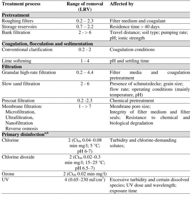
Table 1.1. Treatment processes commonly used individually or in combination to reduce the microbial loads during the production of drinking water (adapted from WHO, 2008). 5
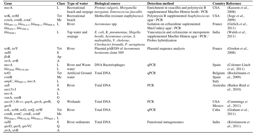
Table 1.2. Examples of antibiotic resistance genes of clinical relevance distributed worldwide in aquatic environments and illustration of some methodological approaches commonly used to detect resistance determinants in the environment (from Manaia et al., 2012) ... 18 Table 3.1. Phylum affiliation of the closest neighbors of the 16S rRNA gene sequences analysed in the DGGE profiles ... 45 Table 3.2. Mean value and variance analysis of Diversity (H’) and Evenness (J) indices calculated on basis of the DGGE profiles. ... 49 Table S1. PCR-DGGE bands identification... 51 Table 4.1. Physicochemical and microbiological characterization of the water sample ... 67
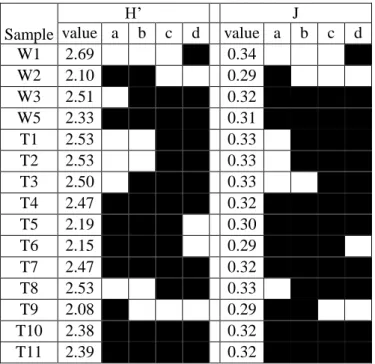
Table 4.2. Shannon’s diversity index (H’) and Pielou’s Evenness index (J) for total and cultivable bacteria ... 73
Table 4.3. Qualitative analysis of cost-benefits for the three methods in study ... 83 Table 5.1. Percentages and types of antibiotic resistance per culture medium, sampling site, and genus ... 100
Table 6.1. Primers used in the study of Pseudomonas spp. diversity ... 119 Table 6.2. Counts of total heterotrophic bacteria on R2A medium, and proportion of
xxiv
Table 6.3. Sequence types obtained for the different housekeeping genes and the combination of them (STfinal) for the Pseudomonas spp. recovered from the different general types of water (W, drinking water treatment plant and respective distribution system; T, domestic tap water; D, dental chairs; Bf, biofilm; M, mineral water) for the different sampling points and sampling dates or batches (A, B and C). ... 125
Table 6.4. Sequence type diversity and evenness indices for the samples from the different sampling sites (W, drinking water treatment plant and respective distribution system; T, domestic tap water; Bf, biofilm; M, mineral water). ... 131 Table 7.1. Distinctive characteristics of strain DS22T and its closest phylogenetic neighbours ... 148
Table 7.2. Cellular fatty acid compositions of strain DS22T and its closest phylogenetic neighbours ... 149
xxv
List of Figures
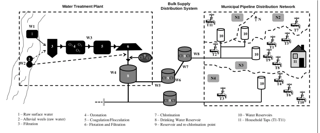
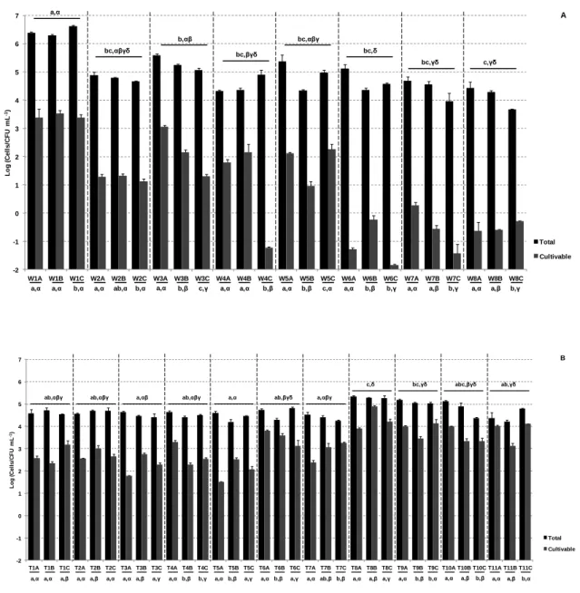
Figure 1.1. The urban water cycle (Marsalek et al., 2006). ... 2 Figure 1.2. The clean (CW) and unclean (UW) water components of the human intervened water cycle. ... 3 Figure 3.1. Schematic representation process of drinking water production and distribution analysed in this study. Numbers 1-11 indicate water source, disinfection, storage and distribution, and W1-W8 and T1-T11 the sampled sites. ... 35 Figure 3.2. Enumeration of the total (black) and cultivable (grey) bacteria over the three sampling dates in the water treatment plant and distribution system (A) and in household tap (B) samples. ... 42
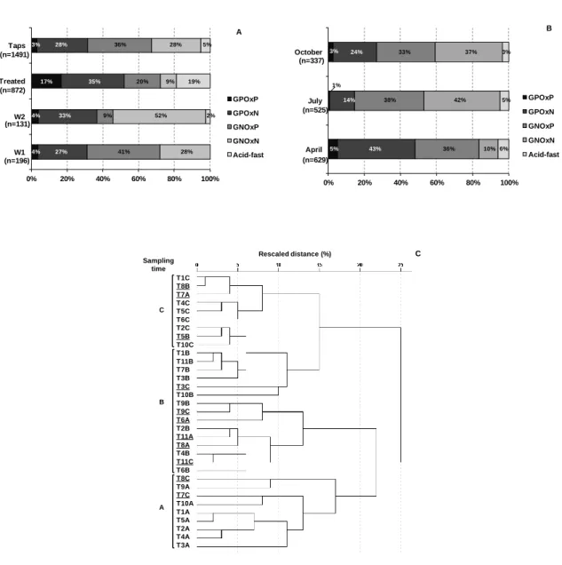
Figure 3.3. Cultivable bacterial diversity in the different types of water (A), and in household taps in the different sampling dates (B). Cluster analysis of the cultivable bacteria patterns of the tap water samples (C). ... 44
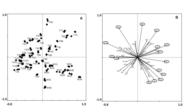
Figure 3.4. Principal components analysis with the PCR-DGGE profiles. A) samples (W1-W3, W5 and T1-T11) distribution and B) PCR-DGGE bands distribution, with the bands presenting the highest Eigenvalues marked with a circle. ... 48
Figure S1. PCR-DGGE gels used for the analysis and band identification ... 50 Figure 4.1. Schematic representation of the study methodology. For each approach, the sample was processed in triplicate. ... 66 Figure 4.2. A) Bacterial diversity of the cultivable bacteria identified at the genus level, for the three different culture media; B) Bacterial diversity at the phylum level obtained with each method used. ... 72
xxvi
Figure 4.3. Dendrogram constructed on basis of partial 16S rRNA gene sequences (111 bp) of the cultivable bacteria (isolates identified with ‘‘R’’ were isolated from R2A, ‘‘T’’ from TTC and ‘‘P’’ from PIA) and of the DGGE bands (marked in bold in the figure). . 77
Figure 4.4. Dendrogram constructed on basis of partial 16S rRNA gene sequences (205 bp) of the cultivable bacteria (isolates identified with ‘‘R’’ were isolated from R2A, ‘‘T’’ from TTC and ‘‘P’’ from PIA) and the OTU obtained by 454 pyrosequencing. ... 79
Figure 5.1. Summary of the numbers of isolates examined in this study according to isolation conditions (A) and site (B). ... 94 Figure 5.2. (Left) Dendrogram constructed on the basis of 16S rRNA gene sequences (1,229 bp), and (Right) Antibiotic resistance profiles ... 97
Figure 5.3. Cluster analysis based on the antibiotic resistance profiles observed for
Sphingomonas and Sphingobium spp. using Euclidean distance and the Ward method for
the aggregation criterion. ... 103 Figure 6.1. Dendrogram constructed on basis of 16S rRNA, rpoD, rpoB and gyrB concatenated gene sequences (1227+568+742+667 bp, respectively). ... 134
Figure 7.1. Transmission electron micrographs of cells of strain DS22T. (a) Cells after growth for 2 days at 30 ºC on nutrient agar, showing cell morphology and endospore positions (asterisks). (b) Detail of an endospore. Bars, 0.5 mm. ... 147 Figure 7.2. Polar lipid profile of strain DS22T after separation by two dimensional TLC, spraying with 50 % (v/v) aqueous sulfuric acid and charring at 160 ºC for 25 min. ... 150 Figure 7.3. Neighbour-joining phylogenetic tree derived from 16S rRNA gene sequences, showing the relationships of strain DS22T with members of the genus Bacillus. ... 151
xxvii
Table of Contents
ABSTRACT ... i RESUMO ... v Acknowledgments... ix Publications ... xi Keywords ... xiii List of Abbreviations ... xv List of Tables ... xxiii List of Figures ... xxvTable of Contents ... xxvii 1. Introduction ... 1
1.1. The urban water cycle ... 1 1.1.1. Water for human consumption ... 4 1.1.1. Water distribution ... 6
1.2. Drinking water bacterial diversity ... 8 1.3. Antibiotic resistance in the environment ... 10
1.3.1. Mechanisms of antibiotic resistance acquisition and dissemination ... 11 1.4. Drinking water as a vehicle of antibiotic resistant bacteria? ... 14 1.5. Tools to assess and track bacterial populations in waters ... 17
xxviii
1.5.2. Bacteria identification and typing... 20 1.6. The hypothesis and objectives of this study ... 24
2. Roadmap for the thesis ... 25 3. Bacterial diversity from the source to the tap: a comparative study based on 16S rRNA-PCR-DGGE and culture-dependent methods ... 29 3.1. Abstract ... 29 3.2. Introduction ... 31
3.3. Materials and methods ... 34 3.3.1. Sampling ... 34
3.3.2. Microbiological characterization and bacterial isolation ... 36 3.3.3. Extraction of total DNA ... 38 3.3.4. 16S rRNA-DGGE analysis ... 38
3.3.5. Statistical analyses ... 40 3.4. Results ... 41
3.4.1. Total and cultivable heterotrophic bacteria counts ... 41 3.4.2. Diversity of cultivable bacteria ... 43 3.4.3. Bacterial diversity based on 16S rRNA-DGGE ... 45
3.5. Discussion ... 55 3.6. Conclusions ... 59
4. Culture-dependent and culture-independent diversity surveys target different bacteria: a case study in a freshwater sample ... 61
xxix
4.1. Abstract ... 62 4.2. Introduction ... 63
4.3. Materials and Methods ... 65 4.3.1. Sampling ... 65
4.3.2. Microbiological characterization ... 65 4.3.3. Bacterial isolation and characterization ... 66 4.3.4. Total DNA extraction ... 68
4.3.5. DGGE analysis ... 68 4.3.6. 454 pyrosequencing ... 69
4.3.7. Sequence analysis and phylogenetic classification ... 69 4.3.8. Richness, diversity and evenness indices ... 71 4.4. Results ... 71
4.4.1. Cultivable bacteria ... 71 4.4.2. Culture-independent methods ... 73
4.4.3. Culture-dependent versus DGGE or 454 pyrosequencing ... 74 4.5. Discussion ... 79 5. Diversity and antibiotic resistance patterns of Sphingomonadaceae isolates from drinking water ... 85 5.1. Abstract ... 86
5.2. Introduction ... 87 5.3. Materials and Methods ... 88
xxx
5.3.1. Sampling ... 88 5.3.2. Bacterial isolation and characterization ... 89
5.3.3. Bacterial identification and typing ... 90 5.3.4. Antibiotic resistance phenotype ... 91
5.3.5. Statistical analyses ... 92 5.3.6. Nucleotide sequence accession numbers ... 93 5.4. Results ... 93
5.4.1. Abundance and diversity of Sphingomonadaceae... 93 5.4.2. Antibiotic resistance phenotypes ... 99
5.5. Discussion ... 106 6. Diversity and antibiotic resistance in Pseudomonas spp. from drinking water... 111 6.1. Abstract ... 112
6.2. Introduction ... 113 6.2. Materials and Methods ... 115
6.2.1. Sampling ... 115 6.2.2. Bacterial isolation and characterization ... 116 6.2.3. Bacterial identification and typing ... 117
6.2.4. Characterization of antibiotic resistance phenotypes ... 121 6.2.5. Diversity and evenness indices and statistical analyses ... 122
6.3. Results ... 122 6.3.1. Isolation and identification ... 122
xxxi
6.3.2. Diversity of Pseudomonas species over the sampled sites ... 127 6.3.3. Antibiotic resistance in Pseudomonas species ... 130
6.4. Discussion ... 135 6.5. Conclusions ... 140
7. Bacillus purgationiresistens sp. nov., isolated from a drinking water treatment plant .. ... 141 7.1. Abstract ... 142
7.2. Introduction ... 143 7.3. Materials and Methods ... 143
7.4. Results and Discussion ... 146 7.5. Description of Bacillus purgationiresistens sp. nov. ... 152 8. General Discussion ... 155
9. Main Conclusions ... 163 10. Proposals for Future Work ... 165
1
1. Introduction
1.1. The urban water cycle
Water is the most common and important chemical compound on Earth, with an unquestionable importance to all the basic biochemical processes, and therefore, for human health and well-being. The hydrological cycle is often referred to as the water cycle, comprising the storage and circulation of water between the biosphere, atmosphere, lithosphere and the hydrosphere. Due to anthropogenic influences and interventions on the urban areas, a more restricted water cycle was proposed, the so called urban water cycle (Figure 1.1). The urban water cycle combines the hydrological with the human intervened parts of the water cycle. Key components of the urban water cycle are the facilities for water treatment and disinfection, the network of pipelines for drinking water distribution and the waste water municipal collectors. In this cycle, humans are important links, either consuming water for bathing, cleaning, drinking and food preparation or producing waste waters and innumerous anthropogenic substances capable of contaminating the water courses (Figure 1.1).
In the urban water cycle it is possible to recognize two complementary parts – the water destined to human use (for simplicity herein designated clean water, CW in Figure 1.2) and that resultant from human activities (herein designated unclean water, UW in Figure 1.2). CW includes the water source, normally surface water (rivers, lagoons, alluvial wells) or groundwater, treatment facilities where the water is prepared for safe human consumption, and distribution networks which can reach several kilometers (Mitchell et al., 2001; Marsalek et al., 2006; Mitchell and Diaper, 2006). UW includes every type of waste waters, including those produced by houses, industries or hospitals. In
2
developed regions, before being discharged into the environment, these waste waters undergo treatment in order to remove the excess of organic matter and diverse types of contaminants. With this purpose the effluents are collected into waste water treatment plants (WWTP) prior to its discharge into a natural water course (e.g. a river or a lagoon). Although it is not possible to track the fate of the treated effluents in the environment, a hypothetical contamination of the “clean” part of the water cycle (CW) by these effluents cannot be discarded. For instance, in major rivers, it is possible to observe the discharge of WWTP not only downstream, but also upstream the sites where drinking water treatment plants are located (Sirivedhin and Gray, 2005; Guo and Krasner, 2009).
Figure 1.1. The urban water cycle (Marsalek et al., 2006).
The current study is focused on the CW part of an urban water cycle, with major emphasis on final drinking water. By definition, drinking water is suitable for human consumption, washing/showering and domestic food preparation (98/83/EC, 1998; Bartram et al., 2003; WHO, 2008). Factors affecting the drinking water compliance
3
include chemical and microbiological factors which can affect the health of the consumers, through ingestion, contact or aerosol inhalation (Lee et al., 2002; Reynolds et
al., 2008; WHO, 2008). Water quality can be affected at different parts of the water cycle
as outlined in Figure 1.1. If it is true that drinking water treatment may largely improve water quality and safety, it is also observed that the treatment efficiency and the quality of the final water depend on the properties of the raw water. Indeed, the contamination of surface waters with fertilizers, pesticides, pharmaceutical products or heavy metals can seriously endanger the final quality of the drinking water (Ritter et al., 2002; WHO, 2008).
Figure 1.2. The clean (CW) and unclean (UW) water components of the human intervened water cycle. W a te r s o u rc e Water
Treatment Plant Consumers
Wastewater Treatment Plant
CW
4 1.1.1. Water for human consumption
According to the definition of drinking water, the microbiological quality of water is an important aspect regarding its safe use. Indeed, pathogenic bacteria, viruses, protozoa and helminthes, are the most common and widespread health risk associated with drinking water (Lee et al., 2002; Reynolds et al., 2008; WHO, 2008). Given the enormous and unrealistic task that would be the search for all possible pathogens in waters, the detection of indicators is recommended, even though this can represent a shortcoming to assess the microbiological water quality. According to the European (98/83/EC, 1998) and the national (DL306-2007, 2007) legislation, the water supplied for human consumption should be exempt of cultivable Escherichia coli and enterococci in 100 mL of water.
Frequently, the elimination or reduction of water biohazards can only be achieved through a combination of treatment processes (Marsalek et al., 2006; WHO, 2008). The destruction and removal of undesired microorganisms involves processes of filtration and disinfection, with chlorine, ozone and UV. Table 1.1 resumes some treatment processes commonly used, individually or in combination, to reduce the microbial loads in waters. Although bacteria are generally regarded as the microbes most sensitive to inactivation by disinfection, some bacterial pathogens are frequently transmitted through water -
Campylobacter jejuni, Salmonella, Shigella, Mycobacterium tuberculosis, Vibrio cholerae and Helicobacter pylori (Rusin et al., 1997; Marsalek et al., 2006).
5
Table 1.1. Treatment processes commonly used individually or in combination to reduce the microbial loads during the production of drinking water (adapted from WHO, 2008).
Treatment process Range of removal (LRV)
Affected by Pretreatment
Roughing filters 0.2 – 2.3 Filter medium and coagulant Storage reservoirs 0.7 – 2.2 Residence time > 40 days
Bank filtration 2 - > 6 Travel distance; soil type; pumping rate; pH; ionic strength
Coagulation, flocculation and sedimentation
Conventional clarification 0.2 - 2 Coagulation conditions
Lime softening 1 - 4 pH and settling time
Filtration
Granular high-rate filtration 0.2 – 4.4 Filter media and coagulation pretreatment
Slow sand filtration 2 - 6 Presence of schmutzdecke; grain size; flow rate; operating conditions (mainly temperature, pH)
Precoat filtration 0.2 -2.3 Chemical pretreatment Membrane filtration:
Microfiltration, Ultrafiltration, Nanofiltration Reverse osmosis
1 - > 7 Membrane pore size;
Integrity of filter medium and filter seals; Resistance to chemical and biological degradation
Primary disinfectiona,b
Chlorine 2 (Ct99 0.04–0.08
min·mg/l; 5 °C; pH 6-7)
Turbidity and chlorine-demanding solutes;
Chlorine dioxide 2 (Ct99 0.02–0.3
min·mg/l; 15–25 °C; pH 6.5–7)
Ozone 2 (Ct99 0.02 min·mg/l)
UV 4 (0.65–230 mJ/cm2) Excessive turbidity and certain dissolved species; UV dose and wavelength; exposure time
The minimum and maximum removal rates are indicated as log10 reduction values (LRV) which
may be observed under failing and optimal treatment conditions, respectively. Ct, product or disinfectant concentration and contact time.
a
Chemical disinfection: Ct values correspond to the required doses to achieve 2 LRV;
b
6 1.1.1. Water distribution
Besides the final quality of the product, water treatment must be also optimized in order to prevent microbial growth, pipes corrosion and the formation of deposits, during storage and distribution. In this respect, the quality of the distribution network, which may include several hundred kilometers of pipes, storage tanks, interconnections and the potential for tampering and vandalism, is also of major relevance. This complex network is full of opportunities for microbial contamination to occur. An intermittent water supply is also critical for microbial contamination, since low water pressure may allow the ingress of contaminants into the system through breaks and joints (Robertson et al., 2003; WHO, 2008).
Disinfection processes such as chlorination, ozonation and UV irradiation reduce significantly the number of bacteria in water. After disinfection, some microorganisms can survive, sometimes as dormant cells and, under favorable conditions, such as the absence of disinfectant residues, can enter regrowth. Under favorable conditions, the planktonic (suspended) surviving bacteria can also form biofilm structures. Biofilms comprise a mixture of microorganisms able to proliferate, often attached to a surface, originating a heterogeneous and discontinuous structure, with a non-uniform distribution over the surface of the materials in contact with water (Batté et al., 2003). The detachment of bacteria from mature biofilms is also know to occur, frequently due to the network pipe walls shearing/erosion, favoring the spreading of biofilm bacteria into the circulating water (Batté et al., 2003). Major factors influencing the bacterial regrowth and biofilm formation are the temperature, nutrients availability, including assimilable organic carbon, the absence of disinfectant residues, hydrodynamic regime (water stagnation or laminar/turbulent flow) and the pipe characteristics (surface, material, etc.) (Camper et al., 1998; Butterfield et al., 2002; Bartram et al., 2003; Chu et al., 2003;
7
Wijeyekoon et al., 2004; Lehtola et al., 2005; Ndiongue et al., 2005; WHO, 2008; Lautenschlager et al., 2010; Manuel et al., 2010). For instance, some pipe materials can stimulate the bacterial growth by releasing bioavailable forms of iron and phosphorous to the water (Morton et al., 2005), and by contributing to neutralize the disinfectants (Hallam et al., 2002; Lehtola et al., 2005). In contrast, some elements such as copper released from the pipes, showed to slow the biofilm development, presumably due to the toxicity or inhibitory effect on microorganisms (Lehtola et al., 2004; van der Kooij et al., 2005). The biofilms are suspected to be the primary source of microorganisms in water distribution systems fed with treated water and with no pipeline breaches. In this respect, the resilience of biofilms to disinfection is a major limitation. For example, the disinfection with chlorite or chlorine dioxide can reduce the loads of free bacteria, but have little or no effect on the density of biofilm bacteria (Gagnon et al., 2005). In general, multispecies biofilms can have even higher resistance to disinfection than mono-species structures (Berry et al., 2006).
For the reasons presented above, biofilms can potentiate considerably the emergence and persistence of waterborne pathogens (Bartram et al., 2003). Some ubiquitous enterobacteria, such as those of the genera Citrobacter, Enterobacter and Klebsiella are referred to as common biofilm members in drinking water distribution systems (Schwartz
et al., 2003; September et al., 2007; WHO, 2008). On the other hand, the low
temperatures and nutrients concentration in the distribution systems do not support the growth of bacteria like E. coli or enteric pathogens in biofilms (Robertson et al., 2003; Tallon et al., 2005; WHO, 2008). Thus, the presence of E. coli in biofilms in the distribution system can be considered an evidence of the occurrence of recent fecal contamination (Robertson et al., 2003; WHO, 2008).
8 1.2. Drinking water bacterial diversity
From the source to the final consumer, the water bacterial diversity suffers successive alterations (Norton and LeChevallier, 2000; Eichler et al., 2006; Lautenschlager et al., 2010). Therefore, the bacterial diversity in the water that reaches the consumer does not necessarily mirrors the bacterial diversity in the water source. In a pilot study conducted by Norton and LeChevalier (2000) the changes in the bacteriological populations due to water treatment and distribution were evident. These authors showed that although the ozonation process did not alter dramatically the composition of the cultivable bacteria in raw water, the chlorination resulted in a rapid shift from predominately Gram-negative bacteria in the raw water to mostly Gram-positive organisms in the chlorinated water. Nevertheless, downstream the distribution system, the disinfectants relief may enable the Gram-negative bacteria regrowth. In fact, the composition of the final water, after bacterial regrowth, is almost unpredictable. Besides the properties of the water and physicochemical factors, such as total organic content or hydrodynamic regime, also the conditions of the pipes, the range of temperatures, the residence times, among others, may induce changes in the bacterial community (Pepper et al., 2004; Lautenschlager et al., 2010). The wide range of cultivable bacteria frequently found in drinking water include members of the genera Acinetobacter, Actinomycetes, Aeromonas, Alcaligenes,
Arthrobacter, Citrobacter, Comamonas, Corynebacterium, Enterobacter,
Flavobacterium, Klebsiella, Micrococcus, Moraxella, Pseudomonas, Serratia,
Sphingomonas, Stenotrophomonas, Xanthomonas, atypical Mycobacterium, Bacillus, Nocardia, among others. (Rusin et al., 1997; Norton and LeChevallier, 2000; Szewzyk et al., 2000; Bartram et al., 2003; WHO, 2008). Although occasional episodes of
pathogenicity may be associated with some of these bacteria, drinking water ingestion presents a very low risk of promoting gastrointestinal infection in the general population
9
(WHO, 2008). The calculated risk is less than 1/10 000 for a single exposure to the bacterial agent (Rusin et al., 1997). In spite of such considerations, some drinking water bacteria may be of concern for people under immunosuppression or undergoing antibiotic therapy (Rusin et al., 1997; Norton and LeChevallier, 2000; Bartram et al., 2003; WHO, 2008).
Having in mind that the cultivable bacteria represent a small part of the water microbiota, over the last decade many studies have used culture-independent approaches in order to have a broader perspective of the drinking water bacterial diversity (Farnleitner et al., 2004; Eichler et al., 2006; Wu et al., 2006; Poitelon et al., 2009; Kormas et al., 2010; Lautenschlager et al., 2010). Proteobacteria (mainly Alpha-, Beta- and Gammaproteobacteria) were frequently observed as the prevailing phylum in treated drinking waters (Eichler et al., 2006; Poitelon et al., 2009; Kormas et al., 2010; Revetta et
al., 2010). Nevertheless, the predominance of other phyla, such as Cyanobacteria, Actinobacteria, Bacteroidetes, and Planctomycetes was also reported (Eichler et al.,
2006; Revetta et al., 2010). The impact of disinfection processes on the bacterial community diversity was also observed using culture-independent methods (Eichler et al., 2006; Revetta et al., 2010). For instance, Eichler et al. (2006) observed that the first steps in the processing of the raw water (i.e. flocculation and sand filtration) did not change the microbiota composition, although chlorination had a significant effect on the bacterial community. After this treatment, phylotypes not detected in previous stages, as for example nitrifying bacteria, were identified. In addition to the reduction of the bacterial counts, water treatment may impose selective pressures capable of selecting bacteria resistant to different types of chemical or physical biocides (e.g. disinfectants, antibiotics, radiation) (Armstrong et al., 1981; Armstrong et al., 1982; Schwartz et al., 2003; Shrivastava et al., 2004; Xi et al., 2009). Hypothetically, the bacteria that can survive the
10
treatment, will be able to regrow downstream the disinfection points where may contribute to spread and increase antibiotic resistance prevalence. This effect was evidenced by Xi et al. (2009) who concluded that the water distribution systems may serve as important reservoirs for the spread of antibiotic resistance.
1.3. Antibiotic resistance in the environment
Most of the antibiotics commercially available nowadays are derivatives of natural compounds produced by bacteria and/or fungi. In nature, it is thought that these microorganisms use the antibiotics, which are secondary metabolites, for microbial cell defense, inhibiting the growth of competitors. However, many bacteria can survive in the presence of natural antimicrobial substances and even benefit from their presence. For example, some bacteria can use antibiotics as biochemical signals, modulators of metabolic activity or even as carbon sources (Davies et al., 2006; Dantas et al., 2008; Martinez, 2009). In other cases, bacteria can tolerate the antibiotics because they have structures similar to the natural substrates and can be inactivated by the bacterial enzymes, leading to a natural form of resistance (Martinez, 2009). These are some evidences that illustrate that antibiotic resistance is a natural property of bacteria, eventually as old as bacteria themselves (Datta and Hughes, 1983; Hughes and Datta, 1983; Aminov, 2010; D'Costa et al., 2011).
Before the introduction of antibiotics in the 1940’s, and their increasing use in bacterial infections therapy, the concentrations of these compounds in the environment were low and confined to the site of their production. The increasing use of antibiotics and other substances with antimicrobial activity changed the equilibrium between fully susceptible and resistant bacteria (Larson, 2007; Davies and Davies, 2010). Gradually,
11
antibiotic resistant bacteria and their specific genetic determinants have reached new habitats, with evident increases on the prevalence of resistance and the extension of the spectrum of antimicrobial substances tolerated (Davies and Davies, 2010). The high prevalence of (multi)-antibiotic resistance has been extensively reported, mainly in clinical environments. Nevertheless, the problem is not limited to the clinical environment, and has been also reported in wild animals, surface waters or agriculture soils, allegedly due to antibiotics use (frequently overuse) and anthropic selective pressures (Literak et al., 2010; Simões et al., 2010; Storteboom et al., 2010; Thaller et al., 2010). Although a relationship between the increase of the antibiotics use and the increase of the antibiotic resistance exists, it was demonstrated that the antibiotic resistance genes already existed in the pre-antibiotic era (Knapp et al., 2010; D'Costa et al., 2011). Nowadays, antibiotic resistance is considered a serious global public health problem, and is receiving the attention of several international health agencies (APUA; CDC; COST-DARE; ECDC; WHO).
1.3.1. Mechanisms of antibiotic resistance acquisition and dissemination
The success of antibiotics as therapeutic agents is due to the capacity of these molecules to interfere with structures and/or functions of the bacterial cell (prokaryotic), which are absent in the host cells (eukaryotic). Antibiotics may interfere with cell wall synthesis, inhibit the protein or nucleic acid synthesis, disrupt the bacterial membrane structure or inhibit a metabolic pathway vital to the cell. In turn, antibiotic resistance mechanisms are related with the ability that bacteria have or may develop to avoid such interferences. Resistance mechanisms are much more diverse than the modes by which a drug can interfere with a cell. These may include the degradation or alteration of the antibiotic by different processes (e.g. hydrolysis, acetylation, phosphorylation,
12
glycosylation), the removal of the antibiotic from the cell (e. g. efflux pumps), or altered targets for the antimicrobial agent (Mazel and Davies, 1999; Scott, 2005; Tenover, 2006; Manaia et al., 2012)
Some bacteria, given the presence of key genes and/or physiological functions, are intrinsically resistant to one or more classes of antibiotics. This is an ancestral property within a group and thus is common to most or all representatives of a genus or species (EUCAST; Davies and Davies, 2010). In contrast, acquired resistance is observed only in some representatives of a species, in which most of the members are susceptible to that antimicrobial agent (EUCAST). Acquired antibiotic resistance may result from gene mutation or genetic recombination (Martinez and Baquero, 2000; Livermore, 2003; Tenover, 2006; Zhang et al., 2009; Davies and Davies, 2010). Gene mutations occur randomly in the genome, often potentiated by mutagens. Examples of resistance phenotypes emerging by mutation include altered targets for an antimicrobial agent (e.g. quinolones, rifampin, linezolid, clarithromycin, amoxicillin, and streptomycin), limited access of the antimicrobial agent to the intracellular target (e.g. penicillin, cephalosporins, glycopeptides, and tetracyclines), or transformation and further broadening of the range of antimicrobial agents that can be inactivated (e.g. extended spectrum beta-lactamases) (Manaia et al., 2012). Under favorable conditions, the clones harboring the gene mutation may have advantage, achieving higher rates of cell division than the non-mutated cells (higher fitness, i.e., the capacity of an individual to survive and reproduce) and, thus, become dominant. In such a situation the genetic determinant of resistance is disseminated by vertical transmission.
In bacteria, genetic recombination is frequently referred to as horizontal gene transfer (HGT). This process, also named “bacterial sex”, is very common among bacteria and represents a major driving force for bacterial evolution (Ochman et al., 2000;
13
Wiedenbeck and Cohan, 2011). This form of genetic recombination involves the transfer of genetic material from a donor to a recipient and requires that both share the same space, but not necessarily the same species. HGT can occur by transformation, consisting on the uptake of naked DNA (on plasmids or as linear DNA), released by dead cells; transduction, mediated by bacteriophages; and conjugation, involving cell-to-cell contact through a pilus (Davison, 1999; Dröge et al., 1999; Trevors, 1999; Andam et al., 2011; Skippington and Ragan, 2011; Stokes and Gillings, 2011; Manaia et al., 2012). In general, HGT processes are potentiated by genetic elements which facilitate the mobilization and integration of exogenous DNA, either between cells or between chromosomal DNA, and extrachromosomal genetic elements and vice versa. Examples of these genetic elements are plasmids, transposons and integrons, in which many of the known antibiotic resistance genes are inserted.
Some studies suggest that the mobile genetic elements are a considerable part of the antibiotic resistome (collection of all the antibiotic resistance genes and their precursors), which means that a high part of the resistome has a high mobility potential (Partridge et
al., 2009; Andersson and Hughes, 2010; Parsley et al., 2010). Indeed, Fondi and Fani
(2010) concluded that apparent geographical or taxonomic barriers are not a limitation for the occurrence of HGT, as they observed that bacteria phylogenetically unrelated and/or inhabiting distinct environments had similar antibiotic resistance determinants.
A high number of reports and studies have shown that the prevalence of antibiotic resistance, as well as the diversity and distribution of resistance genes has increased over the last decades (EARS-Net; ESAC; NARMS; Houndt and Ochman, 2000; Knapp et al., 2010). In this respect, not only the bacterial pathogens but also the environmental bacteria are important reservoirs of antibiotic resistance (Ash et al., 2002; Ferreira da Silva et al., 2006; Ferreira da Silva et al., 2007; Allen et al., 2010; Figueira et al., 2011a; Figueira et
14
al., 2011b; Figueira et al., 2012). The selective pressures present in the environment (e.g.
antibiotics, disinfectants and other antimicrobials, heavy metals, etc.), which supposedly contribute to the increase of the antibiotic resistance, are diverse and act by different mechanisms, most of them still unclear. Some cases of co-selection were reported, for example 1) when the genes specifying the resistant phenotypes for the antibiotic and disinfectant or metal are located together in the same genetic element (co-resistance), or 2) when different antimicrobial agents attack the same target, initiate a common pathway to cell death or share a common route of access to their respective targets (cross-resistance) (Chapman, 2003; Baker-Austin et al., 2006; Martin et al., 2008). Contrary to the initially thought, that in the absence of selective pressures acquired antibiotic resistance genes can be a dead weight for its host, it seems that acquired antibiotic resistance may have a reduced cost for its host and, thus, become stable once acquired (Johnsen et al., 2009; Andersson and Hughes, 2010, 2011). Several factors could contribute to this irreversibility, including the absence of a fitness cost, reduction of the fitness cost, through compensating mutations, and the referred to genetic co-selection between the resistance-conferring gene and another gene under selection (Gullberg et al., 2011). All these factors are supposedly contributing for the continuous increase of the antibiotic resistance dissemination.
1.4. Drinking water as a vehicle of antibiotic resistant bacteria?
Although it is still difficult to establish clear cause-effect relationships, it is widely accepted that chemical pollution, mostly due to anthropic causes, contributes for antibiotic resistance dissemination (McArthur and Tuckfield, 2000; Davies and Davies, 2010; Graham et al., 2011).In this respect, antibiotics seem to be a major, although not
15
the unique, form of pollution, mainly because it is estimated that about 75 % of the antibiotics consumed by humans and animals are eliminated as active substances, contaminating sewage treatment systems and the respective receptors (Kümmerer and Henninger, 2003; Zhang et al., 2009). The WWTP are the main destination of these substances, where they are only partially eliminated. The antibiotics that are not eliminated during the treatment process pass through the system and may end up in the environment, usually in a water course, entering the urban water cycle. Not surprisingly, residues of antibiotics have been detected worldwide in municipal sewage, hospital effluents, influents and effluents of WWTP, surface water and ground water (Kümmerer, 2004, 2009). In drinking water the detection of antibiotic residues is less frequent. Nevertheless, some recent studies reveal that antibiotics are also present in drinking water, including those submitted to the recommended disinfection process (Ye et al., 2007; Benotti and Snyder, 2009; Touraud et al., 2011).
Once in environment, antibiotics may be eliminated by biotic processes (biodegradation by bacteria and fungi) or by non-biotic processes (sorption, hydrolysis, photolysis, oxidation and reduction), reaching very low concentrations, i.e., sub-inhibitory levels (Halling-Sorensen et al., 1998; Kümmerer, 2009). At sub-sub-inhibitory levels, antibiotics may have a hormetic effect and can promote several alterations in housekeeping functions of the cells. Apparently, some of these alterations may not be associated with antibiotic resistance, but contribute for the perturbation of the microbial community, leading, eventually, to an overall resistance increase (Davies et al., 2006; Fajardo and Martinez, 2008; Yergeau et al., 2010; Graham et al., 2011). Indeed, recent studies suggested that the low antibiotic concentrations found in many natural environments are important for enrichment and maintenance of resistance in bacterial populations (Gullberg et al., 2011).
16
Bacteria resistant to antibiotics have been extensively found in the aquatic environment, namely in drinking waters (Armstrong et al., 1981; Schwartz et al., 2003; Pavlov et al., 2004; Zhang et al., 2009). Thus, water may be a vehicle, not only for the dissemination of pollutants, but also of bacteria and resistance genes in the environment. Given their ubiquity, bacteria can move between different environmental niches and, like stickers, drive antibiotic resistance determinants from heavily contaminated sites to places in which selective pressures (no matter which they are) may be inexistent or negligible. Some studies have concluded that the treatment of raw water and its subsequent distribution selects for antibiotic-resistant bacteria, increasing phenotypic resistance rates at drinking water sampling points (Kümmerer, 2004; Scoaris et al., 2008; Xi et al., 2009). Although disinfection processes contribute to reduce the number of bacteria, the persistence or re-colonization of antibiotic resistant bacteria in drinking waters is a reality, worsened by the high potential of many bacteria to produce biofilms in pipelines, reservoirs and taps (Schwartz et al., 2003). The risks can be attenuated using expensive treatment systems (e.g. ultrafiltration or reverse osmosis), but still some resistance genes can persist and enter the food chain via drinking water (Bockelmann et al., 2009).
A single gene is often the basic functional unit responsible for resistance to one or more antibiotics. The same antibiotic resistance genes are detected worldwide in hospital and animal husbandry waste waters, sewage, waste water treatment plants, surface water, ground water and drinking water (Table 1.2) (Zhang et al., 2009; Manaia et al., 2012). In respect to drinking water, many are the examples of antibiotic resistance genes found in different world regions, associated with beta-lactam (ampC, blaTEM and blaSHV),
chloramphenicol (cat and cmr), sulfonamide (sulI and sulII), tetracycline (tetA, tetB and
tetD), aminoglycosides (aphA, aadA1, aadA2 and sat2), trimethoprim (dfrA12 and dfrA17), erythromycin (msrA, ermA and ermC) and vancomycin (vanA) resistance
17
(Schwartz et al., 2003; Cernat et al., 2007; Faria et al., 2009; Xi et al., 2009; Zhang et al., 2009; Figueira et al., 2012; Manaia et al., 2012).
Despite the considerable amount of information published, probably just a little fraction of the resistance genes occurring in waters (and in the environment in general) were characterized till now. One of the reasons is the fact of the antibiotic resistance genes detection is mainly performed by PCR-based approaches and metagenomic analysis, using specific primers, followed by sequence similarity analysis. These procedures limit the detection of resistance genes to those harbored by bacteria which genome is known. Another bias is related with the fact that most of the genes screened were originally described in clinical pathogens, mainly cultivable aerobic bacteria with fast and non-fastidious growth. For these reasons, the current perspective of the antibiotic resistome is still mainly culture-dependent. The simple detection of a gene is not indicative of its expression in its host and in the environment, but it evidences the stability and potential of that gene to spread to other environments or hosts.
1.5. Tools to assess and track bacterial populations in waters
1.5.1. Culture-dependent vs culture independent methods
Over the last decades it became evident that only a small fraction of the bacterial diversity is known and that none of the methods to study bacteria in the environment is able to cover the whole community (Muyzer et al., 1993; Amann et al., 1995; Palleroni, 1997; Kemp and Aller, 2004; Sleator et al., 2008; Zinger et al., 2011).