Oncobiologia
The role of YB-‐1 in acquired resistance to
hormone therapy in breast cancer
Ana Rita de Sousa Noia de Mendonça Bello
Oncobiologia
The role of YB-‐1 in acquired resistance to
hormone therapy in breast cancer
Ana Rita de Sousa Noia de Mendonça Bello
Orientado por:
Prof. Dra. Sandra Cristina Cara de Anjo Casimiro
Abstract
Breast cancer (BC) is the most frequent and lethal malignancy among women. Metastatic disease represents a challenge where systemic therapy is crucial. Hormone therapy (HT) has a major role in the treatment of BC with hormone receptor (HR) expression, denominated by luminal BC. Unfortunately, a percentage of patients will develop acquired resistance. Recently, YB-1, a recognized oncoprotein associated with all cancer hallmarks and chemotherapy resistance, was found to be involved in processes related to HT acquired resistance.
In this study, we aimed to continue a previous project developed in our lab, which purpose was to determine the association between acquired HT resistance in luminal BC and YB-1 expression. In this context, four human BC cell lines were exposed to different therapies. We derived post-therapy clones (released cells) and assessed possible changes in YB-1, HR and HER2, in both resistant and released cells.
Our results show an increase in YB-1’s activated form, p-YB-1, in MCF-7 HT- -resistant cells. When an increase in p-YB-1 was present, it was followed by a rise in HER2. Therapy interruption did not allow phenotype reversion, neither for HT or lapatinib. YB-1 and p-YB-1 expression was raised in released cells, as well as HER2, while ER and cyclin D1 expression decreased in MCF-7 cells exposed to fulvestrant, suggesting the emergence of more aggressive tumor cells, with possible enhanced resistance to therapy. In ER+/HER2+ cells resistant to lapatinib, expression of YB-1 and pYB-1 was increased; this was reversed in released cells, which also showed an increase in HR expression, possibly representing a sensitization to HT. This study must be complemented with cell viability assessments, re-evaluation of therapy resistance and trial of second-line therapies.
Keywords: Breast cancer; Hormone therapy resistance; Y-box binding protein 1
(YB-1).
Resumo
O cancro da mama é a neoplasia maligna mais prevalente e com maior mortalidade na mulher. O tratamento da doença metastática permanece um desafio no qual a terapêutica sistémica tem um papel fundamental. A hormonoterapia tem um impacto importante no tratamento de tumores com expressão de receptores hormonais, denominados de Luminais. Infelizmente, uma percentagem significativa de doentes adquire resistência à terapia com progressão da doença. Recentemente, demonstrou-se que a YB-1, uma oncoproteína associada a todos os hallmarks do cancro e à resistência à quimioterapia, está também envolvida na resistência à hormonoterapia.
Neste estudo, tivemos como objetivo dar continuidade a um projeto anterior desenvolvido pelo nosso laboratório, cujo propósito foi determinar a associação entre a resistência inata e adquirida à hormonoterapia no cancro da mama luminal e a expressão de YB-1. Neste contexto, expusemos quatro linhas celulares humanas de cancro de mama a diferentes fármacos. Obtivemos clones pós-terapêutica (células released) e avaliámos possíveis alterações no padrão de expressão de YB-1, receptores hormonais e HER2, em células resistentes e released.
Os nossos resultados demonstram um aumento na expressão da forma ativada da YB-1, p-YB-1, em células MCF-7 resistentes à hormonoterapia, que se acompanhou de uma maior expressão de HER2. As células released apresentaram um aumento na expressão de YB-1, p-YB-1 e HER2, ocorrendo diminuição da expressão de receptor de estrogénios e de ciclina D1 em células MCF-7 expostas a fulvestrano, sugerindo a emergência de clones mais agressivos, com maior resistência à terapêutica. Nas células HER2+ resistentes ao lapatinib, existe um aumento da expressão de YB-1 e p-YB-1 que é revertido nas células released, com um concomitante aumento de expressão de receptor de estrogénios, possivelmente representando uma sensibilização à hormonoterapia. Este estudo será complementado com ensaios de viabilidade, reavaliação da presença de resistência à hormonoterapia ou lapatinib, bem como a fármacos possivelmente usados numa segunda-linha terapêutica.
Palavras-Chave: Cancro da mama, Resistência à Hormonoterapia; Y-box binding
protein 1 (YB-1).
Resumo Alargado
O cancro da mama é a neoplasia maligna mais frequente na mulher. Apesar dos progressos realizados no sentido do diagnóstico precoce e eficácia terapêutica, é o cancro associado a maior mortalidade neste género no mundo. Existem vários fatores que influenciam o prognóstico, nomeadamente a extensão da doença, a sua histologia, e mais recentemente, biomarcadores moleculares e genéticos. Dentro destes últimos, destacam-se os receptores hormonais de estrogénio (RE) e progesterona (RP), a amplificação do HER2, e análises multigénicas, como por exemplo o Oncotype DX. O estudo destes biomarcadores permitiu subclassificação da doença e o desenvolvimento da terapêutica dirigida, tornando-se a abordagem aos doentes mais individualizada. Do ponto de vista molecular, poderá fazer-se um classificação do cancro da mama em três grandes grupos: 1) luminal, que se caracteriza pela expressão de receptores hormonais, podendo-se subclassificar em A ou B, consoante a presença de características mais agressivas, nomeadamente a sobreexpressão de HER2 e/ou elevado índice proliferativo (Ki67); 2) com sobreexpressão de HER2, em que não existe expressão de receptores hormonais mas sim amplificação do gene ERBB2; e 3) basal, que corresponde a quase todos os casos de tumores triplos-negativos, não possuindo expressão de receptores hormonais ou de HER2.
O cancro da mama metastático permanece um desafio terapêutico, sendo que a utilização de terapêutica sistémica nestes casos tem um papel fundamental. A hormonoterapia, que visa inibir a estimulação estrogénica do tecido mamário, permitiu melhorar o prognóstico de doentes com tumores luminais. A hormonoterapia sistémica inclui inibidores da aromatase, moduladores seletivos dos RE (por exemplo tamoxifeno) e antagonistas seletivos dos RE (por exemplo fulvestrano). Contudo, cerca de 20-30% dos doentes adquirem resistência à hormonoterapia, sendo que vários mecanismos já foram identificados para explicar este fenómeno, nomeadamente mutações e diminuição da expressão do RE, ativação de oncogenes e vias que promovem a proliferação celular, alterações do ciclo celular e processos epigenéticos.
Recentemente, foi demonstrado que a Y-box binding protein 1 (YB-1), uma oncoproteína envolvida em todos os hallmarks do cancro e em processos de resistência à quimioterapia, participa em mecanismos relacionados com a aquisição de resistência à hormonoterapia. A YB-1 é fosforilada no citoplasma, o que permite a sua translocação para o núcleo da célula, onde ativa a transcrição de genes que promovem proliferação e sobrevivência celular. No cancro da mama, foi demonstrado que níveis nucleares
aumentados desta proteína se associam a uma doença mais agressiva e resistente à terapêutica. No contexto da hormonoterapia, a sua sobreexpressão promove a degradação do RE e aumento da transcrição de ERBB2, tornando esta estratégia menos eficaz.
Por conseguinte, o nosso laboratório desenvolveu um projeto para determinar a associação entre a resistência à hormonoterapia em cancro da mama luminal e alterações na expressão da YB-1. Para cumprir esse objetivo, expusemos quatro linhas celulares humanas de cancro da mama luminal, duas sem sobreexpressão de HER2 (ER+/HER2-) e duas que apresentam sobreexpressão desta proteína (ER+/HER2+), a diferentes fármacos in vitro e foram selecionados os clones que adquiriram resistência às mesmas (Anexo 1). As células ER+/HER2- foram expostas a tamoxifeno, fulvestrano e lapatinib, enquanto as células ER+/HER2+ foram apenas expostas ao lapatinib. Os resultados preliminares sugerem que a expressão de YB-1 está aumentada em células resistentes. Concomitantemente, ocorre uma alteração na expressão de receptores moleculares e no perfil de sensibilidade a segunda linha de terapêutica.
Este trabalho tem como objetivo dar continuidade ao estudo anterior, através da obtenção de clones das linhas celulares resistentes, mas agora pós-terapêutica, e caracterização do seu padrão molecular, para avaliar possíveis modificações que possam ocorrer após interrupção da terapêutica. Por conseguinte, as células foram transferidas para um meio sem fármacos, onde foram mantidas durante três meses (células released). Por Western Blot avaliámos a expressão de YB-1, p-YB-1, RE, HER2 e Ciclina D1 nas células resistentes e released.
Os resultados não demonstraram uma variação consistente na expressão de YB-1 e p-YB-1 nas células ER+/HER2- resistentes à hormonoterapia, destacando-se um aumento de expressão de p-YB-1 nas células MCF-7 resistentes a tamoxifeno e fulvestrano, que não ocorre nas células T47D. Apenas nas células MCF-7 se encontraram alterações significativas e consistentes na expressão de outros biomarcadores. Na presença de um aumento na expressão de p-YB-1, ocorreu um aumento concomitante de HER2. Nas células MCF-7 resistentes ao fulvestrano, ocorreu uma diminuição na expressão do RE e Ciclina D1. Uma vez que as células deixam de estar expostas ao fármaco, as células resistentes à hormonoterapia não recuperam o seu fenótipo inicial, com exceção de uma redução da expressão de HER2 em células MCF-7 com resistência a fulvestrano. Destaca-se ainda o aumento significativo da expressão de p-YB-1 e YB-1 nas células released. Deste modo, é possível que uma vez que é
interrompida a terapêutica as células desenvolvam mecanismos de adaptação eficazes, surgindo clones mais agressivos, com maior probabilidade de resistência à terapêutica.
Relativamente às células ER+/HER2- expostas ao lapatinib, não existe alteração na expressão de YB-1 e p-YB-1 nas células resistentes, verificando-se um aumento consistente na expressão de HER2 e diminuição da expressão de RE, que apenas nas células T47D se acompanha de uma diminuição significativa na ciclina D1. Não se verificou nenhuma alteração significativa nas células released em comparação às células resistentes, apoiando a hipótese da irreversibilidade deste processo em células ER+/HER2-.
Por outro lado, as células ER+/HER2+ resistentes ao lapatinib revelaram um aumento na expressão de p-YB-1 e YB-1, que se acompanhou de um aumento de expressão de HER2 e com manutenção ou aumento da expressão do RE. Ao contrário do que se verificou para as células sem sobreexpressão de HER2, aquando da interrupção da terapêutica, as células recuperam o seu fenótipo, com diminuição da expressão de YB-1, p-YB-1 e aumento da expressão de RE. Nas células MDA-MB-361, ocorre também diminuição da expressão de HER2. Não foi possível a avaliação da expressão de ciclina D1 por dificuldades técnicas, o que terá de ser feito posteriormente. Contudo, estes resultados sugerem que uma possível sensibilização à hormonoterapia de células luminal B com sobrexpressão de HER2 ocorre após exposição ao lapatinib.
Este projeto, no entanto, apresenta algumas limitações. Não foram realizados ensaios de viabilidade das células released, pelo que não é possível saber se se mantêm resistentes às terapias de seleção. Será fundamental reavaliar a suscetibilidade das células released à terapêutica a que foram originalmente expostas, para compreender se estas mantêm o seu padrão de resistência na prática. Será ainda importante expor estas células a uma segunda linha de tratamento para avaliar a sua resposta e, na presença de suscetibilidade, o tempo de demora de aquisição de resistências.
Desta forma, os nossos resultados não revelam as alterações que seriam expectáveis de acordo com a literatura anterior, bem como alguns dos resultados preliminares do projeto desenvolvido previamente pelo laboratório. Tornam-se então necessários novos estudos para compreender o papel da YB-1 na hormonorresistência. No entanto, destaca-se a possível irreversibilidade do processo de aquisição de resistência, com a emergência de clones de células tumorais mais agressivas. Do ponto de vista da prática clínica, esta associação poderia levar à utilização da proteína como um novo marcador para prever a resistência à terapêutica e selecionar os doentes que
estão aptos para receber uma segunda linha de hormonoterapia ou repetir um curso de um fármaco previamente utilizado.
Palavras-Chave: Cancro da mama; Resistência à Hormonoterapia; Y-box binding
Index
Abstract ... 2
Index ... 8
1. Introduction ... 9
1.1 Breast Cancer: Epidemiology, Diagnosis and Classification ... 9
1.2 Metastatic Breast Cancer ... 11
1.3 Hormone Therapy ... 12
1.4 Resistance to Hormone Therapy ... 13
1.5 YB-1 and Breast Cancer ... 15
2. Aims ... 17
3. Material and Methods ... 18
3.1 Cell lines ... 18
3.2 Western Blotting ... 18
4. Results ... 20
4.1 YB-1 and p-YB-1 expression in HT-resistant and HT-released cells ... 20
4.2 ER, HER2 and Cyclin D1 expression in HT-resistant and HT-released cells ... 20
4.3 YB-1 and p-YB-1 expression in anti-HER2-resistant and anti-HER2-released cells ... 22
4.4 ER, HER2 and Cyclin D1 expression in resistant and anti-HER2-released cells ... 23 5. Discussion ... 24 6. Conclusions ... 26 7. Acknowledgements ... 27 8. References ... 28 9. Annex 1 ... 30
1. Introduction
1.1 Breast Cancer: Epidemiology, Diagnosis and Classification
Breast cancer (BC) is the most common malignancy among women, and it is estimated that over 2 million new cases were diagnosed in 2018 worldwide, accounting for approximately 25% of female cancers (1, 2). In the last years, many progresses have been made in what concerns early diagnosis and treatment of BC, and these have had a major impact in BC patients’ prognosis and life-quality. In spite of these advances, BC remains the leading cause of cancer-related deaths in women globally, representing approximately 15% of all cancer deaths in women worldwide in 2018 (627,000 women) (2).
BC can manifest by a nodule that is detected by the patient; however, it can be a symptomless disease, even in advanced stages, and is identified by routine screening procedures. Nonetheless, symptoms can occur from lymph node or distant organ involvement. Considering these facts, the consequences of the disease and the impact of early treatment in disease progression and survival, screening programs have been developed to detect BC in early stages (3, 4). The most robust method supported by evidence is universal screening of women with no additional risk factors with mammography in ages between 50-70 years old (5). New evidence and other expert consensus and guidelines suggest beginning mammography at an earlier age (3, 5). Importantly, screening should be adapted to each individual’s risk factors (5).
Many risk factors have been identified for BC, such as age of menarche, menopause, first pregnancy/nulliparity, obesity and family history. Some of these reflect the promoter effect of estrogen exposure, as breast tissue is hormone-dependent. Overall, risk can be estimated by using specific models such as Gail’s model (3, 4).
BC is a heterogeneous and potentially systemic disease, and several variables are implied in predicting outcomes and therefore making treatment decisions. The diagnosis is suggested by the combination of clinical data and imaging, but confirmatory histopathologic diagnosis, characterization and staging are essential (3, 4, 5). Anatomopathological studies will evaluate the histologic subtype, basement membrane invasion, multicentricity and focality, lymphovascular invasion, cells proliferation rate, and most importantly, the tumor’s grade according to cell differentiation. These will contribute to an accurate pathological staging (4, 5). BC can be localized, or affect lymph nodes or/and distant organs. Axillar lymph node involvement is traditionally
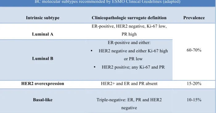
Table 1. BC molecular subtypes recommended by ESMO Clinical Guidelines
Adapted from (5, 8)
described as the most important prognosis factor (4). Metastatic disease occurs more commonly in the lymph nodes, bone, lung, liver and brain, and is associated with a poor prognosis. The TNM system compiles all information and is used for staging (3, 4, 5).
Although histopathological BC classifications have a fundamental role, the study of tumor biology has allowed further characterization of BC and better understanding of its heterogeneity. Several molecular biomarkers for BC have been described and, consequently, therapeutic targets and new prognostic markers were identified, which has led to improved and individualized therapeutic decisions. In spite of the existence of many others, as mentioned above, currently, molecular classification BC (Table 1) is mainly based on three parameters: Hormone Receptor (HR) status (Estrogen Receptor (ER) and Progesterone Receptor (PR)), Human Epidermal growth factor Receptor 2 (HER2) status and Ki-67, which must be quantified in all tumors (3-8).
BC molecular subtypes recommended by ESMO Clinical Guidelines (adapted)
Intrinsic subtype Clinicopathologic surrogate definition Prevalence
Luminal A
ER-positive, HER2 negative, Ki-67 low, PR high
60-70%
Luminal B
ER-positive and either:
• HER2 negative and either Ki-67 high or PR low
• HER2 positive; any Ki-67 and PR
HER2 overexpression HER2+ and ER and PR absent 15-20%
Basal-like Triple-negative: ER, PR and HER2
negative
10-15%
This molecular classification is used for prognosis, such as risk of recurrence, and to predict treatment response and choose the most appropriate systemic therapy regimen (6).
Additionally, it is important to mention that prognostic multigene assays are available (6, 7, 9). For example, Oncotype DX analyses 21 genes and the result will help physicians to decide whether a patient should undergo chemotherapy or not (6). It has
been approved for women with ER+ and HER2– BC, with no evidence of lymph node invasion (9). Recently, tumor histologic grade and biomarker profile are being integrated in staging systems for BC to refine classification (10).
1.2 Metastatic Breast Cancer
Concerning disease’s extent, BC can be localized, regional, locally advanced or metastatic. As aforementioned, this will affect patients’ outcome. Although most BC are diagnosed at an early stage, a smaller percentage (6-19% in some studies) will present with distant metastasis (11, 12). The presence of metastasis in asymptomatic patients occurs more often in large tumors (>5 cm) and extensive nodal disease (>3 involved lymph nodes); in these cases, appropriate imaging studies and lab tests should be used to search for distant disease (9). In addition, it is estimated that one third of the patients with initially localized disease will develop metastasis, most often in distant organs (3, 4).
For metastasized disease, the prognosis is poor, with a median survival of less than 3 years (3, 4). Treating metastatic BC remains a challenge and it is considered an incurable disease. Therefore, the goals of metastatic BC therapy are not only prolonging survival, but also providing symptom control and quality of life. Systemic therapy is usually the first option at this stage; however, there are specific indications for loco regional treatments (9). Therapeutic decision depends on molecular biomarkers, and therefore may include:
• Hormone therapy: this option will be described more thoroughly below; it is the standard treatment for metastatic disease positive for hormone receptors (in absence of visceral crisis) (3, 13).
• Chemotherapy: mainly based on the use of anthracyclines, taxanes and cyclophosphamide. This is the most important type of systemic therapy available for triple-negative tumors, and is also used in other subtypes with more aggressive features (according to the presence of nodal disease and gene expression, for instance) (3, 4, 9, 13).
• Targeted-therapy: the most notorious example is anti-HER2 therapy, which uses antibodies (trastuzumab, pertuzumab) and tyrosine kinase receptors inhibitors (TKI) (eg, lapatinib) against HER2 in patients that present amplification of
tumors in the presence of BRCA gene mutation or signature (14). mTOR and cell cycle inhibitors will be discussed below. Many other target-therapies were or are being studied (3-5, 13).
1.3 Hormone Therapy
BC is classically a hormone-dependent disease (15). As aforementioned, nearly 70% of all BC express HR, ER and/or PR, detected by immunohistochemistry (IHC), which led to the development of hormone therapy (HT) (15). Inhibiting estrogen stimulation can be accomplished by several methods. The first being used was ovarian ablation in pre-menopausal women (15, 16). Nowadays, instead of surgery, ovarian suppression can be achieved through administration of gonadotropin-release hormone (GnRH) analogues. Ovarian ablation or suppression can be used in pre-menopausal women with concomitant use of other HT agents (8, 13, 16); however, its use is controversial in women who must undergo chemotherapy, since side effects of this option will cause ovarian insufficiency (5, 16). Women who did not become amenorrheic after chemotherapy may be candidates for ovarian suppression (16). Different types of antiestrogens have been developed. The choice of the most appropriate treatment regimen will depend on many factors, one of the most important being the pre or post-menopausal status. The three main classes of antiestrogens currently used are described below:
• Selective estrogen receptors modulators (SERMs): SERMS have agonistic and antagonistic actions in ER, by inducing conformational changes in this protein. Tamoxifen is the most important example in this class. It is the first-line of HT in pre-menopausal women; and used in BC prophylaxis in women at high risk (3-5, 16).
• Selective estrogen receptors downregulators (SERDs): SERDs are ER antagonists and, in addition, accelerate ER degradation. Unlike SERMs, SERDs do not possess any agonistic activity. Fulvestrant is the most commonly used drug of this class. Currently it is a second-line option for HT; however, several studies have shown a great clinical benefit in its use, and may soon become a first-line treatment (3, 5, 11, 16).
• Aromatase inhibitors (AIs): This latter group has a crucial role in treatment of BC in post-menopausal women. AIs, such as letrozole or anastrozole, work by
inhibiting extra-ovarian production of estrogens, and thereby by reducing hormonal stimulation of breast tissue (3, 5, 16).
1.4 Resistance to Hormone Therapy
Although HT has become a cornerstone in the treatment of ER and PR positive breast tumors and had a great role in changing the prognosis in advanced disease, unfortunately, about 20 to 30% of the patients are resistant to this type of therapy (3, 11, 14). This will manifest by early disease progression or recurrence. It will be designated by early progression if it develops <12 months of therapy or late progression if opposite (9, 12). This resistance is most frequently acquired, meaning that the tumor cells become resistant after being exposed to HT. Nevertheless, intrinsic HT resistance also occurs, associated with ER mutations and epigenetic changes, driving a poor response to HT from the beginning (15).
Several key-players have been proposed to explain acquired resistance to HT in BC. The most studied include:
• ER-related mechanisms: The ER has two isoforms (ERα and β, encoded by ESR1 and ESR2 respectively) and its major function is as a transcription factor, although it may also act through non-genomic pathway. The main causes of ER-related HT resistance include loss of expression or mutation. Epigenetic mechanisms such as CpG islands methylation and histone acetylation contribute to loss of function of ER, thought to be the cause of acquired resistance in 10-20% of the cases. More importance is recently being given to ESR1 mutations. Several studies have assessed the prevalence of these mutations in tumors that received HT, which ranged from to 11 to 55%. The most commonly described mutations are Y537S and D538G, which alter the conformation of ER 1’s ligand binding domain. These mutations can be identified using liquid biopsies (12, 14, 15, 17, 18).
• PR: it has been demonstrated that PR negative tumors have a worse response to HT. The exact molecular mechanisms that explain these findings remain to be clarified (11, 15).
• EGFR/HER activation: several studies have shown that the presence of a cross-talk between ER and growth-factor receptors results in increased
ERS1 gene expression, cell proliferation and resistance to HT. The most
studied, EGFR and HER2, become activated after exposure to tamoxifen and stimulate ER genomic activity and increase gene expression. HER2 amplification is present in 30% of metastatic BC, and is associated with greater risk for metastasis and decreased disease free and overall survival (DFS and OS, respectively) (15).
• Bypass of ER signaling: this can occur through down-regulation of tumor suppressors, such as PTEN, or up-regulation of drug resistance drivers and oncogenes (Akt, for instance), and consequent activation of PI3K/AKT/mTOR pathway (11, 12, 18, 19).
• Cell cycle regulation: this has become an attractive target, especially cyclin D axis, cyclin dependent kinases (CDK) 4/6 and retinoblastoma proteins in ER+/HER2- BC. Cyclin D1 overexpression has been demonstrated to increase ligand-independent ER signaling activation, and was linked to tamoxifen resistance (11, 12, 18, 19).
Other mechanisms may include changes in metabolism, changes in tumor microenvironment and immune system modulation (11, 19). As a result, other therapies have been proposed and developed to inhibit these pathways. Therapy with anti-HER2 agents, such as trastuzumab, in association with HT, in patients with HR+/HER2+ tumors resulted in an increased progression-free survival (PFS), in spite of having no benefit in OS (15); other studies have also shown clinical benefit in associating lapatinib and letrozole, even in tumors without HER2 overexpression that have become resistant to HT (15).
Options for HT resistant tumors include mTOR inhibitors, since PI3K-AKT- -mTOR pathway activation has a role in HT resistance (14, 16, 19). Association of everolimus and sirolimus to HT has been studied, and the combination of everolimus and exemestane has improved PFS in early relapse in BOLERO-2 trial (12, 13, 18, 19).
CDK4/6 inhibitors, such as palbociclib, have shown good results in association with fulvestrant in pre and post-menopausal women (PALLOMA 3 trial) in early relapse (10, 11, 14). Association of palbociclib with letrozole for late relapse after HT in post-menopausal also had improved outcomes, and has been approved as first-line treatment option for ER+/HER2- metastatic BC (12-14, 18, 19). Other CDK4/6 inhibitors approved include ribociclib and abemaciclib (18, 19).
Additionally, selective histone deacetylase inhibitors (HDAC) and inhibitors of PI3K are also being studied in this context (3, 11-14, 17-19).
Nonetheless, ER may still have a key role in patients with HT resistance, which is supported by tumor response under second and third trials of HT and justifies the widespread use of sequential therapy (12, 13, 19). Thus, HT sequence is also an important factor for preventing relapse under HT. However, solid evidence concerning this matter is lacking (12, 13, 18, 19).
1.5. YB-1 and Breast Cancer
Y-box binding protein 1 (YB-1) is a member of the Y-box binding protein family of transcription factors that is characterized by the presence of a cold-shock domain. This protein is encoded by YBX1 gene located on chromosome 1p34.2. YB-1 is a recognized oncoprotein, overexpressed in many types of cancer and is regarded as a valuable oncologic marker (20).
In what concerns this protein’s intracellular functions, YB-1 is involved in several mechanisms of cell biology, most of them related to DNA and RNA regulation and protein expression. It is important to notice, however, that YB-1 assumes different roles according to its location. The majority of the protein is located in the cytoplasm, where it is associated with mRNA. An increase in cytoplasmatic YB-1 can have a protective role in oncogenesis, by inhibiting PI3K/AKT/mTOR pathway; on the other hand, YB-1 can cause acquisition of genomic instability and promote epithelial-mesenchymal transition (EMT) and therefore metastatic processes (20, 21).
YB-1 can suffer many post-transcriptional changes, one of them being phosphorylation at serine 102 (S102) in the cold-shock domain. Phosphorylated YB-1 (pYB-1) can then be transferred to the nucleus, where it regulates the transcription of several genes involved in cell division, apoptosis, immune response, multidrug resistance, stress response, and tumor growth. Moreover, its actions in DNA repair also favor cell survival and resistance to xenobiotics and ionizing radiation (20, 21). YB-1 also induces a “stem cell-like” phenotype and represses tumor suppressor genes (21, 22).
In addition to these two intracellular forms, YB-1 can also be secreted in a non-canonic pathway, which has been described particularly in an inflammatory context (23).
YB-1’s role in BC has been studied and the protein is regarded as a potential prognostic biomarker. In this context, nuclear YB-1 has been linked to worst prognosis, greater risk of metastasis development and therapy resistance, whilst an increase in cytoplasmatic YB-1 may lead to the development of a more aggressive phenotype (22). Moreover, it has also been demonstrated that overexpression of YB-1 has an association with an unfavorable molecular pattern and therefore a more aggressive disease (24). Additionally, detection of secreted YB-1in the serum of patients with metastatic disease was linked to higher tumor burden and accelerated bone disease (25).
In what concerns therapy resistance, YB-1 was found to be involved in a large number of molecular pathways; it has been linked to multidrug resistance (MDR) phenomena, through P-glycoprotein activity (20, 21, 26). In BC, it has been demonstrated that YB-1 reduced ER activity, decreasing sensitivity to antiestrogens, promoted cell cycle arrest and had an association with HER2 expression in tumors that underwent chemotherapy; conversely, YB-1 knockout increased ER expression (27). The protein has further been linked to acquired resistance to HT by increasing degradation of ER expression and promoting ERBB2 transcription (28).
2. Aims
A recent study developed by our group aimed to determine if resistance to HT in Luminal BC was associated with a change in expression of YB-1. For this purpose, we exposed two human ER+/HER2- BC cell lines, MCF-7 and T47D, and two human ER+/HER2+ BC cell lines, BT474 and MDA-MB-361, to different therapies in vitro and selected clones with acquired resistance to such therapies (29). The preliminary analysis does support that YB-1 expression is increased in resistant cells, along with molecular receptor status alterations and a different sensitivity profile to possible second-line therapies. Therefore, the aim of this work was to derive post-therapy clones and to characterize their molecular pattern, in order to address which further modifications may occur after interrupting exposure to therapy.
3. Material and Methods 3.1 Cell Lines
Human BC cell lines MCF-7 and MDA-MB-361 were obtained from ATCC. T47D and BT474 BC cell lines were kindly provided by Dr. Phillippe Clézardin (INSERM). Cells were maintained in Dulbecco’s modified Eagle’s medium (DMEM, Gibco) containing 1% (v/v) Penicillin/Streptomycin (Pen/Strep, 10,000 U/mL Penicillin, 10,000 µg/mL Streptomycin, Gibco), 10% (v/v) foetal bovine serum (FBS, Gibco), and 0,01mg/mL Insulin (Gibco), except for MDA-MB-361, where 20% (v/v) FBS was used. Medium was replaced twice a week and cells were incubated at 37⁰C with 5% CO2 in humidified atmosphere.
Therapy-resistant clones (Drug_Res) were selected upon 5 month-exposure to tamoxifen, fulvestrant or lapatinib ((29), Annex 1). Therapy-released clones (Drug_Rel) were selected upon 3 month-culture in drug-free medium.
3.2 Western blotting
Proteins were extracted from under confluent cells growing in T25 flasks. Cells were trypsinized and harvested by centrifugation at 450g and 4°C for 5 min. Cells were lysed in 100µl RIPA buffer (SIGMA), containing protease and phosphatase inhibitors cocktails (1:100; Santa Cruz), incubated for 10 min on ice, and centrifuged at 16,000g and 4°C for 10 min. The supernatants were collected, and proteins quantified using Bradford Reagent (VWR Life Science) according to manufacturer’s instructions. Protein extracts were diluted in 2X Laemmli buffer, denatured for 10 min at 95°C and stored at -20°C.
Equal amounts of protein (5µg per lane) were separated by SDS-PAGE and transferred onto nitrocellulose membranes (Invitrogen), using an iBlot2 system (Invitrogen) according to manufacturer’s instructions.
Membranes were incubated for 1h in 5% BSA (Santa Cruz) or 5% non-fat milk in PBS-T (1X PBS with 0,05% Tween, Sigma-Aldrich), and then, incubated with the primary antibodies. Membranes were washed with PBS-T (3X 10 min) and incubated 1h with secondary antibodies conjugated with horseradish peroxidase (HRP). Antibodies used, and incubation details are listed in Table 2. The washing step was repeated with PBS-T (3X 10 min) and proteins were detected using the HRP
chemiluminescent substrate reagent kit (Invitrogen) on Amersham Imager 680 equipment, according to manufacturer’s instructions.
β-Actin was used as loading control. Band intensity was normalized using ImageJ software.
Table 2. Antibodies used in Western Blotting
Primary Antibody Dilution Diluent
Incubation period, Incubation temperature
Rabbit anti-human YB-1 (D299, Cell Signaling) 1:1000
5% BSA in PBS-T
Overnight, 4°C
Rabbit anti-human p-YB-1 (Ser102) (Cell Signaling)
1:500
Rabbit anti-human ER (Cell Signaling) 1:1000
Rabbit anti-human HER2 (Cell Signaling) 1:1000
Rabbit anti-human CyclinD1 (Cell Signaling) 1:1000
Mouse-anti-human β-Actin (Cell Signaling) 1: 25000 5% non-fat milk in PBS-T
2 hours, RT
Secondary Antibody
Goat anti-rabbit IgG (HRP Conjugate) (Bio Rad)
1:5000 5% non-fat milk in PBS-T
1 hour, RT
4. Results
4.1. YB-1 and pYB-1 expression in HT-resistant and HT-released cells
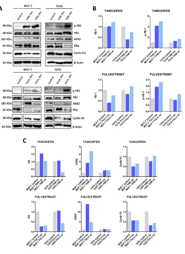
Our first aim was to determine if the expression of YB-1 and/or its activated form (p-YB-1) was different in HT-resistant cells. Our results show that p-YB-1, but not YB-1, is increased only in MCF-7 cells resistant to tamoxifen or fulvestrant, in comparison with parental cell lines (Figure 1A, B). This increase in p-YB-1 was not abrogated by drug release.
4.2. ER, HER2 and Cyclin D1 expression in HT-resistant and HT-released cells
Next we questioned which alterations in molecular receptors would accompany resistance to HT, and assessed the expression of ER and HER2 plus their downstream effector cyclin D1 (Figure 1A, C). We only observed significant and consistent alteration in MCF-7 cells, where HER2 was remarkably up-regulated in HT-resistant clones. Moreover, ER and cyclin D1 were down-regulated in MCF-7 cells upon exposure to fulvestrant. Again, these alterations were mostly not abrogated by drug release, with the exception of HER2 up-regulation in MCF-7 fulvestrant-resistant cells, which was decreased by ca. 50% in fulvestrant-released cells.
Figure 2. Protein expression in BC cells with acquired resistance to HT and after drug release. A.
Western blot analysis showing p-YB-1, YB-1, HER2, ERα and cyclin D1 expression in MCF-7 and T47D cell lines. β-Actin was used as loading control. B and C. Relative protein expression normalized to control.
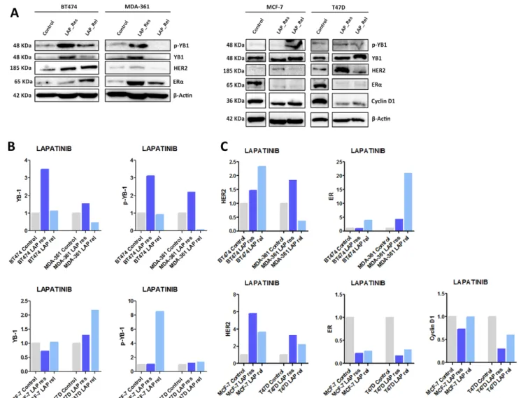
4.3 YB-1 and pYB-1 expression resistant and anti-HER2-released cells
Anti-HER2 therapy is also used to treat HER2+ BC, with or without HR positivity. Therefore we next assessed YB-1 and p-YB-1 expression in ER+/HER2+ cells, resistant to lapatinib, but also in ER+/HER2- cells, from which we could derive lapatinib-resistant clones.
HER2+ cells resistant to lapatinib suffer an increase in expression of both YB-1 and p-YB-1 (Figure 2A, B), reverted by drug release. p-YB-1, but not YB-1, was also highly increased in MCF-7 cells upon release from lapatinib.
Figure 2. Protein expression in BC cells with acquired resistance to Lapatinib and after drug release. A.
Western Blot analysis showing p-YB-1, YB-1, HER2, ERα and Cyclin D1 expression BT474 and MDA-MB-361 (referred as MDA-MDA-MB-361) cell lines. β-Actin was used as loading control. B and C. Relative protein expression, normalized to control.
4.4 HER2, ER and Cyclin D1 expression in HER2-resistant and anti-HER2-released cells
Again, we interrogated which alterations in molecular receptors would accompany resistance to lapatinib, and assessed the expression of ER and HER2 plus their downstream effector cyclin D1 (Figure 2A, C).
HER2 expression was consistently increased in all lapatinib-resistant cells, HER2+ or HER2-, and only reversed by drug release in MDA-MB-361 cells. ER was increased in ER+/HER2+ cells after drug release and decreased in ER+/HER2- lapatinib-resistant cells. These last changes were followed by alterations in cyclin D1 only for T47D cells.
5. Discussion
As we stated before, previous studies have demonstrated and explored the role of YB-1 in tumor acquired resistance to chemotherapy and HT (20, 22, 24, 28). In the context of BC and HT, it was shown that YB-1 accelerates ER proteossomal degradation and promotes ERBB2 transcription in cells with acquired resistance to tamoxifen and fulvestrant. However, this did not result in HER2 overexpression and it was inhibited by the presence of estrogen (28).
Considering these results, our lab developed a project to further characterize the prognostic role of YB-1 and its association with HT acquired resistance. Preliminary data corroborates the importance of YB-1 as a biomarker of poor prognosis and shows that it is associated with ER negativity in clinical samples of BC (29). Moreover, we provide the first data that shows that adjuvant HT may be implicated in the selection of clones with elevated YB-1 expression, which may be associated with the poor outcome of YB-1 high patients. In vitro results suggest that YB-1 overexpression decreases expression of HR, and that cells become more resistant to HT and more sensitive to lapatinib (29). Moreover, cells with acquired resistance to tamoxifen, fulvestrant or lapatinib show some alterations in gene transcription, but HT-resistant cells remain sensitive to other therapies such as lapatinib, everolimus or palbociclib (29).
This work aimed to derive post-therapy clones and to characterize their molecular pattern, in order to address further modifications that may occur after interrupting exposure to therapy.
Our results have shown an increase in p-YB-1 expression only in MCF-7 cells resistant to HT. On the other hand, YB-1 expression does not show significant variations in resistant cells. This dissociation might be related to an increment in nuclear YB-1. Moreover, the difference between MCF-7 and T47D cells might be related with the presence of mutant p53 in T47D, which might favour other pathways for resistance.
Increase in pYB-1 was accompanied by an up-regulation of HER2 and decrease in ER. After release, YB-1 and p-YB-1’s expression increased, and only HER2 up- -regulation in MCF-7 exposed to fulvestrant was partially reversed.
Together, these findings suggest that after HT interruption, luminal A cells with acquired resistance may not be able to recover their previous, sensitive phenotype, and may even develop a more aggressive one. Exposure to HT drugs may activate resistance mechanisms that are further enhanced once the therapy is interrupted, and consequently, allow the emergence of more resistant cells, which might become a therapeutical
challenge. The phenotype of MCF-7 fulvestrant released cells (increased YB-1, p-YB-1 and HER2, and decreased ER and cyclin D1) would be expected to be resistant to HT and is consistent with previous findings and mechanisms of resistance involving YB-1 proposed in HT resistant cells, as described above (28). However, these cells might reveal increased sensitivity to anti-HER2 agents, considering the increase in HER2 expression in some clones.
In what concerns ER expression, it is either maintained or diminished in cells resistant to HT. A reduction in expression would be an expected result, especially in the presence of greater expression of YB-1 (28).
As aforementioned, we repeated this analysis in cells exposed to lapatinib, a TKI of HER2 receptor.
In ER+/HER- lapatinib-resistant cells, there is an increase in HER2 expression and a decrease in ER, followed by a decrease in cyclin D1 expression in T47D cells, without alterations in what concerns YB-1 and p-YB-1. However, once the cells are released, there is no clear and consistent reversion in expression of any protein. Therefore, the mechanism of resistance of these cells to lapatinib seems to occur by a compensatory increase in HER2 expression, independent of YB-1, and inhibition of estrogen signalling. In released cells, these mechanisms of resistance seem to be maintained.
In ER+/HER2+ cells, we demonstrated an increase in expression of YB-1, p-YB-1 and HER2 in lapatinib-resistant cells, without alteration in ER.
After release, opposite to what we found for ER+/HER- cells, there was a reversion in the cells’ phenotype: YB-1 and p-YB-1 expression diminishes, whereas ER expression rises. It is important to notice that there is a greater expression of ER after release, when compared to control. In MDA-MB-361 released cells there is also a reduction in HER2 expression. Based on this, one can question if lapatinib exposure of ER+/HER2+ cells and subsequent release might therefore allow the cells to develop a greater sensitivity to HT. Previous studies have shown this effect in cells ER+/HER2- with acquired resistance to tamoxifen; lapatinib may restore sensitivity in these cells by reactivating the functional target for hormone therapy (30). On the other hand, recent works have shown that lapatinib induces ER degradation, and re-expression of ER restores detoxification capacity and sustains survival in lapatinib-resistant cells (31). This same study also suggested that ER inhibition could overcome lapatinib-acquired
resistance (31). We were not able to assess cyclin D1 expression in ER+/HER2+ cells due to technical difficulties, and we shall take on a repetition of this protocol.
To complement these results, it is necessary to assess the sensitivity profile of drug-released cells. Without this information, it is not possible to conclude if this change in the molecular pattern reflects a change in cellular behaviour. Additionally, it would be interesting to expose release cells to a second line of HT and study the presence of resistance, to confirm that this new phenotype is resistant to all types of HT. Should the cells be susceptible to these therapies, it will be relevant to evaluate the time necessary to acquire resistance. In clinical practice, this would allow select patients with acquired resistance who would be amenable to a second-line of HT.
6. Conclusion
In conclusion, we were not able to demonstrate a clear link between acquired resistance to HT and YB-1. Nonetheless, we showed that an increase in p-YB-1 expression in HT resistant cells is associated with a rise in HER2 expression. Furthermore, we found that the interruption of HT or therapy with lapatinib in ER+/HER2- cells does not allow the recovery of the initial phenotype and may lead to the emergence of more aggressive cells, which might remain resistant to HT. For HER2+/ER+ cells, lapatinib exposure may sensitize them to HT. This must assessed and confirmed in further studies, in order to further understand the role of YB-1 in HT resistance and if it could be used as a biomarker to select patients amenable to a second-line of HT.
7. Acknowledgments
To Professor Doutor Luís Costa and his lab for allowing me to participate in this project.
To my supervisor Sandra Casimiro for all the guidance, support and availability. To Patrícia Alves, for all the help with the methodology, patience and, most of all,
commitment to this project.
To José Rocha, my partner in a previous project, and hopefully in many others, for all the support and his friendship.
8. References
1. Bray, F., Ferlay, J. & Soerjomataram, I. (2018). Global Cancer Statistics 2018 : GLOBOCAN Estimates of Incidence and Mortality Worldwide for 36 Cancers in 185 Countries. CA: A Cancer Journal for Clinicians, 68: 394-424.
2. WHO reports 2018 - https://www.who.int/cancer/prevention/diagnosis-screening/breast-cancer/en/ (09/05/2019).
3. Lipman ME. Breast Cancer. In: Kasper, D. et al. Harrison’s Principles of Internal Medicine. McGraw-Hill Education, 19th edition, 2015, 108: 523-532.
4. Hunt KK et al. The Breast. In: Brunicardi, F., Schwartz’s Principles of Surgery, McGraw Hill Education, 10th edition, 2014, 17: 497-557.
5. Senkus E., et al. (2015). Primary breast cancer: ESMO Clinical Practice Guidelines for diagnosis, treatment and follow-up. Annals of Oncology 26 (5): 8-30.
6. Rakha E., Green A. (2017). Molecular classification of breast cancer: what the pathologist needs to know. Pathology, 49(2): 111-119.
7. Prat, A. et al. (2015). Clinical implications of the intrinsic molecular subtypes of breast cancer. The Breast, 24: 26-35.
8. Tang. Y, et al. (2016). Classification, Treatment Strategy, and Associated Drug Resistance in Breast Cancer. Clinical Breast Cancer, 16(5): 335-43.
9. Harbeck N, Gnant M. (2017). Breast Cancer. Lancet; 389: 1134–50.
10. Hortobagyi, GN. (2018). New and Important Changes in the TNM Staging System for Breast Cancer. American Society of Clinical Oncology Educational Book, 38: 457-467.
11. Glück S. (2016). Consequences of the Convergence of Multiple Alternate Pathways on the Estrogen Receptor in the Treatment of Metastatic Breast Cancer. Clinical Breast Cancer, 17 (2): 79-90.
12. Brufsky, A. (2017). Long-term management of patients with hormone receptor-positive metastatic breast cancer: Concepts for sequential and combination endocrine-based therapies. Cancer Treatment Reviews, 59: 22–32.
13. Cardoso F., et al. (2017) 3rd ESO–ESMO International Consensus Guidelines for Advanced Breast Cancer (ABC 3). Annals of Oncology 28: 16–33, Published online 5 December 2016. 14. Gu G, Dustin D and Fuqua SAW. (2016). Targeted therapy for breast cancer and molecular
mechanisms of resistance to treatment. Current Opinion in Pharmacology, 31: 97–103.
15. Fan, Chang & Fu. (2015). Endocrine therapy resistance in breast cancer: current status, possible mechanisms & overcoming strategies, Future Med. Chem. 7(12): 1511-9.
16. M. Drãgãnescu et al. (2017). Hormone Therapy in Breast Cancer, Chirurgia, 112 (4): 413-17. 17. Jeselsohn, R., De Angelis, C., Brown, M. et al. (2017). The Evolving Role of the Estrogen
Receptor Mutations in Endocrine Therapy-Resistant Breast Cancer, Curr Oncol Rep 19: 35. 18. Murphy C., Dickler M. (2016). Endocrine resistance in hormone-responsive breast cancer:
mechanisms and therapeutic strategies. Endocrine-Related Cancer, 23: 337–352.
19. Liu, Chun-Yu, et al. (2017). Treatment for the endocrine resistant breast cancer: current options and future perspectives. Journal of Steroid Biochemistry and Molecular Biology, 117: 166-175. 20. Eliseeva, I.A., Kim, E.R., Guryanov, S.G., Ovchinnikov, L.P. & Lyabin, D.N. (2011).
Y-box-binding protein 1 (YB-1) and its functions. Biochemistry (Mosc) 76(1): 402-33.
21. Kosnopfel, C., Sinnberg, T. & Schittek, B. (2014). Y-box binding protein 1- a prognostic marker and target in tumour therapy. Eur J Cell Biol 93: 61-70.
22. Mylona, E. et al. (2014). Y-box-binding protein 1 (YB1) in breast carcinomas: relation to aggressive tumor phenotype and identification of patients at high risk for relapse. Eur J Surg Oncol, 40: 289-96.
23. Frye, B.C. et al. (2009). Y-box protein-1 is actively secreted through a non-classical pathway and acts as an extracellular mitogen. EMBO Rep, 10: 783-9.
24. Maciejczyk, A. et al. (2012). Elevated nuclear YB1 expression is associated with poor survival of patients with early breast cancer. Anticancer Res 32: 3177-84.
25. A.R. Ferreira et al. (2017). Serum YB-1 (Y-box binding protein 1) as a biomarker of bone disease progression in patients with breast cancer and bone metastases. Journal of Bone Oncology, 6: 16–21.
26. Huang, J. et al. (2005). Y-box binding protein, YB-1, as a marker of tumor aggressiveness and response to adjuvant chemotherapy in breast cancer. International Journal of Oncology, 26:607-13.
27. Ito, T. et al. (2012). Alteration of Y-box binding protein-1 expression modifies the response to endocrine therapy in estrogen receptor-positive breast cancer. Breast Cancer Res. Treat., 133: 145–159.
28. Shibata, T. et al. (2017). Breast Cancer Resistance to Antiestrogens Is Enhanced by Increased ER Degradation and ERBB2 Expression. Cancer Research, 77(2): 545–557.
29. Patrícia Alves, “Impact of YB-1 expression in breast cancer outcomes under estrogen receptor (ER) and human epidermal growth factor receptor 2 (HER2)-targeted therapies”, Masters in Oncobiology, Lisbon Medical School, University of Lisbon, Submitted, 2019.
30. Leary AF, et al. (2010). Lapatinib restores hormone sensitivity with differential effects on estrogen receptor signaling in cell models of human epidermal growth factor receptor 2-negative breast cancer with acquired endocrine resistance. Clinical Cancer Research 16(5): 1486-97.
31. Debois G. (2016). ERRα mediates metabolic adaptations driving lapatinib resistance in breast cancer. Nature Communications, 12(7):121-56.
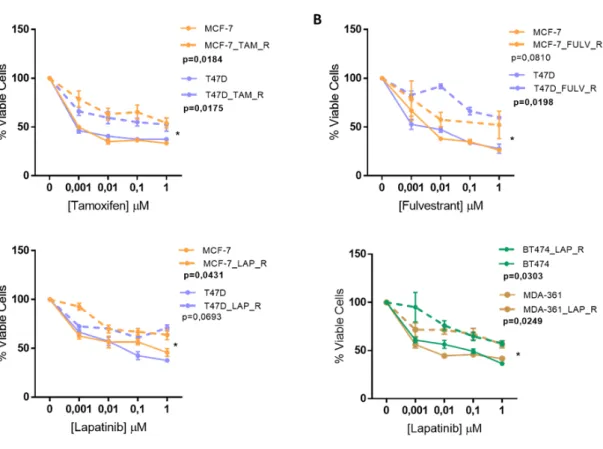
Annex 1
Figure A1: Sensitivity profiles after 5 months of drug’s exposure assessed by Alamar blue viability assay. (A) Tamoxifen; (B) Fulvestrant; (C) Lapatinib. Viability Assays were measured by Alamar
Blue assay after 7 days of exposure to drugs. The experiments were done with four replicates per assay and presented as the mean ± SEM. p-values were calculated usind paired t-test and *p<0,05.
(From (29))