UNIVERSIDADE DE LISBOA
FACULDADE DE FARMÁCIA
Synthetic antibodies targeting HIV-1 infectivity
Andreia Domingos Couto
DOUTORAMENTO EM FARMÁCIA
ESPECIALIDADE DE MICROBIOLOGIA
UNIVERSIDADE DE LISBOA
FACULDADE DE FARMÁCIA
Synthetic antibodies targeting HIV-1 infectivity
Andreia Domingos Couto
DOUTORAMENTO EM FARMÁCIA
ESPECIALIDADE DE MICROBIOLOGIA
2011
Todas as afirmações efectuadas no presente documento são da exclusiva responsabilidade da sua autora.
O trabalho apresentado nesta dissertação foi realizado na Unidade de Retrovírus e Infecções Associadas da Faculdade de Farmácia, Universidade de Lisboa, sob a orientação do Professor Doutor João Gonçalves.
A autora foi financiada por uma Bolsa de Doutoramento (SFRH/BD/28211/2006) atribuida pela Fundação para a Ciência e Tecnologia (FCT), Lisboa, Portugal.
De acordo com o disposto no ponto 1 do artigo nº 40 do Regulamento de Estudos Pós-Graduados da Universidade de Lisboa, deliberação nº 961/2003, publicado em Diário da República - II série nº 153 – 5 de Julho de 2003, a autora desta dissertação declara que participou na concepção e execução do trabalho experimental, interpretação dos resultados obtidos e redacção dos manuscriptos.
i
ACKNOWLEDGEMENTS
First and foremost I would like to thank my supervisor Prof. Doutor João Gonçalves for allowing me to go on this journey. For all the support and commitment to the development of this work, especially since this work was not a smooth ride.
To all my laboratory colleagues, present and past. Ana Catarina Santos (ACS), Catarina Santos, Cátia Cantante, Carina Pereira, Luís Ferreira, Nuno Saraiva, Inês Soeiro, Frederico Aires da Silva, Sara Maia, Paula Brito, Soraia Oliveira, Acilino Freitas Vieira, Lídia Fonseca, André Ramos, Leonor Resende, Sylvie Rato, Iris Couto, Mariana Santa-Marta, Pedro Perdigão, Rita Nogueira. Thank you for all your support, for all the help and companionship.
I would also like to give a special thanks to Nuno Taveira’s Group, in particular to Cheila Rocha and Pedro Borrego who performed the inhibition assays with the HIV-1 primary isolates, and also to Andreia Martins, Rita Almeida and Inês Bártolo for all the exchange of ideas and friendship.
I would also like to give a special thanks to Lídia M. D. Gonçalves, António J. Almeida and Lara Figueiredo for their precious collaboration for the nanoparticles and vaccine assays.
To my son, Afonso, who inspires me every day to do more and to be a better person, you are my sunshine baby boy.
To my husband, Pedro, who continues to encourage me every day and who has always been my safe harbor. I love you more every day.
To my mother and my father who have always encouraged me to follow my dreams and who are always present and without who’s help it would have been impossible to come this far.
To my grandmother, Maria Delfina, who continues to contribute and make our lives easier everyday in every way she can. You are truly a super grandmother, like my friends called you in high school.
iii
RESUMO
A Síndrome da Imunodeficiência Adquirida (SIDA) representa hoje um dos principais problemas de Saúde Pública a nível mundial, tendo contraído a doença mais de 60 milhões de pessoas em todo o mundo desde a sua descoberta em 1981, um terço das quais faleceram subsequentemente.
Apesar dos grandes progressos realizados no tratamento da SIDA, especialmente desde a introdução do regime terapêutico HAART em 1996, as alternativas terapêuticas actuamente disponíveis não permitem erradicar completamente o VIH-1 do organismo, o que resulta em toxicidade a longo prazo e leva eventualmente à emergência de estirpes de VIH-1 resistentes à terapêutica disponivel. Estes problemas levam à procura e desenvolvimento de novos fármacos activos contra o VIH, nomeadamente contra as estirpes de VIH resistentes aos fármacos actualmente utilizados. Entre os novos fármacos actualmente disponíveis encontra-se uma nova classe, os inibidores de entrada.
O processo de fusão do VIH-1 é mediado pelas proteínas Env (gp120 e gp41) e inicia-se através da ligação da gp120 ao receptor celular CD4. A interacção gp120-CD4 dá origem a uma alteração conformacional na loop V3 da gp120 que expõe o epitopo de ligação ao receptor de quimiocinas, o que por sua vez induz alterações conformacionais na gp41 que culminam na conformação activa do péptido de fusão (conformação fusogénica). Portanto, a interação gp120-CD4 é fundamental para o processo de fusão vírus-célula.
Igualmente, uma descoberta recente identificou na gp120 uma região
conformacionalmente invariável que se sobrepõe parcialmente ao local de ligação ao CD4, esta região corresponde à zona de ligação do anticorpo b12 e possui a particularidade que se encontrar constitutivamente exposta no domínio exterior da gp120. Esta região encontra-se envolvida na ligação metaestável ao CD4, antes de ocorrer o rearanjo necessário para a existência de uma ligação estável. Este é por conseguinte um local de vulnerabilidade, relacionado com um requisito funcional para existência de uma associação eficiente com o CD4.
iv O anticorpo b12 é um dos poucos anticorpos monoclonais humanos conhecidos que pode eficientemente neutralizar uma ampla gama de isolados primários de VIH-1 in vitro e pode proteger contra o desafio viral in vivo.
O objetivo deste trabalho foi a avaliação da sequência alvo do anticorpo neutralizante b12 ou o epítopo de ligação ao CD4 fora do contexto da glicoproteína gp120. Para tal procedeu-se à construção de anticorpos de domínio, por meio de “grafting” da sequência alvo do anticorpo neutralizante b12 ou o epítopo de ligação ao CD4 num dos CDR’s, CDR1 ou CDR3 de um anticorpo de coelho de único domínio VL,.altamente estável. O potencial das construções VLB12 e VLCD4 foi testado para ligação ao CD4 por ELISA e por Citometria de Fluxo. A Citometria de Fluxo foi efectuada recorrendo a células HEK293T, HeLa-P4 e Jurkat, ambas as construções apresentam ligação ao receptor CD4, e o VLB12 CDR1, em particular, e parece ter uma ligação ao CD4 de elevada afinidade. A partir dos resultados de Citometria de Fluxo e dos rendimentos obtidos para as purificações as construções VLB12 CDR1 e VLCD4 CDR3 foram selecionados para análise posterior. Pretendeu-se também caracterizar o local de ligação
das construções VLB12 CDR1 e VLCD4 CDR3 no receptor CD4, nomeadamente
identificar qual o domínio do CD4 ao qual se ligam os anticorpos VLB12 e VLCD4. Para tal, realizaram-se ensaios de ELISA utilizando como antigénios que o receptor celular humano CD4 na sua forma solúvel, quer uma construção em que os domínios 1 e 2 do CD4 humano foram colocados em fusão com uma IgG2 humana na qual as regiões variáveis das cadeias leves e das cadeias pesadas foram substituidas pelos domínios 1 e 2 do CD4 humano dando origem a uma proteína de fusão designada CD4-IgG2. Dos ensaios de ELISA foi possivel concluir que ambos os anticorpos VLB12 e VLCD4 se ligam ou ao domínio 1 ou ao domínio 2 do CD4 humano. Para identificar especificamente a qual dos domínios se ligavam o VLB12 e o VLCD4, foram realizadas experiências em que células HEK293T foram transfectadas com CD4 humano (hCD4), CD4 de murganho (mCD4) ou com CD4 humano em que o domínio 1 foi substituido pelo domínio 1 de murganho (hCD4mD1). Estas células transfectadas com os diferentes receptores celulares CD4 foram submetidas a ensaios de Citometria de Fluxo para avaliação da ligação dos anticorpos VLB12 e VLCD4 aos diferentes receptores celulares CD4 e concluiu-se que o VLB12 se liga ao domínio 2 e que o VLCD4 se liga ao domínio
v 1 do CD4 humano. Realizaram-se também ensaios de ELISa em que se verificou que os epitopos reconhecidos pelo VLB12 e pelo VLCD4 são ambos conformacionais.
A fim de avaliar a capacidade do VLB12 e do VLCD4 na inibição da infecção por VIH-1, foram realizados ensaios de inibição com o clone molecular NL4-3 do VIH-1 (subtipo B) e com os subtipos J e H de isolados primários de VIH-1. Os ensaios de inibição foram realizados em células TZM-bl num ensaio de um ciclo único de infecção. O VLB12 foi capaz de inibir o VIH-1NL4-3 com um IC50 = 3,89 M e o VLCD4 foi capaz de inibir o HIV-1NL4-3 com um IC50 = 6,57 M. Realizaram-se igualmente ensaios de inibição com o T20 para o qual se obteve um IC50 = 0,0694 M.Quanto aos isolados primários, o VLB12 foi capaz de inibir o VIH-1 subtipo H com um IC50 = 3,97 M e o VIH-1 subtipo J com um IC50 = 3,86 M, mas o VLCD4 não foi capaz de inibir os subtipo J e H de isolados primários de VIH-1. Para o T20 obteve-se um IC50 = 1,33 nanoM para o subtipo H do VIH-1 e para o subtipo J do VIH-1 obteve-se um IC50 = 0,42 nanoM.
Estes resultados indicam que o VLB12 apresenta um amplo potencial de inibição ao contrário do VLCD4 que não foi capaz de inibir qualquer um dos isolados primários dos subtipo não B.
Devido à elevada afinidade de ligação ao receptor CD4 apresentada pelo VLB12, uma das abordagens desta Tese teve por objectivo avaliar o potencial de um novo sistema de entrega de moleculas para terapia génica em que se utilizaram nanopartículas revestidas com VLB12 para avaliar a entrega de biomoléculas a células que expressam o receptor CD4.
Foram testados dois tipos diferentes de formulações de nanopartículas, de quitosano e de PEI, para a entrega do plasmídeo FUGW-dsRed em células Jurkat. Ambas as formulações de nanopartículas foram bem sucedidos na entrega do plasmídeo FUGW-dsRed às células Jurkat, conforme determinado por imunofluorescência.
A formulação de nanopartículas de quitosano provou globalmente ser mais eficaz, devido à reduzida toxicidade celular. A quantidade ideal de VLB12 adsorvido às nanopartículas deu origem a um aumento de 16% da população dsREd positiva
vi fluorescente com um direccionamento das nanopartículas para o receptor alvo (CD4) devido à presença do VLB12.
Numa abordagem distintadas anteriores, pretendeu-se avaliar as propriedades antigénicas do VLB12 para utilização numa formulação de vacina. Deste modo, a imunidade das mucosas foi testada utilizando o VLB12 encapsulado em nanopartículas de quitosano, por comparação com a via subcutânea, utilizando como adjuvante alumínio e realizando as mesmas formulações sem adjuvante. A resposta de IgG total específico nas amostras de soro foi avaliada por ELISA utilizando como antigénio o VLB12. Foram também avaliadas as respostas Th1 ou Th2 através do racio dos antigénios específicos IgG1-IgG2 nas amostras de soro.
Para avaliar o potencial antigénico do VLB12 como vacina, foram realizados ensaios de inibição de VIH-1 usando os soros dos diferentes grupos de murganhos, aos quais foi administrado cada uma das formulações. Os ensaios de inibição foram realizados em células TZM-bl, com uma MOI de 0,1, num ensaio de um ciclo único de infecção. Apesar de os grupos vacinados apresentarem títulos de IgG específico elevados, nenhum dos soros obtidos apresentou efeito inibitório sobre o VIH-1NL4-3. Confirmou-se por ELISA que os soros obtidos não reconheciam a gp120 o que indica que a framework do VL é altamente imunogénica, não tendo sido obtida uma proporção de anticorpos dirigidos contra a região alvo b12 suficiente para se obter um efeito inibitório nos ensaios de inibição do VIH-1NL4-3.
Palavras-chave: VIH-1; gp120; anticorpos de domínio único VL; inibidores de entrada;
vii
ABSTRACT
The HIV-1 entry process is mediated by the Env proteins, and begins with the binding of gp120 to the CD4 cellular receptor. A recent discovery has identified in gp120 a conformationally invariant surface that overlaps a distinct subset of the CD4-binding site, which is a binding site for the b12 natural antibody.
The goal of this work was to evaluate both the b12 target sequence and the CD4 binding epitope outside of the gp120 context and to evaluate the potential for CD4 binding of these two epitopes individually. This was done through grafting of the b12 target sequence or CD4 binding epitope into either CDR1 or CDR3 of a highly stable VL antibody. The potential of the VLB12 and VLCD4 constructs was tested for CD4 binding by ELISA assay and Flow Citometry analysis; both graftings present binding to the CD4 receptor and VLB12, in particular.
In order to evaluate the VLB12 and VLCD4 ability to inhibit HIV-1 infection, inhibition
assays were performed with the HIV‐1NL4‐3 subtype B molecular clone and with HIV‐1
subtypes J and H primary isolates. VLB12 was able to inhibit HIV‐1NL4‐3 with an IC50 = 3,89 M and VLCD4 was able to inhibit HIV‐1NL4‐3 with an IC50 = 6,57 M. As for the HIV‐1primary isolates, VLB12 was able to inhibit subtype H with an IC50 = 3,97 M and subtype J with an IC50 = 3,86 M but VLCD4 was not able to inhibit either subtype J or subtype H. These results indicate that VLB12 presents potentially a broad inhibition spectrum as opposed to VLCD4 that was not able to inhibit either of the non subtype B primary isolates.
Due to the VLB12 high binding affinity to the CD4 receptor the potential of VLB12 coated nanoparticles as a new therapeutic delivery system was evaluated. Chitosan and PEI nanoparticle formulations were tested, for the delivery of FUGW-dsRed plasmid to Jurkat cells. Both nanoparticle formulations were successful in delivery of specific CD4 targeted FUGW-dsRed as determined by immunofluorescence. Chitosan nanoparticle formulation proved to be more effective due to the lower cell toxicity. The optimal amount of VLB12 adsorbed to the nanoparticles gave a 16 % positive dsREd fluorescent population with specific VLB12 targeting.
viii To evaluate the vaccine antigen properties, of VLB12 the mucosal immunity was tested by using encapsulated VLB12 in nanoparticles of chitosan in comparison with subcutaneous routes with aluminium adjuvant and without adjuvant. Th1 or Th2 responses were evaluated by the ratio of antigen-specific IgG2a-IgG1 in the serum samples. HIV-1 neutralization assays using serum from different groups to evaluate the VLB12 as potential vaccine antigen were performed but the serum presented no inhibitory effect on HIV-1NL4-3.
ix
LIST OF ACRONYMS AND ABBREVIATIONS
Reagents and Techniques
ABTS 2,2'-Azinobis [3-ethylbenzothiazoline-6-sulfonic acid]-diammonium salt CPRG ChloroPhenolRed‐β‐D‐Galactopyranoside
CS Chitosan
CS/DS Chitosan/Deoxycholate
DMEM Dulbecco’s modified Eagle’s Medium ECL Enhanced ChemiLuminescence reagent ELISA Enzyme‐Linked ImunoSorbent Assay FBS Fetal Bovine Serum
MOI Multiplicity Of Infection PBS Phosphate Buffer Saline PCR Polymerase Chain Reaction
PEI Polyethylenimine
RPMI Roswell Park Memorial Institute medium
SDS‐PAGE Sodium Dodecyl Sulfate PolyAcrylamide Gel Electrophoresis HRP HorseRadish Peroxidase
General
ADCC antibody-dependent cell-mediated cytotoxicity
AIDS Acquired Immune Deficiency Syndrome
APOBEC3G apolipoprotein B mRNA-editing, enzyme-catalytic, polypeptide-like 3G
ART Antiretroviral Therapy
ARV Anti Retroviral
CA Capsid
CCR5 CC Chemokine Receptor 5
CD4 Cluster of Differentiation 4
CD4bs CD4 binding site
CD4i CD4 induced
cDNA complementary DNA
CDR Complementarity Determining Region
x
CDRH3 third complementarity determining region of antibody heavy chains
CDRL complementarity determining region of antibody light chains
CDRL1 first complementarity determining region of antibody light chains
CDRL3 third complementarity determining region of antibody light chains
CH Constant Heavy Chain
CL Constant Light Chain
CXCR4 CXC Chemokine Receptor 4
DNA Deoxyribo Nucleic Acid
dsRNA double‐stranded RNA
E. coli Escherichia coli
Env Envelop polyprotein
ER Endoplasmic Reticulum
ESCRT endosomal sorting complex required for transport
Fab Fragment antigen binding
Fc Fragment crystallizable
FcRn neonatal Fc receptor
FR framework region
Fv Fraction variable
Gag Group specific antigen polyprotein
h hour
HAART Highly Active Antiretroviral Therapy
hcAbs heavy chain antibodies
hCD4 Human Cluster of Differentiation 4
HIV Human Immunodeficiency Virus
HIV-1 Human Immunodeficiency Virus type 1
HIV-2 Human Immunodeficiency Virus Type 2
HMG-1(Y) High Mobility Group protein I(Y)
hmAbs Human monoclonal antibodies
HR1 Heptad Repeat sequence 1
HR2 Heptad Repeat sequence 2
hTRIM5α human TRIM5α
Ig Immunoglobulin
IL‐1 InterLeukine 1
xi
IN Integrase
kDa kiloDalton
LBD lipid-binding domain
LTR Long Terminal Repeats
MA Matrix
MALT Mucosa Associated Lymphoid Tissue
MHC Major histocompatibility complex
min minute
ml millilitre
mM miliMolar
MPER membrane-proximal external region
mRNA messenger RNA
nanoM nanoMolar
NC Nucleocapsid
Nef Negative regulator factor
NES Nuclear Export Signal
ng nanogram
NLS Nuclear Localization Signal
nm nanometer
ºC Degrees Celsius
PBD Pocket-Binding Domain
PBS Primer Binding Site
PIC PreIntegration Complex
POL Polimerase
PR Protease
Rev Regulator of Expression of viral proteins
RNA RiboNucleic Acid
RNAPol II RNA Polymerase II
RNase H RiboNuclease enzyme H
RRE Rev Responsive Element
RT Reverse Transcriptase
RTC Reverse Transcription Complex
scFv Single chain Fv
xii
SIV Simian Immunodeficiency Virus
SU Surface
TAR TransActivation Response
Tat Transcriptional transactivator protein
TM transmembrane
TNF‐α Tumor Necrosis Factor‐alpha
TRIM5α Tripartite Motif Protein 5 alpha
tRNA transference RNA
U3 Unique 3´ sequence
U5 Unique 5´sequence
VH Variable Heavy Chain
VHH Variable domain of camelid heavy chain antibody
Vif Viral Infectivity Factor
VL Variable Light Chain
VNAR Variable domain of the shark new antigen receptor
Vpr Viral protein R
Vpu Viral Protein U
g microgram
M microMolar
Amino acids
A - Alanine G - Glycine M - Methionine S - Serine
C - Cysteine H - Histidine N - Asparagine T - Threonine
D – Aspartic acid I - Isoleucine P - Proline Y – Tyrosine
E – Glutamic acid K - Lysine Q - Glutamine V - Valine
xiii
INDEX
ACKNOWLEDGEMENTS ... i
RESUMO ... iii
ABSTRACT ... vii
LIST OF ACRONYMS AND ABBREVIATIONS ... ix
INDEX ... xiii
Index of Figures ... xviii
Index of Tables ... xxi
1 General Introduction ... 2
1.1 Human Immunodeficiency Virus type 1 ... 4
1.1.1 Genetic structure ... 4
1.1.2 Morphology ... 5
1.1.3 Replication Cycle ... 6
1.2 The fusion process: a crucial step in HIV infection ... 11
1.2.1 The CD4 receptor – targets of opportunity ... 12
1.2.2 Viral surface glycoprotein (gp120) ... 14
1.2.3 Viral transmembrane protein gp41 ... 18
1.3 Antiretroviral therapy for treatment of HIV-1 infection ... 19
1.4 Entry inhibitors ... 21 1.4.1 Inhibitors of gp120-CD4 interaction ... 21 1.4.2 CCR5 antagonists ... 22 1.4.3 CXCR4 antagonists ... 23 1.4.4 Entry inhibitors ... 24 1.5 Antibodies ... 26
1.5.1 The immune system and the origin of antibodies ... 26
xiv
1.5.3 Antibodies - Mechanisms of in vivo action ... 28
1.5.4 Production of polyclonal, monoclonal and recombinant antibodies ... 30
1.5.5 Recombinant antibody fragments ... 31
1.6 Antibody response to HIV-1 ... 37
1.6.1 Gp120 CD4bs Antibodies: the b12, VRC01 and HJ16 antibodies. ... 38
1.6.2 Gp120 V2/V3 Antibodies: the PG9/PG16 antibodies. ... 42
1.6.3 Antibodies against gp120 glycans: 2G12 ... 42
1.6.4 MPER antibodies : 2F5, 4E10 and Z13e1 ... 43
1.6.5 CD4 receptor antibodies: Ibalizumab ... 44
1.6.6 CD4-Induced (CD4i) Antibodies ... 44
1.7 Vaccines ... 45
1.7.1 AIDS vaccine: Present status and future possibilities ... 45
1.8 Aims ... 51
2 A new approach for CD4 targeting – grafted single domain antibodies ... 54
2.1 Introduction ... 54
2.2 Materials and methods ... 56
2.2.1 Construction of recombinant single domain antibodies, dAbs ... 56
2.2.2 Cloning of the CD4 sequence in the CDR1 of the VL antibody ... 57
2.2.3 Cloning of the CD4 sequence in the CDR3 of the VL antibody ... 58
2.2.4 Cloning of the b12 sequence in the CDR1 of the VL antibody ... 58
2.2.5 Cloning of the b12 sequence in the CDR3 of the VL antibody ... 58
2.2.6 Cloning of the CD4 sequence in the CDR3 of the VLB12(CDR1) antibody ………...58
2.2.7 Expression and purification of VLB12 or VLCD4 antibodies cloned in pComb3X ... 59
2.2.8 Expression and purification of VLB12 or VLCD4 antibodies cloned in pET28a……… ... 60
xv
2.2.9 Western blot ... 61
2.2.10 Coomassie staining ... 62
2.2.11 Enzyme-linked immunosorbent assay (ELISA) to evaluate the dAbs binding to human CD4 ... 62
2.2.12 ELISA assay to evaluate the dAbs binding to b12 mAb ... 63
2.2.13 ELISA assay for epitope characterization ... 63
2.2.14 Production of HEK293T expressing mCD4, hCD4 or hCD4mD1 ... 64
2.2.15 Flow cytometry analysis ... 64
2.2.16 Statistical analysis ... 66
2.3 Results ... 67
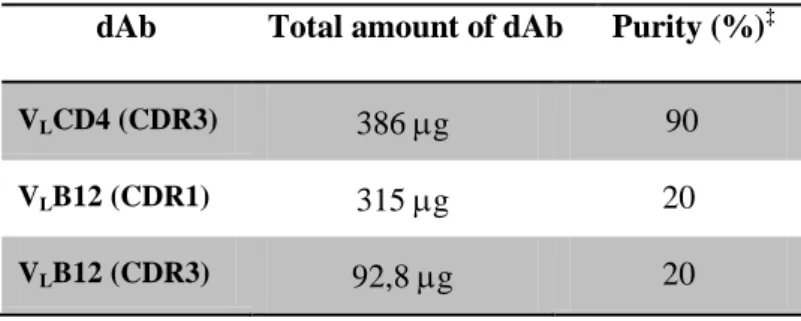
2.3.1 Construction, expression and purification of the recombinant domain antibodies, dAbs ... 67
2.3.2 ELISA assays to evaluate the dAbs binding to human CD4 ... 70
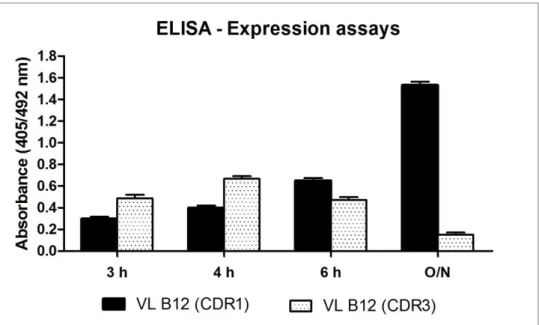
2.3.3 Flow cytometry assay to evaluate VLB12 binding to CD4 ... 72
2.3.4 Flow cytometry assay to evaluate VLB12 (CDR1), VLB12 (CDR3) and VL CD4 (CDR3) binding to CD4 in Jurkat cells ... 74
2.3.5 Alternate expression and purification method of recombinant domain antibodies, dAbs ... 77
2.3.6 VLB12 and VLCD4 binding to hCD4 – characterization and domain identification ... 80
2.3.7 ELISA assay to evaluate the dAbs binding to b12 mAb ... 83
2.3.8 VL B12 and VL CD4 binding to hCD4 – epitope characterization ... 84
2.3.9 Flow cytometry assay to identify the hCD4 domain recognized by VLB12 and VLCD4 ... 85
2.4 Discussion ... 90
2.5 Acknowledgments ... 92
xvi 3.1 Introduction ... 94 3.2 Materials and Methods ... 95 3.2.1 Cell Lines and culture conditions ... 95 3.2.2 NL4-3 Viral production ... 95 3.2.3 Primary isolates viral production ... 95 3.2.4 Virus inhibition assay using TZM-bl cells. ... 96
3.2.5 Assessment of cell viability in the presence of the recombinant
antibodies.. ... 97 3.2.6 Statistical analysis ... 98 3.3 Results ... 99 3.3.1 HIV-1 inhibition assays ... 99 3.4 Discussion ... 105 3.5 Acknowledgments ... 107 4 VL B12 coated nanoparticles - A new therapeutic delivery system... 110 4.1 Introduction ... 110 4.1.1 Nanoparticles ... 110 4.1.2 Chitosan (CS) nanoparticles ... 111 4.1.3 Polyethylenimine (PEI) nanoparticles ... 112 4.1.4 Nanoparticle formulations with VL B12 for FUGW-dsRed delivery to CD4 positive cells. ... 112 4.2 Materials and Methods ... 113 4.2.1 Cell Lines and culture conditions ... 113
4.2.2 Determination of the optimal pDNA/nanoparticle ratio for CS/DS and PEI
nanoparticles ... 113
4.2.3 Preparation of chitosan-sodium deoxycholate (CS/DS) nanoparticles... 113
4.2.4 Preparation of PEI based nanoparticles ... 114 4.2.5 Transfection assays for immunofluorescence assays ... 115
xvii 4.2.6 Immunofluorescence ... 115
4.2.7 VLB12 coated nanoparticles targeting CD4 cell receptor for FUGW-dsred
plasmid delivery to Jurkat cells ... 116 4.2.8 Flow cytometry analysis of nanoparticle delivery to Jurkat cells ... 116 4.3 Results ... 117
4.3.1 Determination of the optimal pDNA/nanoparticle ratio for CS/DS and PEI
nanoparticles ... 117 4.3.2 Immunofluorescence ... 118 4.3.3 VLB12 coated nanoparticles for delivery of FUGW-dsREd plasmid to CD4 positive Jurkat cells. ... 125 4.4 Discussion ... 129 4.5 Acknowledgments ... 130 5 VL B12 as a vaccine antigen ... 132 5.1 Introduction ... 132 5.1.1 Vaccine formulations and vaccine administration... 132 5.1.2 Nanoparticles for delivery of vaccines ... 133 5.1.3 Antiviral activities of antibodies in vivo – their role in vaccine development ... 133 5.2 Materials and Methods ... 135 5.2.1 Cell Lines and culture conditions ... 135 5.2.2 Viral production ... 135 5.2.3 Virus neutralization assay using TZM-bl cells. ... 135 5.2.4 Preparation of CS/DS ... 136 5.2.5 Nanoparticles Physicochemical Characterization ... 137 5.2.6 Mice immunisation schedule ... 137 5.2.7 Quantification of antigen-specific IgG and subtypes by ELISA ... 138 5.2.8 ELISA assay for mice serum IgG specificity to gp120 ... 139
xviii 5.2.9 Statistical analysis ... 139 5.3 Results ... 140 5.4 Discussion ... 146 6 General Discussion ... 150 7 Bibliography ... 158
Index of Figures
Figure 1.1– Genetic organization of HIV-1. ... 4 Figure 1.2 - Schematic representation of the HIV-1 virion. ... 6 Figure 1.3 - Steps involved in HIV-1 replication. ... 7 Figure 1.4 – Simplified schematic representation of the current model for the fusion mechanism between HIV-1 and the target cell. ... 11 Figure 1.5 - Schematic representation of the CD4 molecule. ... 12 Figure 1.6 - Model of the gp120 component of the trimeric HIV-1 envelope spike based on the structure of core gp120, with three gp120 monomers shown in purple, green, and pink. ... 15 Figure 1.7 - Core gp120 showing the inner domain, outer domain, and bridging sheet 15 Figure 1.8 – Tridimensional representation of the interaction between CD4 and gp120. ... 16 Figure 1.9 - Approved anti-retroviral drugs for the treatment of HIV-1 infection. ... 20 Figure 1.10 - Simplified schematic representation of the current model for the fusion mechanism between HIV-1 and the target cell, and its inhibition by entry inhibitors. .. 21 Figure 1.11- Modular structure of immunoglobulins. ... 27 Figure 1.12- Schematic diagram of the structure of immunoglobulin G... 27 Figure 1.13 – Schematic representation of different antibody formats. ... 33
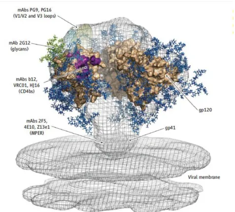
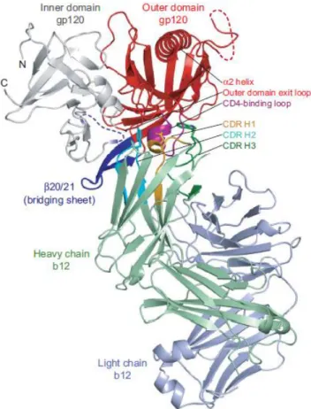
xix Figure 1.14 – Tridimensional model of gp120. Location of ENV neutralizing monoclonal antibodies is depicted. ... 37 Figure 1.15 - The structure of the b12 monoclonal antibody. ... 39 Figure 1.16 - Structure of b12 in complex with an HIV-1 gp120 core. ... 40 Figure 2.1 – Model of the contact surfaces of CD4 and b12 on gp120. ... 54 Figure 2.2 – Predicted structure of the single domain VL antibody. ... 55 Figure 2.3 – 1st round of PCR for grafting of b12 target epitope or CD4 binding epitope. ... 57 Figure 2.4 – Western blot analysis of the expression levels (soluble fraction) of the dAbs. Equal amounts of protein were loaded onto the gel. The dAbs detection was performed with -HA-HRP. ... 68 Figure 2.5 - The VL B12 (CDR1) and VL B12 (CDR3) protein expression assays were compared by ELISA assay. ... 68 Figure 2.6 - ELISA assays to evaluate the dAbs binding to hCD4. ... 70 Figure 2.7 – Flow cytometry assay to evaluate that VLB12 (CDR1) binds specifically to the CD4 receptor. ... 72 Figure 2.8 - Flow cytometry assay to evaluate VLB12 (CDR1), VLB12 (CDR3) and VLCD4 (CDR3) binding to CD4 in Jurkat cells. ... 74 Figure 2.9 - Graphical representation of the percentage of CD4 positive cells obtained for the three independent Flow cytometry assays performed. Single domain Ab amounts range from 1,0 nmol to 50,0 nmol. ... 75 Figure 2.10 - Graphical representation of the percentage of CD4 positive cells obtained for the three independent Flow cytometry assays performed.(continues on the next page) ... 76 Figure 2.11 - Optimized expression conditions for VL B12 in the pET-28a vector. ... 78 Figure 2.12 - VLB12 protein recovered after each purification step using the protocol for purification of insoluble dAbs . ... 78 Figure 2.13 - ELISA assays were performed to evaluate VL B12, VL CD4 and the original VL binding to either soluble hCD4 or a CD4-IgG. ... 81
xx
Figure 2.14 - ELISA assays were performed to evaluate b12 mAb binding to VLB12 and
VLCD4. ... 83 Figure 2.15 - VLB12 and VLCD4 binding to soluble hCD4 – Epitope characterization. 84 Figure 2.16 - Schematic representation of the HEK293T cells transfected with mCD4, hCD4 or hCD4mD1. ... 86
Figure 2.17 - Flow cytometry assay to identify the hCD4 domain recognized by VLB12
and VL CD4 ... 87 Figure 3.1 – HIV‐1NL4‐3 inhibition assays performed in TZM-bl cells. ... 100 Figure 3.2 – TZM-bl cell viability was evaluated using alamarBlue. ... 102 Figure 3.3 – HIV‐1 primary isolates inhibition assays using TZM-bl cells. ... 104 Figure 4.1 – Nanoparticle encapsulation as determined by agarose gel electrophoresis. ... 117 Figure 4.2 – Immunofluorescence staining of FUGW-dsRed/CS/DS nanoparticle complexes using VLB12 for specific delivery to Jurkat cells. ... 119 Figure 4.3 – Immunofluorescence staining of FUGW-dsRed/PEI nanoparticle complexes using VLB12 for specific delivery to Jurkat cells. ... 121 Figure 4.4 - Normalized fluorescence units/105 cells for CS/DS and PEI VLB12 coated nanoparticles delivery of FUGW-dsREd. ... 123 Figure 4.5 – Flow cytometry analysis of chitosan nanoparticles coated with VLB12 for delivery of FUGW-dsREd plasmid to CD4 positive Jurkat cells. ... 125 Figure 4.6 – Flow cytometry analysis of PEI nanoparticles coated with VLB12 for delivery of FUGW-dsREd plasmid to CD4 positive Jurkat cells. ... 127 Figure 5.1 – ELISA assay performed to determine antigen specific total IgG titers. .. 140 Figure 5.2 – ELISA assay performed to determine antigen specific IgG1 and IgG2a titres. ... 142 Figure 5.3 - Inhibition assay performed with serial dilutions of G1 to G7 serum from Bleed 6 for HIV-1NL4-3 inhibition. ... 143 Figure 5.4 - ELISA assay for serum binding to gp120. ... 144
xxi
Index of Tables
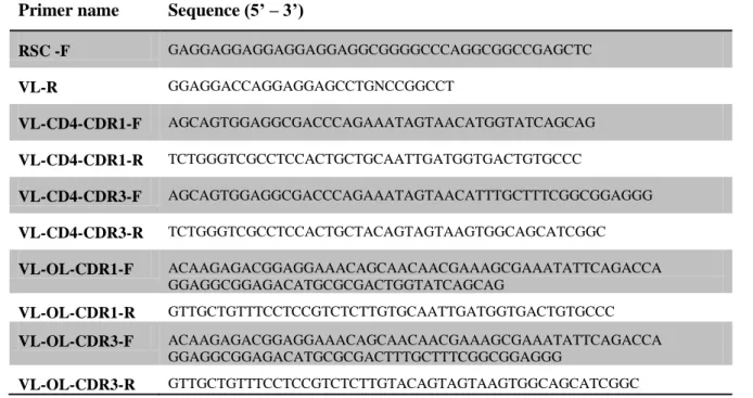
Table 2.1– Coding sequences used for CDR grafting of gp120 epitopes. ... 56 Table 2.2 - Primers used to clone the b12 target sequence, the CD4 binding epitope sequence, or both sequences in the CDRs of the VL antibody. ... 56 Table 2.3 - Primers used to amplify VL B12 or VL CD4 for cloning in the pET28a vector. ... 60 Table 2.4 - Optimal expression conditions for the dAbs cloned in pComb3X. ... 67
Table 2.5 – Comparison between the CDR1 and CDR3 sequences of the original VL and
the CD4 and b12 epitopes. ... 69 Table 2.6 - Results of the dAbs purifications per liter of bacteria culture. ... 69 Table 2.7 - % of CD4 positive cells ... 75 Table 2.8 - Results of the dAbs purifications per liter of bacteria culture, using the insoluble dAb purification protocol. ... 79 Table 2.9 - % of binding to CD4 transfected cells ... 88 Table 2.10 - % of binding to CD4 transfected cells. ... 89 Table 4.1 -VLB12 coated CS/DS nanoparticle formulations with encapsulated FUGW-dsred plasmid ... 114 Table 4.2 - VLB12 coated PEI nanoparticle formulations with encapsulated FUGW-dsred plasmid ... 114 Table 4.3 - Normalized results from the fluorescence assay, with CS/DS nanoparticles and PEI Nanoparticles. Assays are representative of n = 4 ... 122 Table 4.4 - Calculations from the fluorescence assay, with CS/DS nanoparticles and PEI Nanoparticles ... 124 Table 5.1 – Mice groups used for the immunization assay of the VL B12 antigen. ... 140
1
2
Introduction
1 General Introduction
The Acquired Immune Deficiency Syndrome (AIDS) is now a major public health problem worldwide, having contracted the disease more than 60 million people since its discovery in 1981, one third of whom subsequently died.1,106
The etiological agents responsible for AIDS are the human immunodeficiency virus type-1 and type-2 (HIV-1 and HIV-2). The first agent to be isolated was HIV-121,113 in 1983. In 1986 HIV-2 was identified,69,70 having similar biological properties to those of HIV-1, but a greater genetic homology with Simian Immunodeficiency Virus than with HIV-1. This translates into a significant difference in some of its molecular and antigenic properties.
HIV-1 is more virulent and has higher transmission rates than HIV-2, which explains why HIV-1 is responsible for a worldwide pandemic and HIV-2 accounts for more localized epidemics, particularly in countries of West Africa and in some European countries such as Portugal.69,70 Only HIV-1 will be studied in this Thesis.
HIV-1 has the ability to infect cells that have a specific receptor, cluster of differentiation 4 (CD4), and chemokine co-receptors, especially CCR5 and CXCR4, which are mainly on the surface of CD4+ T lymphocytes.26,162 Other cells that express these receptors and likely to be susceptible to HIV-1 are, for example, cells of the mononuclear phagocytic system, particularly monocytes, macrophages, B lymphocytes and dendritic cells.
The cell tropism of HIV suggests that once infection is established it will be very difficult to eliminate the virus from the body. This leads to progressive destruction of the immune system, leading to AIDS, with its multiple opportunistic infections, and/or complications such as neurological disorders and cancer. With this type of epidemiological characteristics it is imperative a commitment to a preventative therapy or acting in the early stages of infection.106,323
3 Despite the stunning advances in the treatment of AIDS, especially since the introduction of highly active antiretroviral therapy (HAART) in 1996, current therapy cannot completely eradicate HIV-1 from the body, resulting in long-term toxicity and eventually leading to the emergence of drug-resistant HIV-1 strains. These problems prompt the search for potent new drugs that are active against drug-resistant viral strains. Among these drugs appears a new class, the entry inhibitors.
The monoclonal antibody therapy has had an important development in recent years particularly with regard to new therapeutic approaches for treating HIV infection and AIDS. This form of therapy can be complementary and add to the effectiveness of the existing drugs. Studies with "natural" antibodies (hmAbs) that neutralize the virus by binding to conserved epitopes of gp120 or gp41 proteins have shown a half-life of 50 to 100 times higher than fusion inhibitor peptides, showing no toxicity associated with pharmacological anti-HIV drug therapy.17,171,239,266,320
This type of strategy is a priority in the activities of the Unit of Retrovirus and Associated Infections (URIA) of the Faculty of Pharmacy of Lisbon, particularly in identifying new forms of biological therapies against HIV-1, and specifically the identification of recombinant antibodies against various therapeutic targets, whose large-scale production is easily optimized. Thus, it will be presented and discussed in this Thesis a new way to inhibit the infectivity of HIV-1 using synthetic recombinant antibodies with inhibitory capacity of the entry process. Moreover the potential for other therapeutic applications was also explored for one of these molecules, in particular as part of a new therapeutic delivery system and also as a vaccine antigen.
4
1.1 Human Immunodeficiency Virus type 1 1.1.1 Genetic structure
HIV-1 belongs to the genus Lentivirus and family Retroviridae. These are enveloped viruses with a nucleocapsid that resembles a truncated cone. They have a diploid positive-strand RNA genome which replicates via a double strand DNA intermediate (reverse transcription), being afterwards integrated in the host genome in the form of a provirus. These features are common to all retroviruses.222,322,344
Figure 1.1– Genetic organization of HIV-1.
(Reprinted with permission from: W.H. Freeman and Company, copyright (2006)).176
The genome of HIV-1 is composed of three essential genes, gag, pol and env, the regulatory genes tat and rev, four accessory genes, nef, vif, vpr and vpu, and two
identical sequences, called long terminal repeats (LTRs) flanking the
genome.222,322,323,352
The gag and pol genes are translated into long precursor polyproteins (Figure 1.1). The gag gene codes for a precursor polyprotein of 55 kDa (p55) that is subsequently cleaved by the action of the viral protease giving rise to the major internal structural proteins, designated matrix (p17, MA), capsid (p24, CA), nucleocapsid p7 (NC) and p6. After cleavage the Gag-Pol precursor polyprotein gives rise to the protease (p10, PR), reverse transcriptase (p66/p51, RT) and integrase (p31, IN) .322
The product of the env gene is translated in the form of a precursor of 160 kDa (gp160) that is subsequently cleaved generating the two subunits of the viral envelope, the surface glycoprotein (gp120, SU) and the transmembrane glycoprotein (gp41, TM).93
5 The tat and rev regulatory genes give rise, respectively, to the Tat (p14) and Rev (p19) proteins. The Tat protein is a trans-activator of transcription that increases the processivity of RNA polymerase II. The Rev protein is the regulator of expression that inhibits the splicing of viral RNAs and is also involved in the translocation to the cytoplasm of RNAs that did not undergo splicing.322,352
The down-regulating factor (Nef), the viral infectivity factor (Vif), viral protein R (Vpr) and protein U (Vpu) are all small accessory proteins encoded respectively by genes nef, vif, vpr and vpu, and each play more than one function in the replication cycle of HIV-1.322,323
The Vif protein averts the lethal threat of deamination, precluding the packaging of APOBEC3G (A3G) into assembling virions by mediating its proteasomal degradation.238 Vpr functions early in the viral life cycle, in the transport of the PIC to the nucleus. Vpr has several other critical functions including activation of HIV-1 LTR transcription, cell-cycle arrest due to DCAF-1 binding, and both direct and indirect contributions to T-cell dysfunction. 165,233 Vpu enhances virus budding and degrades cellular CD4.37 Nef also has similar functions, and appears to be required for disease induction in vivo.108
The LTR regions are important for replication and transcription of viral RNA. These sequences are divided into three parts: U3 (derived from a unique sequence of the 3' region of the viral RNA), R and U5 (derived from a unique sequence of 5' region of the viral RNA). The LTR sequences include promoters, enhancer elements and other genomic sequences used to connect to different cellular transcription factors.222
1.1.2 Morphology
HIV-1 is a virus whose virions have an icosahedral structure of about 100 nm in diameter. The mature virion consists of an outer layer, called the viral envelope, which derives from the host cell membrane and is acquired during their release (budding) of the cell. The viral envelope is formed by a lipid bilayer impregnated with some human proteins, such as Major Histocompatibility Complex (MHC) proteins, and viral
6 glycoproteins gp120 and gp41 that form a trimer of heterodimers and whose function is to mediate the entry process allowing the virus to infect target cells.327
The coating of the inner viral envelope is the matrix protein (MA). In the center there is a nucleo-shaped truncated cone, formed by p24 (CA), within which there are two copies of the viral genome. The single-stranded RNA copies associate with one another by base-pairing near its 5 'end. Each of these molecules is coupled with a transfer RNA (tRNA lysine) which acts as an oligonucleotide to initiate reverse transcription of viral RNA. The viral RNA forms a ribonucleoprotein complex with NC, p6 and a few molecules of IN, PR and RT. Nef, Vif and Vpr are also incorporated in the viral particle (Figure 1.2).72,176,327
Figure 1.2 - Schematic representation of the HIV-1 virion. (Reprinted with permission from Elsevier science, copyright (2009)).179
1.1.3 Replication Cycle
HIV-1 has a typical retroviral life cycle, divided into two phases: the early phase and the late phase. The early phase begins with the recognition of the target cell by the mature virion and involves all processes leading to and including integration of the genomic DNA into the chromosome of the host cell. The late phase begins with the regulated expression of the integrated proviral genome and involves all processes up to and including virus budding and maturation (see Figure 1.3).322
7 The first step in HIV-1 infection is the Env mediated direct fusion of the viral membrane with the plasma membrane of the host cells. The Env glycoproteins62,89,114,355 are organized into trimeric spikes,342 anchored to the viral membrane by the gp41 transmembrane protein. The Env glycoprotein gp120, forms surface trimeric spikes, which are associated by noncovalent interactions with each subunit of the also trimeric, and normally hidden, gp41.169
Figure 1.3 - Steps involved in HIV-1 replication.
(Reprinted by permission from Macmillan Publishers Ltd: Nat Rev Immunol, copyright (2004).) 364
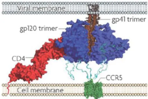
The fusion process begins with the binding of gp120 to the CD4 cellular receptor. The binding of gp120 to CD4 induces conformational changes in gp120, exposing the chemokine receptor binding site and promoting the interaction between gp120 and the chemokine receptor, usually CXCR4 or CCR5. This interaction subsequently induces conformational changes in gp41 that result in an active conformation of the fusion peptide (fusogenic conformation) promoting the fusion of the cellular and viral membranes.16,237,277,285,322,369 Membrane fusion is followed by the release and uncoating of the HIV capsid into the cytoplasm of the host cell. Completion of uncoating leads to the formation of the reverse transcription complex (RTC), composed of viral RNA, reverse transcriptase, integrase, tRNAlys3, MA, nucleocapsid (p7), Vpr (Viral protein R)
8 and several host proteins.111,317,327 Reverse transcription is performed by RT, which copies the viral RNA template into a complementary double strand DNA. To initiate the synthesis of viral DNA, a oligonucleotide of cellular origin is used, tRNAlys3. The binding site of the lysine tRNA is designated primer binding site (PBS) and is located in the 5 'end of the unspliced viral RNA.163,193 The process of synthesis also depends on NC, MA and the accessory protein Vif.322
Once synthesized, the viral DNA is transported to the nucleus as part of a preintegration complex (PIC), containing the viral proteins IN, MA, RT and Vpr and the cellular protein High Mobility Group protein I(Y) (HMG-I (Y)).317,322 The integration of HIV DNA into the host cell chromossome, giving rise to the provirus, is promoted by the catalytic activity of IN.111,190,317,322
The provirus thus becomes an integral part of the host cell genome and is treated by the cell as a cellular gene. After integration, the provirus may either remain latent (post‐integration latency) or become transcriptionally active. The provirus acts as a template for the synthesis of viral RNA that can be subsequently used as genomic RNA for virion progeny or translated into viral proteins.322 HIV‐1 transcription is controlled by the inducible viral promoter located at U3 of 5´LTR.300
The transcription of the viral DNA is mediated by cellular RNA polymerase II and involves regulatory sequences in the region of the 5'- LTR U3 viral promoter, which functions as an activator of transcription, since it has binding sites for several cellular transcription factors.317,322,327
The late phase of the replication cycle begins with the synthesis of spliced and unspliced mRNAs that are transported out of the nucleus to be translated. Transcription from the HIV-1 LTR leads to the generation of three major classes of viral RNAs:264
1) unspliced RNAs, which function as the mRNAs for the Gag and Gag-Pol polyprotein precursors, and are packaged into progeny virions as genomic RNA;
2) partially spliced mRNAs, which are around 5 kb in size and encode the Env, Vif, Vpu, and Vpr proteins;
9 3) small (1.7 to 2.0 kb), multiply spliced mRNAs, which are translated into Rev, Tat, and Nef.
Initially, only short spliced RNAs are translated and these code for proteins Tat, Rev and Nef.322,327 Since most cellular mRNAs are fully spliced before their transport out of the nucleus, the need for unspliced and partially spliced RNAs in the cytoplasm has been overcome through the evolution of viral protein, Rev (for “regulator of expression of viral proteins”), and a cis-acting RNA element, the Rev responsive element (RRE). The RRE is a large (250 nucleotide), highly structured RNA element that is located in the env gene and is present in all unspliced and partially spliced HIV-1 RNAs.111,255
The basal transcriptional activity from the HIV LTR is very low and RNA synthesis is greatly increased (by more than two logs) when the transcriptional transactivator protein Tat is present.111,149When sufficient amounts of Rev protein are produced, Rev binds to the the Rev‐responsive element (RRE) of the unspliced or single‐spliced RNAs, leading to the formation of a protein complex that interacts with the cellular nuclear export machinery, consequently promoting unpliced or partially spliced RNA transport out of the nucleus, for translation in the cytoplasm.111,338
Following the synthesis of the full complement of viral proteins, the assembly process begins. The major player in virus assembly is the Gag precursor polyprotein, Pr55Gag.115,149 The Gag polyprotein is synthesized in the ribosomes from unspliced RNAs and a translation frameshift leads to the formation of smaller amounts of Gag‐Pol precursor proteins.322 Gag polyprotein is the major responsible for the assembly of new immature viral particles. This protein contains determinants that target it to the plasma membrane, bind the membrane itself, promote Gag-Gag interactions, encapsidate the viral RNA genome, associate with the viral Env glycoproteins, and stimulate budding from the cell.30,41,111
The assembly of lentiviruses, including HIV-1, takes place at the plasma membrane of the infected cell. During assembly, Gag is targeted to the plasma membrane. The anchored Gag leads to the induction of Gag multimerization and subsequently incorporation of the viral genomic RNA, Env glycoproteins and Gag‐pol precursor into the viral particle.41,111,322
10 The MA domain is responsible for the targeting of Gag to the plasma membrane and for the incorporation of gp120‐gp41 complexes and Gag‐Pol precursor into the viral particle, whereas the C‐terminal domain of CA (CA‐CTD) and NC are responsible for Gag multimerization.111 In addition,NC as part of the Gag polyprotein, specifically binds the genomic RNA via tight interactions between its zinc‐finger motifs and the Psi packaging signal located in the 5’ leader sequence. These specific NC–RNA molecular interactions are thought to promote genomic RNA dimerization leading the RNA genome for packaging into the virions.86,236 Viral enzymes, accessory viral proteins, the cellular tRNAlys3 primer and cellular proteins also associate to the immature core for incorporation into the viral particle.3,30,149 Subsequently, the immature core associated to the plasma membrane suffers budding through the plasma membrane. The P6 protein helps in this process by recruiting the Endosomal Sorting Complex Required for Transport (ESCRT), facilitating fission of virions from the plasma membrane to the extracellular medium.82,221,310
In contrast to all the other viral proteins, the Env precursor polyprotein (gp160) is synthesized in the endoplasmic reticulum (ER) where CD4 molecules are also present. To prevent the premature binding of Env to CD4, the CD4 molecules are targeted for removal by Vpu and Vpu signals their degradation via the ubiquitin-proteasome pathway.36,322 The binding of Env to CD4 in the ER would prevent the translocation of Env to the cell membrane and the formation of fully functional gp120‐gp41 trimers.36,322 Gp160 is translated from the single spliced env mRNA and suffers posttranslational modifications in the ER and in the Golgi apparatus. During trafficking through the Golgi, gp160 is cleaved by a host cell protease generating the mature gp120 and gp41 proteins that subsequently form trimeric non-convalent complexes (gp120‐gp41). These complexes are transported to the cell surface via the secretory pathway and gp41 protein anchors the complexes in the cell membrane for virus assembly.36,322
New viral particles can be produced and released through the cell membrane (budding), which after suffering a process of maturation become infectious, and can infect new cells. However, when integrated into the host's genetic material, there is the possibility of HIV remaining dormant for many years.322
11
1.2 The fusion process: a crucial step in HIV infection
To initiate a new infection, mature virus particles need to encounter a potential target cell that expresses the appropriate receptor structures. In the case of HIV, these are the CD4 molecule found primarily on T lymphocytes, macrophages and dendritic cells. Although the virus binds to CD4 on the cell surface, this interaction alone is not sufficient for entry and productive infection. Expression of other cell-surface molecules, designated co-receptors, is required for HIV-1 infection.176
The entry of HIV-1 into a target cell involves three distinct stages: binding of gp120 to CD4, binding of gp120 to a co-receptor, and gp41-mediated fusion of the viral and host membranes.
The discovery that CXCR4 and CCR5 serve as co-receptors for HIV-1 on T cells and macrophages, respectively, explained why some strains of HIV-1 preferentially infect T cells (tropic strains) while others prefer macrophages (M-tropic strains). A T-tropic strain uses CXCR4, while the M-T-tropic strains use CCR5.176
As previously mentioned the fusion process begins with the binding of gp120 to the CD4 cellular receptor. CD4 binding results in a major reorganization of the Env trimer, causing an outward rotation and displacement of each gp120 monomer. 195
Since the first step in HIV infection involves both cellular and viral proteins, to understand the process it is necessary to identify all the proteins involved and the part each protein represents in this crucial step of the virus life cycle.
Figure 1.4 – Simplified schematic representation of the current model for the fusion mechanism between HIV-1 and the target cell.
(Adapted with permission from ©Dooms et al., 2000. Originally published in The Journal of Cell
12
1.2.1 The CD4 receptor – targets of opportunity
Cluster determinant 4 (CD4) is a transmembrane glycoprotein of 58 kDa, composed of four immunoglobulin-like extracellular domains spanning 370 amino acids, a transmembrane segment of 25 amino acids and a cytoplasmic tail of 38 amino acids at the C-terminal end (Figure 1.5).36,201 The other three domains are less closely related to Ig molecules at the level of primary structure but fold similarly to Ig family domains, confirming that CD4 is a member of the Immunoglobulin like superfamily.68,201,202,349 Post-translational modifications of CD4 include the formation of disulfide bonds which stabilize the D1, D2, and D4 domains and the addition of two N-linked glycans between D3 and D4.201
Figure 1.5 - Schematic representation of the CD4 molecule.
The major structural features of the CD4 molecule are shown, including the D1-to-D4 immunoglobulin-like domains and the two N-glycosylation sites (●——●). The cysteine residues involved in disulfide bonds (S– – –S) are indicated by arrows together with their location in the
amino acid sequence. The T-lymphocyte-specific protein tyrosine kinase p56lck is also partially
13 As an important component of the immune system, CD4 functions as a co-receptor of the T cell receptor (TCR) on the surface of CD4+ T cells for stronger association with the class II major histocompatibility complex (MHC II) on antigen-presenting cells (APCs). This association is sufficient to trigger T-cell signaling transduction resulting in activation of the CD4+ T cells.64The CD4 molecule uses its D1 domain to interact with the β2-domain of MHC class II molecules.53,167
More importantly, epitope mapping of CD4 substitution mutants with MAbs showed that a small region, containing amino acids 41 to 52 of human CD4 D1, was involved in gp120 binding.288 Despite extensive homology with human CD4, mouse CD4 (L3T4) cannot bind gp120.36
Other studies, performed with substitution of nonconserved amino acids from the mouse CD4 D1 domain onto human CD4 showed that residues 38 to 57, located in a region analogous to an Ig light-chain variable domain (CDR2), were indispensable for gp120 binding.71,185 Studies, in which CD4 mutants were selected through loss of reactivity with MAbs against different CD4 epitopes, showed that binding of gp120, as measured by syncytium formation, required residues 42 to 49 in the CDR2 domain.250
Finally, with the resolution of the atomic structure of the D1 and D2 domains of
CD4 282,335 it was possible to assess that four charged residues (Lys-29, Lys-35, Lys-46, and Arg-59) and one hydrophobic phenylalanine at position 43 were essential for gp120 binding.66,214,321 These five amino acids are predicted to form a hydrophobic pocket by holding the four charged amino acids around the hydrophobic phenylalanine residue, a structure that may be involved in direct contact with gp120.214,283
Several studies have demonstrated that only aminoacids located in the D1 domain of hCD4 are required for gp120 binding.183,282 Interestingly, a monoclonal antibody that blocks HIV-1 entry by binding to D2 domain of human CD4 has been identified.88,181,301 This antibody has the commercial designation of Ibalizumab and is a humanized IgG4 monoclonal antibody that was engineered from its mouse progenitor (5A8) by grafting
the mouse complementary-determining region (CDR) onto a human IgG4 construct. 301
This shows that in spite of the fact that only the D1 domain of hCD4 is required for gp120 binding, antibodies that bind the D2 domain can impair the CD4-gp120
14 interactions and effectively blocking HIV-1 entry into the target cell.112 On the contrary, antibodies that bind the D3 domain of hCD4, like the OKT4 clone,59,116 do not block HIV-1 entry.269
To date only antibodies that target D1 or D2 domains of hCD4 have been found to possess the ability to block HIV-1 entry into the target cell, as these domains appear to be the ideal targets for an antibody based HIV-1 inhibition strategy that targets the CD4 receptor.
1.2.2 Viral surface glycoprotein (gp120)
Env is a heterodimer of a transmembrane glycoprotein (gp41) and a surface glycoprotein (gp120) and forms trimers on the surface of the viral membrane. There is, however, evidence that other envelope species may also be present on the surface of HIV-1.242 Although trimers may likely represent the functional envelope spike, both functional and nonfunctional forms of the envelope may be present on the virion surface. These nonfunctional envelope entities may be monomers, dimers, or tetramers and could arise as the result of the dissociation of functional gp120-gp41 complexes, perhaps causing gp120 to be shed from the viral surface, or inefficient trimerization of the spike in the Golgi.46,244,356
CD4 binding results in a major reorganization of the Env trimer, causing an outward rotation and displacement of each gp120 monomer. This appears to be coupled with a rearrangement of the gp41 region along the central axis of the trimer, leading to closer contact between the viral and target cell membranes.195
In its native state, gp120 is composed of two distinct regions: an inner domain, involved in interactions with gp41 and the formation of trimeric envelope spikes, and an outer domain that forms a large part of the exposed surface of the spikes and is extensively glycosylated (see Figure 1.6 and Figure 1.7).187,241
15
Figure 1.6 - Model of the gp120 component of the trimeric HIV-1 envelope spike based on the structure of core gp120, with three gp120 monomers shown in purple, green, and pink.
Carbohydrate chains are shown in yellow.( Reprinted with permission, Copyright (2010) National
Academy of Sciences, USA).95
The gp120 protein contains five variable regions (V1-V5)213,304,348 and five conserved regions (C1 –C5). The conserved domains contribute to the core of gp120 (inner domain), while the variable domains (and numerous N-linked glycosylation sites) are located near the surface of the molecule (outer domain).315
Figure 1.7 - Core gp120 showing the inner domain, outer domain, and bridging sheet (Reprinted with permission from Elsevier science, copyright (2010)).315
16 The gp120 C1 and C5 regions are considered to be the main areas on gp120 for contact with gp41, since these regions are accessible to antibody on monomeric gp120 but not on gp120-gp41 complexes.132,218,220 Major segments of the C2, C3, and C4 regions were proposed to form a buried, relatively hydrophobic core within the gp120 molecule. This gp120 core harbors several discontinuous neutralizing antibody epitopes that overlap the binding sites for CD4, and for the coreceptor.218,219,235,254 In contrast to the conserved regions, the variable regions (in particular, V1, V2, and V3) are predicted to be well exposed on the surface of monomeric gp120.218,241 Deletion of V1/V2 and V3 generally increases the binding affinity of antibodies to epitopes that overlap the binding sites for CD4 and the coreceptor, which suggests that these variable regions may shield conserved epitopes from efficient antibody recognition.54,311,354,357 Regarding the V4 and V5 variable regions, no definitive role has been ascribed; although deletion of the V4 region has been shown to disrupt gp160 folding,254,357 V4 also seems to tolerate insertion of foreign antibody epitopes which increase virus neutralization efficiency.270,337
The gp120-CD4 interaction induces a conformational change in the V3 loop of gp120 exposing an envelope epitope that binds a chemokine receptor, inducing conformational changes in gp41 that result in an active conformation of the fusion peptide (fusogenic conformation).277,369
Figure 1.8 – Tridimensional representation of the interaction between CD4 and gp120.
17 Binding of CD4 to gp120 (Figure 1.8) induces significant conformational changes that result in the formation of a third domain termed the bridging sheet. This domain consists of two pairs of anti-parallel -sheets that link the inner and outer domains, and plays a major role in interacting with the viral co-receptors.273
1.2.2.1 The CD4 binding site
The binding site for CD4 on the liganded gp120 structure is formed by the interface between the inner domain, bridging sheet, and outer domain.183,353 At the center of this interface lies a hydrophobic cavity that has been dubbed the Phe43 cavity.183
However, most of the CD4 contact residues are located on the outer domain of the liganded HIV-1 gp120 structures and form a contiguous binding region. The location of these conserved residues likely minimizes their immediate recognition by antibodies, while preserving the ability to contact CD4. The CD4 binding site (CD4bs) is not coherently present on the unliganded structure. It indeed seems likely that gp120 transiently samples conformations that are reflective of the liganded structure; upon interaction with CD4, the gp120 structure is locked in the bound conformation.242
1.2.2.2 The co-receptor binding site
The region that is important for the interaction with the β-chemokine receptor CCR5 has been mapped to residues in the bridging sheet and near the V3 stem.272,274 Only one amino acid change is necessary for co-receptor switch from CCR5 to CXCR4. These residues are closer together on the liganded HIV-1 gp120 structure, than in the predicted unliganded gp120 structure.63,336 These differences are consistent with the notion that CD4 binding is required to lock these areas into a contiguous binding site. The fact that the coreceptor site is not presented until after CD4 binding suggests that the site may be susceptible to antibody recognition.242
18
1.2.3 Viral transmembrane protein gp41
The viral transmembrane protein gp41 promotes HIV-1 entry by mediating the fusion of viral and cellular membranes. The primary structure of gp41 contains an amino-terminal domain (ectodomain), a single transmembrane domain (TM) and a cytoplasmic tail (Cyto).114,277,369
The gp41 ectodomain consists of an amino-terminal fusion peptide and two segments of hydrophobic heptad repeat (HR) sequences.98 The fusion peptide is approximately 20 amino-acid in lenght, rich in glycine and hydrophobic residues and is believed to insert into the target cell membrane following receptor activation of Env.277,285
The heptad repeat sequences are designated first heptad repeat (HR1) and second heptad repeat (HR2), these are segments with 40 to 60 amino acids and are found immediately after the fusion peptide (HR1) and preceding the gp41 transmembrane domain (HR2). 120,277,369
In its native conformation the core of the isolated gp41 ectodomain is a bundle of six -helices derived from three gp41 ectodomains. The N-peptides (HR1) form a central, three-stranded coiled coil. The C-peptides (HR2) pack as -helices in an antiparallel direction into hydrophobic grooves formed at the outer interface of two N-peptides.114
When gp41 is in its native conformation most of its extracellular amino-terminal domain is “inaccessible” due to the presence of the gp120 surface protein. The gp41 structural changes that result in the fusogenic conformation start with the extension of gp41, exposing the HR1 and HR2 regions and enabling the insertion of the fusion peptide into the target cell. Afterwards, this intermediary structure collapses, forming the trimer of heterodimers, or six-helix bundle, and the fusion between the viral and cellular membranes occurs.277,369
The fusion mechanism (Figure 1.4) is not yet fully understood, it is still unclear if the membrane fusion occurs before, concurrent with, or after gp41 collapses into the six-helix bundle. But it is clear that both the binding to CD4 by gp120 and the gp41 fusion-mediated structural changes are crucial to the fusion process. 98,114,369
19
1.3 Antiretroviral therapy for treatment of HIV-1 infection
Over the past decades, the growing knowledge of the mechanisms of HIV-1 infection, led to the development of antiretroviral drugs that until recently were restricted to reverse transcriptase inhibitors and protease inhibitors. The reverse transcriptase inhibitors are divided into two groups: nucleoside analogues, which act as alternative substrates, being incorporated by reverse transcriptase in the nascent DNA chain, interrupting the polymerization reaction, and non-nucleosides, which inhibit the functioning of the enzyme by other mechanisms.222
Protease inhibitors block the morphogenesis of the virion, inhibiting the cleavage of Gag polyproteins and Gag-Pol. The evolution of antiretroviral therapy has led to the development of a treatment regimen called HAART (Highly Active Antiretroviral Therapy) which is the combination of three or more drugs of the following antiretroviral classes: reverse transcriptase inhibitors, protease inhibitors and more recently an entry inhibitor. The use of combination HAART can increase the therapeutic effectiveness but cannot eradicate HIV-1 from the body which results in long-term toxicity and inevitably leads to the emergence of resistant strains, as well as being associated with a high level of severe side effects.222,249,306 These problems lead to the demand and development of new drugs against HIV-1, especially against HIV-1 strains resistant to currently used drugs. There are several compounds in development and in clinical or preclinical trials, acting at various stages of the life cycle of HIV-1: the virus adhesion to the cell, the binding of gp120 to the CD4 receptor, gp120 binding to the co-receptor, fusion of the viral envelope with the cell membrane, formation of the viral particle, reverse transcription, nuclear import of the PIC, integration of proviral DNA, among others.4,104,151,253,263,267,298
From the demand for new therapeutic approaches emerged a new class of drugs, the HIV-1 entry inhibitors (see Section 0). These compounds have a mechanism of action different from other classes of antiviral drugs since they act outside the CD4+ T cells in order to prevent HIV-1 from infecting the cell.256,277,316
20 Due to the characteristics and nature of HIV-1 infection, including the ability to establish reservoirs in various cell lines, progressive and irreversible destruction of the immune system associated with an increased prevalence of strains resistant to drug therapy, one of the priority areas in the development of new drugs has been the development of new molecules, which prevent viral entry into cells, including peptides and antibodies that prevent the entry process.
Figure 1.9 - Approved anti-retroviral drugs for the treatment of HIV-1 infection. (Reprinted with permission from Elsevier science, copyright (2010)) 315
21
1.4 Entry inhibitors
Drugs that block HIV-1 entry are collectively known as entry inhibitors, but comprise a complex group of drugs with multiple mechanisms of action. Entry inhibitors can target the gp120-CD4 interaction, coreceptor binding or the fusion process (Figure 1.10).
Figure 1.10 - Simplified schematic representation of the current model for the fusion mechanism between HIV-1 and the target cell, and its inhibition by entry inhibitors. (Adapted with permission
from Biomed Central, Copyright (2009)). 211
1.4.1 Inhibitors of gp120-CD4 interaction
Several strategies have been pursued in order to block the interaction between gp120 and CD4. So far none has resulted in a clinically useful anti-HIV drug. One of the first strategies was the development of recombinant soluble CD4 (rsCD4) molecules, which function as molecular decoys inhibiting the ability of gp120 to attach to cell-associated CD4. Despite good activity in vitro against lab-adapted HIV-1 strains, in vivo the levels of recombinant sCD4 were too low to inhibit primary isolates.78,316 Other attempts were made, using variations that mimic the CD4 receptor, like a tetravalent CD4-IgG2 fusion protein, comprising human IgG2 in which the Fv portions of both heavy and light chains have been replaced by the V1 and V2 domains of human CD4 (PRO-542).7,363 PRO-542 presented only modest reductions in HIV-1 viremia in phase I and II clinical trials and it was not persued.180