Faculdade de Medicina de Lisboa
The role of LRRK2 in Parkinson’s disease:
from function to dysfunction
Patrícia I. da Silva Guerreiro
Doutoramento em Ciências Biomédicas
Neurociências
Faculdade de Medicina de Lisboa
The role of LRRK2 in Parkinson’s disease:
from function to dysfunction
Patrícia I. da Silva Guerreiro
Tese orientada por: Prof. Doutor Tiago Fleming Outeiro
Doutoramento em Ciências Biomédicas
Neurociências
Todas as afirmações efectuadas no presente documento são da exclusiva responsabilidade do seu autor, não cabendo qualquer responsabilidade à Faculdade de Medicina de Lisboa pelos conteúdos nele apresentados.
A impressão desta tese foi aprovada pelo Conselho Científico da
Faculdade de Medicina de Lisboa em reunião de 26 de Maio de
2015.
Acknowledgements ……… VI
Abstract ………. VII
Resumo ………. X
List of abbreviations ……….. XIII Chapter 1. General Introduction
1. The neurodegenerative disorder of Parkinson’s disease ………. 2
1.1 Clinical symptoms and pathogenesis of PD ………. 2
1.2 Etiology of PD ……… 4 1.2.1 Environmental factors ………. 5
1.2.2 Genetic factors ………. 5
1.3. α-synuclein and PD ………. 7
1.4 Association of Tau with PD ………. 9
1.5 Leucine-Rich Repeat Kinase 2: a key player in PD ……... 10
1.5.1 Functional structure of LRRK2 protein ……… 11
1.5.2 LRRK2 mutations and PD ………... 12
1.5.3 Neuropathology of LRRK2 mutations in PD ……….. 14
1.5.4 Interplay between GTPase and Kinase domains ……….. 15
1.5.3 LRRK2 interacting proteins and putative functions ……….. 16
1.6 Cellular Quality Control Systems ……… 18
1.6.1 The ubiquitin-proteasome system (UPS) ………... 18
1.6.2 Autophagy ………... 20
1.7 References ………... 22
Chapter 2. Aims of the project ……… 28
Chapter 3. LRRK2 interactions with α-synuclein in Parkinson’s disease brains and in cell models 3.1 Introduction and main goals ……… 30
3.2 Materials and Methods ………. 31
3.3 Results ……… 36
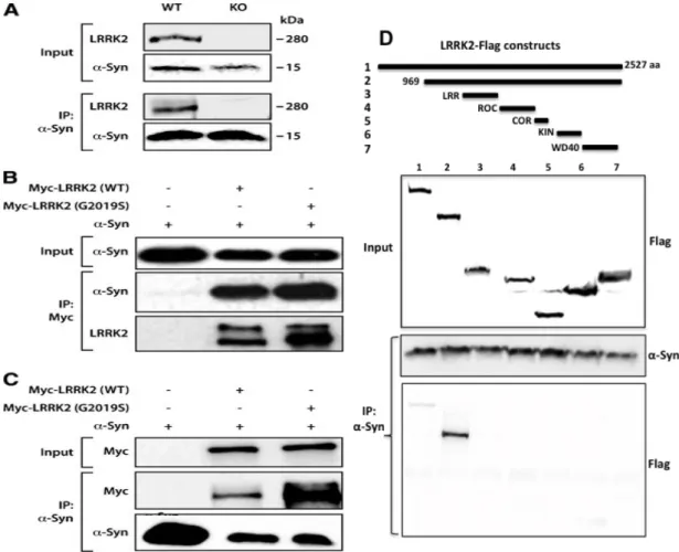
3.3.1 LRRK2 co-immunoprecipitates with α-synuclein ……….. 36
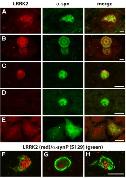
3.3.2 LRRK2 co-localizes with α-synuclein in PD brain and cell model ……… 37
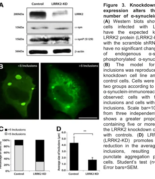
3.3.3 Knocking down LRRK2 expression reduces α-synuclein aggregation ………... 39
3.5 References ………. 46
Chapter 4. LRRK2 promotes Tau accumulation, aggregation and release 4.1 Introduction and main goals ……… 49
4.2 Materials and Methods ………. 50
4.3 Results ……… 54
4.3.1 Tau levels are decreased in LRRK2 knock-out mice ……… 54
4.3.2 LRRK2 physically interacts with Tau ……… 55
4.3.3 Increased levels of Tau depend on LRRK2 expression but not on its kinase activity ………. 56
4.3.4 LRRK2 promotes the accumulation of high-molecular weight Tau species……… 58
4.3.5 LRRK2 impairs proteasomal protein degradation independently of its kinase activity……… 60
4.3.6 LRRK2 impairs the proteasomal degradation of Tau but does not interfere with the autophagy pathway ……… 62
4.3.7 LRRK2 promotes the cellular release of Tau ……….. 62
4.4 Discussion ……….. 64
4.5 References ………. 68
Chapter 5. LRRK2 interactors and their biological significance 5.1 Introduction and main goals ……… 71
5.2 Materials and Methods ……… 71
5.3 Results ……… 75
5.3.1 Network of protein interactions……….. 75
5.3.2 Gene Ontology Analysis ………. 75
5.3.3 The effect of LRRK2 expression on the mechanical properties of the cell……… 81
5.3.4 Different LRRK2 distribution patterns result in different cell stiffness ……….. 81
5.4 Discussion ……….. 84
5.5 References ………. 87
Chapter 6. General discussion and conclusions 6.1 The implications of interaction of LRRK2 with α-synuclein and Tau 90
6.2 LRRK2-interacting protein network and its particular role on cytoskeleton dynamics ……….. 93
6.3 Conclusions ………... 94
6.4 References ………. 96
During my PhD I was exposed to a variety of challenges, developing my passion for working in science and having the opportunity to meet amazing people, who became amazing friends. During this journey, I had the essential support of several people, which allowed me to grow and learn a lot on how to be a scientist and to whom I am extremely greatful.
I would like to thank my supervisor Prof. Dr. Tiago Outeiro, for taking me as a graduate student, for his support since the beginning of my project, for always being available to discuss my ideas and for encouraging me to work hard to achieve my goals. Also, for givinging me the opportunity to join the UNCM, at IMM in Lisbon, where I am proud to have been part of this extraordinary working group. Later on, for supporting my move to his group in Goettingen, allowing me to belong to an outstanding community of scientists.
I am thankful to all the members of UNCM group, that in one way or another helped me during my PhD. A very friendly thank to my colleagues and good friends: Teresa Pais, Leonor Fleming, Ana Oliveira, Oldriska Chutna, Elisa Basso, Sandra Jacinto, Hugo Miranda and Sandra Tenrreiro, with whom I shared fruitful scientific discussions and for the good moments and unconditional support in the lab. Also a special thank to Federico Herrera and Rita Oliveira for being so patient in helping me with the scientific writing, always with a very constructive criticism.
I would also like to thank all the members from the group in Göttingen, for receiving me so well, and having jointly started a new working group. I owe my gratitude to my german colleagues and good friends Ellen Gerhardt, Christiane Fahlbusch, Sonja Reisenauer and Omar Diaz, and to my special collaborator and friend Katrin Eckermann, for all the essential support, for always making my life easier, in and outside the lab, and for making me feel at home in the (sometimes) cold Germany.
A very special thank to my buddy Pauline, with whom I shared my time in Germany, for our long and hard hours in the lab, and also all the good moments outside the lab, and for always being with me in overcoming many Germans hills.
Lastly, I am extremely thankful to all my family for their unconditional support, in particular to João, for always supporting my life decisions, and to Mariana, for helping me to have a four-hand writing of this thesis.
Parkinson’s disease (PD) belongs to the group of neurodegenerative disorders and it is currently considered the most common progressive movement disorder. Neurodegenerative disorders, such as Alzheimer’s, Huntington’s, fronto-temporal dementia and amyotrophic lateral sclerosis, share several dysfunctional molecular pathways and impairments in basic cell mechanisms. Despite intense efforts to understand to decipherthe triggers underlying these disorders, to date, there is no effective cure. This results in a growing number of cases and, consequently, in a complex social and economic problem. Therefore, it is of extreme importance to understand the common biological mechanisms involved in the pathogenesis of this devastating group of diseases, in order to develop effective therapies. The majority of the PD cases are sporadic, however, in the last decades, it has been recognized that rare genetic mutations are patholgical for PD in a number of inherited cases. Futhermore, these mutations can be as well a risk factor for sporadic PD, supporting the idea that familial and sporadic PD can share common pathlogical mechanisms.
This study focused on a key player protein in PD, Leucine-rich repeat kinase 2 (LRRK2). Mutations in LRRK2 gene are the most frequent cause of autosomal dominant forms of PD and they are also consider a risck factor for sporadic cases. A central catalytic GTPase and kinase core, flanked by protein interaction domains, composes this large and complex multi-domain protein. The most frequent LRRK2 PD-related mutation occurs at the animoacid 2019, a glycine subtitution for a serine (G2019S), precisely on the kinase domain of the protein resulting in its toxic gain of function. LRRK2 is known to play a role in distinct cellular mechanisms such as vesicular trafficking, microtubule network regulation and mitochondrial morphology. However, the function of LRRK2 in these important mechanisms and their related pathways is not fully understood, which is crucial for developing new therapeutic targets. Here, we investigated LRRK2 function by characterizing/identifying its protein interactors and, in particular, by exploring its relationship with two central proteins in neurodegenerative disorders, α-synuclein and Tau. In PD brain samples, we show that levels of LRRK2 are positively correlated to an increase in α-synuclein phosphorylation and aggregation in affected brain regions, where both proteins co-localize in neurons and Lewy body
inclusions and knocking down LRRK2 promotes formation of smaller inclusions. Moreover, we show an interaction between α-synuclein and LRRK2 under endogenous and over-expression conditions. These results shed light on the complex interaction of these two central PD proteins and, in particular, on underlying molecular mechanisms involved in a disease scenario. Furthermore, we demonstrate that LRRK2 also interacts with Tau protein in a cell line model, in which co-expression of both proteins promotes accumulation of Tau protein. This accumulation occurs independently of LRRK2 kinase activity and it gives rise to formation of high molecular weight Tau species and increased levels of Tau secretion. Moreover, we suggest that these effects are a consequence of an impairment of proteasomal Tau degradation and that this impairment is promoted by LRRK2. Consistently, a LRRK2-knockout mouse displayed lower levels of Tau in the brain, when compared with transgenic animals expressing human wild-type LRRK2. Our results highlight the compromised status of cellular and molecular neurodegenerative mechanisms. The identification of LRRK2 interactors is crucial to placing the protein in known biochemical pathways. To that end, we performed a screen to identify LRRK2-interacting proteins. The results obtained confirmed that this is a multifaceted protein, involved in a variety of molecular functions and biochemical pathways. α-synuclein and Tau are two proteins present in the list of interactors, which validates previously reported results. The role of LRRK2 on the cytoskeleton is also highlighted by the presence of several protein interactors linked to microtubule dynamics, which lead us to explore the effect of LRRK2 on mechanical properties of the cell. Applying a combined microscopy tecniques in cell indentation experiments, we confirmed that different distribution patterns of LRRK2 result in differential states of cell stiffness. We found that the stiffest cells exhibit a diffuse pattern of LRRK2 distribution, such that LRRK2 is dispersed throughout the entire cell, interacting with microtubule-related proteins and compromising cytoskeletal dynamics. The identification of novel interactos resulted in a better understanding of LRRK2 patho-physiological role.
Taken together, our results presented in this thesis provide novel insight into the function of LRRK2 and its particular role in neurodegenerative diseases. Ultimately, this knowledge is essential for the understanding of the molecular underpinnings of PD and for the development of novel therapeutics.
A doença de Parkinson (DP) pertence ao grupo das doenças neurodegenerativas, sendo atualmente considerada a doença neurodegenerativas motora progressiva mais comum. As doenças neurodegenerativas, como a doença de Alzheimer, a demência frontotemporal ou a esclerose lateral amiotrófica, partilham várias disfuncionalidades em importantes vias de sinalização molecular e mecanismos celulares. Apesar dos esforços desenvolvidos para compreender os factores que estão na origem e na progressão destas doenças, presentemente ainda não foi encontrada uma cura eficaz. O resultante crescente número de casos destas doenças, consequentemente contribui para um complexo problema socioeconómico. É assim de extrema importância identificar os mecanismos biológicos envolvidos na patogénese deste devastador grupo de doenças, a fim de desenvolver terapias eficazes para o combate das mesmas. A maioria dos casos de DP são esporádicos, no entanto nas últimas décadas têm sido identificadas várias mutações genéticas ligadas a casos hereditários. Estas mutações podem ainda ser consideradas um factor de risco para o desenvolvimento de casos esporádicos da DP, o que suporta a ideia que os casos hereditários e esporádicos partilham os mesmos mecanismos patológicos.
Este estudo foca-se numa proteína chave na DP, Leucine-rich repeat kinase
2 (LRRK2). Mutações na proteína LRRK2 são consideradas a causa mais
frequente em casos autossómicos dominantes da doença, ocorrendo também em casos esporádicos. Esta grande e complexa proteína com múltiplos domínios, é composta por um núcleo catalítico central de GTPase e quinase, flanqueado por vários domínios de interação proteica. A mutação mais frequente em LRRK2 é a substituição de uma glicina por uma serina, que ocorre no aminoácido 2019 (G2019S). Esta mutação localiza-se precisamente no domínio da quinase da proteína, promovendo um tóxico ganho de função da mesma. É conhecido o envolvimento de LRRK2 em distintos mecanismos celulares como o tráfego vesicular, regulação da rede de microtúbulos e morfologia mitocondrial. No entanto, não é completamente conhecido o papel de LRRK2 nestes importantes mecanismos e suas vias de sinalização, o que é crucial para o desenvolvimento de novos alvos terapêuticos. Neste trabalho investigamos a função de LRRK2 através da caracterização/identificação de proteínas interatuantes, em particular
neurodegenerativas, alpha-sinucleína (α-sinucleína) e Tau. Em amostras de cérebro de pacientes com DP, mostramos que os níveis de LRRK2 são positivamente corelacionados com um aumento de fosforilaçao e agregação de α-sinucleína fosforilada e agregada, nas regiões do cérebro mais afectadas. Também nas regiões de cérebro mais afectadas, se verifica uma co-localização destas duas proteínas em neurónios e em inclusões de corpos de Lewy. Num modelo de linha celular, esta co-localização também ocorre em inclusões de α-sinucleína, onde o knockdown de LRRK2 promove a formação de inclusões mais pequenas. A interação entre α-sinucleína e LRRK2 é ainda confirmada em condições endógenas e de sobre-expressão. Estes resultados contribuem para uma melhor compreensão sobre a complexa interação destas duas proteínas centrais na DP, em particular sobre os mecanismos moleculares subjacentes, envolvidos num cenário de doença. Em seguida e usando um modelo celular, demostramos que a LRRK2 interatua com a Tau, sendo que a co-expressão destas proteínas promove uma acumulação de Tau. Esta acumulação ocorre independentemente da atividade de quinase da LRRK2, e promove a formação de espécies de Tau com elevado peso molecular, bem como um aumento de secreção de Tau. Estes efeitos serão a consequência de uma falha ao nível da degradação de Tau pelo proteassoma, que por sua vez será promovida pela LRRK2. Em cérebros de ratinhos knockout para LRRK2, verifica-se um decréscimo dos níveis de Tau, quando comparado com animais transgénicos para LRRK2 humana. Estes resultados realçam a disfunção de mecanismos celulares e moleculares, envolvidos nas doenças neurodegenerativas. A identificação de proteínas interatuantes com LRRK2 é crucial para posicionar esta proteína nas conhecidas vias de sinalização bioquímica. Com este objectivo, desenvolvemos um screen para identificar novas proteínas interatuantes com LRRK2. Os resultados obtidos confirmam que esta é uma proteína multifacetada, envolvida em várias funções moleculares e vias de sinalização bioquímicas. A presença de α-sinucleína e Tau nesta lista de proteínas interatuantes, vem validar os resultados acima descritos. Também a presença de várias proteínas relacionadas com a dinâmica de microtúbulos, vem realçar o papel de LRRK2 ao nível do citoesqueleto celular, o que nos levou a explorar o efeito de LRRK2 nas propriedades mecânicas das células. Aplicando uma técnica combinada em
LRRK2, resultam em diferentes estados de rigidez celular. Descobrimos que as células com maior rigidez são as que exibem um padrão difuso de distribuição de LRRK2, onde a proteína está dispersa por toda a célula, interagindo com proteínas relacionadas com os microtúbulos, comprometendo assim a dinâmica do citoesqueleto. A identificação de novas proteínas interatuantes resulta num melhor conhecimento da função pato-fisiológica de LRRK2. Em resumo, os resultados apresentados nesta tese, fornecem novos conhecimentos sobre as funções da LRRK2 e o seu particular papel nas doenças neurodegenerativas. Por fim, estes conhecimentos são essenciais para a compreensão das bases moleculares da DP e consequentemente para o desenvolvimento de novas terapêuticas.
Palavras-chave: LRRK2, Doença de Parkinson, α-sinucleína, Tau, interação proteica.
AD - Alzheimer’s disease AFM - atomic force microscopy ALP- autophagy-lysosome pathway ALS- amyotrophic lateral sclerosis
BiFC- bimolecular fluorescence complementation (assay) CMA - chaperone mediated autophagy
DA - dopamine
FTD- fronto-temporal dementia GO - gene ontology
GWAS- genome wide association studies HD- Huntington’s disease
HMW - high molecular weight
L-dopa - L-3,4-dihydroxyphenylalanine LBs - Lewy bodies
LNs - Lewy neurites
LRRK2 - leucine-rich repeat kinase 2 MAPT- microtubule-associated protein tau
MPTP- 1-methyl-4-phenyl-1,2,3,6-tetrahydropyridine mRNA - messenger RNA
MS - mass spectometry NFTs- neurofibrillary tangles PD - Parkinson’s disease
SEC- size exclusion chromatography shRNA - small hairpin RNA
SNCA - alpha-synuclein gene
SNpc - substantia nigra pars compacts
TIRFM- Total Internal Reflection Fluorescence Microscope UPS- ubiquitin proteasome system
Chapter 1.
Chapter 1. General Introduction
1. The neurodegenerative disorder of Parkinson’s disease
The earliest records about Parkinson’s disease date back to 1817 when a British surgeon, James Parkinson, described in the monograph “An Essay on the Shaking Palsy”, the first observations of patients affected by a disease he called “paralysis agitans”. Years later, the terminology was updated by a French neurologist, Jean-Martin Charcot, that in memory of James Parkinson, named this disorder Parkinson’s disease (PD).
PD is the second most common progressive neurodegenerative disorder, affecting 1-2% of people over 65 years old and 4-5% above the age of 85. The onset of PD is intimately related with age, which increases the expected number of cases due to an increased lifespan of the population in developed countries [1,2].
Almost two centuries after the first descriptions of PD, and despite intense research efforts in the field, there is still no effective cure and our knowledge about the etiology and the development of this disease is still incomplete.
The pathogenic mechanisms of PD, do not seem exclusive of this disease but actually share several common features with other neurodegenerative diseases like Alzheimer’s (AD), Huntington’s, fronto-temporal dementia and amyotrophic lateral sclerosis. Although these diseases present different features and hallmarks, they share several dysfunctional molecular pathways and impairments in basic cell mechanisms. Thus, it is very important to deeply investigate these common molecular pathways to determine the function of the key proteins. This cross knowledge is crucial to discover the basis of neurodegenerative mechanisms common to several dramatic diseases for targeting efficient therapies.
1.1 Clinical symptoms and pathogenesis of PD
PD is typically known for its characteristic motor symptoms such as resting tremor, bradykinesia, muscle rigidity and postural instability. Before the appearance of the first motor symptoms and the final diagnosis of the disease, there is already cognitive deterioration accompanied by pre-motor symptoms like
mood disturbances, rapid-eye-movement, sleep disorder, loss of smell (hyposmia) and depression [3,4].
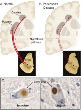
The described clinical symptoms result from an increasing and selective loss of dopaminergic neurons from a particular midbrain region, the substantia
nigra pars compacta, and consequently a massive depletion of striatal dopamine,
an extremely important neurotransmitter (Figure 1A and 1B). Another typical pathological hallmark of PD, described in the early nineties by Friederich H. Lewy, is the presence of cytoplasmic protein-containing inclusions in the surviving dopaminergic neurons called Lewy bodies (LBs) (Figure 1C). LBs are agglomerates of several proteins, with α-synuclein being one of the major components and mostly present in the phosphorylated and fibrillar forms [3,5,6]. The presence of LBs is not only restricted to the dopaminergic neurons, they appear in other areas of the brain, such as spinal cord. Although considered a hallmark of PD, the LBs are also found in other neurodegenerative diseases like AD and Lewy body disease or even in healthy aged brains [7-9]. Curiously, patients with autosomal recessive forms of PD, in particular with mutations in the
PARKIN gene, do not present LBs inclusions [10]. Despite the extensive research
in the field, the question whether LBs confer toxicity or on the other hand are protective to the cells, by sequestering potential toxic species, is a hot topic for debate without a consensual answer [8,11,12].
Figure 1. Neuropathology of PD.
Representation of a healthy brain (A) versus a PD brain (B). Highlighted in red are the compromised nigrostrital pathways and the depigmentation (three arrows), due to the loss of melanine present in the dying dopaminergic neurons, that projects to the striatum (putamen and caudate nucleus). (C) Detail of a Lewy body inclusion in dopaminergic neurons, showing the Immunoreactivity against α-synuclein and ubiquitin [6].
To date, there is no effective treatment to cure PD or stop the progressive degeneration of the dopaminergic neurons. While gene and stem cell therapies are being heavily studied and not yet available, the gold standart for the treatment of PD continues to be the pharmacological approached introduced in the eighties, aimed at amelioreating the motos symptoms of the disease. This therapy consists in the replacement of the depleted striatal dopamine by the administration of a dopamine percursor, L-3,4-dihydroxyphenylalamine, usually known as levodopa (L-dopa). Although it is considered an effective drug therapy for the motor symptoms, especialy when administered in early stages of the disease, L-dopa is not suitable for all PD cases and long-term treatment can promote severe side effects [13-15].
Currently, PD is no longer exclusively considered a disorder of the dopaminergic neurons of the substantia nigra. Although its onset and progression are still unclear, the disease is thought to happen in different brain regions and even outside the nervous system [7,15,16]. The progressive and cumulative symptoms confirm that this is a multisystem disorder that needs to be approached from a broad perspective.
1.2 Etiology of PD
PD is a typical late onset disorder and most cases (90-95%) occur sporadically, without a defined cause or relation with patient’s life style, characterizing it as an idiopathic disease. It is consensual that the major risk factor for PD is ageing. However, initial finding linked the disease with environmental factors, such as the chronic exposure to several neurotoxic pesticides. Later on, in the late twenties, the discovery of rare familial genetic mutation linked with PD, brought a new perspective for the research of this disease. These findings leaded to the development of a variety of animal models, in attempt to elucidate the molecular pathogenesis of the disease [6,17]. Despite the increasing knowledge about the etiology and the development steps of PD, resulting from the intensive research in the field, the underlying molecular mechanisms of the disease are still not completely understood.
Nowadays, there is solid evidence that a synergistic combination of environmental and genetic factors is determinant to the onset and development of the disease.
1.2.1 Environmental factors
The first identified environmental factors for the development of PD was the continuous exposure to neurotoxins and chemical substances present in several pesticides. 6-hydroxydopamine was one of the first neurotoxins identified. It promotes a selective degeneration of the catecholaminergic neurons due to an increase in reactive oxygen species [18]. Another neurotoxin, MPTP (1-methyl-4-phenyl-1,2,3,6-tetrahydropyridine), was identified in a synthetic drug consumed by a group of drug addicts who started to develop typical parkinsonism symptoms. This neurotoxin crosses the blood brain barrier and is converted into MPP+ in glial cells, which then displays selective toxicity in dopaminergic neurons [19]. Also, the chronic exposure to pesticides widely used in agriculture, like rotenone and paraquat, were found to be a potential risk factor for PD. Rotenone inhibits the mitochondrial complex I promoting degeneration of nigral-striatal neurons. Paraquat directly crosses the blood brain barrier, contributes for an increase of reactive oxygen species, which lead to degeneration in dopaminergic neurons [20, 21]. The knowledge about these neurotoxic chemicals was used to develop the first models of PD, which continue to be a valuable tool available for mimicking parkinsonism symptoms and consequently to test new therapeutics for impact on the disease [18].
Interestingly, there are some studies suggest that nicotine and caffeine contribute for a decrease in the incidence of PD [22], but these will not be explored further in this document, as it would be out of the scope.
1.2.2 Genetic factors
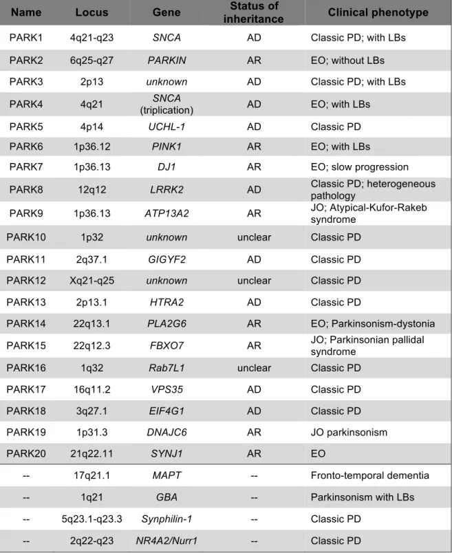
In the last two decades, genome-wide studies in several populations identified a growing number of genes associated with familial PD, reinforcing the importance of genetics as a risk factor for the disease [23-25]. The regions of the genome to which these genes map are known as the PARK loci. In addition, several genes associated with non-familial forms of PD were identified (Table 1). The PARK loci correspond to genes that might be divided into two large groups: genes responsible for autosomal dominant or autosomal recessive forms of the disease [26,27].
Table 1. Genes and loci associated PD associated genes.
AD: autosomal dominant; AR: autosomal recessive; LBs: Lewy bodies; JO: juvenile onset (age< 20); EO: early onset (age 20-40); Classic PD has a late onset (age >40). Adapted from [26] and [28].
Name Locus Gene inheritance Status of Clinical phenotype
PARK1 4q21-q23 SNCA AD Classic PD; with LBs PARK2 6q25-q27 PARKIN AR EO; without LBs PARK3 2p13 unknown AD Classic PD; with LBs
PARK4 4q21 SNCA
(triplication) AD EO; with LBs
PARK5 4p14 UCHL-1 AD Classic PD
PARK6 1p36.12 PINK1 AR EO; with LBs
PARK7 1p36.13 DJ1 AR EO; slow progression
PARK8 12q12 LRRK2 AD Classic PD; heterogeneous pathology
PARK9 1p36.13 ATP13A2 AR JO; Atypical-Kufor-Rakeb syndrome
PARK10 1p32 unknown unclear Classic PD
PARK11 2q37.1 GIGYF2 AD Classic PD
PARK12 Xq21-q25 unknown unclear Classic PD
PARK13 2p13.1 HTRA2 AD Classic PD
PARK14 22q13.1 PLA2G6 AR EO; Parkinsonism-dystonia PARK15 22q12.3 FBXO7 AR JO; Parkinsonian pallidal
syndrome
PARK16 1q32 Rab7L1 unclear Classic PD
PARK17 16q11.2 VPS35 AD Classic PD
PARK18 3q27.1 EIF4G1 AD Classic PD
PARK19 1p31.3 DNAJC6 AR JO parkinsonism
PARK20 21q22.11 SYNJ1 AR EO
-- 17q21.1 MAPT -- Fronto-temporal dementia
-- 1q21 GBA -- Parkinsonism with LBs
-- 5q23.1-q23.3 Synphilin-1 -- Classic PD -- 2q22-q23 NR4A2/Nurr1 -- Classic PD
SNCA was the first gene associated with familial forms of PD. Although rare,
missense mutations in SNCA such as A53T, A30P and E46K as well as duplications and triplications, are found in familial cases of PD [29-31]. New studies are constantly updating the SNCA mutations related with PD, and the most
recent ones described are: A53E, H50Q and G51D [32-35]. Also several polymorphisms in the SNCA gene can constitute risk factors for the development of sporadic PD [36,37].
Mutations in the LRRK2 gene are the most frequent cause of familial PD, although it is also associated with sporadic cases [38,39]. Several point mutations were identified within the multiple domains of the protein, however the G2019S substitution, in the catalytic core of the protein, is by far the most frequently identified [38].
PARKIN, PINK1, DJ-1 and ATP13A2 are associated with autosomal
recessive forms of PD, usually linked with juvenile and early-onset cases of the disease [27,40,41]. PARKIN mutations are responsible for the majority of the autosomal recessive PD cases, which interestingly are characterized by the absence of LBs [10].
Although familial mutations are rare, present in only up to 10% of the cases, it is known that both familial and sporadic forms of PD share common pathogenic mechanisms, which are still not clear. Studying these mutated genes and their respective encoded proteins, to know more about their physiological functions and their dysfunction in PD, is crucial to identify molecular pathways that might be used as targets for therapeutic intervention.
1.3 α-synuclein and PD
The α-synuclein protein is the main component of LBs and LNs, present in different synucleinopathies. Importantly, mutations and multiplications in the SNCA gene (which encodes for α-synuclein) were the first known genetic causes associated with familial forms of PD [42, 43]. These facts make this protein a central player in PD and one of the most studied proteins in the field.
α-synuclein is an abundant protein in the brain and it is particularly enriched in pre-synaptic terminals. Although its function is not completely understood, it appears to be involved in several cellular processes like synaptic activity, vesicle recycling and, as a chaperone it is involved in the formation of SNARE complexes [44, 45]. This small (140 amino acids), thermostable and natively unfolded protein is structurally composed of three different domains. The N-terminal region is usually unstructured in solution, forming amphipathic α-helices when it interacts with phospholipid membranes. The central non-amyloid-β component (NAC) is a
highly hydrophobic region, responsible for the amyloidogenic properties of the protein and the formation of β-sheet structures, which consequently potentiate aggregation. The C-terminus is a highly acidic region, and possibly responsible for the chaperone activity of the protein [46-48].
The first identified familial SNCA mutation was the A53T substitution, followed by two other point mutations A30P and E46K, all promoting early onset of the disease with an extremely aggressive progression [29-31]. While these mutations are considered rare, the duplications and triplications of wild-type SNCA are more prevalent and directly toxic via higher expression of the protein [36,37]. More studies are required to better characterize the recently described SNCA mutations (A53E, H50Q and G51D), which interestingly seem to be related with the aggregation capacity of the protein [32-35].
Familial mutations, multiplications, polymorphisms and post-translational modifications (particularly phosphorylation) of the protein are considered key factors contributing to α-synuclein accumulation and subsequent aggregation. As α-synuclein is such a predominant component of LBs, the mechanism through which this protein leads to aggregation and confers effects on onset and development of neurodegeneration, has been extensively studied [49-53]. Though α-synuclein is a monomeric and unfolded protein, its central hydrophobic region (NAC) has a tendency to oligomerize [54]. Briefly, the α-synuclein aggregation processes initiates with the formation of dimers that, due to a continuous oligomerization propensity, evolve into bigger oligomers, followed by protofibrils and amyloid fibrils, which ultimately are deposited in LBs. This aggregation process occurs together with neuronal dysfunction. However, there is an intense debate regarding which are the most toxic species formed along this pathway that contributes to an increase in cell toxicity, culminating in neuronal death [52-55]. To better understand this toxic oligomerization and aggregation process occurring in PD, it is also important to consider the relevance of several α-synuclein-interacting proteins. Synphilin-1 is a protein that co-localizes with α-synuclein in the LBs and is described to contribute to α-synuclein aggregation [56-58]. Some isoforms from the 14-3-3 chaperone-like protein family interact with genetic PD-associated proteins, including α-synuclein, and are present in the LBs from human PD patients [58-60].
and aggregation and its role in PD, this is not entirely known. This will be crucial to identifying therapeutic targets in an early phase, avoiding a massive neuronal death and disease progression.
1.4 Association of Tau with PD
The Tau protein is encoded by the microtubule-associated protein tau (MAPT) gene. There are six isoforms of the protein generated by alternative splicing [61]. This predominantly neuronal protein is highly expressed in the adult central nervous system, where it binds to and stabilizes microtubules. Tau interaction/stabilization with microtubules occurs through the C-terminal of the protein and is regulated by phosphorylation of specific epitopes, some of which have been described as pathogenic in some neurodegenerative disorders like AD and PD [62-64].
The Tau protein was initially related to AD, being one of the main components of neurofibrillary tangles, a defined pathological hallmark of the disease, together with extracellular plaques of amyloid-β peptides [65]. Frontotemporal dementia with parkinsonism linked to chromosome 17 (FTDP-17) was the first neurodegenerative disorder associated with Tau mutations, therefore belonging to the group of tauopathies [66]. Common to tauopathies, is the presence of hyperphosphorylated and insoluble aggregated forms of Tau, observed inside neuronal cells in different brain regions [67,68].
Several MAPT mutations were described to affect the ability of Tau to bind to microtubules and to increase its aggregation propensity. Moreover these mutations were recently confirmed by GWAS as a risk factor for PD [66, 69]. Further insight into the role of Tau as a key player in PD, can be gained from understanding how this protein interacts with the other two central PD proteins: α-synuclein and LRRK2.
The link between Tau and α-synuclein was first highlighted through the co-occurrence of these proteins in insoluble protein deposits in PD and AD brains [70-73]. In addition, it was shown that the majority of AD cases display some α-synuclein enriched LBs and Tau tangles can, as well, be identified in PD brains [74-77]. In vivo studies confirmed the presence of hyper-phosphorylated Tau species in a mouse model of over-expressed α-synuclein [78]. In a Drosophila model of PD, the interaction between these two proteins resulted in the disruption
of cytoskeletal organization and increased neurotoxicity [79]. Moreover, in vitro studies show that α-synuclein and Tau are able to influence each other’s polymerization. In cell models, both proteins interact through GSK-3beta and Tau enhances α-synuclein aggregation and toxicity [80-82].
The link between Tau and LRRK2 is another interesting point that correlates Tau with PD. Being the most frequent cause of the autosomal dominant form of PD, LRRK2 cases are characterized by a pleomorphic pathology including LBs, LNs and Tau tangles [83,84]. Initial studies in a mutated LRRK2 mouse, showed an extensive Tau tangle pathology, and Tau positive axonal swellings were observed in rat neuronal cultures overexpressing a fragment of LRRK2 [85]. Years later, LRRK2 as a kinase, was suggested to phosphorylate Tau in a tubulin-dependent manner [86]. Also in a mouse model of tauopathy, LRRK2 expression results in increased Tau aggregation and phosphorylation of different residues [87]. More recently in vitro studies demonstrate that this phosphorylation happens in the presence of tubulin and indirectly via GSK-3β [88].
1.5 Leucine-Rich Repeat Kinase 2: a key player in PD
The human LRRK2 gene is located on chromosome 12 and encodes for a large protein named Leucine-rich repeat kinase (LRRK2). LRRK2 is also known as Dardarin, from the Basque word dardara (tremor), although this term is less used. The first LRRK2 mutations associated with PD were reported in 2004 and are currently considered the most common genetic cause of PD, being responsible for a high number (5-15%) of all familial cases [83,84]. Only a few mutations, concentrated at the enzymatic domains of the protein, segregate with familial disease. Importantly, there are several LRRK2 variants reported in all other domains of the protein, which are a risk factor for sporadic PD [89-90]. The G2019S mutation is the most frequently found in PD patients, and results in a kinase gain of function of the protein [91-93].
Clinically, the symptomatology of LRRK2-related PD cases is indistinguishable from sporadic cases, presenting an average late onset with a slower progression and not frequently associated with dementia. The LRRK2-PD cases are characterized by a pleomorphic neuropathology with the presence of pure classical nigral neuronal degeneration or LBs, LNs and positive ubiquitin and Tau phosphorylated inclusions [94-95].
The clinical and pathological similarity of LRRK2-related familial cases to sporadic cases, together with the identification of LRRK2 mutations as a common risk factor for sporadic PD cases, qualifies this protein as a candidate in bridging the gap between inherited and sporadic PD. Therefore, LRRK2 represents an extremely important target for research, with the potential to uncover the common mechanisms of familial and sporadic PD.
1.5.1 Functional structure of LRRK2 protein
LRRK2 is a multi-domain and large (2527 amino-acid) protein of approximately 285 kDa, expressed in various tissues including the brain. This multifunctional protein belongs to the Roco protein family, which is characterized by having a conserved domain containing a Ras-like GTPase domain, called ROC, and a characteristic COR domain (C-terminal of ROC). Neighbouring this GTPase domain is a serine/threonine kinase domain (Kinase), and together these two domains compose the central enzymatic core of LRRK2. Flanking these central core, the protein has additional protein-protein interaction domains such as ankirin-like repeats (ANK), leucine-rich repeats (LRRs) at N-terminal and a β-propeller-like domain (WD40) at C-terminal (Figure 2) [38, 96].
As a GTPase, LRRK2 binds GTP through its ROC domain, leading to a change of conformation and facilitating GTP hydrolysis. However, this GTPase activity is weaker when compared to other members of the Ras related GTPase family [97]. Several pathogenic mutations were found in the GTPase domain, located within the ROC (R1441G/C/H) and the COR (Y1699C) subdomains, which were associated with decreased GTPase enzymatic activity of the protein [98,99].
The kinase domain of LRRK2 shares a high similarity with mixed-lineage kinases (MLKs) and receptor-interacting protein kinases (RIPKs) [100]. Some of the studies to evaluate the kinase activity of LRRK2 are based on its capacity to phosphorylate myelin basic proteins (MBPs) like moesin and pseudo-substrate (single peptides or proteins) [101]. However, the most widely used assays to evaluate LRRK2 activity rely on its autophosphorylation capacity [102,103]. The most frequently reported mutation, G2019S, occurs precisely in the activation segment of the kinase domain and is responsible for a 2-3 fold gain-of-function of the protein. This point mutation is thought to interfere with the activated “ON-OFF” state of the kinase, due to the negative charge of the serine residue which
compromises the structural flexibility, prolonging the activated state of the kinase [104-105].
Figure 2. Domain organization of LRRK2 and cellular pathways associated with its function.
Schematic representation of LRRK2 structural domains, highlighting the functional dimeric conformation of the protein. The protein has a central enzymatic core composed by a GTPase domain (ROC) and its C-terminal (COR), together with a serine/threonine kinase domain (Kinase). Flanking these central core, the protein has several protein-protein
interaction domains; at N-terminal an ankirin-like repeats (ANK) and leucin-rich repeats (LRRs) and at C-terminal a β-propeller-like domain (WD40). In the central core of the protein are placed the most frequent and pathogenic mutations thought to be responsible for a decrease in GTPase activity and an increase of kinase activity. The study of LRRK2 mutations has shed light on the protein function and on the cellular mechanisms where it could be involved in a PD scenario, ultimately contributing to neuronal damage. Adapted from [96].
1.5.2 LRRK2 mutations and PD
Since the discovery of first LRRK2 mutations related with the autosomal dominant forms of PD, several other mutations have been identified within the multiple domains of the protein. Thus far, more than 40 LRRK2 variants have been reported, however only seven mutations are considered pathogenic: N1437H, R1441C/G/H, Y1669C, G2019S and I2020T [89,88].
N" " C" " ANK LRRs ROC
GTPase COR Kinase WD40
R1441G/C/H Y1699C G2019S I2020T Decreased GTPase activity Increased kinase activity Vesicular trafficking
(synaptic vesicles) Protein translation/ degradation Autophagy Apoptosis Neuronal damage Mitochodrial morphology & activity
Microtubule network regulation - Cytoskeleton - Neurite outgrowth 1" 2527"aa" LRRs COR Kinase WD40 Altered DA neurotransmission N1437H!! 713"ANK 1035" 1335" 1879" 2168" ROC GTPase
Among the LRRK2 mutations identified, G2019S is located in the kinase domain and promotes its gain of function, confirmed by increased phosphorylation of LRRK2 and known generic substrates [102]. G2019S is the most frequent mutation found in PD patients, being responsible for up to 7% of the familial cases and also 1-3% of sporadic cases worldwide [107]. The frequency of the G2019S mutation varies among different populations across the globe, being particularly high in genetically isolated populations where it can account for up to 40% of total PD cases [108]. A study with data collected from over 133 families, reported a higher occurrence of the G2019S mutation in southern than northern European countries [89]. The age-dependent penetrance of the G2019S mutation, results in a probability of disease onset of 28% at age 59, rising up to 74% at age 79 [107, 109, 110]. Overall, the high but incomplete penetrance of G2019S mutation results in the existence of some carriers of the mutation, who do not develop PD in their lifetime [111, 112].
Neighbouring the G2019S, is the I2020T mutation, which seems to have a very modest effects on kinase activity of the protein, so the relationship of this mutation with the LRRK2 biochemical activities is unclear [113].
The three mutations, reported in the “hotspot” R1441 (R1414G/H/C), make this the second most common site of pathogenic LRRK2 substitutions. The R1441C mutation was initially founded in two autosomal dominant PD families. Although R1441C is found in different populations, R1414G is particularly common in the Basque region of Spain and R14141H was only found in four individual from diverse ethnicities [114]. These three mutations in the GTPase domain of LRRK2 are generally associated with a decreased GTPase activity [115, 116]. In the COR domain, a tyrosine to cysteine or guanidine mutation (Y1699C) was reported in one family from UK and other family with German heritage [99]. These mutations present a highly variable penetrance and clinical features resemble the idiopathic PD cases, with a late onset of the disease.
The N1437H mutation, in the COR domain of LRRK2, is the most recently identified mutation, in a large Norwegian family, and curiously it presents a very young age of onset (approximately 48 year old) [117]. Interestingly other two LRRK2 polymorphisms (G2385R and R1628P), which are almost absent in Caucasians, represent almost 10% risk for sporadic PD in Asian populations [118, 119].
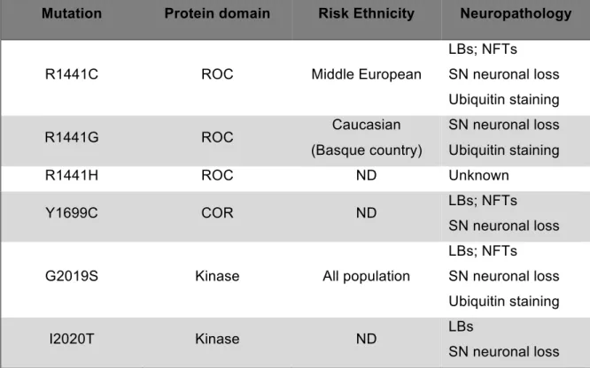
1.5.3 Neuropathology of LRRK2 mutation in PD
The different LRRK2 mutations promote a wide spectrum of neuropathology features, resumed in table 2 [107]. The post-mortem analysis of LRRK2-associated PD patients, revealed a pleomorphic neuropathology LRRK2-associated with different LRRK2 mutations and even within the same mutation. Indeed, the analysis of LRRK2-associated PD brains revealed a variety of pathological features consisting in a pure nigral neuronal degeneration, similar to classical PD cases, together with typical LBs and LNs [83,120,121]. There are also some cases were LBs and LNs are absent, but instead is displayed hyperphosphorylated or ubiquitinated single proteins, in particular Tau, indicative of frontotemporal dementia [122].
The presence of LBs is the most typical and widely spread pathological feature in LRRK2-associated PD cases, occurring in the brainstem, cortex and limbic system. Indeed, LRRK2 patients who bear the most frequent mutation (G2019S) mainly exhibit α-synuclein positive LBs pathology, which is a typical feature of idiopathic PD [107].
Table 2. Pathological features of LRRK2 mutations.
Table representing the pleomorphic neuropathological features associated to the most frequent PD-LRRK2 mutations. Adapted from Lie J., et al, 2014 [107].
Mutation Protein domain Risk Ethnicity Neuropathology
R1441C ROC Middle European
LBs; NFTs SN neuronal loss Ubiquitin staining R1441G ROC Caucasian (Basque country) SN neuronal loss Ubiquitin staining R1441H ROC ND Unknown Y1699C COR ND LBs; NFTs SN neuronal loss
G2019S Kinase All population
LBs; NFTs SN neuronal loss Ubiquitin staining
I2020T Kinase ND LBs
SN neuronal loss
The wide variety of pathological features associated with different LRRK2 mutations, demonstrate that this protein influences different mechanisms of cell viability. The effect of LRRK2 mutations may still be conditioned by genetic variations of other loci such as MAPT or SNCA, which encodes for Tau and α-synuclein, two other central proteins in neurodegeneration. The fact that LBs inclusions are a pathological feature common to LRRK2-related familial PD cases and sporadic cases, also highlight the importance of LRRK2 as a crucial protein to bridge the common features between idiopathic and familial PD pathology [112].
However, the characterization of LRRK2-associated neuropathology is still dependent on the systematic evaluation of large number of LRRK2 PD patients and respective families, in different populations.
1.5.4 Interplay between GTPase and Kinase domains
The central catalytic unit of LRRK2 confers to the protein a particular dual enzymatic activity: GTPase and kinase. Several lines of evidence suggest a potential intrinsic regulatory mechanism between these two domains [123-125]. Interestingly, it is also in this ROC-COR-kinase catalytic core, where the most pathogenic and frequently observed LRRK2 mutations are found. In particular, the G2019S mutation at the kinase domain of LRRK2 is the most frequent mutation related with autosomal dominant forms of PD. The biological consequence of this glycine substitution for a serine has been extensively investigated, and the consensus is that it promotes an exacerbated increase in the kinase activity of the protein, resulting in neurological toxicity [126, 127]. Still, in the same kinase domain, the effect of the I2020T mutation is not well established. Contradictory results showing that this mutation promotes an increase, decrease or even a null effect on the kinase activity of the protein have been reported [128, 129]. The ROC domain of LRRK2, houses three different substitutions in the Arg1441 residue, R1441C/G/H, which appear to affect GTPase efficiency, leading to a decreased ability of LRRK2 to hydrolyze GTP [130-132]. These mutations are thought to alter the folding properties of the protein and, consequently, interfere with the known ability of LRRK2 to dimerise. This occurs due to the particular location of the R1441 residue, which might be responsible for disrupting the hydrogen bond between two GTPase domains [133, 134]. More recently, the Y699C mutation at the COR domain of LRRK2 has also been shown to affect the GTPase activity of
the protein [99]. This mutation causes a decrease in GTP hydrolysis by strengthening the interaction between the ROC-COR domains, which leads to a weaker bond between LRRK2 monomers. The described alterations on the GTPase activity of LRRK2 will also subsequently modulate the downstream kinase activity of the protein, in particular, by stimulating LRRK2 autophosphorylation [131,133,134]. Interestingly two mutations in the ROC domain of the protein, K1347A and T1348N, were described to totally abolish its kinase activity [134]. Moreover, the N and C-terminal of LRRK2 were suggested to act as modulators of its kinase activity. Thus, the N-terminus is suggested to have an inhibitory effect on kinase activity, while the C-terminal tail is required for full kinase activity [129, 135]. Summing up, there are several LRRK2 mutations located within the GTPase and kinase domains of the protein, which reciprocally condition the double enzymatic activity of the protein and consequently, the downstream events. Therefore, the modulation of LRRK2 GTPase and kinase enzymatic activity is an appealing candidate for therapeutic intervention, aiming at preventing LRRK2-dependent neuronal toxicity and progression of the disease [136-138].
1.5.5 LRRK2 interacting proteins and putative function
The complex multi-domain structure of LRRK2 encompasses a central GTPase and kinase core, flanked by additional putative protein interacting domains. These last domains suggest that LRRK2 may act as a scaffold protein involved in several signalling pathways and protein complexes.
A number of in vitro screens revealed several potential LRRK2 substrates and/or interactor proteins, however their validation and biological meaning in in
vivo models is not frequently achieved. To date, there are some identified and
confirmed LRRK2 interactors that help to build a picture about the putative role of this complex multifunctional protein in several pathways and cellular mechanisms [139-142].
The potential role of LRRK2 in the cytoskeleton architecture and microtubule network dynamics was initially reported by the identification of ezrin/radixin/moesin, and β-tubulin, as LRRK2 substrates [101,143]. More recently, LRRK2 was reported to phosphorylate tubulin-associated Tau at the Thyrosine 181 residue, which may regulate neurite outgrowth, by promoting neurite retraction [86]. In the present thesis, we explored the biological
consequences of this interaction and proposed a mechanism through which LRRK2 regulates intracellular levels and Tau biochemical species by compromising Tau-proteasomal degradation (fully explored in Chapter 4).
The structural similarity of LRRK2 with MLKs as well as the in vitro results showing its capacity to phosphorylate MKK3/6 and 4/7, suggest that this protein is upstream of the MAP kinase pathways. This idea is strongly supported by the in
vivo results obtained in LRRK2 transgenic mice, where hyper-phosphorylation of
MKK4 by the mutant G2019S, activates the MKK4-JNK-c-Jun pathway, leading to degeneration of dopaminergic neurons [144]. Studies in a Drosophila
melanogaster revealed that LRRK2 phosphorylates the transcription factor FoxO1
at Ser319. It is known that this phosphorylation promotes an enhancement of transcriptional activity, triggering a cascade of mechanisms, like oxidative stress and programed cell death. However, other direct downstream targets are poorly identified [145].
The ADP-ribosylation factor GTPase-activating protein 1 (ArfGAP1) was also identified as a robust substrate of LRRK2. The two proteins interact in vivo in the brain and co-localize at Golgi membranes. ArfGAP1 regulates LRRK2 GTPase activity and thereby modulate its kinase activity. Moreover, in primary cortical neurons, silencing of ArfGAP1 expression rescues the neurite shortening phenotype induced by LRRK2-G2019S, and neurite shortening induced by ArfGAP1 overexpression is also attenuated by silencing of LRRK2 [146].
The role of LRRK2 in the synaptic environment was highlighted by the discovery of EndophilinA (EndoA) as a LRRK2 substrate. EndoA is a crucial protein involved in the vesicle formation during the endocytosis process [147]. When LRRK2 is phosphorylated at serine 75, it inhibits the role of EndoA on membrane tubulation, increasing its affinity to bind to membranes and so compromising synaptic vesicle endocytosis [148]. α-synuclein is another synaptic protein that was initially reported to be a LRRK2 substrate [149]. Although this idea was very appealing, the biological meaning of this interaction was not further explored. More recently, we reported that α-synuclein interacts LRRK2 [150]. In Chapter 3, we fully explore how LRRK2 models α-synuclein aggregation pattern in
1.6 Cellular Quality Control Systems
The misfolding and accumulation of certain proteins is a pathology hallmark common to several neurodegenerative disorders, where it could act as an underlying cause of the disease mechanism [151]. In fact, the accumulation of misfolded proteins leads to the formation of intermediate oligomeric species and, ultimately, protein aggregates. Whether these protein aggregates promote a toxic or a defensive effect on the cells remains a topic of intense debate in the field [152]. Neurons, as post-mitotic cells with a high metabolic activity, are extremely sensitive to protein accumulation. Thus, the post-mortem confirmation of the presence of protein aggregates in the brain of patients with neurodegenerative diseases emphasises the importance of protein turnover in neuronal homeostasis.
To avoid the deleterious process of protein accumulation, cells have specific quality control mechanisms wish include molecular chaperons, the ubiquitin-proteasome system (UPS) and autophagy. While chaperons help other proteins in the folding process, the UPS and autophagy are responsible for the targeting and degradation of unfolded or mutated proteins. The degradation of proteins is critical to clear the cytosolic space, from proteins that might be harmfull for essential cellular processes, and also to recycle amino acids. Impairment in the cell quality control mechanisms is usually related with neurodegenerative diseases and therefore the focus of intensive research in the field [153].
1.6.1 The ubiquitin-proteasome system (UPS)
The UPS is responsible for the degradation of misfolded, mutated and excess cytoplasmic short-lived proteins. Protein substrates are tagged with a poly-ubiquitin chain and targeted for proteasomal degradation (Figure 3). This complex mechanism depends on a cascade of enzymatic events involving specific proteins for the degradation of a substrate. This mechanism requires the involvement of ubiquitin-activating enzymes (E1), ubiquitin-conjugating enzymes (E2) and protein-ubiquitin ligase (E3). These enzymes specifically recognize the substrates, covalently attach multiple ubiquitin molecules, which will make the protein recognized and degraded by the 26S subunit of the proteasome [151, 155].
The PARKIN and UCH-L1 genes encode two proteins associated with familial forms of PD (Table 1). These two proteins play roles in the UPS. Parkin is a typical ubiquitin ligase enzyme (E3) and UCHL-1 is an ubiquitin
carboxyl-terminal esterase, involved in deubiquitylation and the recycling of ubiquitin [156, 157]. Mutations in these PD-related genes, as well as exposure to stressful environmental conditions impair the UPS, leading to the accumulation of protein aggregates and intermediary protein species, detrimental for neuronal survival [151-155]. By itself, proteolytic stress caused by large amounts of non-degraded proteins also inhibits the regular function of the UPS. This creates a vicious cycle that alters the regular mechanism of protein degradation, leading to continuous accumulations and consequent aggregations of non-degraded proteins.
Figure 3. Cellular Quality Control Systems.
Schematic representation of the ubiquitin proteasome system (UPS), chaperone mediated autophagy (CMA) and macroautophagy. Native and misfolded protein can be targeted with a poly-ubiquitin chain to be degraded by the proteasome (UPS) or even a chaperon protein that lead them to be degraded in the lysosome (CMA). Protein inclusions and bigger aggregates are engulfed in a phagophore that after merged with a lysosome, results in an autophagosome, characterizing the macroautophagy process.
The UPS activity decreased in aged neurons. Accordingly, age-related impairment of the proteasomal activity is implicated in several neurodegenerative diseases [153]. Moreover, the presence of highly ubiquitylated protein inclusions in
Native
proteins Unfolded & Misfolded proteins Protein aggregates
Phagophore Autophagosome Lysosome Ubiquitin Macroautophagy Proteasome CMA UPS Lysosome Chaperones
different neurodegenerative diseases, suggests that impairment of protein degradation might be a common feature of these disorders [151,158].
Thus, being the UPS an essential protein quality control mechanism, disturbances on its function might lead to pathological conditions, such as those occurring in neurodegenerative disorders as PD and AD. However the exact mechanisms underlying proteasome impairment in neurodegeneration are still elusive.
1.6.2 Autophagy
Autophagy is another cellular quality control mechanism, responsible for the clearance of cytosolic components, in lysosomes. Its importance in the central nervous system has been emphasized in recent years [159]. Macroautophagy, microautophagy, and chaperone mediated autophagy (CMA) are the three types of autophagy co-existing in animal cells, which differ depending on the size of the cargo delivered to the lysosomes. Both macroautophagy and microautophagy involve the direct sequestration of the cytosolic cargo. In macroauthophagy, this sequestration happens by a vacuole that seals to form a double-membraned vesicle (autophagosome). In microauthophagy, the cargo sequestration happens by invaginations at the lysosomal membrane. The CMA does not involve sequestration of cytosolic cargo, which instead is selectively recognised by a complex of chaperones that mediates its delivery to a receptor at the lysosomal membrane (Figure 3) [159,160]. This translocation process is limited to soluble proteins that are able to be completely unfolded. Nowadays, the important role of autophagy in neural cells is well accepted and there is evidence confirming the altered autophagy in major neurodegenerative disorders. This is especially due to accumulation of autophagic vesicles in multiple diseased-brains and particularly in SNpc neurons from PD patients [161].
There are also genes related to familial forms of the disease, which are directly involved in autophagy, such as the ATP13A2, that encodes for a transmembrane lysosomal protein [162].
As already mentioned, age is the most important risk factors for neurodegenerative disorders. Interestingly, with ageing there is an impairment of the protein degradation mechanisms, leading to protein accumulation that gradually contributes to an imbalance in protein homeostasis. Consequently, this
results in inadequate response to stress, increased toxicity and overall reduced cell lifespan, which comprise the basis of neurodegenerative diseases [153,163].
1.7 References
1 de Lau LM, Breteler MM (2006) Epidemiology of Parkinson's disease. Lancet Neurol. 5(6):525-35.
2 Zawadka-Kunikowska M, et al (2014) Age-related changes in cognitive function and postural control in Parkinson's disease. Aging Clin Exp Res. 26(5):505-10.
3 Goedert M, Spillantini MG, Del Tredici K, Braak H (2013) 100 years of Lewy pathology. Nat Rev Neurol. 9(1):13-24.
4 Chaudhuri KR, Healy DG, Schapira AH (2006) Non-motor symptoms of Parkinson's disease: diagnosis and management. Lancet Neurol. 5(3):235-45.
5 Hornykiewicz O (2006) The discovery of dopamine deficiency in the parkinsonian brain. J Neural Transm Suppl. (70):9-15.
6 Dauer W, Przedborski S (2003) Parkinson's disease: mechanisms and models. Neuron. 39(6):889-909. 7 Braak H, et al (2003) Staging of brain pathology related to sporadic Parkinson's disease. Neurobiol Aging. 24(2):197-211.
8 Jellinger KA (2009) Formation and development of Lewy pathology: a critical update. J Neurol. 256 Suppl 3:270-9.
9 Dickson DW, et al (2008) Evidence that incidental Lewy body disease is pre-symptomatic Parkinson's disease. Acta Neuropathol. 115(4):437-44.
10 van de Warrenburg BP, et al (2001) Clinical and pathologic abnormalities in a family with parkinsonism and parkin gene mutations. Neurology. 56(4):555-7.
11 Lee JT, et al (2008) Ubiquitination of alpha-synuclein by Siah-1 promotes alpha-synuclein aggregation and apoptotic cell death. Hum Mol Genet. 17(6):906-17.
12 Tanaka M, et al (2004) Aggresomes formed by alpha-synuclein and synphilin-1 are cytoprotective. J Biol Chem. 279(6):4625-31.
13 Munhoz RP, Werneck LC, Teive HA (2010) The differential diagnoses of parkinsonism: findings from a cohort of 1528 patients and a 10 years comparison in tertiary movement disorders clinics. Clin Neurol Neurosurg. 112(5):431-5.
14 Schrag A, Quinn N (2000) Dyskinesias and motor fluctuations in Parkinson's disease. A community-based study. Brain. 123 (Pt 11):2297-305.
15 Connolly BS, Lang AE (2014) Pharmacological treatment of Parkinson disease: a review. JAMA. 311(16):1670-83.
16 Braak H, et al (2007) Parkinson's disease: lesions in dorsal horn layer I, involvement of parasympathetic and sympathetic pre- and postganglionic neurons. Acta Neuropathol. 113(4):421-9.
17 Thomas B, Beal MF (2007) Parkinson's disease. Hum Mol Genet. 16 Spec No. 2:R183-94.
18 Betarbet, R, Sherer, T.B., Greenamyre, J.T (2002) Animal models of Parkinson's disease. BioEssay News and Reviews in Molecular, Cellular and Developmental Biology, 24(4), 308-318.
19 Langston JW, Ballard PA Jr (1983) Parkinson's disease in a chemist working with 1-methyl-4-phenyl-1,2,5,6-tetrahydropyridine. N Engl J Med. 4;309(5):310.
20 Richardson J.R., et al 2005 Paraquat neurotoxicity is distinct from that of MPTP and rotenone. Toxicological Sciences, 88(1), 193-201.
21 Lai BC, Marion SA, Teschke K, Tsui JK. (2002) Occupational and environmental risk factors for Parkinson's disease. Parkinsonism Relat Disord. 8(5):297-309.
22 Ross GW, et al (2000) Association of coffee and caffeine intake with the risk of Parkinson disease. JAMA. 24-31;283(20):2674-9.
23 Satake W,et al (2009) Genome-wide association study identifies common variants at four loci as genetic risk factors for Parkinson's disease. Nat Genet. 41(12):1303-7.
24 Simón-Sánchez J, et al (2009) Genome-wide association study reveals genetic risk underlying Parkinson's disease. Nat Genet. 41(12):1308-12.
25 Do CB,et al (2011) Web-based genome-wide association study identifies two novel loci and a substantial genetic component for Parkinson's disease. PLoS Genet. 7(6):e1002141.
26 Hardy J (2010) Genetic analysis of pathways to Parkinson disease. Neuron. 68(2):201-6.
27 Corti O, Lesage S, Brice A (2011) What genetics tells us about the causes and mechanisms of Parkinson's disease. Physiol Rev. 91(4):1161-218.
28 Lin MK, Farrer MJ (2014) Genetics and genomics of Parkinson's disease.Genome Med. (6):48.
29 Polymeropoulos MH, et al (1997) Mutation in the alpha-synuclein gene identified in families with Parkinson's disease. Science. 276(5321):2045-7.
30 Krüger R, et al (1998) Ala30Pro mutation in the gene encoding alpha-synuclein in Parkinson's disease. Nat Genet. 18(2):106-8.
31 Zarranz JJ, et al (2004) The new mutation, E46K, of alpha-synuclein causes Parkinson and Lewy body dementia. Ann Neurol. 55(2):164-73.
32 Pasanen P, et al (2014) Novel α-synuclein mutation A53E associated with atypical multiple system atrophy and Parkinson's disease-type pathology. Neurobiol Aging. 35(9):2180.e1-5.
33 Appel-Cresswell S, et al (2013) Alpha-synuclein p.H50Q, a novel pathogenic mutation for Parkinson's disease. Mov Disord. 28(6):811-3.
34 Proukakis C, et al (2013) A novel α-synuclein missense mutation in Parkinson disease. Neurology. 80(11):1062-4.