A functional
metagenomics
approach to identify
novel ultraviolet
resistance genes
Marta Cortesão
Dissertação de Mestrado apresentada à
Faculdade de Ciências da Universidade do Porto em
Mestrado em Biologia Celular e Molecular
2015
IA f un cti on al met ag en omi cs ap proa ch t o ident if y no v el ult rav iolet resi stanc e genes M ar ta Co rt es ão FCUP 2015 2.º CICLOA functional
metagenomics
approach to identify
novel ultraviolet
resistance genes
Marta Cortesão
Mestrado em Biologia Celular e Molecular
Departamento de Biologia 2015
Orientador
José Eduardo González Pastor, Lab Chief, Department of Molecular
Ecology, Centro de Astrobiología (CAB), CSIC-INTA, Madrid
Coorientador
Olga Maria Oliveira da Silva Lage, Professora Auxiliar, Faculdade de
Ciências da Universidade do Porto (FCUP)
O Presidente do Júri,
Acknowledgements
Someone once told “If you want to go fast, you go alone. If you want to go far, you go together”. This was what I learned, above all, during this past year – teamwork is what counts to achieve more and better.
It wasn’t just in terms of lab work, but also regarding the European students’ association I am proudly part of - BEST. This amazing NGO has taught me very much, not only to develop myself, but also to learn how to help others to develop themselves. In the past four years I have managed to gain strengths to come by many challenges, and believe me, it wouldn’t be possible to do it all alone.
Por isso, obrigada a todos os membros do BEST Porto por me fazerem crescer de baby, a full e a possivelmente não-old bastard.
Thanks to BEST in general for raising my awareness for so many issues, for so many cultures, social activities, and for making me want to be an actual part of the world.
Gracias a todos de BEST Madrid, al CAB y todas las personas simpáticas por la calle, tiendas, restaurantes, porque al final de todo los Españoles han sido justo mis hermanos. Un muchas gracias a todos los del Invernadero: Eduardo, Maria, Caro, Ale, Salvador, Sara, Javi, Kiko, Stefano, Maca por mucha ayuda, mucho trabajo y muchas risas. Gracias a los aparatos que no han fallado cuando los necesitaba, dias de sol, lluvia, nieve, viento fuerte y calor (pero todos muy felices) que he pasado por Madrid.
Obrigada Professora Olga por me ter apoiado nesta aventura, e ao LEMUP em geral por me acolher tão bem. Obrigada Mafalda por seres uma colega, amiga, angel e confidente e pêras durante já alguns anitos. E obrigada coleguinhas de Biologia por fazerem estes 5 anos serem ainda mais especiais.
Um obrigada aos meus avós, Maria, tios, primos, pai, mãe e mirma por me aturarem e me fazerem seguir em frente, seja qual for o caminho que eu escolha. E por fim, obrigada Tiago por nunca desistires de mim e me ajudares a seguir os meus sonhos.
Resumo
O conhecimento de estratégias moleculares e de mecanismos responsáveis pela resistência de microorganismos a ambientes extremos é fundamental para descobrir os limites da vida. As técnicas independentes de cultivo fornecem informação sobre os microorganismos não cultiváveis, desempenhando um papel fulcral no estudo da biodiversidade terrestre. Sabe-se que a Terra primordial tolerou condições extremas de exposição aos raios ultravioletas (UVs), e que estes incidem actualmente sobre diversos ambientes tanto em superfícies de naves espaciais ou corpos planetários como em ambientes de elevada altitde na Terra. Apesar de os raios UVB e UVC serem prejudiciais à vida, existem microorganismos capazes de sobreviver à sua irradiação, surgindo o interesse pela evolução e adaptação da vida aos UVs. Deste modo, o presente estudo consiste na aplicação de metagenómica funcional na identificação de novos genes responsáveis pela resistência aos Uvs.
Neste trabalho, foram construidas e analisadas três bibliotecas metagenómicas a partir de comunidades microbianas expostas aos UVs em ambientes hipersalinos (lagos nos Andes, Argentina e uma salina, Mallorca, Espanha), usando E. coli DH10B como hospedeiro. Cada biblioteca foi rastreada para resistência aos UVB e UVC, permitindo a identificação de clones recombinantes com fragmentos de DNA ambiental que conferem resistência aos UVs. No total foram identificados cinco clones resistentes: pML5, pML6, pML56, pML84 (lagos), and pML105 (salina), com uma taxa de sobrevivência aos UVB cerca de 15% superior ao controlo E. coli DH10B.
Para cada clone, os fragmentos de DNA ambiental foram sequenciados e as respectivas Open Reading Frames (ORFs) identificadas. O clone pML84-orf1 codifica para um domínio C-terminal de uma proteína, o clone pML56-orf1 codifica para uma ribonuclease III enquanto o pML56-orf2 codifica para um factor de transcrição. Por sua vez, o clone pML5-orf1 codifica para a proteína RecA - uma recombinase previamente identificada e que se mostra envolvida na resistência aos UV através da SOS response na reparação do DNA. Tanto o clone pML6-orf1 como o pML105-orf1, originários de locais geograficamente distantes, codificam para proteínas hipotéticas que partilham 32% de identidade.
De modo a entender o mecanismo de resistência, cada clone foi tratado com 4-nitroquinolina 1-óxido (um composto que imita o efeito da radiação UV no DNA),
mostrando taxas de sobrevivência cerca de 16% superiores ao controlo E. coli DH10B. Isto sugere o seu involvimento na reparação de DNA.
Contudo, uma caracterização posterior destes genes permitirá um melhor conhecimento sobre os mecanismos moleculares e as vias metabólicas por detrás da resistência aos UVs.
Palavras-chave: metagenómica, radiação ultravioleta, ambientes extremos, recA, reparação de DNA, astrobiologia, mecanismos de adaptação
Abstract
To disclose the limits of life, it is fundamental to study the molecular strategies and adaptation mechanisms of microorganisms to extreme environments on Earth. Culture independent techniques have recently unveiled information on the resistance mechanisms of uncultured organisms, correcting our biased understanding of Earth's biodiversity. Extreme ultraviolet (UV) radiation exposure conditions are believed to have existed on early Earth, and are currently affecting surfaces of spacecrafts and planetary bodies as well as high altitude environments on Earth. Although UVB and UVC are harmful to life, microorganisms have been found striving under high doses of UV radiation, triggering the curiosity of scientists on how life on Earth has evolved to adapt and resist to such conditions. In this project a functional metagenomic approach was used to identify novel genes responsible for UV-resistance.
Three metagenomic libraries were constructed and analysed using E.coli DH10B as a host from microbial communities highly exposed to UV radiation in hypersaline environments (two Andean ponds in Argentina and a saltern in Spain). Each library was screened for resistance to UVB and UVC, allowing the identification of recombinant clones harbouring an environmental DNA fragment conferring UV-resistance. In total, five resistant clones were identified: pML5, pML6, pML56, pML84 (Andean ponds), and pML105 (saltern), with survival rates around 15% higher than the control E. coli DH10B.
The environmental DNA fragments in these clones were sequenced and the open reading frames (ORF) were identified and annotated. The clone pML84 showed to harbour a single ORF each, encoding a C-terminal domain protein. The clone pML56 was shown to harbour two ORFs encoding a ribonuclease III and a transcription factor. In turn, the pML5 contains an ORF encoding for the RecA protein, a recombinase previously identified as involved in UV-resistance through DNA repair, mainly within the SOS response. Interestingly, the clones pML6 (Andean pond) and pML105 (saltern), from a distant geographical origin, encode each for hypothetical proteins sharing 32% identity.
Toelucidate the mechanism of resistance, the clones were treated with 4-nitroquinoline 1-oxide, a compound that mimics the effect of UV radiation on DNA. In the presence of this compound, the survival rates of the clones were around 16% higher than those of the control, suggesting their direct involvement in DNA repair.
Nevertheless, further characterization of the identified UV-resistance genes will improve the knowledge of the molecular mechanisms and metabolic pathways behind them.
Key-words: metagenomics, ultraviolet radiation, extreme environments, recA, DNA repair, astrobiology, adaptation mechanisms
List of tables and Figures
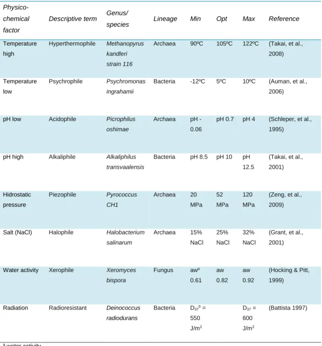
Table 1. Extremophiles and their characteristics. Adapted from Pearce (2012). ... 15
Table 2. Characteristics of the Metagenomic libraries ... 35
Table 3. Characteristics of the identified clones and their DNA fragments ... 38
Table 4. DNA-binding predicted results for each identified ORF ... 41
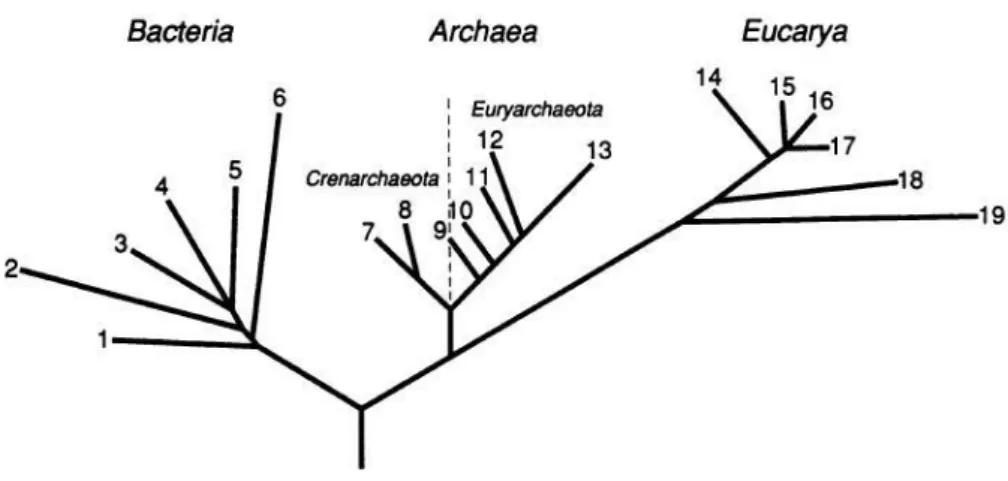
Figure 1. The three domains of life: Bacteria, Archaea and Eucarya, defined by Woese (1987) ... 16
Figure 2. Effects triggered by UV irradiation on bacterial cells and respective DNA repair mechanisms. (UV) Ultra-violet radiation; (IR) Ionizing radiation; (ROS) Reactive Oxygen Species; (SSB) Single-strand break; (DSB) Double-strand break; (CPD) Cyclobutane dymer; (AP-site) Apurinic/apyrimidinic site ; (BER) Base excision repair; (SP-BER) short-patch BER; (LP-BER) Long patch BER; (HR) Homologous recombination; (NHEJ) Non-homologous end joining; (NER) Nucleotide excision repair; (TC-NER) Transcription-coupled NER; (GG-NER) Global genome NER; (MMR) Mismatch repair. Adapted from (Rastogi et al. 2010). ... 18
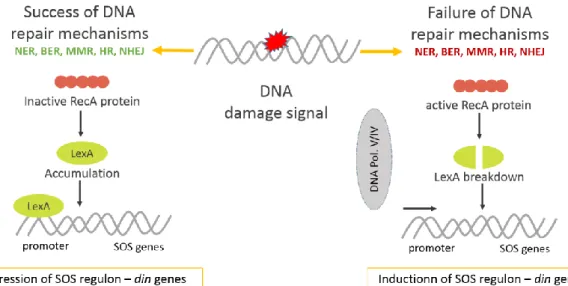
Figure 3 - Overview of SOS response mechanism in E. coli. Adapted from Rastogi et al. (2010). When there is DNA damage, there are several DNA repair mechanisms that ensure survival. These can be (NER) Nucleotide Excision repair, (BER) Base excision repair; (MMR) Mismatch repair, (HR) Homologous recombination; or (NHEJ) Non-homologous end joining, among others. But when these mechanisms fail to repair the damage, there is a genomic wide mechanism that comes into action, known as SOS response. The SOS response activates a series of genes through the interaction of two main proteins: lexA and RecA. So when there is a failure of DNA repair mechanisms the RecA protein cleaves LexA, which will no longer repress the SOS regulon, leading to the expression of damage induced genes that assure the repair of the DNA. In case the damage is too much, then SOS response skips from the initial repair stage to the mutagenesis stage, where RecA activates the DNA Polymerase V that is able to perform Translesion synthesis, ensuring the replication and survival of the cell. ... 20
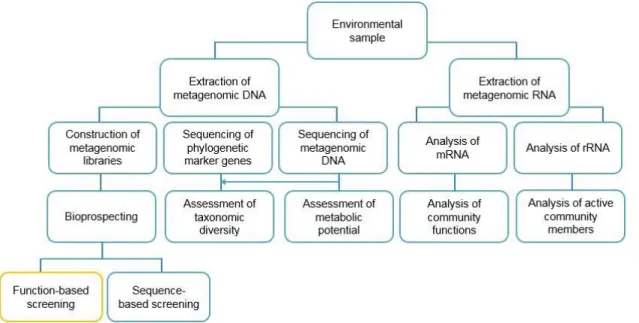
Figure 4 – Diagram of Metagenomics techniques. Function-based screening is marked in yellow. Adapted from Simon & Daniel (2011). ... 23
Figure 5. Scheme of the construction of metagenomic libraries. a) Environmental sample from three different environments; b) DNA isolation; c) DNA fragmentation – digestion with Sau3AI enzyme resulting in the optimal insert size 1-8 kb; d) vector preparation – the vector used was pSKII+ digested with BamHI; e) ligation –
pSKII+ with the fragmented DNA in order to originate a recombinant plasmid; f) electroporation; g) library tittering; h) library amplification; i) functional screening – in this case, the screening was exposure to UVB radiation (312 nm) for 150 seconds. ... 26 Figure 6. Map of pSKII+ (Stratagene) with the multiple cloning site region. ... 27
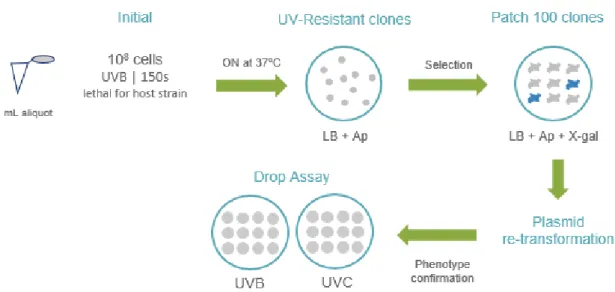
Figure 7. Steps of the UV screening. In general for 1 ml aliquot the procedure was the following: the initial screening consisted in spreading 108 cells in LB-Ampicillin
plates, and irradiate them with UVB for 150 seconds. The plates were incubated ON at 37ºC, and the clones that grow are UV resistant. The resistant clones were grown in patch in an LB+Ap+X-gal in order to select them for transformed cells and recombinant plasmids. The white ones were selected for further re-transformation of the plasmid in E.coli to assure the resistance was due to the environmental DNA and not due to spontaneous mutations. The re-transformed clones were screened by Drop Assay to confirm their resistance to UV radiation. ... 30 Figure 8. (1.) Drop assay results. E. coli DH10B does not grow when exposed to UVB radiation for 80 seconds or to UVC radiation for 15 seconds. In turn, the recombinant clones are shown to be resistant, up to 10-2 dilution. (2.) Graphic
representation of the results of the functional screenings of OS1(a) and D1 (b) metagenomic libraries by methodological stages (Initial, Patch, Re-transformation, Drop Assay and UVC). ... 37 Figure 9. Representation of the organisation of the identified genes within the plasmid pSKII+. E-values in brakets. (HP) Hypothetical protein. ... 39
Figure 10. Alignment of pML6-orf1 and pML105-orf1. The two HPs share 32% of identity, revealing potential common function in UV-resistance. ... 40 Figure 11. Survival rate to 120 s of UVB (312 nm). *** indicate the statistical significance of Tukey test at a level of 0.05 ... 42 Figure 12. Survival rate of the five resistant-clones to 60’ of incubation with 4NQO (50μM). * indicates the significance of Dunnett test at a level of 0.05. ... 43 Figure 13. Biochemically documented template-dependent DNA polymerases found in E. coli (Sutton & Walker 2001). ... 45
Abreviations
ºC Degrees Celsius
µL Microlitre
µM Micromolar
4NQO 4-Nitroquinoline 1-oxide
6-4PPs Pyrimidine 6-4 pyrimidone photoproducts
aa Amino acid
Ap Ampicillin
AP site Apurinic/apyrimidinic site APE-1 AP endonuclease-1
ATP Adenosine triphosphate
ATPase Adenosinetriphosphatase BER Base excision repair
BLAST Basic Local Alignment Search Tool
bp Base-pair
CDS Coding sequence (nucleotide) CPDs Cyclobutane pyrimidine dimers
din Damage induced genes
DNA Deoxyribonucleic acid
dNTPs Deoxyribonucleotide triphosphates DSBs Double Stranded breaks
GC% Guanine/Cytosine content GFP Green Fluorescent Protein
GG-NER Global genome NER
h Hour
HP Hypothetical protein
HR Homologous recombination
IPT Immunoglobin-like fold shared by plexins and transcription factors domains
kb Kilo-base-pair
kV Kilovolts
LB Luria Bertrani
mRNA Messenger RNA
NER Nucleotide excision repair
ON Overnight
ORF Open Reading Frame
PCD Programed cell dead
PCR Polymerase Chain Reaction pSKII+ pBluescript SKII+
RNA Ribonucleic acid
ROS Reactive Oxygen Species rpm Revolutions per minute
s Seconds
SOC Super Optimal broth with Catabolite repression
SSB Single Stranded breaks
TAE Tris-acetate-Ethylenediaminetetraacetic acid TBP TATA-box binding protein
TC-NER Transcription-coupled NER TFIIH Transcription factor-IIH
TLS Translesion DNA synthesis
Tm Melting temperature UV Ultraviolet radiation UVA Ultraviolet-A UVB Ultraviolet-B UVC Ultraviolet-C V Volts v/v Volume/volume w/v Weight/volume X-gal 5-bromo-4-chloro-3-indolyl-β-D-galactopyranoside
Table of contents
Acknowledgements ... 1
Resumo ... 2
Abstract ... 4
List of tables and Figures ... 6
Abreviations ... 9 Table of contents ... 11 1. Introduction ... 13 1.1. Astrobiology ... 13 1.2. Limits of Life ... 14 1.3. UV Radiation ... 17 1.4. Functional Metagenomics ... 22 1.5. Objectives ... 24
2. Materials and Methods ... 25
2.1. Bacterial strains, media and culture conditions ... 25
2.2. Construction of metagenomic libraries ... 25
2.2.1. DNA isolation and extraction ... 25
2.2.2. DNA fragmentation ... 25 2.2.3. Vector preparation ... 27 2.2.4. Ligation ... 28 2.2.5. Electroporation... 28 2.2.6. Library tittering ... 28 2.2.7. Library amplification ... 29
2.3. Functional screening of UV-resistance ... 30
2.3.1. UV irradiation ... 30
2.3.2. Initial plate assay ... 30
2.3.3. Drop Assay ... 31
2.3.4. Average insert size characterization ... 32
2.3.5. UV survival rate ... 33
2.4. Identification and analysis of resistance-conferring genes ... 34
2.4.1. Primer design and PCR reactions ... 34
2.4.2. Gene identification and in silico analysis ... 34
3. Results ... 35
3.1. Metagenomic libraries ... 35
3.2. Functional Screening ... 36
3.3. Gene identification and in silico analysis ... 38
3.4. UV survival rate ... 42
3.5. 4NQO survival rate ... 43
4. Discussion ... 44 4.1. Gene identification ... 44 4.2. pML5 ... 44 4.3. pML6 and pML105 ... 45 4.4. pM56 ... 46 4.5. pML84 ... 46
4.6. UV survival rate and 4NQO survival rate ... 47
5. Conclusion and Future perspectives ... 48
6. References ... 49
Appendix ... 56
1. Introduction
1.1.
Astrobiology
Astrobiology might strike as a new science but the possibility of life beyond Earth is an ancient Human question. Yet, it was the 50s’ space age that revolutionized the modern debate over life on other worlds. In 1960 the term “exobiology” was first introduced by Joshua Lederberg as the study of life beyond Earth (Chyba & Hand 2005).The term “bioastronomy” was also used. By the end of the 20th century the term “astrobiology” took place, defined as a broader and multidisciplinary field addressing life in the context of its planetary history (Dick 2006). Today, Astrobiology is defined as the study of the origins, evolution, distribution, and future of life in the universe (Des Marais et al. 2008; Billings et al. 2006).
The NASA Astrobiology Roadmap emphasizes the multidisciplinary aspect of astrobiology, both in its content and execution. It requires the sciences of molecular biology, ecology, geology, planetary science, astronomy, information science, space exploration technologies, and others, to work together to (i) understand the origin and emergence of life in the universe, (ii) to know the limits to the Earth’s biosphere, (iii) to understand the evolution of life on Earth, and (iv) to discover if life on Earth is unique and if there are other forms of intelligences in the universe (Des Marais et al. 2008).
Generally an agreed-upon definition of life is still lacking, but astrobiology depends on some kind of life definition which allows the differentiation of living beings when searching for life in other planets (Chodasewicz 2014). Definitions of life should be open and not limited by our current state of knowledge, thus establishing a framework from diverse perspectives (from the origin of life on Earth, to synthetic biology, to the search for extra-terrestrial life) rather than a precise definition (Tirard 2010). The definition accepted by NASA nowadays is that life is a self-sustained chemical system capable of undergoing Darwinian evolution, although this view has been a target of criticism (Chodasewicz 2014).
1.2.
Limits of Life
Life on Earth is, by now, our only analogue to what we might expect to find in other worlds. This makes Earth analogy studies to be our most important methods when speculating about extraterrestrial environments. The study of the Earth’s biogeochemistry and microbiome that characterise extreme environments of our planet are of great importance to the design and development of robotic systems that will be needed to search for biosignatures and evidence of past or present life on Mars, Europa, Titan and other prime targets for Astrobiology research (Pikuta et al. 2007; Gleeson et al. 2012). A biosignature is defined by Des Marais et al. (2008) as “an object, substance and/or pattern whose origin specifically requires a biological agent”, and it can be, for instance, organic biomarker compounds, stable isotopes, atmospheric gases or biosedimentary structures (Mustard et al. 2013).
On Earth, the lack of liquid water is likely to represent the most fundamental constraint for life, but combinations of other stressors (e.g., radiation and vacuum conditions) may also control the habitability of extraterrestrial environments. In fact, desiccation and radiation were found to exert the strongest deleterious impacts on bacterial survival, acting as dominant selective pressure on microorganisms exposed to the terrestrial stratosphere and thermosphere (Harm 1980).
On the search for establishing the limits of life on Earth, every year scientists identify and characterize extremophiles - organisms that strive in extreme environments – as shown in Table 1. Extremophiles reveal the ranges of habitability and possibility of life beyond Earth, demonstrating the incredible adaptability of life. There are several examples of extremophiles. Archaea, for instance, are known as extreme-lovers, including hyperthermophilic (high temperatures), halophilic (high salt concentrations), acidophilic (low pH) and UV-resistant microorganisms (Leuko et al. 2014; Mastascusa et al. 2014). Cyanobacteria can also cooperate with extreme temperatures and radiation exposure, even though they are photosynthetic organisms (Cowan et al. 2015). Bacillus subtilis has been identified to resist several extreme conditions, mainly due to endospore formation (Nicholson et al. 2000). And Deinococcus radiodurans (Battista 1997), is one of the most well-known species capable of surviving extreme radiation, withstanding lethal and mutagenic effects of DNA damage.
Table 1. Extremophiles and their characteristics. Adapted from Pearce (2012).
Physico-chemical factor
Descriptive term Genus/
species Lineage Min Opt Max Reference
Temperature high
Hyperthermophile Methanopyrus
kandleri strain 116
Archaea 90ºC 105ºC 122ºC (Takai, et al.,
2008)
Temperature low
Psychrophile Psychromonas
ingrahamii
Bacteria -12ºC 5ºC 10ºC (Auman, et al.,
2006)
pH low Acidophile Picrophilus oshimae
Archaea pH -
0.06
pH 0.7 pH 4 (Schleper, et al., 1995)
pH high Alkaliphile Alkaliphilus transvaalensis Bacteria pH 8.5 pH 10 pH 12.5 (Takai, et al., 2001) Hidrostatic pressure Piezophile Pyrococcus CH1 Archaea 20 MPa 52 MPa 120 MPa (Zeng, et al., 2009)
Salt (NaCl) Halophile Halobacterium salinarum Archaea 15% NaCl 25% NaCl 32% NaCl (Grant, et al., 2001)
Water activity Xerophile Xeromyces bispora Fungus awa 0.61 aw 0.82 aw 0.92
(Hocking & Pitt, 1999)
Radiation Radioresistant Deinococcus radiodurans Bacteria D37b = 550 J/m2 D37 = 600 J/m2 (Battista 1997) a water activity
b dose required to kill 63% of cells
Knowledge of Earth’s microbial diversity has influenced taxonomic classification over the years from early approaches in the 18th century based on physical traits (Paterlini
2007), towards recent advances in molecular phylogenetics (Fox et al. 1977), metagenomics (Handelsman 1998; Chistoserdova 2010), metaproteomics (Wilmes & Bond 2006) and single-cell sequencing (Raghunathan et al. 2005; McLean & Lasken 2014) that allowed direct access to microbial genomes and made it possible to rectify the bias of known biodiversity (Woese et al. 1990). Nowadays, the tree of life contains three primary lines of descent referred to as “domains” by Carl Woese: Bacteria, Archaea and Eukarya (Figure 1).
Understanding the physico-chemical limits of life on Earth, made it possible to turn out to the solar system, which is considered as of exobiological interest. Our solar system has been evaluated in terms of habitable zone and potential for planets other than Earth to harbour life. Defining reliable habitable zones (Kasting et al. 2013), can eventually lead to the detection of habitable exoplanets, and perhaps to evidence of extraterrestrial life. In the 1990’s, the confirmation of a thick sheet of ice covering Europa, and the possibility that an ocean exists beneath it, focused renewed attention on the possibility of life beyond the presumed habitable zone of our solar system (Khurana et al. 1998). Moreover, the existence of organic molecules in the atmosphere of the Saturnian moon Titan (Lorenz et al. 2008), fostered the interest on the origin of life, and the limits of life as we know it (Ali et al. 2015; Lorenz et al. 2008).
Nevertheless, it will be crucial to evaluate whether any biological material potentially found beyond Earth represents an independent origin or another branch in the family tree of Earth life (Worth et al. 2013).
1.3.
UV Radiation
UV radiation is divided in three types: ultraviolet-A (UVA) (315-400 nm), ultraviolet-B (UVB) (280–315 nm) and ultraviolet-C (UVC) (280-100 nm), as defined by the International Commission on Illumination (Sliney 2007). On earth, most of the UVB radiation is attenuated by the ozone layer. Nevertheless, the exposure of short-wavelengths of ultraviolet radiation UVB and UVC causes direct and indirect damage to cells, particularly in DNA and photosystems (Sinha & Häder 2002; Cadet et al. 2014). High UV radiation exposure is believed to have existed in early Archaean Earth, posing a challenge to life’s protection and repair processes (Cockell 1998) and is currently prevailing in space environments such as surfaces of spacecrafts and planetary bodies (Mancinelli & Klovstad 2000). Nevertheless microorganisms such as Bacillus subtilis or Deinococcus radiodurans, and others such as planctomycetes have been found striving under these extreme conditions (Nicholson et al. 2000; Battista 1997). This has triggered scientists to question how life has evolved to resist to UV-light (Cockell 2000).
There are several strategies of UV radiation mitigation and UV screening providing a first line of defence against UV radiation. These can be followed by repair processes used to deal with damage. In fact there is a sequence of effects triggered by UV irradiation on bacterial cells such as mutagenesis and reactive oxygen species (ROS) formation that mainly affect DNA (Figure 2). UV radiation is responsible for most of the mutagenesis due to a process of DNA translesion synthesis (TLS) in which a polymerase encounters a noncoding or miscoding lesion, inserts an incorrect nucleotide opposite the lesion and then continues elongation. UV can also originate cyclobutane pyrimidine dimers (CPDs), pyrimidine 6-4 pyrimidone photoproducts (6-4PPs) and their Dewar isomers, apurinic/apyrimidinic sites (AP site), single-stranded breaks (SSB) and double-stranded breaks (DSB) (Rastogi et al. 2010). Consequently, there are several tolerance and damage-control mechanisms that allow organisms to cope with UV radiation (Figure 2).
These damage-control mechanisms can be: Nucleotide excision repair (NER), mismatch repair (MMR), homologous recombination (HR), non-ending homologous recombination (NEHJ) and photoreactivation, acting in a lesion-directed level. Whereas base excision repair (BER) and SOS response act in a general line of DNA damage repair (Harm, 1980). Post-replication repair, de novo synthesis of proteins and lipids and programmed cell death (PCD) or apoptosis may also become effective for the
recovery of genome integrity (Cockell & Knowland 1999; Cohen & Walker 2011; Rastogi et al. 2010).
Regarding AP-site repair, the AP-site is removed by the action of AP endonuclease-1 (APE-1) along with phosphodiesterase that breaks the DNA strand along 5’ or 3’ to the AP site, respectively, and subsequently the gap is filled by a repair DNA polymerase and the strand is joined by a DNA ligase (Rastogi et al. 2010).
NER is one of the most versatile and flexible repair systems found in most organisms, being highly conserved in eukaryotes. Discovery of NER was first described in E. coli where about six proteins such as UvrA, B, and C (known as ABC-complex, which shows exonuclease activity), UvrD (helicase II), DNA polymerase I (pol. I), and DNA ligase are recruited to complete the repair (Rastogi et al. 2010). NER is critically important in the repair of UV-induced DNA lesions, sorting out a wide range of DNA damages such as CPDs and 6-4PPs. Depending on the damage location, two sub-pathways of NER coexist and are mechanistically conserved from prokaryotic to eukaryotic cells: global genome NER (GG-NER) removes lesions anywhere in the genome, whereas transcription-coupled NER (TC-NER) specifically removes lesions Figure 2. Effects triggered by UV irradiation on bacterial cells and respective DNA repair mechanisms. (UV) Ultra-violet radiation; (IR) Ionizing radiation; (ROS) Reactive Oxygen Species; (SSB) Single-strand break; (DSB) Double-strand break; (CPD) Cyclobutane dymer; (AP-site) Apurinic/apyrimidinic site; (BER) Base excision repair; (SP-BER) short-patch BER; (LP-BER) Long patch BER; (HR) Homologous recombination; (NHEJ) Non-homologous end joining; (NER) Nucleotide excision repair; (TC-NER) Transcription-coupled NER; (GG-NER) Global genome NER; (MMR) Mismatch repair. Adapted from (Rastogi et al. 2010).
from the transcribed strand of active genes (Alekseev & Coin 2015; Logette et al. 2011).
In turn, the BER pathway against UV radiation is done via generation of ROS which efficiency and specificity are determined by several forms of DNA glycosylase. These glycosylases remove different types of modified base by cleaving the N-glycosidic bond between the abnormal base and deoxyribose creating either an abasic site or an SSB
which is further processed by short-patch repair or long-patch repair (Krokan & Bjøra 2013; Rastogi et al. 2010).
On one hand, homologous recombination can help repair double-stranded breaks, by using foreign DNA as a template (Bernstein et al., 1981). The strand-exchange reaction is mediated mainly by RecA family proteins both in Bacteria and Eukarya. Evidence of homologous recombination can be found in multilocus sequence typing studies (Didelot et al. 2010). On the other hand, non-homologous end joining (NHEJ) is an evolutionarily conserved repair system that helps to maintain the gene integrity for DNA damage (Ochi et al. 2014).
The process of DNA repair by photoreactivation is done through photoreactivating enzymes known as photolyases, which are conserved and can be found throughout the three domains of life, but seem to be absent or non-functional in humans. These enzymes are present in archaea suggesting their role as ancient repair proteins, which may have helped in the evolution of the earliest organisms on primordial Earth (Sinha & Häder 2002).
The SOS response is activated when there is severe DNA damage impeding DNA replication and repair from proceeding effectively, as it is exemplified in Figure 3. The primary task of the SOS response is to restart replication before the cell dies. This system is regulated by both the LexA transcriptional repressor and the RecA recombinase. In E. coli, the UV radiation firstly damages the DNA by creating lesions that mechanically interrupt the process of DNA duplication by stalling the DNA-polymerase (Pol III) in a moving replication fork. This results in the production of single-stranded DNA breaks (SSB). These breaks are recognised by the protein RecA involved in the non-mutagenic coating of SSB via homologous recombination. RecA also catalyses the cleavage of LexA diminishing its levels. The cleaved LexA de-represses the regulon involved in the SOS response – din (damage induced) genes -
and allows expression of SOS responding genes (Krishna et al. 2007; Rastogi et al. 2010).
The UmuD protein, which is part of the UmuC/UmuD2 complex of the error-prone DNA
polymerase (Pol V), is also cleaved by RecA, directly resulting in mutagenesis in UV-irradiated E. coli cells. Pol V inserts several random base pairs in the DNA strand directly opposite a lesion, thus helping a replication fork to quickly bypass the lesion (TLS) after which Pol III can take over and continue replication. DNA polymerase IV (dinB) is also involved in TLS in E. coli (Rastogi et al. 2010).
SOS response is complex, in fact SOS regulon is known to comprise more than 40 genes, including those encoding the mutagenesis proteins UmuD and UmuC, RecA, and LexA. Also part of the SOS regulon are genes encoding UvrA, B, C—a group of nucleotide excision repair (NER) proteins that locate and excise damaged regions from the DNA (Shah & He 2015; Aksenov 1999; Janion 2008).
Figure 3 - Overview of SOS response mechanism in E. coli. Adapted from Rastogi et al. (2010). When there is DNA damage, there are several DNA repair mechanisms that ensure survival. These can be (NER) Nucleotide Excision repair, (BER) Base excision repair; (MMR) Mismatch repair, (HR) Homologous recombination; or (NHEJ) Non-homologous end joining, among others. But when these mechanisms fail to repair the damage, there is a genomic wide mechanism that comes into action, known as SOS response. The SOS response activates a series of genes through the interaction of two main proteins: lexA and RecA. So when there is a failure of DNA repair mechanisms the RecA protein cleaves LexA, which will no longer repress the SOS regulon, leading to the expression of damage induced genes that assure the repair of the DNA. In case the damage is too much, then SOS response skips from the initial repair stage to the mutagenesis stage, where RecA activates the DNA Polymerase V that is able to perform Translesion synthesis, ensuring the replication and survival of the cell.
Although SOS response has been studied for decades, new discoveries are being made every year. However, most of the currently known mechanisms of adaptation to UV radiation are based on studies that address less than 1% of the total amount of microorganisms that exist on Earth, the ones that can be cultured and maintained in a laboratory. There is still 99% that remain unstudied, giving us scientists a biased understanding of the microbial and functional diversity (Staley & Konopka 1985).
Thus, to better explore the diversity of adaptation mechanisms, there is a need to study the other 99% of uncultured microorganisms. This can be done by using culture independent techniques, associated with earths’ extreme environments.
1.4.
Functional Metagenomics
Metagenomic strategies comprise analysis of both DNA – construction of metagenomics libraries, sequencing of DNA, sequencing of phylogenetic markers (such as 16S rRNA) – and RNA – analysis of mRNA or rRNA, as shown in Figure 4 (Simon & Daniel 2011). The direct sequence analysis of metagenomic DNA is presently considered the most accurate method for assessing the structure of an environmental microbial community, since it does not involve any selection (by cultivation and/or enrichment) and minimizes technical biases, as the ones introduced by PCR amplification of the 16S rRNA (Lewin et al. 2013). Previously identified genes found through metagenomics include chaperons, transporters, DNA-binding proteins, (Chistoserdova 2010; Shestakov 2012), bacteriorhodopsins (Beja et al. 2000), among others.
Function-based metagenomics, or functional metagenomics, has become an efficient tool to find novel genes involved in stress adaptation processes by expressing the metagenome of a microbial community in a foreign host (Ekkers et al. 2012). Metagenomic libraries can thus be screened for genes coding for proteins with a particular function: antibiotics (Gillespie et al. 2002), enzymes of commercial interest (Jeon et al. 2009), quorum sensing inhibitors or inducers (Williamson et al. 2005), aromatic compounds (Uchiyama & Miyazaki 2013), or compounds conferring resistance to extreme conditions (Allen et al. 2009). Moreover, hypothetical and unknown genes not previously assigned to confer resistant to these conditions, can be annotated with the help of functional metagenomics.
By assuring that uncultured microorganisms are taken into account as an important source of unknown genes, metagenomics plays a key-role in understanding microbial adaptation mechanisms and shows great potential in the area of biotechnology. In fact, the use of metagenomics in the search for novel molecular mechanisms of adaptation of the microorganisms to extreme conditions has been popular in recent studies, contributing to uncover the limits of life (Cowan et al. 2015). These studies involve resistance to toxic metals (González-Pastor & Mirete 2010), acidic pH (Guazzaroni et al. 2013), low and high temperatures (Lewin et al. 2013) or high salt concentrations (Culligan et al. 2013).
Figure 4 – Diagram of Metagenomics techniques. Function-based screening is marked in yellow. Adapted from Simon & Daniel (2011).
1.5.
Objectives
The current project is set within the goal number five of the Astrobiology Roadmap, which strives to “Understand the evolutionary mechanisms and environmental limits of life. Determine the molecular, genetic, and biochemical mechanisms that control and limit evolution, metabolic diversity, and acclimatization of life” (Des Marais et al. 2008). In this way, this project aims to:
1. Identify novel genes responsible for UV-resistance from the metagenome of different environmental samples (representing different pools of microorganisms adapted to both hypersaline and UV-exposure environments) by using a functional metagenomic approach;
2. Characterize these genes and their involvement in the adaptation mechanisms of microorganisms to UV radiation.
The main goal of this 10-month project was to improve the knowledge of microbial molecular strategies and metabolic function of adaptation mechanisms involved in UV-resistance, taking uncultured microorganisms as an important source of yet unknown genes. Enlightening the molecular basis of adaptation of microorganisms to UV radiation will allow us to disclose aspects of the evolution of life on Earth and other planets, and like other metagenomic studies, it will gives us the chance to apply the knowledge to biotechnological applications (Shestakov 2012; Simon & Daniel 2011).
2. Materials and Methods
2.1.
Bacterial strains, media and culture conditions
Escherichia coli DH10B was grown in Luria-Bertani (LB) medium, over-night (ON) at 37 ºC. The growth LB medium for recombinant E. coli strains was supplemented with 80 µg/ml ampicillin (Ap).
2.2.
Construction of metagenomic libraries
The construction of metagenomic libraries was performed, following the González-Pastor & Mirete protocol (González-González-Pastor & Mirete 2010), consisting of: DNA isolation and extraction, DNA fragmentation, vector preparation, ligation, electroporation, library tittering and library amplification. A schematic overview of metagenomic library construction is shown in Figure 5.
2.2.1.
DNA isolation and extraction
Environmental samples were previously collected from several high-altitude hypersaline environments. These were two Andean ponds from Argentina (Diamante at 4600 m and Ojo seco at 3900 m), and one Spanish saltern in Mallorca (Es Trenc). Each of these samples contain a different pool of microorganisms highly exposed to UV radiation. Metagenomic DNA was isolated using BIO101 FastDNA spin kit (Qbiogene, Carlsbad, CA, USA) assessing the quality, quantity and concentration by spectrophotometer (dilution 1/12). To extract the genomic DNA the Wizard R Genomic DNA purification Kit (Promega) was used, following the manufacturer’s instructions.
2.2.2.
DNA fragmentation
To obtain DNA fragments between 1-8 kb which ensure the presence of one or more genes, the metagenomic DNA was digested using the frequent-cutting restriction enzyme Sau3AI at a concentration that permitted a partial digestion of the DNA.
Digestion was analysed in a low melting point agarose gel electrophoresis (1%, TAE), which allowed the recovery of intact DNA fragments after electrophoresis. DNA was purified using QIAquickR Gel Extraction kit, Qiagen, and DNA sample was concentrated on SpeedVac. DNA concentration values were determined using a spectrophotometer.
Figure 5. Scheme of the construction of metagenomic libraries. a) Environmental sample from three different environments; b) DNA isolation; c) DNA fragmentation – digestion with Sau3AI enzyme resulting in the optimal insert size 1-8 kb; d) vector preparation – the vector used was pSKII+ digested with BamHI; e) ligation – pSKII+
with the fragmented DNA in order to originate a recombinant plasmid; f) electroporation; g) library tittering; h) library amplification; i) functional screening – in this case, the screening was exposure to UVB radiation (312 nm) for 150 seconds.
2.2.3.
Vector preparation
The vector pBluescript SKII+ (pSKII+) was prepared for cloning by BamHI digestion,
which provides a restriction site compatible with that of the Sau3AI enzyme. To access the information contained in the metagenome, the environmental DNA fragments were inserted in pSKII+ by ligation. The resulting recombinant plasmids were used to
transform the Escherichia coli DH10B strain, allowing their expression and subsequent screening for UV-resistance. By using pSKII+ the selection of correct incorporation of
the plasmid in the E. coli strain is enabled by ampicillin resistance and alpha-complementation (Langley et al. 1975). In this way, pSKII+ was digested with restriction
enzymes allowing further correct cloning of DNA fragments.
2.2.4.
Ligation
Ligation of the metagenomic DNA fragments (100 ng) and pSKII+ (100 ng) was done
using 1 µL of T4 DNA ligase (Roche, Mannheim, Germany), and 1 µL of T4 DNA ligase buffer (10x) in a total volume of 10 µL. For each library, independent ligation reactions were set up (x3) and incubated ON at 16 ºC in a heating block. Ligase was inactivated at 65 ºC for 15 min in a heating block. The three ligation reactions were transferred by pipetting to a fresh microcentrifuge tube and water was added up to 200 µL. DNA was precipitated following the protocol described in (González-Pastor & Mirete 2010) except at the final dissolution, done by adding 10 µL of distilled water and incubated at 55 ºC for 5 min.
2.2.5.
Electroporation
Transformation of 3 µl of recombinant plasmid (ligation) in 20 µl of electrocompetent E.coli DH10B cells (Invitrogen, Carlsbad, CA, USA) was performed by electroporation using Micropulser (Bio-Rad, Hemel Hempstead, UK) at 1.8 kV according to the manufacturer’s instructions. DNA, electroporation cuvettes and filter tips were previously cooled on ice or on fridge for at least 30 minutes. To assure enough transformants each electroporation was performed 3 times. After electroporation, 1ml S.O.C medium (Hanahan 1983) was added and samples were incubated at 37 ºC for 1:15 h at 225 rpm. 1/20 dilutions in LB-Ap (80mg/ml) and X-gal (40mg/ml) were plated and incubated at 37 ºC ON.
2.2.6.
Library tittering
Library tittering was done by preparing serial dilutions of 100 µL of the metagenomic library, up to 10-8 dilution. 100 µL of the last 3 dilutions were plated in LB-Ap, and
incubated at 37ºC ON. The tittering value allows us to determine the precise number of cells to be exposed to UV treatment. In this case, 108 cells/plate were used in the
UV-screening. Average insert size of the metagenomic library was determined by isolating and digesting 10 random clones with restriction enzymes flanking the insert: XhoI and XbaI (Roche) [1 µg de plasmid, 1,5 µl buffer H (Roche), 0,5 µl XbaI, 0,5 µl XhoI, adding water up to 15 µl]. Incubate at 37 ºC for 3h in the heating block. Fragment length polymorphism was analysed with a 1% agarose gel TAE, stained with Pronasafe. Control used was non-recombinant vector pSKII+ digested with XhoI and XbaI.
2.2.7.
Library amplification
To assure enough library sample for multiple functional screenings, library amplification must be performed, either in solid or liquid medium, ending with up to 108-1010 cfu/ml. A
metagenomic library aliquot of 20 µl was kept at -80ºC for further studies.
i) Plate amplification: the metagenomic library was directly plated on LB-Ap solid medium. To get the final number of plates to be used, the following formula was applied: total number of clones (tittering value) x 3 / colonies per plate. Plates with LB-Ap were incubated at 37 ºC ON. Afterwards, cells from each plate were mixed with 3,5 ml LB and 10% glycerol (w/v), being pooled in a flask with cells from the same library, mixed again. The pool was divided in several 1ml/eppendorf aliquots which were stored at -80 ºC.
ii) Liquid amplification: the metagenomic library was centrifuged at 4 ºC, 8000 rpm for 15 min. The supernatant was discarded and the remaining sample was resuspended in 1 ml of LB+Ap. The sample was added to 50 ml LB+Ap (80 µL/µg) and incubate at 37 ºC for 6 h in the water bath with agitation (no measurements to avoid loss of sample). When the culture reached A600=2 glycerol was added at 10% (w/v) and aliquots of 1 ml/eppendorf were stored at -80 ºC.
2.3.
Functional screening of UV-resistance
2.3.1.
UV irradiation
In order to select the clones harbouring genes conferring UV-resistance, the metagenomic libraries were exposed to UV radiation. The UV lamp used is VL6-MC from Vilber Lourmat, and can irradiate at 254 nm (UVC) or at 312 nm (UVB). The UV irradiation chamber has an upper aperture, where the lamp fits, and a side aperture where the Petri dish is placed. The distance from the UV lamp and the plate is approximately 24 cm, which gives us the chance to irradiate with 1.56 W m-2 at 254 nm
and 2.26 W m-2 at 312 nm. A general scheme of the steps of the UV-screening is
presented bellow in Figure 7.
2.3.2.
Initial plate assay
Each metagenomic library was plated on LB-Ap solid medium (108 cells) and exposed
to UV radiation, 312 nm for 150 s, conditions that we previously observed that were lethal to the E. coli DH10B strain, host of the metagenomic libraries. The UV resistant clones were isolated and patched on LB agar medium supplemented with Ap and X-gal Figure 7. Steps of the UV screening. In general for 1 ml aliquot the procedure was the following: the initial screening consisted in spreading 108 cells in LB-Ampicillin plates, and irradiate them with UVB for 150 seconds. The plates were
incubated ON at 37ºC, and the clones that grow are UV resistant. The resistant clones were grown in patch in an LB+Ap+X-gal in order to select them for transformed cells and recombinant plasmids. The white ones were selected for further re-transformation of the plasmid in E.coli to assure the resistance was due to the environmental DNA and not due to spontaneous mutations. The re-transformed clones were screened by Drop Assay to confirm their resistance to UV radiation.
and incubated ON at 37 ºC. These two supplements originate a selective medium for an antibiotic (only transformed cells can grow) and alpha-complementation selectivity by blue and white colonies (the colonies harbouring an environmental DNA fragment cloned in the pSKII+ will be white, whereas the colonies without DNA fragment will be
blue) (Langley et al. 1975).
To rule out potential chromosomal spontaneous mutants conferring UV-resistance, the resistant colonies were pooled by adding 3,5 mL of LB-Ap to each plate and collecting the clones into several aliquots of 1mL. Plasmids were isolated from the aliquots, and used to transform DH10B cells by the heat shock method. 1/10 and 1/20 dilutions of the transformations were plated on LB-agar supplemented with Ap and X-gal and incubated ON at 37 ºC. The resulting colonies will be first streaked on LB-Ap-Xgal and incubated ON at 37 ºC, and kept at 4 ºC until the Drop Assay.
2.3.3.
Drop Assay
To select the clones harbouring a recombinant vector containing an environmental DNA fragment conferring UV resistance, a drop assay was used. For that, a culture is established from a single colony, using Ap as the selective medium, and incubated ON at 37 ºC, 140 rpm. The cultures were normalized to get an A600=1, to exclude false resistance phenotypes due to the presence of higher amounts of cells in some cultures. Afterwards, 10 µL of each culture and the negative control (E. coli DH10B) were spread on LB-Ap-X-gal plates, and exposed to UV radiation (312 nm) for 80 s, and incubated ON at 37 ºC. These UV exposure conditions are lethal for E. coli.
To exclude clone resistance conferred by different cell growth or higher amounts of cells, cultures from each resistant clones at A600=1 were serially diluted up to 10-5, and drops from each dilution were exposed to UVB radiation at 312 nm, 80 s and to UVC radiation at 254 nm, 15 s. In this assay, two controls were included: i) a non-irradiated growth of the dilutions, to evaluate if the differences in UV resistance are due to differences in growth of the clones, and ii) dilutions of a culture of the strain host of the library (DH10B) at A600=1.
2.3.4.
Average insert size characterization
To characterize the insert size of the resistant clones, they were grown in liquid LB-Ap-X-gal and incubated ON at 37 ºC. This culture allowed plasmid extraction using QIAprep R Spin Miniprep Kit (250) (Quiagen), determination of plasmid concentration using Nanodrop (Thermo), and insert size characterization by performing a double digestion of the plasmid with XhoI and XbaI enzymes, followed by result analysis in 0,8% TAE agarose gel electrophoresis, ran at 80V for 45 min. A small amount of culture of each clone was stored in 15% (w/v) glycerol at -80 ºC for further analysis. Unique clones were recovered by plasmid digestion with XhoI, XbaI [2 µL DNA, 1.5 µL buffer H (Roche), 0,5 µL XhoI, 0.5 µL XbaI, adding H2O up to a total of 15 µL] and
HindIII [2 µL of DNA, 1.5 µL of Buffer B (Roche), 0,5 µL of HindIII and H2O up to a total
of 15 µL]. Pattern analysis was done through in 0,8% TAE agarose gel electrophoresis, ran at 80 V for 45 min.
2.3.5.
UV survival rate
To compare the survival rate among the resistant clones and the negative control the E. coli strain DH10B, a culture of each clone was grown in LB-broth-Ap and incubated ON at 37 ºC 140 rpm until A600=1. 100 µL of each culture on Eppendorf lids attached to a petri dish were exposed to 120 s of UVB (312 nm). This was followed by serial dilutions up to 10-6. Dilutions 10-4, 10-5 and 10-6 were plated on solid LB-Ap. Controls were assured by serial dilutions up to 10-7 of unexposed cultures, having the 10-5, 10-6 and 10-7 dilutions plated in LB-Ap. All plates were incubated at 37 ºC ON and survival rate was further calculated. Each experiment was performed at least three times for each clone.
2.4.
Identification and analysis of resistance-conferring genes
2.4.1.
Primer design and PCR reactions
Once selected, the environmental DNA fragments from the resistant clones were sequenced by primer walking to search for the genes conferring resistance. For that, plasmid sequencing (STAB vida) using M13 forward and reverse primers was performed. This allowed the design of specific primers to “walk” along the sequence, using DNA start and ApE software, to obtain a trustful sequence of the insert, which in turn will guarantee the right in silico analysis of the insert.
2.4.2.
Gene identification and in silico analysis
In silico analysis mainly consisted on the identification and characterization of putative Open Reading Frames (ORFs) using ORF finder and fgenesB (Solovyev & Salamov, 2011) and further similarity check using Basic Local Alignment Search Tool (BLAST) (Altschul, et al., 1997). Protein characterization and function determination were carried out using bioinformatics tools, such as to find protein domains with NCBI conserved domain search (http://www.ncbi.nlm.nih.gov/Structure/cdd/wrpsb.cgi) (Marchler-Bauer et al. 2014) as well as to assess DNA-binding properties with iDNA-Prot|dis
(http://bioinformatics.hitsz.edu.cn/iDNA-Prot_dis/) (Liu et al. 2014), iDNA-prot
(http://www.jci-bioinfo.cn/iDNA-Prot) (Lin et al. 2011), DNAbinder
(http://www.imtech.res.in/raghava/dnabinder/submit.html) (Kumar et al. 2007) and
DNABIND (http://dnabind.szialab.org/) (Szilágyi & Skolnick 2006).
2.4.3.
4NQO survival rate
To further characterize the role of each DNA fragment in UV-resistance, the survival rate clones and E. coli DH10B cells were analysed when exposing 100 µL of A600=1 cells to 100 µM of 4-Nitroquinoline 1-oxide (4NQO) for 60 minutes, in comparison with unexposed cells. The treatment was performed in a ventilated reaction chamber using the appropriate equipment. Serial dilutions of the exposed 100 µL of A600=1 cells were performed up to 10-6. Dilutions, 10-4, 10-5 and 10-6 were plated in LB-Ap. Controls were
assured by serial dilutions up to 10-7 of unexposed cultures, having the 10-5, 10-6 and
10-7 dilutions plated in LB-Ap. All plates were incubated at 37 ºC ON and survival rate
was further calculated. Each clone’s treatment with 4NQO was repeated at least three times.
3. Results
3.1.
Metagenomic libraries
In this project, two metagenomic libraries were constructed, from DNA extracted from planktonic microorganisms from brines of two hypersaline environments: Ojo Seco (OS1) and Diamante (D1) as described in material and methods. The metagenomic library from Mallorca (PMB) was previously constructed by Salvador Mirete, being analysed in this study.
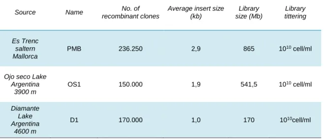
Characteristics of the Metagenomic libraries analysed in this study are shown in Table 2. Approximately 240,000 clones were obtained from PMB, 150,000 from OS and 170,000 from D, and an estimation of 685, 541 and 170 MB of environmental DNA respectively was cloned in each library. Library amplification was done as described in material and methods. Average insert size was calculated as described in Materials and Methods resulting in 2.9 kb for PMB, 1.9 kb for OS1 and 1.0 kb for D1.
Table 2. Characteristics of the Metagenomic libraries
Source Name No. of
recombinant clones
Average insert size (kb) Library size (Mb) Library tittering Es Trenc saltern Mallorca PMB 236.250 2,9 865 1010 cell/ml
Ojo seco Lake Argentina 3900 m OS1 150.000 1,9 541,5 1010 cell/ml Diamante Lake Argentina 4600 m D1 170.000 1,0 170 1010cell/ml
3.2.
Functional Screening
The functional screening of the PMB metagenomic library was done previously to this work by Maria Lamprecht Grandio (unpublished data). Only one clone (pML105) was shown to harbour a recombinant plasmid and resist to both UVB and UVC radiation using Drop Assay as described in Materials and Methods and depicted in Figure 8.1.
The screening of the metagenomic library of OS (Figure 8.2.a)) has resulted in few clones (13 in total), each of which was isolated in patch in LB-AP-X-gal plates to check for the ones carrying the recombinant plasmid. 6 out of the 13 clones grew and were selected for re-transformation of E. coli. This re-transformation assured that the resistance was conferred by the environmental DNA fragment and not by spontaneous mutations caused by UVB exposure. After re-transformation, only 1 clone (pML6) was shown to harbour the recombinant plasmid and resist to both UVB and UVC radiation using Drop Assay as described in Materials and Methods (Figure 8.1).
The initial screening of the metagenomic library of Diamante (Figure 8.2.b)) has resulted in 76 resistant clones, each of which was isolated in patch in LB-AP-X-gal plates to check for the ones carrying the recombinant plasmid. The 67 grown patches were gathered in several 1 mL pools of LB-Ap, being afterwards re-transformed in E. coli. This re-transformation assured that the resistance was due to the environmental fragment and not to spontaneous mutations caused by UVB exposure. After re-transformation, only 26 clones were shown to still harbour the recombinant plasmid. 17 out of the 36 were shown to survive to UVB exposure conditions. Out of these 17, 3 of them (pML5, pML56 and pML84), were also shown to resist to UVC exposure using Drop Assay (Figure 8.1).
In total, there were 4 UV-resistant clones identified in this study: pML5 (D), pML6 (OS), pML56 (D), pML84 (D). And 1 clone from a previous study was further analysed pML105 (PMB).
1.
2. a)
2. b)
Figure 8. (1.) Drop assay results. E. coli DH10B does not grow when exposed to UVB radiation for 80 seconds or to UVC radiation for 15 seconds. In turn, the recombinant clones are shown to be resistant, up to 10-2 dilution. (2.) Graphic
representation of the results of the functional screenings of OS1(a) and D1 (b) metagenomic libraries by methodological stages (Initial, Patch, Re-transformation, Drop Assay and UVC).
3.3.
Gene identification and in silico analysis
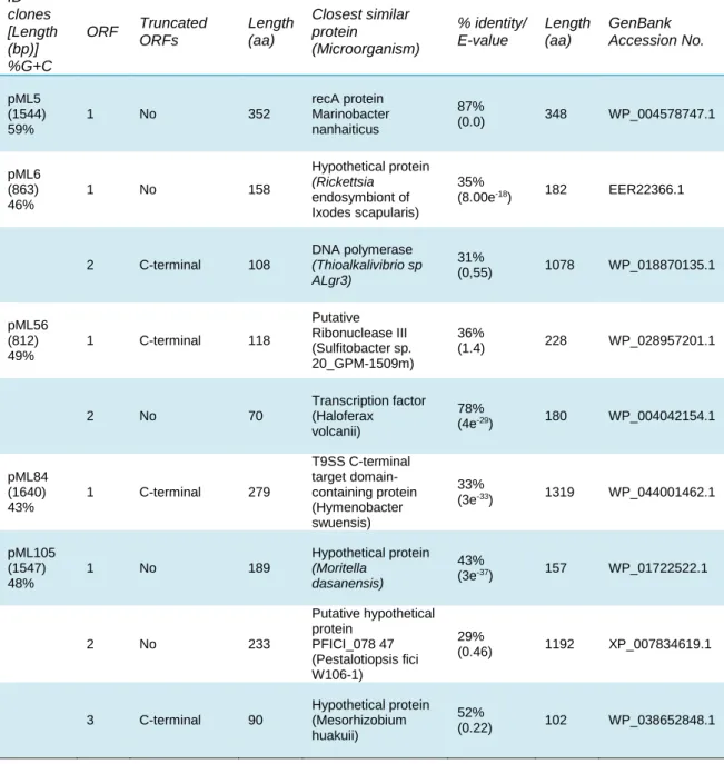
Each of the 5 clones identified in this study harbours an environmental DNA fragment, conferring resistance to UV radiation. Each fragment contained one or more ORFs which sequences were ran through BLAST in order to identify the genes involved in UV-resistance. The BLAST results are presented in Table 3 and the genetic organization of the 5 clones is presented in Figure 9 along with a short description of the best blast hit for the identified ORFs.
Table 3. Characteristics of the identified clones and their DNA fragments
ID clones [Length (bp)] %G+C ORF Truncated ORFs Length (aa) Closest similar protein (Microorganism) % identity/ E-value Length (aa) GenBank Accession No. pML5 (1544) 59% 1 No 352 recA protein Marinobacter nanhaiticus 87% (0.0) 348 WP_004578747.1 pML6 (863) 46% 1 No 158 Hypothetical protein (Rickettsia endosymbiont of Ixodes scapularis) 35% (8.00e-18) 182 EER22366.1 2 C-terminal 108 DNA polymerase (Thioalkalivibrio sp ALgr3) 31% (0,55) 1078 WP_018870135.1 pML56 (812) 49% 1 C-terminal 118 Putative Ribonuclease III (Sulfitobacter sp. 20_GPM-1509m) 36% (1.4) 228 WP_028957201.1 2 No 70 Transcription factor (Haloferax volcanii) 78% (4e-29) 180 WP_004042154.1 pML84 (1640) 43% 1 C-terminal 279 T9SS C-terminal target domain-containing protein (Hymenobacter swuensis) 33% (3e-33) 1319 WP_044001462.1 pML105 (1547) 48% 1 No 189 Hypothetical protein (Moritella dasanensis) 43% (3e-37) 157 WP_01722522.1 2 No 233 Putative hypothetical protein PFICI_078 47 (Pestalotiopsis fici W106-1) 29% (0.46) 1192 XP_007834619.1 3 C-terminal 90 Hypothetical protein (Mesorhizobium huakuii) 52% (0.22) 102 WP_038652848.1
ORF1 closest BLAST hit was RecA protein from Marinobacter nanhaiticus, with an identity of 87% and an E-value of 0.0. The closest BLAST hit for pML6-orf1 was shown to be a hypothetical protein (HP) from Rickettsia endosymbiont of Ixodes scapularis, with an identity of 35% and an E-value of 8.00e-18. A lower BLAST hit for pML6-orf1
showed a HP from Moritella dasanensis with 31% identity and an E-value of 2e-11
(WP_017221522.1). Interestingly pML105-orf1 closest BLAST hit was a HP from M. dasanensis with an identity of 43% and an E-value of 3e-37. Further analysis between
the latter two ORFs was made by alignment in NCBI, revealing a 32% identity between the two HPs (see Figure 10).
The BLAST hit of pML6-orf2 was shown to be a DNA polymerase from Thioalkalivibrio sp ALgr3, a Gammaproteobacteria, with an identity of 31% and an E-value of 0,55. In turn, the closest BLAST hit for pML105-orf2 was a putative HP PFICI_078 47 (Pestalotiopsis fici W106-1). For pML105-orf3 the closest BLAST hit was a HP (Mesorhizobium huakuii).
pML56-orf1 was shown to be a putative ribonuclease III Sulfitobacter sp (20_GPM-1509m) whereas pML56-orf 2 closest BLAST hit was a transcription factor from Haloferax volcanii with an identity of 78% and an E-value of 4e-29. Interestingly the
second BLAST hit showed to be a TATA-binding transcription initiation factor from Haloquadratum walsbyi with an identity of 58% and an E-value of 5e-23. Several
Figure 9. Representation of the organisation of the identified genes within the plasmid pSKII+. E-values in brakets.
conserved domain hits were found for pML56-orf2, including archaeal TATA box binding protein (TBP) and Transcription factor TFIID (or TATA-binding protein, TBP.
pML84-orf1 closest BLAST hit was a T9SS C-terminal target domain-containing protein from the organism Hymenobacter swuensis, with an identity of 33% and E-value of 3e -33. A conserved domain was found to be a non-specific hit with E-value of 1.88e-04.
Pfam assession number is 07610, representing a protein of unknown function (DUF1573). This domain is present in bacteria such as Rhodopirellula baltica, Bacteroides thetaiotaomicron, and Porphyromonas gingivalis which share a region of conserved sequence towards their N-termini.
To assess the DNA binding proteins of each identified ORF, the four programmes described in Materials and Methods section were used to analyse the protein sequences (Appendix 1) and results are depicted in Table 4. It was found that both pML6-orf1 is consistently predicted as DNA-binding proteins on the four analysis, and that pML56-orf2 and pML105-orf2 were predicted as DNA-binding proteins in three out of the four analysis.
Figure 10. Alignment of pML6-orf1 and pML105-orf1. The two HPs share 32% of identity, revealing potential common function in UV-resistance.
Table 4. DNA-binding predicted results for each identified ORF
iDNA-Prot|dis iDNA-prot DNAbinder DNABIND
pML5-orf1 DNA-binding protein - - -
pML6-orf1 DNA-binding protein DNA-binding protein DNA-binding protein DNA-binding protein
pML6-orf2 - - DNA-binding protein DNA-binding protein
pML56-orf1 - - - -
pML56-orf2 DNA-binding protein DNA-binding protein DNA-binding protein -
pML84-orf1 - - - -
pML105-orf1 DNA-binding protein - DNA-binding protein DNA-binding protein
pML105-orf2 - DNA-binding protein DNA-binding protein DNA-binding protein
3.4.
UV survival rate
In order to quantitatively determine the levels of resistance conferred by the selected clones (represented in Table 3) their survival rate under UV exposure was assayed as described in Materials and Methods section. This experiment also ruled out the possibility of resistance due to variation of cell numbers. When compared to the E.coli DH10B survival when exposed to 120 s of UVB radiation (5,3%), all the clones have shown an average of around 15% more of survival rate than the control: pML5 (20%), pML6 (19,7%), pML56 (18,1%), pML84 (19,3%), pML105 (27,3%) as can be seen in Figure 11. The results show values that are statistically significance, according to the Tukey test, at a level of 0.05.
Figure 11. Survival rate to 120 s of UVB (312 nm). *** indicate the statistical significance of Tukey test at a level of 0.05
3.5.
4NQO survival rate
In order to explore the molecular mechanism of resistance of each gene in UV resistance, the clones were tested for their survival rate when exposed to 4NQO, a compound that only induces DNA lesions mimicking the effect of UV radiation on DNA. Thus, if the retrieved genes conferred also resistance to 4NQO means that they could have a specific role in DNA repair. The treatment was done as described in Materials and Methods.
E.coli DH10B survival rate, when exposed to 50 µM of 4NQO for one hour was shown to be 18,1%. All the clones have shown an average of around 16% survival rate above the one from E.coli: pML5 (31,9%), pML6 (34,1%), pML56 (38,1%), pML84 (38,3%), pML105 (30,7%) as can be seen in Figure 12. The results show values that are statistically significance, according to the Dunnett test, at a level of 0.05.
Figure 12. Survival rate of the five resistant-clones to 60’ of incubation with 4NQO (50μM). * indicates the significance of Dunnett test at a level of 0.05.