Value of Systemic Staging in Asymptomatic
Early Breast Cancer
Valor do estadiamento sistêmico no câncer de mama precoce
assintomático
Gregório Pinheiro Soares
1Allan Andresson Lima Pereira
1Mariana Silva Vilas Boas
1Victor Van Vaisberg
1Maria Cristina Figueroa Magalhães
1Rudinei Diogo Marques Linck
1Max Senna Mano
11Faculty of Medicine, Instituto do Câncer do Estado de São Paulo, Universidade de São Paulo, São Paulo, SP, Brazil
Rev Bras Ginecol Obstet 2018;40:403–409.
Address for correspondence Max Senna Mano, MD, PhD, Instituto do Câncer do Estado de São Paulo, Faculdade de Medicina, Universidade de São Paulo, Av. Dr. Arnaldo 251, Cerqueira César, 01246-000, São Paulo, SP, Brazil (e-mail: max.mano@usp.br).
Keywords
►
breast neoplasms
►
metastases
►
neoplasm staging
Abstract
Objective
Metastases are rare in early breast cancer (EBC), and international guidelines
recommend against routine systemic staging for asymptomatic patients. However,
imaging exams remain widely employed in the clinical practice. The aim of the present
study is to evaluate the value of imaging for systemic staging in EBC.
Methods
A retrospective analysis of newly-diagnosed breast cancer (BC) patients was
performed. Clinical data including BC subtype, stage, presence of symptoms at
diagnosis and instrumental procedures performed for staging were recorded.
Results
A total of 753 patients were included, with a median age of 57 years. The
majority of the patients underwent at least 1 imaging procedure (91%); had invasive
ductal carcinoma (83.5%); histological grade 2 (51.4%); stage II (61.8%); and luminal
subtype (67.9%). Among the 685 (91%) patients who underwent any radiologic
staging, distant metastases (DMs) were detected in 32 (4.7%). In the univariate
analyses, stage IIb and pathological lymph node involvement (pN1) showed a
statisti-cally signi
fi
cant association with the presence of DMs, versus only a trend for triple
negative and human epidermal growth factor receptor 2 (Her2) positive subtype. In an
exploratory analysis performed in this same subgroup, when unfavorable biology
(triple negative or Her2 positive) was present, patients had a DM rate of 14.4%, one of
the highest reported at this stage of the disease.
Conclusion
Early breast cancer has a low prevalence of DM at the initial evaluation,
and systemic staging of asymptomatic, unselected patients is not warranted as a
routine practice. However, we have identi
fi
ed subgroups of patients to whom a full
staging could be indicated.
Resumo
Objetivo
Metástases são de ocorrência rara no câncer de mama precoce, e as
diretrizes internacionais não recomendam o estadiamento sistêmico de rotina para
pacientes assintomáticos. Apesar disso, exames de imagem continuam sendo
received
November 21, 2017 accepted
May 17, 2018 published online July 10, 2018
DOIhttps://doi.org/ 10.1055/s-0038-1666997. ISSN 0100-7203.
Copyright © 2018 by Thieme Revinter Publicações Ltda, Rio de Janeiro, Brazil
Introduction
Breast cancer (BC) is the second most frequent cancer worldwide, and the most common amongst women.1Distant metastases (DMs) are found in 4% of newly-diagnosed patients with early breast cancer (EBC)—defined as stage I-II by the American Joint Committee on Cancer (AJCC) Cancer Staging Manual, Seventh Edition (2010)2—when they under-go imaging procedures for initial staging.3Because DMs are uncommon in this setting,4–7international guidelines advise against routine imaging of asymptomatic EBC patients.8,9 The National Comprehensive Cancer Network (NCCN) only recommends imaging procedures in cases of EBC when guided by symptoms (such as bone, respiratory or abdominal pain), or laboratory abnormalities (such as elevated alkaline phosphatase, abnormal liver function), but recommends staging for all stage III patients2due to the higher prevalence of occult DMs in this population.10
Recently, the American Society of Clinical Oncology (ASCO) published recommendations advising against the use of baseline staging in EBC patients. Actually, it was considered one of the“topfive”opportunities for improve-ment in cancer care and cost reduction.11 Simos et al12 evaluated the impact of this recommendation in their clinical practice; however, no significant change was observed after the ASCO’s statement, which indicates a certain“addiction”
of the oncology community to comprehensive staging–often reinforced by strong demands by the patients to be compre-hensively staged.
Imaging procedures are not harmless. Previous research show that significant proportions of patients require further
procedures to clarify equivocal scan results,7,12which could increase the risk of iatrogenic events. For instance, a study evaluating staging in EBC observed abnormal scanfindings in 86% of patients. However, only 12% were eventually diag-nosed with DM.13Furthermore, this approach may lead to delays in the treatment delivery, have negative impact on the cost of care, and cause unnecessary psychological distress to patients. On the other hand, failure to properly diagnose DMs during the initial workup may also lead to an inappropriate treatment, such as unnecessary local surgery and/or radia-tion and adjuvant chemotherapy.
The aim of the present study is to assess the value of systemic staging through imaging procedures in asymptom-atic patients with EBC in a tertiary, high-demand public institution and help to establish a more cost-effective ap-proach for budget-constricted public health services.
Methods
We have performed a retrospective review of all newly-diag-nosed BC cases registered between 2010 and 2012 at the Instituto do Câncer do Estado de São Paulo. The expression EBC was used to define specifically stage I-II tumors according to the AJCC Cancer Staging Manual, Seventh Edition.2Stage III-IV patients were excluded, but we were careful as not to exclude patients with apparent stage I-II who subsequently had DMs detected by imaging procedures. Patients who underwent imaging procedures for other conditions (such as concurrent cancer diagnosis, surveillance for other cancers) were also excluded. Patients who underwent either neoadju-vant or adjuneoadju-vant therapy were included; for the neoadjuneoadju-vant
largamente empregados na prática clínica. O objetivo do presente estudo é avaliar o
valor do estadiamento por imagem no câncer de mama precoce.
Métodos
Análise retrospectiva de pacientes recém-diagnosticados com câncer de
mama. Foram registrados os dados clínicos dos pacientes, incluindo subtipo da
neoplasia de mama, estadiamento, presença de sintomas no momento do diagnóstico
e procedimentos de estadiamento.
Resultados
Um total de 753 pacientes foram incluídos, com idade média de 57 anos.
Grande parte deles se submeteu a pelo menos um exame de imagem (91%); tinha
carcinoma ductal invasivo (83,5%); grau histológico 2 (51,4%); estádio II (61,8%); e
subtipo luminal (67,9%). Entre os 685 (91%) pacientes que realizaram algum exame de
imagem, metástases à distância foram detectadas em 32 (4,7%). Na análise univariada,
estádio IIb e acometimento linfonodal (pN1) tiveram uma associação estatisticamente
signi
fi
cativa com a presença de metástase, enquanto os subtipos triplo negativo e
receptor tipo 2 do fator de crescimento epidérmico humano (Her2) positivo
demons-traram apenas uma tendência para a identi
fi
cação de metástases. Na análise
explora-tória deste mesmo subgrupo, diante da presença de biologia desfavorável (triplo
negativo e Her2 positivo), os pacientes apresentaram uma taxa de metástase à
distância de 14,4%, uma das mais altas relatadas nesse estádio.
Conclusão
Neoplasia de mama precoce apresenta baixa prevalência de metástase à
distância no momento do diagnóstico, e o estadiamento sistêmico de rotina de
pacientes assintomáticos e não selecionados não é justi
fi
cável. Contudo, identi
fi
camos
subgrupos de pacientes para os quais o estadiamento completo poderia ser indicado.
Palavras-chave
►
neoplasia de mama
►
metástases
►
estadiamento de
cases, the most advanced stage (clinical or pathological) was considered for analysis.
The molecular subtypes of BC were defined according to immunohistochemical (IHC) parameters: luminal A (estrogen receptor [ER]>1% and/or progesterone receptor [PR]>1%, human epidermal growth factor receptor 2 (Her2) negative and Ki67 index<15%); luminal B (ER>1% and/or PR>1%, Her2 negative and Ki67 index15%); triple positive (Her2 positive [Her2 score 3 and/or FISH positive] and hormonal receptor [HR] positive [ER and/or PR>1%); Her2 positive and HR negative (ER and PR <1%); Her2 positive and hormonal receptor negative (Her2 positive; HR negative; ER and PR negative, Her2 score 3 and/or FISH positive); and triple nega-tive (TN) (ER, PR and Her2 neganega-tive).
The characteristics of the patients were summarized by descriptive statistics. The categorical parameters were com-pared by sided Pearson X2-test or Fisher exact test, as appropriate, and the t-test was used for the continuous variables. The Shapiro-Wilk test was used to test for normal-ity of the variable age, which was analyzed by applying the t-test or the Mann-Whitney t-test, as appropriate. For all analy-ses, a two-sidedp-value<0.05 was considered as statisti-cally significant. The analyses were performed using the Statistical Package for the Social Sciences (SPSS, IBM Corp., Armonk, NY, US) software, version 20.0. Approval by the institutional ethics in research committee was obtained before the beginning of the present study.
Results
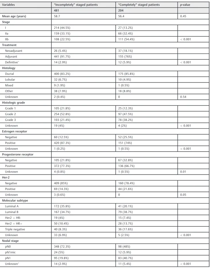
We have identified 753 patients with EBC, with a median age at diagnosis of 57 years (range: 26–93). Most patients (629) had invasive ductal carcinoma (83.5%), histological grade 2 (51.4%), stage II disease (61.8%), no pathological lymph node involvement (pN0) (67.1%), and luminal A or B subtypes (67.9%). At least one imaging procedure for systemic staging was performed in 685 patients (91%) (►Table 1). The patients who underwent high-quality diagnostic procedures (com-puted tomography [CT], magnetic resonance imaging [MRI] or bone scan [BS]) directed to the most common sites of DMs (bone, lung and liver) were defined as the “completely”
staged group (CSG) and represent 29.8% of all staged patients (204 of 685) (►Table 2).
For patients undergoing chest imaging, chest X-ray (CXR) was more frequently performed (57.8%), followed by CT (42.2%). Regarding abdominal imaging, abdominal ultraso-nography (AUS) was the preferred method (59.8%), and bone scan (BS) was the procedure of choice for skeleton staging (96.3%). Additional staging modalities required for confi rma-tion of metastatic disease—including biopsy, imaging or both
—were performed in 19 patients (2.8%).
Among those who underwent any radiologic staging, DMs were detected in 32 patients (4.7%). The bones were the most frequent site of metastases in 20 (62.5%) patients, and all of the cases were identified by conventional BS. Despite CXR and AUS being the most frequently used methods, only one case of DM was diagnosed by these means. The CT scan was responsible for the detection of all cases of lung (10 cases)
and the majority of liver metastases (10 in 11 cases). Metastases were found in 9.3% of the CSG versus 2.7% of the patients not submitted to a“complete”staging.
In the population of staged patients (n¼685), disease stage (p<0.001) and pathological lymph node involvement Table 1 Patient and tumor characteristics
Variables Patients (%) At least one imaging procedure for staging perfomed (%)
Systemic staging not perfomed (%)
753 (100) 685 (91) 68 (9)
Age (years)
<40 52 (6.9) 48 (92.3) 4 (7.7)
40–59 351 (46.6) 327 (92.9) 24 (7.1)
60–69 181 (24.0) 160 (88.4) 21 (11.6)
70 169 (22.5) 150 (88.8) 19 (11.2)
Stage (TNM)
I 288 (38.2) 241 (83.7) 47 (16.3)
II 465 (61.8) 444 (95.5) 21 (4.5)
Treatment
Neoadjuvant 64 (8.5) 63 (98.4) 1 (1.6) Adjuvant 661 (87.8) 596 (90.2) 65 (9.8) Definitive
28 (3.7) 26 (92.9) 2 (7.1)
Histology
Ductal 629 (83.5) 575 (91.4) 54 (8.6)
Lobular 49 (6.5) 42 (85.7) 7 (14.3)
Mixed 11 (1.5) 10 (90.9) 1 (9.1)
Other 62 (8.2) 56 (90.3) 6 (9.7)
Unknown 2 (0.3) 2 (100) 0 (0)
Histologic grade
Grade 1 147 (19.5) 130 (88.4) 17 (11.6)
Grade 2 387 (51.4) 351 (90.7) 36 (9.3)
Grade 3 195 (25.9) 181 (92.8) 14 (7.2)
Unknown 24 (3.2) 23 (95.8) 1 (4.2)
Molecular subtype
Luminal A 240 (31.8) 213 (88.8) 27 (11.2) Luminal B 272 (36.1) 246 (90.4) 26 (9.6)
Her2þHR- 39 (5.2) 34 (87.2) 5 (12.8)
Her2þHRþ 84 (11.2) 78 (92.9) 6 (7.1) Triple negative 80 (10.6) 76 (95) 4 (5)
Unknown 38 (5.1) 38 (100) 0 (0)
Nodal Stage
pN0 505 (67.1) 446 (88.3) 59 (11.7)
pN1mic 38 (5.0) 36 (94.7) 2 (5.3)
pN1 183 (24.4) 178 (97.3) 5 (2.7)
Unknown
27 (3.5) 25 (92.3) 2 (7.7)
Abbreviations: Her2þHRþ, human epidermal growth factor receptor 2 positive and hormonal receptor positive; Her2þHR-, human epi-dermal growth factor receptor 2 positive and hormonal receptor negative; pN0, no pathological nodal invasion; pN1, macrometastatic nodal invasion; pN1mic, micrometastatic nodal invasion; TNM, TNM staging system.
Table 2 Patient and tumor characteristics according to radiologic staging
Variables “Incompletely”staged patients “Completely”staged patients p-value
481 204
Mean age (years) 58.7 56.4 0.45
Stage
I 214 (44.5%) 27 (13.2%)
IIa 159 (33.1%) 66 (32.4%)
IIb 108 (22.5%) 111 (54.4%) <0.001
Treatment
Neoadjuvant 26 (5.4%) 37 (18.1%)
Adjuvant 441 (91.7%) 155 (76%)
Definitive
14 (2.9%) 12 (5.9%) <0.001
Histology
Ductal 400 (83.2%) 175 (85.8%)
Lobular 32 (6.7%) 10 (4.9%)
Mixed 9 (1.9%) 1 (0.5%)
Other 38 (7.9%) 18 (8.8%)
Unknown 2 (0.4%) 0 0.54
Histologic grade
Grade 1 105 (21.8%) 25 (12.3%)
Grade 2 254 (52.8%) 97 (47.5%)
Grade 3 103 (21.4%) 78 (38.2%)
Unknown 19 (4%) 4 (2%) <0.001
Estrogen receptor
Negative 60 (12.5%) 52 (25.5%)
Positive 420 (87.3%) 151 (74%)
Unknown 1 (0.2%) 1 (0.5%) <0.001
Progesterone receptor
Negative 105 (21.8%) 67 (32.8%)
Positive 372 (77.3%) 136 (66.7%)
Unknown 4 (0.8%) 1 (0.5%) 0.01
Her-2
Negative 409 (85%) 160 (78.4%)
Positive 69 (14.3%) 44 (21.6%)
Unknown 3 (0.6%) 0 0.05
Molecular subtype
Luminal A 172 (35.8%) 41 (20.1%)
Luminal B 167 (34.7%) 79 (38.7%)
Her2þHR- 19 (4%) 15 (7.4%)
Her2þHRþ 50 (10.4%) 28 (13.7%)
Triple negative 40 (8.3%) 36 (17.6%)
Unknown 33 (6.9%) 5 (2.5%) <0.001
Nodal stage
pN0 348 (72.3%) 98 (48%)
pN1mic 24 (5%) 12 (5.9%)
pN1 95 (19.8%) 83 (40.7%)
Unknown
14 (2.9%) 11 (5.4%) <0.001
Abbreviations: Her2, human epidermal growth factor receptor 2; Her2þHRþ, human epidermal growth factor receptor 2 positive and hormonal receptor positive; Her2þHR-, human epidermal growth factor receptor 2 positive and hormonal receptor negative; pN0, no pathological nodal invasion; pN1, macrometastatic nodal invasion; pN1mic, micrometastatic nodal invasion.
(p<0.001) were identified as risk factors by univariate analysis. Metastatic disease was proportionally more fre-quent in patients with Her2 positive or TN molecular sub-types, but these findings did not achieve statistical significance (►Table 3).
In an exploratory analysis, the combination of CSG and unfavorable biology (TN or Her2 positive) resulted in a DM rate of 14.4%.
Discussion
In a review of data from prospective and retrospective studies evaluating the role of staging by imaging in cases of EBC (7 studies for stage I and 11 studies for stage II), the presence of occult DMs was rare, with a reported median prevalence of 0.2% (range 0–5.1%) in stage I BC after conven-tional imaging tests (excluding positron emission
tomogra-phy [PET]/CT), and 1.2% (range 0–34.3%) in stage II BC after imaging tests that included PET/CT. Conversely, DMs were found in14% of all stage III BCs.5
Interestingly, our results suggest that the prevalence of occult metastases (4.7%) is slightly higher than in previous studies; however, the rate remains low. Potential explana-tions for ourfindings include: 1) the retrospective nature of most studies could lead to biases caused by poor data quality (such as difficulties in classifying apparent EBC patients who were identified as metastatic by imaging staging but were registered simply as stage IV); 2) heterogeneity between study populations and tumor features (such as when infor-mation about BC subtypes is not reported in older studies); 3) heterogeneity of the imaging methods employed, because the accuracy of imaging methods has improved over time, and might influence the diagnostic ability to detect small metastatic lesions.
Table 3 Association between clinical characteristics and metastases between all patients staged and only“completely”staged patients
All staged patients Metastases p-value “Completely”staged patients Metastases p-value
Variables 685 32 (4.7%) 204 19 (9.3%)
Age (mean) 58.1 56.3 0.47 56.4 54.2 0.31
Stage (TNM)
I 241 3 (1.2%) <0.001 27 1 (3.7%)
IIa 225 7 (3.1%) 66 2 (3.0%)
IIb 219 22 (10.0%) 111 16 (14.4%) 0.02
Histology
Ductal 575 28 (4.9%) 0.61 175 17 (9.7%)
Lobular 42 3 (7.1%) 10 1 (10.0%)
Mixed 10 0 1 0
Other 56 1 (1.8%) 18 1 (5.6%) 1.0
Histologic grade
Grade 1 130 5 (3.8%) 0.83 25 3 (12.0%)
Grade 2 351 17 (4.8%) 97 9 (9.3%)
Grade 3 181 10 (5.5%) 78 7 (9.0%) 0.89
Molecular subtype
Luminal A 213 9 (4.2%) 0.46 41 5 (12.2%)
Luminal B 246 9 (3.7%) 79 5 (6.3%)
Her2þHR- 34 2 (5.9%) 15 1 (6.7%)
Her2þHRþ 78 5 (6.4%) 28 3 (10.7%)
Triple negative 76 6 (7.9%) 36 5 (13.9%) 0.64
Nodal stage
pN0 446 6 (1.3%) <0.001 98 3 (3.1%)
pN1mic 36 1 (2.8%) 12 1 (8.3%)
pN1 178 13 (7.3%) 83 8 (9.6%) 0.13
Abbreviations: Her2þHRþ, human epidermal growth factor receptor 2 positive and hormonal receptor positive; Her2þHR-, human epidermal growth factor receptor 2 positive and hormonal receptor negative; pN0, no pathological nodal invasion; pN1, macrometastatic nodal invasion; pN1mic, micrometastatic nodal invasion; TNM, TNM staging system.
From another perspective, Simos et al14found that most BC patients would not feel comfortable if they were not referred to systemic staging to rule out metastatic diseases, even if the physician recommendation was in compliance with evidence-based guidelines. However, in the same study, when patients were asked about the ranges of chances of detecting metastatic disease, 57.1% chose the range between 6–10%,14an expectation that is clearly in conflict with the data from the literature.7,15In fact, no single cut-off point has ever been established to define when and which methods of imaging should be employed as a function of the clinical impact and cost-effectiveness. Cancer Care Ontario (CCO) subjectively established, during the development of its guidelines, that methods for BC staging should be able to detect DMs in at least 1% of all patients.15Modern methods such as CT and PET/CT have an enhanced performance for the detection of DMs, being more likely to achieve this goal. However, they also have higher costs and are not easily available everywhere.
Few studies directly discussed thefinancial aspects of EBC staging. A recently published retrospective population-based cohort study from Ontario, Canada, assessing the cost of unwarranted imaging in cases of EBC, based on ASCO and CCO recommendations, reported a substantial cost implication. Among 26,547 stage I-II BC patients, around half of them underwent at least 1 imaging test, resulting in an excess cost of 6.8 million Canadian dollars (CA$258.6 per capita). In the present study, isotopic BS represented the number one cost driver.8
While it is important to consider the cost of unnecessary image staging, it is also important to consider the conse-quences of underdiagnosing metastatic diseases at the initial workup. Metastatic BC is indeed an incurable disease, as supported by numerous studies. In a recent Indian study, resection of the primary tumor failed to improve outcomes in metastatic BC patients.16 Therefore, the detection of metastatic diseases—even if in a small fraction of the EBC population—could spare futile surgery and/or radiation ther-apy for the primary tumor as well as other aggressive treat-ments, such as adjuvant chemotherapy (especially for luminal subtypes, as Her2 positive and triple negative breast cancer [TNBC] would need chemotherapy with or without the addition of targeted therapies anyway). This would help cutting costs and avoid unnecessary side effects. Therefore, an accurate estimation of the risk of occult DMs in cases of EBC is critically important. With the risk being negligible, the adherence to the ASCO guidelines and to the guidelines of other societies will likely be enhanced.
As compared with CXR, CT has a higher capacity to detect small nodules. The American College of Radiology (ACR) recommends CT scans for tumor types with higher propen-sity for lung metastases, such as BC, even in the presence of a normal CXR.17Regarding the detection of liver metastases, the sensitivity of AUS ranges from 50 to 76%, versus 68 to 85% for CT.18In the present study, many patients with presumed stage I or II BC did not undergo CT, which may be a problem due to the low accuracy of AUS and CXR. Our results are in line with these recommendations, with all cases of lung
metastases identified by CT, and only 1 out of 11 liver metastases identified by AUS (10 were identified by CT).
Nodal involvement is a strong and independent negative prognostic factor. In a recent study, the 5-year survival rate of non-metastatic patients was 85%, and, for patients with or without lymph node involvement, 99%.1Even tumors smaller than 2 cm have a worse prognosis if there is lymph node involvement; in 1 series of 24,740 patients, the 5-year survival rate was 96%, 86% and 66% with none, 1–3 and more than 4 nodes involved respectively.19The prognostic significance of micrometastatic nodal invasion (pathological lymph node involvement [pN1] micromestatasis; invasive component
>0.2 mm and/or involving more than 200 cells, but not<2.0 mm) remains under investigation, but it has been suggested by some studies.20–22Our results are in line with these data, indicating a higher risk of occult DMs in cases of EBC with nodal involvement (1.3% in no regional lymph node metastasis [pN0], 2.8% in pN1 micrometastasis, and 7.3% in pN1). In the present study, pathological involvement of lymph nodes (pNþ) outperformed cancer stage as a risk factor for the presence of DM (1.3% for stage I and 6.5% for stage II).
Limited data are currently available about the molecular classification of BC and the prevalence of occult DMs in cases of EBC. A Chinese retrospective study with information on molecular subtype in 3,411 patients with stage I–III BC showed that luminal a, luminal b, luminal B plus Her2, Her2 overexpression, and basal-like have a statistically dif-ferent risk for bone (1.4%, 0.7%, 2.5%, 2.7%, and 0.9% respec-tively; p<0.05), liver (0.1%, 0.1%, 1.0%, 1.1%, and 0.9% respectively;p<0.01) and lung metastases (0.2%, 0%, 0%, 0.27%, and 0.9% respectively;p<0.05); however, the risk of occult metastases was generally low for all subtypes.6 Con-versely, we report a numerically, non-statistically significant higher rate of DM in Her2 positive and TN disease (6.4% in Her2 positive and HR positive; 5.9% in Her2 positive and HR negative, and 7.9% in TN). The lack of statistical significance could simply be due the small sample size for subgroup analyses.
We speculated that the combination of unfavorable biology and lymph node involvement could indicate a group of EBC patients at a higher risk of having DM, and performed exploratory analyses combining high-risk tumors (higher pretest value) with high-sensitivity imaging (►Table 3). In this group, we detected an occult DM rate of 14.4%. Due to the heterogeneity between the“completely” and “ incom-pletely”staged population and to the small relative size of the CSG (►Table 2), it was difficult to establish the relative contribution of each factor (tumor features and radiologic methods).
conceivable that a large, prospective, randomized clinical trial would definitively clarify the role of modern imaging studies in this setting. However, due to competing funding priorities, it is unlikely that such a trial will ever be per-formed. Finally, radiological evaluation with CT, MRI or PET-CT are certainly more accurate, but the expenses related to these procedures also need to be measured in a cost-effec-tiveness study before they are implemented into the routine practice. Despite these limitations, our results strongly sug-gest that, should an imaging staging be required in EBC patients, BS and either CT or MRI (or PET-CT) may be the preferred procedures.
Conclusion
Regardless of the recommendations against image staging in cases of EBC, 91% of our patients underwent radiologic staging. Despite having found a slightly higher prevalence than previ-ously reported, our results confirm that stage I–II BC has a low rate of metastatic disease at presentation (4.7% of staged patients). The most frequent metastatic site was the bones (3.9%), followed by the liver (1.8%) and the lungs (1.6%). Although frequently used in the clinical practice, CXR and AUS were futile methods in our hands–almost all metastatic lesions were diagnosed by BS and/or CT scan. Stage II and pNþ
were identified as risk factors in the univariate analyses, while Her2 positive and the TN subtype showed only a non-signifi -cant trend. When focusing only on patients staged with highly effective methods, stage I–II patients with unfavorable biology (TN or Her2 positive) had a DM prevalence of 14.4%, one of the highest reported in these stages of the disease. Overall, our results confirm that systemic staging of asymptomatic, unse-lected patients with stage I–II BC is not warranted as a routine practice. However, we have identified subgroups of patients to whom a full staging could be indicated.
Contributors
Soares G. P., Pereira A. A. L., Boas M. S. V., Vaisberg V. V., Magalhães M. C. F., Linck R. D. M. and Mano M. S. contributed with the project and interpretation of data, writing of the article, critical review of the intellectual content andfinal approval of the version to be published.
Conflicts of Interest
The authors have no conflicts of interest to declare.
References
1 Siegel RL, Miller KD, Jemal A. Cancer statistics, 2015. CA Cancer J Clin 2015;65(01):5–29. Doi: 10.3322/caac.21254
2 Edge SB, Compton CC. The American Joint Committee on Cancer: the 7th edition of the AJCC cancer staging manual and the future of TNM. Ann Surg Oncol 2010;17(06):1471–1474
3 Ravaioli A, Pasini G, Polselli A, et al. Staging of breast cancer: new recommended standard procedure. Breast Cancer Res Treat 2002; 72(01):53–60
4 Barrett T, Bowden DJ, Greenberg DC, Brown CH, Wishart GC, Britton PD. Radiological staging in breast cancer: which asympto-matic patients to image and how. Br J Cancer 2009;101(09): 1522–1528. Doi: 10.1038/sj.bjc.6605323
5 Brennan ME, Houssami N. Evaluation of the evidence on staging imaging for detection of asymptomatic distant metastases in newly diagnosed breast cancer. Breast 2012;21(02):112–123. Doi: 10.1016/j.breast.2011.10.005
6 Chen X, Sun L, Cong Y, et al. Baseline staging tests based on molecular subtype is necessary for newly diagnosed breast cancer. J Exp Clin Cancer Res 2014;33:28. Doi: 10.1186/1756-9966-33-28 7 Linkugel A, Margenthaler J, Dull B, Cyr A. Staging studies have limited utility for newly diagnosed stage I-II breast cancer. J Surg Res 2015;196(01):33–38. Doi: 10.1016/j.jss.2015.02.065 8 Thavorn K, Wang Z, Fergusson D, van Katwyk S, Arnaout A,
Clemons M. Cost implications of unwarranted imaging for distant metastasis in women with early-stage breast cancer in Ontario. Curr Oncol 2016;23(Suppl 1):S52–S55. Doi: 10.3747/co.23.2977 9 Senkus E, Kyriakides S, Ohno S, et al; ESMO Guidelines Committee. Primary breast cancer: ESMO Clinical Practice Guidelines for diagnosis, treatment and follow-up. Ann Oncol 2015;26(Suppl 5): v8–v30. Doi: 10.1093/annonc/mdv298
10 Anderson BO. Breast cancer–thinking globally. Science 2014;343 (6178):1403. Doi: 10.1126/science.1253344
11 Schnipper LE, Smith TJ, Raghavan D, et al. American Society of Clinical Oncology identifies five key opportunities to improve care and reduce costs: the topfive list for oncology. J Clin Oncol 2012;30(14):1715–1724. Doi: 10.1200/JCO.2012.42.8375 12 Simos D, Hutton B, Clemons M. Are physicians choosing wisely
when imaging for distant metastases in women with operable breast cancer? J Oncol Pract 2015;11(01):62–68. Doi: 10.1200/ JOP.2014.000125
13 Brothers JM, Kidwell KM, Brown RK, Henry NL. Incidental radi-ologicfindings at breast cancer diagnosis and likelihood of disease recurrence. Breast Cancer Res Treat 2016;155(02):395–403. Doi: 10.1007/s10549-016-3687-1
14 Simos D, Hutton B, Graham ID, et al. Patient perceptions and expectations regarding imaging for metastatic disease in early stage breast cancer. Springerplus 2014;3:176. Doi: 10.1186/2193-1801-3-176
15 Myers RE, Johnston M, Pritchard K, Levine M, Oliver T; Breast Cancer Disease Site Group of the Cancer Care Ontario Practice Guidelines Initiative. Baseline staging tests in primary breast cancer: a practice guideline. CMAJ 2001;164(10):1439–1444 16 Badwe R, Hawaldar R, Nair N, et al. Locoregional treatment versus
no treatment of the primary tumour in metastatic breast cancer: an open-label randomised controlled trial. Lancet Oncol 2015;16 (13):1380–1388. Doi: 10.1016/S1470-2045(15)00135-7 17 Mohammed TL, Chowdhry A, Reddy GP, et al; Expert Panel on
Thoracic Imaging. ACR Appropriateness Criteria® screening for pulmonary metastases. J Thorac Imaging 2011;26(01):W1–W3. Doi: 10.1097/RTI.0b013e3182010bf9
18 Cantisani V, Grazhdani H, Fioravanti C, et al. Liver metastases: Contrast-enhanced ultrasound compared with computed tomo-graphy and magnetic resonance. World J Gastroenterol 2014;20 (29):9998–10007. Doi: 10.3748/wjg.v20.i29.9998
19 Carter CL, Allen C, Henson DE. Relation of tumor size, lymph node status, and survival in 24,740 breast cancer cases. Cancer 1989;63 (01):181–187. Doi: 10.1002/1097-0142(19890101)63:1<181:
AID-CNCR2820630129>3.0.CO;2-H
20 Andersson Y, Frisell J, Sylvan M, de Boniface J, Bergkvist L. Breast cancer survival in relation to the metastatic tumor burden in axillary lymph nodes. J Clin Oncol 2010;28(17):2868–2873. Doi: 10.1200/JCO.2009.24.5001
21 de Boer M, van Dijck JA, Bult P, Borm GF, Tjan-Heijnen VCG. Breast cancer prognosis and occult lymph node metastases, isolated tumor cells, and micrometastases. J Natl Cancer Inst 2010;102 (06):410–425. Doi: 10.1093/jnci/djq008