Alberto Consolaro1,2, Renata Bianco Consolaro3
How to cite: Consolaro A, Consolaro RB. There is no pulp necrosis or calcic metamorphosis of pulp induced by orthodontic treatment: biological basis. Den-tal Press J Orthod. 2018 July-Aug;23(4):36-42.
DOI: https://doi.org/10.1590/2177-6709.23.4.036-042.oin
Submitted: May 18, 2018 - Revised and accepted: May 29, 2018
» The authors report no commercial, proprietary or financial interest in the products or companies described in this article.
Contact address: Alberto Consolaro E-mail: consolaro@uol.com.br
There is no pulp necrosis or calcific metamorphosis of
pulp induced by orthodontic treatment:
biological basis
1 Universidade de São Paulo, Faculdade de Odontologia de Bauru (Bauru/SP, Brazil). 2 Universidade de São Paulo, Faculdade de Odontologia de Ribeirão Preto, Programa
de Pós-graduação em Odontopediatria (Ribeirão Preto/SP, Brazil).
3 Centro Universitário de Adamantina (Adamantina/SP, Brasil).
To biologically explain why the orthodontic treatment does not induce pulp necrosis and calcific metamorphosis of the pulp, this paper presents explanations based on pulp physiology, microscopy and pathology, and especially the cell and tissue phe-nomena that characterize the induced tooth movement. The final reflections are as follows: 1) the orthodontic movement does not induce pulp necrosis or calcific metamorphosis of the pulp; 2) there is no literature or experimental and clinical models to demonstrate or minimally evidence pulp alterations induced by orthodontic movement; 3) when pulp necrosis or calcific metamorphosis of the pulp is diagnosed during orthodontic treatment or soon after removal of orthodontic ap-pliances, its etiology should be assigned to concussion dental trauma, rather than to orthodontic treatment; 4) the two pulp disorders that cause tooth discoloration in apparently healthy teeth are the aseptic pulp necrosis and calcific metamorphosis of the pulp, both only induced by dental trauma; 5) the concussion dental trauma still requires many clinical and laboratory studies with pertinent experimental models, to increasingly explain its effects on the periodontal and pulp tissues.
Keywords: Dental concussion. Tooth movement. Orthodontics. Pulp necrosis. Calcific metamorphosis of the pulp. DOI: https://doi.org/10.1590/2177-6709.23.4.036-042.oin
Para fundamentar biologicamente por que o tratamento ortodôntico não induz necrose pulpar e metamorfose cálcica da polpa, apresentou-se explicações com base na fisiologia, microscopia e patologia pulpar, bem como, e principalmente, nos fenômenos celulares e teciduais que caracterizam a movimentação dentária induzida. As reflexões finais foram: 1) o movi-mento ortodôntico não induz necrose pulpar ou metamorfose cálcica da polpa; 2) não há literatura e modelos experimen-tais e clínicos que comprovem ou minimamente evidenciem alterações pulpares induzidas pelo movimento ortodôntico; 3) quando a necrose pulpar ou metamorfose cálcica da polpa for diagnosticada durante o tratamento ortodôntico ou logo após a remoção dos aparelhos ortodônticos, a sua etiologia deve ser atribuída ao traumatismo dentário do tipo concussão, e não ao tratamento ortodôntico; 4) as duas doenças pulpares que levam ao escurecimento coronário em dentes aparentemente hígidos são a necrose pulpar asséptica e a metamorfose cálcica da polpa, ambas induzidas exclusivamente pelo traumatismo dentário; 5) o traumatismo dentário do tipo concussão requer, ainda, muitos estudos clínicos e laboratoriais, com modelos experimentais pertinentes, para fundamentar cada vez mais os seus efeitos sobre os tecidos periodontais e pulpares.
WHAT TO READ AND WHO TO LISTEN TO
ABOUT THIS SUBJECT?
The search for biological and clinical basis of pulp al-terations induced by orthodontic forces is almost always conducted using keywords as “pulp”, “pulp changes”, “pulp pathologies”, “pulp biology”, “Endodontics” and other keywords related to the pulp. Similarly, when ask-ing someone on the “possible changes, opinions, dog-mata and beliefs” on the effect of orthodontic treatment on the pulp tissues, it is very common to search for who putatively investigates and researches the pulp biology and diseases – almost always, endodontists. This occurs because, simply put, there seems to be a logical direct relationship with the dental pulp.
However, if we want to find researches to achieve basis and search for specialists to whom we should listen, we should search for those who specifically in-vestigate the periodontal ligament, because the orth-odontic movement is an exclusive phenomenon of the periodontal ligament, rather than of the dental pulp. Textbooks of Endodontics and Dental Traumatology rarely discuss the biology of orthodontic movement, which occurs in several texts of Periodontology.
To deeply understand why the dental pulp is not affected by orthodontic movement, there is the need
to deepen into periodontal biology and changes, since the pulp does not participate in tooth movement.
THE ACTIVE ORTHODONTIC FORCES ARE
NECESSARILY LIGHT AND MODERATE
The orthodontic movement is achieved by the application of forces on the tooth, promoting a bio-logical stirring of periodontal ligament cells, known as cellular stress, which may evolve to a mild initial
inflammation for some hours or days,1 characterized
by a mild inflammatory exudate and incipient inflam-matory infiltrate (Figs 1 to 4).
The orthodontic forces are very light in any situ-ation, because the goal is to induce these phenomena of cellular stress and periodontal ligament inflamma-tion in the periodontal ligament, which represents a membranous structure of only 0.25mm – which cor-responds to the thickness of a paper leaf – formed by specialized fibrous connective tissue.1,2
Besides being very light and moderate, the forc-es applied on the tooth over the periodontal liga-ment are dissipating, i.e. they gradually reduce in intensity and disappear in 2 to 7 days, allowing periodontal reorganization, with return to normal-ity between 10 to 15 days after the activation of
orthodontic appliances.1
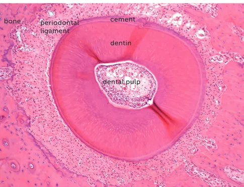
Figure 1 - Microscopic aspects of rat molar root in axial or transverse section, revealing the root structures, including the pulp, alveo-lar bone and periodontal ligament (HE, 10X magnification).
bone periodontal
ligament
cement
dentin
of the periodontal space, which is narrowed by the
tooth compression on its structures.1 This
phenom-enon is stimulated by the chemical mediators released by the compressed and hypoxic cells in the periodon-tal ligament.
After 7 to 10 days, there are nearly no active forc-es moving the teeth in which the orthodontic forcforc-es were applied. After this period, the periodontal phe-nomena are predominantly reparative, to reorganize the normality of tissues, preparing them to receive another cycle of forces.
Since the first minutes, it is not possible to break the vascular and neural bundles that cross the peri-odontal ligament and penetrate into the apical
fora-men to nourish the pulp tissue with blood.3 There
are no sudden orthodontic movements that might promote partial or total lesions in the blood vessels responsible for the pulp blood supply.
THE SEVERE OR HEAVY ORTHODONTIC
FORCES ARE UNABLE TO MOVE THE TEETH
Since the first moment, the orthodontic forces are dissipating and tend to disappear after 5 to 7 days, in a gradual and decreasing process regarding their intensity.
Immediately, the orthodontic force applied on the tooth is reduced or partly dissipated by two mechanisms:
1. The liquids and gel represented by the ex-tracellular matrix are displaced to the marrow and perivascular spaces, partially cushioning the effects of the orthodontic forces applied. This represents a physiological mechanism of the periodontal
liga-ment to cushion the heavy masticatory forces.1,2
Considering that this applies to the occlusal loads, which are very intense and incomparably extreme or heavy, by deduction it may be inferred that they apply even more to orthodontic forces, which are extremely light or moderate.
2. Usually, the orthodontic forces applied are re-duced in 20 to 30% almost immediately after appli-cation, because the alveolar bone crest, which sup-ports the tooth to be moved, undergoes a deflection or deformation due to its elastic or plastic capacity1. The alveolar bone tissue is thin and, alike any bone tissue, it presents a high ratio of organic components, liquids and cells.
Over 10 to 12 hours, several clasts or osteoclasts appear on the periodontal bone surface, which initi-ate the process of bone resorption and enlargement
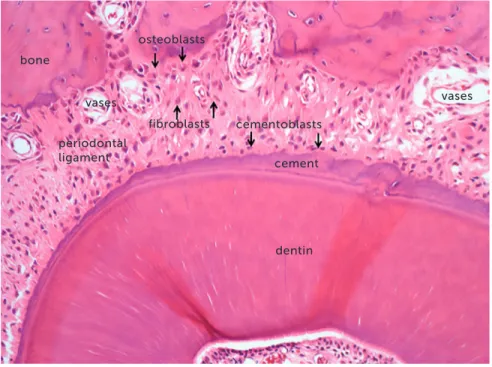
Figure 2 - Microscopic aspects, at greater magnification, of the same rat molar root in axial or transverse section of Figure 1, reveal-ing the root structures, includreveal-ing the pulp, alveolar bone and periodontal ligament (HE, 40X magnification).
periodontal ligament
cementoblasts bone
vases osteoblasts
fibroblasts vases
cement
The orthodontic movement of a tooth requires an alive or biologically viable periodontal ligament, that may receive and nourish the clasts that will promote bone resorption on the periodontal surface of the
tooth socket (Figs 1 to 4). Without vessels with blood supply, without extracellular matrix and mediators, there are no tools necessary for tooth movement in the bone (Figs 5 and 6).
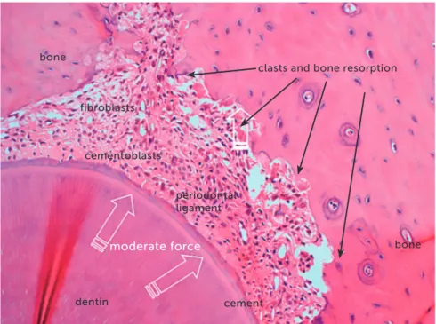
Figure 3 - Microscopic aspects of rat molar root, in axial section, after 4 days under mod-erate orthodontic forces that induced the re-lease of mediators to stimulate the clastic ac-tivity on the periodontal bone surface. In this situation, tooth movement occurs slowly, and the forces dissipate without inducing changes in the vascular and neural bundles that pen-etrate into the pulp via the apical foramen (HE, 25X magnification).
Figure 4 - Microscopic aspects of rat molar root presented in Figure 3, at greater magnifi-cation, in axial section, after 4 days under mod-erate orthodontic forces. The image reveals the activity of clasts and other cells, thanks to the maintenance of periodontal structures, without hyalinization (HE, 40X magnification).
periodontal ligament cementoblasts
clasts and bone resorption bone
bone fibroblasts
cement dentin
moderate force
periodontal ligament
cementoblasts
clasts and bone resorption
vases
bone fibroblasts
cement dentin
When the forces are very severe or heavy, due to accidental or intentional application of forces that compress the periodontal ligament to an extent enough to occlude the periodontal blood vessels, the
cells migrate to neighboring areas to the site of anoxia (Figs 5 and 6).
The orthodontic movement never occurs suddenly! This sudden characteristic is observed in dental
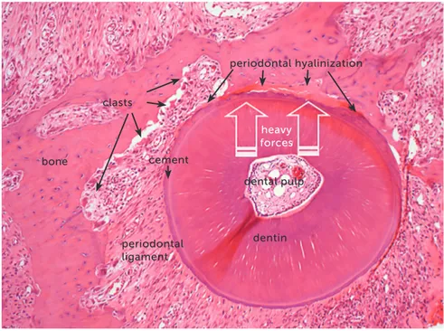
trau-Figure 5 - Microscopic aspects of rat molar root presented in axial section after 4 days un-der heavy orthodontic forces, which induced hyalinization of a segment of periodontal liga-ment. In this situation there is no tooth move-ment, because the clasts are unable to resorb the bone on the periodontal surface (HE, 10X magnification).
periodontal ligament
dental pulp
bone cement
clasts
periodontal hyalinization
dentin heavy forces
Figure 6 -Microscopic aspects of the same rat molar root of Figure 5, at greater magnifica-tion, in axial section after 4 days under heavy orthodontic forces, which induced hyaliniza-tion of a segment of periodontal ligament. In this situation there is no tooth movement, because the clasts are unable to resorb the bone on the periodontal surface (HE, 40X magnification).
periodontal ligament bone
cement periodontal hyalinization
dentin heavy
ma, not in orthodontic movement. The orthodontic movement and dental trauma promote completely dif-ferent tissue changes, even though both are caused by forces, which are yet entirely different concerning their intensity, duration and field of action. In the area where heavy forces act on the periodontal ligament, there will remain only the extracellular matrix, without cells nor blood vessels with blood supply, which will be occlud-ed. At this site, the clasts are unable to resorb the alveo-lar bone and it is impossible to enalveo-large the periodontal space and achieve tooth movement. Microscopically, these areas that only present extracellular matrix, with-out cells, are named hyaline areas, or the process is iden-tified as periodontal hyalinization (Figs 5 and 6).
That is to say: if the forces are not light or
mod-erate, there is no tooth movement;1,3 i.e., severe or
heavy forces do not allow orthodontic tooth move-ment. Therefore, if heavy forces are applied by pro-fessionals without proper orthodontic training, the risk of pulp necrosis and calcific metamorphosis of the pulp is reduced to zero.
ORTHODONTIC MOVEMENT AND DENTAL
TRAUMA: THEY ARE NOT COMPARABLE
Once again: the orthodontic movement never occurs suddenly! This sudden characteristic is ob-served in dental trauma, not in orthodontic move-ment. The orthodontic movement and dental trauma promote completely different tissue changes, even though both are caused by forces, which are yet en-tirely different concerning their intensity, duration and field of action.
Similarly, since the pulp alterations supposedly as-signed to orthodontic treatment are actually related to dental trauma, it is pertinent to investigate the literature or question investigators of dental trauma about this issue.
Dental concussion is the type of dental trauma that may lead to silent aseptic pulp necrosis and calcific
metamorphosis of the pulp.1,3,4 During orthodontic
treatment, if there is aseptic pulp necrosis or calcific metamorphosis of the pulp, it may be surely stated that the cause was dental concussion.
The forces of dental trauma induce sudden move-ments of teeth in the socket, possibly damaging the vascular and neural bundles that penetrate into the
apical foramen to nourish the pulp tissues.4
As previously mentioned in this paper, the orthodon-tic forces are dissipating, and the effective occurrence of tooth movement requires light or moderate forces. The intense forces cause hyalinization of the periodontal liga-ment and do not allow the tissue and cellular phenomena that characterize the tooth movement.
When specialists and investigators of dental trau-ma are questioned on the periodontal and pulp ef-fects of dental concussion, their responses are usually evasive. The literature about dental concussion and its tissue effects is still underestimated in the field of dental traumatology, which understandably still fo-cuses on experimental models of fracture, luxation, avulsion and replantation.
One reason is the clinical relevance of these most severe types of dental trauma. Another reason is the difficulty to establish clinical and laboratory experi-mental models to reproduce dental concussion, con-sidering its incipience and subtility.4
Dental concussion is the type of dental trauma that does not immediately induce clinical changes af-ter a knock on the affected teeth, and some cases only present mild painful symptomatology or discomfort for some hours, which disappear spontaneously.
The patient that suffers a concussion does not consciously remind the dental trauma and will rarely report it during an anamnesis after some months or years. The chief complaint of the patient that suf-fered concussion will only appear after some months
or years, with discoloration of the tooth crown.3
The coronal discoloration of apparently healthy teeth may only be caused by two pulp pathologies, both induced by the same cause — dental trauma, especially concussion —, namely: aseptic pulp
ne-crosis and calcific metamorphosis of the pulp.3,5,6
TWO EXAMPLES OF MORPHOLOGICAL
EVIDENCES IN HUMANS AND ANIMALS
In 2000, Valadares Neto7 microscopically
ana-lyzed the dentin-pulp complex and external root surfaces of human teeth of twelve teenagers, ex-tracted after rapid maxillary expansion, and com-pared them to the teeth of other three teenagers not submitted to tooth movement. The following could be concluded about the dentin-pulp complex:
2. There was no dentin and pulp alteration, con-sidering two (0.45mm) and four (0.9mm) daily acti-vations of the expanding screw.
3. The rapid maxillary expansion using a modi-fied Haas expander was considered a biologically safe procedure for the dentin-pulp complex.
It should be highlighted that the forces employed in rapid maxillary expansion were sufficiently intense to hyalinize the buccal periodontal ligament and pre-clude the tooth movement. Despite the severe orth-odontic forces applied on the teeth, there were no microscopic pulp alterations.
In 2005, Consolaro8 described the tooth
move-ment in 39 rats, with periods of 1 to 7 days, using
the model initially developed by Heller and Nanda,9
recognized worldwide as the most employed in stud-ies on this subject. The study analyzed pulp tissues,8,10 compared to those of teeth of other 9 animals not sub-mitted to tooth movement. It was concluded that in-duced tooth movement did not promote morphologi-cal alterations in the dental pulp detectable by light microscopy, either degenerative or inflammatory.
Frequently, due to the methodological limitations of studies on the pulp or due to the experimental model employed, studies attempt to detect alterations in molecular oxygenation, as well as biochemical and enzymatic changes, in orthodontically moved pulps, yet the results are not visible and associated with de-tectable morphological alterations, and even less mi-croscopically.11
In nearly all studies on photomicrographs, the dental pulps of orthodontically moved teeth are mor-phologically normal.
CONCLUDING REMARKS
1. The orthodontic movement does not induce pulp necrosis or calcific metamorphosis of the pulp.
2. There is no literature nor experimental and clini-cal models that demonstrate or minimally evidence pulp alterations induced by orthodontic movement.
3. When pulp necrosis or calcific metamorphosis of the pulp is diagnosed during orthodontic treat-ment or soon after removal of orthodontic appliances, its etiology should be assigned to concussion dental trauma, rather than to orthodontic treatment.
4. The two pulp pathologies that cause coronal discoloration in apparently intact teeth are aseptic pulp necrosis and calcific metamorphosis of the pulp, both exclusively induced by tooth movement.
5. The concussion dental trauma still requires fur-ther clinical and laboratory studies using pertinent experimental models, to provide more information on its effects on the periodontal and pulp tissues.
REFERENCES
1. Consolaro A. Reabsorções dentárias nas especialidades clínicas. 3a ed.
Maringá: Dental Press; 2012.
2. Osborn JW, Ten Cate AR. Histologia dental avançada. 4a ed. São Paulo:
Quintessence; 1988.
3. Consolaro A. Biopatologia da polpa e do periápice. Maringá: Dental Press;
2018.
4. Queiroz AF, Hidalgo MM, Consolaro A, Panzarini SR, França AB, Pires WR,
et al. Calcific metamorphosis of pulp after extrusive luxation. Dent Traumatol. 2018. In press.
5. Consolaro A. Tratamento ortodôntico não promove necrose pulpar. Dental
Press Endod. 2011 Jan-Mar;1(1):1-11.
6. Consolaro A. Alterações pulpares induzidas pelo tratamento ortodôntico:
dogmas e falta de informações. Rev Dental Press Ortod Ortop Facial. 2007 Jan-Fev;12(1):15-7.
7. Valadares Neto J. Análise microscopia do complexo dentinopulpar e da
superfície radicular externa após a expansão rápida da maxila em adolescents [dissertação]. Goiania (GO): Universidade Federal de Goiás; 2000.
8. Consolaro RB. Análise do complexo dentinopulpar em dentes submetidos
à movimentação dentária induzida em ratos. [dissertação]. Bauru (SP): Universidade de São Paulo; 2005.
9. Heller IJ, Nanda R. Effect of metabolic alteration of periodontal fibers on
orthodontic tooth movement. An experimental study. Am J Orthod. 1979 Mar;75(3):239-58.
10. Massaro CS, Consolaro RB, Santamaria Jr M, Consolaro MFMO, Consolaro A. Analysis of the dentin-pulp complex in teeth submitted to orthodontic movement in rats. J Appl Oral Sci. 2009;17(Spec. issue):35-42. 11. Santamaria M Jr, Milagres D, Stuani AS, Stuani MB, Ruellas AC. Initial