R E S E A R C H
Open Access
Presynaptic neuromuscular action of a methanolic
extract from the venom of
Rhinella schneideri
toad
Sandro Rostelato-Ferreira
1*, Cháriston A Dal Belo
2, Gildo B Leite
1, Stephen Hyslop
1and Léa Rodrigues-Simioni
1Abstract
Background:Rhinella schneideri, previously known asBufo paracnemis,is a common toad in many regions of Brazil. Its venom exerts important cardiovascular effects on humans and other animals. Although this toad venom has been the subject of intense investigations, little is known about its neuromuscular activity.
Methods:The neurotoxicity of a methanolic extract ofR. schneiderivenom was tested on mouse phrenic nerve-diaphragm (PND) preparations mounted for conventional twitch tension recording–in response to indirect stimulation–and for electrophysiological measurements.
Results:Venom extract (50μg/mL) increased the muscle twitch tension in PND preparations but did not significantly
alter the resting membrane potential values. Electrophysiological evaluations showed that the extract (50μg/mL)
significantly augmented the frequency of miniature end-plate potential (from 38 ± 3.5 to 88 ± 15 after 60 minutes; n = 5; p< 0.05) and quantal content (from 128 ± 13 to 272 ± 34 after five minutes; n = 5;p< 0.05). Pretreatment with ouabain (1μg/mL) for five minutes prevented the increase in quantal content (117 ± 18 and 154 ± 33 after five and 60 minutes,
respectively).
Conclusion:These results indicate that the methanolic extract ofR. schneiderivenom acts primarily presynaptically to enhance neurotransmitter release in mouse phrenic-diaphragm preparations.
Keywords:Neurotransmitter release, Ouabain, Presynaptic,Rhinella schneideri, Toad venom
Background
Amphibians produce cutaneous secretions that serve as a defense against predators [1]. Toads in particular have highly toxic venom that is produced by well-developed postorbital parotid glands [2]. The cardiovascular effects of toad venom on vertebrates are well known and have been extensively investigated, particularly in the genus Rhinella [3,4]. The main venom toxins responsible for its cardiac ef-fects are bufadienolides and bufotoxins that increase car-diac contractility and decrease carcar-diac rate by inhibiting the Na+/K+-ATPase pump in a manner similar to digoxin [3,4]. In contrast, little is known about the neuromuscular activity of toad venoms, although it was previously observed that envenoming in dogs may be accompanied by neurological
manifestations such as mydriasis, nystagmus and opisthot-onus [5].
Rhinella schneideri(formerly known asBufo paracnemis Lutz, 1925) is a common toad in several South American countries [6]. A previous study observed that rats injected intraperitoneally withR. schneiderivenom (2–5 mg) show uncoordinated movements, dyspnea, convulsions and par-alysis, followed by respiratory and cardiac arrest [7]. This sequence of events indicates that in rats neurotoxic mani-festations precede cardiac effects, an order that is peculiar to this venom. In the present work, we examined the neurotoxicity of a methanolic extract obtained from Brazil-ianR. schneiderivenom on mouse neuromuscular prepara-tionsin vitro.
Methods
Venom was collected by manual compression of large postorbital parotid glands from two toads. An amount of
* Correspondence:sandrorostelato@yahoo.com.br
1Departamento de Farmacologia, Faculdade de Ciências Médicas,
Universidade Estadual de Campinas (UNICAMP), CP 6111, Campinas, SP 13083-970, Brasil
Full list of author information is available at the end of the article
2 g was then extracted with methanol (50 mL) for three days at room temperature, after which the resulting ex-tract was lyophilized in a SpeedVac® centrifuge (Savant, USA) [8]. The methanolic extract was lyophilized and dissolved in Tyrode solution prior to testing on neuro-muscular preparations.
Male Swiss white mice (25–30 g) were obtained from the Multidisciplinary Center for Biological Investigation (CEMIB/UNICAMP). The animals were housed at 23 ± 3°C on a 12-hour light/dark cycle with free access to food and water.
The diaphragm and its phrenic nerve were dissected from male Swiss mice killed with isoflurane (Cristália, Brazil). The preparations were mounted under a resting tension of 5 g in a 5 mL organ bath containing aerated (95% O2and
5% CO2) Tyrode solution (composition, in mM: NaCl 137,
KCl 2.7, CaCl2 1.8, MgCl2 0.49, NaH2PO4 0.42, NaHCO3 11.9 and glucose 11.1) at 37°C, as described by Bülbring [9]. Supramaximal stimuli (0.1 Hz and 0.2 ms for indirect stimulation) were delivered from a Grass S88 stimulator (Grass Instrument Co., USA) and the muscle twitches were recorded using a Load Cell BG-25 g force displacement transducer coupled to a Gould RS 3400 recorder (both from Gould Inc., USA). The preparations were allowed to stabilize for at least 20 minutes before the addition of meth-anolic extract (50μg/mL).
End-plate potentials (EPPs), miniature end-plate po-tentials (MEPPs) and resting membrane popo-tentials (RPs) were measured with a high input impedance electrom-eter (World Precision 750, USA) in mouse diaphragm muscle preparations using conventional microelectrode techniques. The dissected muscle was mounted in a lucite chamber containing aerated (95% O2and 5% CO2)
Tyrode solution (pH 7.4, at room temperature of 23-27°C) with or without methanolic extract (50μg/mL).
Intracellu-lar microelectrodes filled with 3 M KCl (resistance 15–25 MΩ) were used. The EPPs, MEPPs and muscle RPs were recorded on an oscilloscope (Tektronix, USA) and subse-quently documented as described below. The RP recordings were taken at the end-plate regions in the absence or pres-ence of methanolic extract at t0 (basal), t5, t15, t30 and t60 minutes.
EPPs were recorded in muscles previously subjected to the cut muscle technique in order to uncouple muscle con-tractions from nerve stimulation. [10]. A direct-current channel was used to record the RPs and an alternate-current channel was used to record the EPPs. The EPPs were magnified (AM 502 Tektronix amplifier, gain ¼ 100), low-pass filtered (3 kHz) and digitized (15 kHz sampling rate) using an analog-to-digital converter (Lynx, Brazil; CAD12/36, resolution: 12 bits) coupled to a microcom-puter (Microtec, Brazil) loaded with AqDados 5 software (Lynx) that enabled digital storage of the EPPs online and their subsequent retrieval for measurement and analysis.
For measurement of the quantal content of EPPs, a stimulus rate of 1 Hz for one minute was generated at t0 (basal), t5, t15, t30 and t60 minutes and 30 to 60 potentials were measured at each interval. The quantal content (QC) was estimated as the quotient between the squared average of the EPPs and the variance of the EPPs (indirect method), as described by Dal Beloet al.[11].
MEPPs were recorded in uncut muscle without gener-ating electric stimuli. MEPP measurements were ob-tained before (t0) and at various intervals (t5, t15, t30 and t60) after methanolic extract addition.
Each experimental protocol was repeated 3 to 8 times and the results were reported as the mean ± S.E.M. Stu-dent’s t-test and repeated-measures analysis of variance (ANOVA) were used for statistical comparison of the data, with a value ofp< 0.05 indicating significance. All data analyses were done using OriginPro 8.
Results and discussion
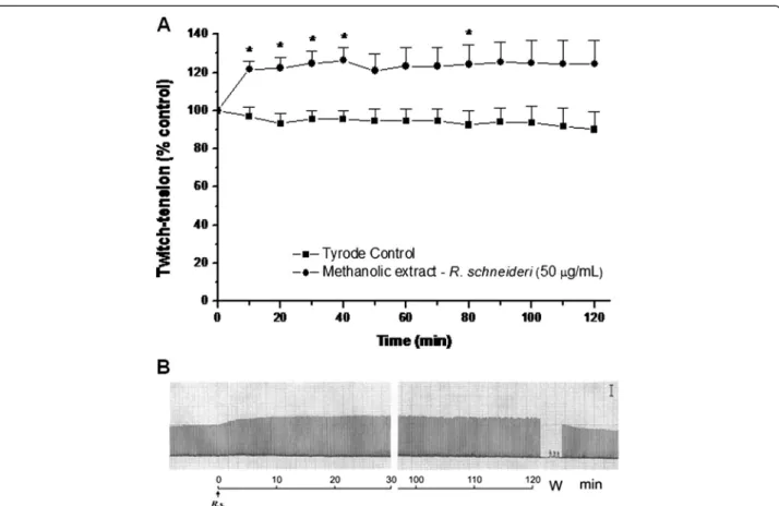
The methanolic extract (50 μg/mL) produced an increase
in muscle twitch tension in PND preparations (Figure 1) during a 120-minute observation. There was little concen-tration dependence in this effect since the responses at lower (25 μg/mL) and higher (200 μg/mL) concentrations
of extract were not significantly different from those ob-served with 50μg/mL (data not shown).
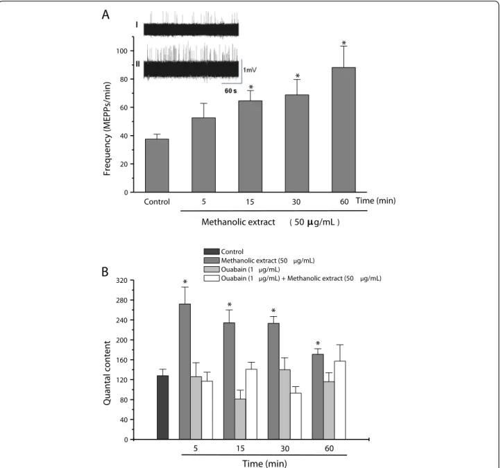
The methanolic extract (50 μg/mL) significantly
in-creased the frequency of MEPPs from 38 ± 3.5/minute (control) to 88 ± 15/minute after 60 minutes (Figure 2A), without affecting the amplitude of these potentials (data not shown). Incubation with extract resulted in a signifi-cant increase in the quantal content within five minutes followed by a gradual decrease towards basal (control) values over the next 60 minutes, although the level after 60 minutes was still significantly greater than the control (Figure 2B). Pretreatment of the preparations with a non-toxic concentration of ouabain (1μg/mL), an inhibitor of
Na+/K+-ATPase pump activity, for five minutes prior to incubation with the methanolic extract prevented the in-crease in quantal content (Figure 2B), although ouabain alone had no effect on this parameter [12].
Incubation with the methanolic extract (50μg/mL) did
not affect the resting membrane potential of the prepa-rations [81 ± 0.9 mV (control) vs. 78 ± 0.9 mV after 60-minute incubation with extract].
Rostelato-Ferreiraet al. [13] employed the methanolic ex-tract (3–30μg/mL) on chick biventer cervicis preparations
and observed a concentration-dependent neuromuscular blockade that was preceded by significant facilitation of neurotransmission at 10 μg/mL. However, this response
The results described herein show that a methanolic ex-tract fromR. schneiderivenom contains substances capable of affecting neurotransmission in PND preparations. The extract caused sustained muscle facilitation that apparently resulted from enhanced presynaptic neurotransmitter re-lease since electrophysiological measurements indicated an increase in MEPP frequency; such a response is generally associated with a facilitatory effect on neuromuscular trans-mission [14,15]. In contrast, variations (increase or de-crease) in MEPP amplitude–such as those that occur with curare and anticholinesterase drugs – are indicative of a postsynaptic action [16]. The absence of these variations in PND preparations incubated with the extract suggested that there was no postsynaptic action involved. Further evidence for a presynaptic involvement was the marked increase in quantal content within five minutes after the extract addition to the preparations. Dal Beloet al.[11] observed a similar increase in the quantal content of PND preparations after ten-minute incubation with MiDCA1, a toxin isolated from coral snake (Micrurus dumerilli carinicauda) venom, and concluded that a presynaptic action was involved.
Digoxin, a cardiac glycoside, and ouabain, a cardiotonic steroid derivative structurally similar to digoxin, potentiate
cardiac contractions by inhibiting the Na+-K+-ATPase pump in cardiomyocytes which, in turn, leads to the accu-mulation of intracellular calcium through indirect blockade of the Na+/Ca2+ antiport system. Low concentrations of these substances facilitate the spontaneous and evoked re-lease of acetylcholine, thereby increasing the frequency of MEPPs, the amplitude of single end-plate potentials and their quantum content [17,18].
Toad venoms are highly cardiotoxic and exert their cardi-otoxicity by a digoxin-like effect [19,20]. Marinobufagenin, an extensively studied component of Rhinella marina (previously Bufo marinus), is a specific inhibitor of Na+-K+-ATPase that acts through the cardiac glycoside binding site of this pump [21]. The experiments with oua-bain described herein showed that the presynaptic effect of the methanolic extract was associated with the inhibition of Na+-K+-ATPase since preincubation with ouabain prevented the release of acetylcholine (increase in quantal content) in mouse PND preparations.
Previous reports have demonstrated that the quantal release of neurotransmitters from nerve terminals may be controlled by a ouabain-sensitive mechanism not dir-ectly related to the pump function of Na+-K+-ATPase
[22]. On the other hand, ouabain-sensitive neuronal Na+-K+-ATPase apparently plays an important role in the
frequency-dependent modulation of the quantum release of neurotransmitters [12]. Our experiments showed that the presynaptic effect of the extract was inhibited by a non-toxic concentration of ouabain (1μM). While this finding
suggests the involvement of Na+-K+-ATPase in this pre-synaptic action, the experimental protocol used here does not exclude the possibility of a non-specific interaction be-tween ouabain and components of the methanolic extract.
Albeit the blockade of the Na+pump by a non-toxic con-centration of ouabain (1 μM) is insufficient to impair the
intracellular ionic homeostasis at low frequencies of stimu-lation, and the precise role of the Na+-K+-ATPase pump in the cholinergic modulation of presynaptic functions is still unclear [23]. Krivoi [24] suggested that activation of the Na+-K+ electrogenic pump could hyperpolarize nerve endings, thereby decreasing repolarization of the K+current
in presynaptic spikes and weakening the inactivation of sodium channels.
0 40 80 120 160 200 240 280 320
* *
*
Time (min)
60 30
15 5
Quantal content
Control
Methanolic extract (50 µg/mL) Ouabain (1 µg/mL)
Ouabain (1 µg/mL) + Methanolic extract (50 µg/mL)
*
0 20 40 60 80 100
Time (min)
Methanolic extract ( 50 g/mL)
60 30
15 5
Control
*
*
*
Frequency (MEPPs/min)
A
B
Figure 2Electrofisiological measurements in PND preparations. (A)Changes in the frequency of miniature end-plate potentials (MEPPs) in phrenic nerve-diaphragm preparations incubated with a methanolic extract ofR. schneiderivenom (50μg/mL). Insets I and II inA: recordings at
time zero (0) and after incubation with extract for 60 minutes, respectively.(B)Changes in the quantal content (end-plate potentials) of phrenic nerve-diaphragm preparations incubated with a methanolic extract ofR. schneiderivenom (50μg/mL) for up to 60 minutes and the effect of
Conclusion
In conclusion, our results suggest that the methanolic ex-tract of R. schneiderivenom acts primarily presynaptically to enhance neurotransmitter release in mouse phrenic-diaphragm preparations and these effects were prevented through the inhibition of neuronal Na+-K+-ATPase.
Ethics committee approval
The present study was approved by the institutional Com-mittee for Ethics in Animal Use (CEUA/UNICAMP, proto-col no. 1552–1) and was performed in accordance with the ethical guidelines established by the Brazilian Society of La-boratory Animal Science (SBCAL, formerly the Brazilian College of Animal Experimentation–COBEA).
Competing interests
The authors declare that there are no competing interests.
Authors’contributions
SRF and LRS are the lead researchers of this study. SRF, CADB and LRS designed the study. SRF and GBL performed experiments and their analysis. SRF, CADB, SH and LRS participated in the analysis of the results and article writing. All authors read and approved the final manuscript.
Acknowledgments
We thank Geraldino Cunha Filho and Aracélio Viana Colares for technical assistance and Prof. John B. Harris for helpful suggestions and discussion of the results. This work is part of a PhD thesis by S. R. F. presented to the Graduate Program of Pharmacology (School of Medical Sciences, UNICAMP) and was supported by a scholarship from the National Council for Scientific and Technological Development (CNPq, Brazil).
Author details
1
Departamento de Farmacologia, Faculdade de Ciências Médicas, Universidade Estadual de Campinas (UNICAMP), CP 6111, Campinas, SP 13083-970, Brasil.2Laboratório de Neurobiologia e Toxinologia, (LANETOX), Universidade Federal do Pampa, (UNIPAMPA), Av. Antônio Trilha, 1847, Centro, CEP 97300-000 São Gabriel, RS, Brazil.
Received: 17 January 2014 Accepted: 30 June 2014 Published: 4 July 2014
References
1. Daly JW, Witkop B:The venoms of amphibians.Mem Inst Butantan1966, 33(2):425–432.
2. Toledo RC, Jared C, Brunner Junior A:Morphology of the large granular alveoli of the parotid glands in toad (Bufo ictericus) before and after compression.Toxicon1992,30(7):745–753.
3. Chen KK, Kovaríková A:Pharmacology and toxicology of toad venom. J Pharm Sci1967,56(12):1535–1541.
4. Sakate M, Oliveira PCL:Toad envenoming in dogs: effects and treatment. J Venom Anim Toxins2000,6(1):52–62 [http://www.scielo.br/scielo.php? script=sci_arttext&pid=S0104-79302000000100003]
5. Camplesi CA:Avaliações clínicas e laboratoriais da intoxicação experimental por veneno de sapo em cães.InMSc Dissertation.Botucatu: Universidade Estadual Paulista–UNESP, Faculdade de Medicina Veterinária e Zootecnia; 2006:103.
6. Frost D:Amphibian species of the world: an online reference, version 5.4. [http://research.amnh.org/vz/herpetology/amphibia/Amphibia/Anura/ Bufonidae/Rhinella/Rhinella-schneideri]. 2009 Available at. Accessed 2 Feb 2010.
7. Riet F:Consideraciones sobre la farmacología y toxicología del veneno deBufo paracnemisLutz.An Fac Vet1966,11(9):17–29. láms. 1–5. Montevideo.
8. Gao H, Zehl M, Leitner A, Wu X, Wang Z, Kopp B:Comparison of toad venoms from differentBufospecies by HPLC and LC-DAD-MS/MS. J Ethnopharmacol2010,131(2):368–376.
9. Bülbring E:Observations on the isolated phrenic nerve diaphragm preparation of the rat.Br J Pharmacol Chemother1946,1(1):38–61. 10. Prior C, Dempster J, Marshall G:Electrophysiological analysis of
transmission at the skeletal neuromuscular junction.J Pharmacol Toxicol Methods1983,30(1):1–17.
11. Dal Belo CA, Leite GB, Toyama MH, Marangoni S, Corrado AP, Fontana MD, Southan A, Rowan EG, Hyslop S, Rodrigues-Simioni L:Pharmacological and structural characterization of a novel phospholipase A2 fromMicrurus dumerilii carinicaudavenom.Toxicon2005,46(7):736–750.
12. Maeno T, Hara N, Enomoto K, Ichinose M, Sawada M:Effects of inhibitors of ouabain-sensitive Na+, K(+)-ATPase and Li+ions on the neuromuscular
transmission of the frog.Jpn J Physiol1995,45(3):397–410. 13. Rostelato-Ferreira S, Dal Belo CA, Cruz-Höfling MA, Hyslop S,
Rodrigues-Simioni L:Presynaptic effect of a methanolic extract of toad (Rhinella schneideri) poison in avian neuromuscular preparation.J Venom Res2011,2:32–36.
14. Lundh H, Schiller HH, Elmqvist D:Correlation between single fibre EMG jitter and endplate potentials studied in mild experimental botulinum poisoning.Acta Neurol Scand1977,56(2):141–152.
15. Rodrigues-Simioni L, Silva-Carvalho I, Heluany NF, Leite GB, Prado-Franceschi J, Cruz-Höfling MA, Ballejo G, Corrado AP:Novel effects of guanidine on the neuromuscular junction.Gen Pharmacol1997,28(4):599–605. 16. Fatt P, Katz B:An analysis of the end-plate potential recorded with an
intracellular electrode.J Physiol1951,115(3):320–370.
17. Haimann C, Torri-Tarelli F, Fesce R, Ceccarelli B:Measurement of quantal secretion induced by ouabain and its correlation with depletion of synaptic vesicles.J Cell Biol1985,101(5 Pt 1):1953–1965.
18. Balezina OP, Lapteva VI:Digoxin facilitates neuromuscular transmission in mouse diaphragm.Bull Exp Biol Med2007,144(4):487–490.
19. Zelnik R:A natureza química do veneno de sapo.Ciênc Cult1965, 17:10–14.
20. Brownlee AA, Johnson P, Mills IH:Actions of bufalin and cinobufotalin, two bufadienolides respectively more active and less active than ouabain, on ouabain binding and86Rb uptake by human erytrocytes.
Clin Sci1990,78(2):169–174.
21. Krivoi II, Drabkina TM, Kravtsova VV, Vasiliev NA, Eaton MJ, Skatchkov SN, Mandel F:On the functional interaction between nicotinic acetylcholine receptor and Na+, K+-ATPase.Pflugers Arch2006,452(6):756
–765. 22. Nikol’skii EE, Bukharaeva EA, Badrutdinov LR:The effects of carbacholine
on spontaneous quantum secretion of mediator from frog motor nerve endings in the presence of ouabain and in a potassium-free medium. Neirofiziologiia1989,21(4):558–561 [Article in Russian].
23. Busse F, Lullmann H, Peters T:Concentration dependence of the binding of ouabain to isolated guinea pig atria.J Cardiovasc Pharmacol1979, 1(6):687–698.
24. Krivoi II:Mechanisms of the non-neurotransmitter actions of acetylcholine in the neuromuscular apparatus.Neurosci Behav Physiol2002,32(2):149–156.
doi:10.1186/1678-9199-20-30
Cite this article as:Rostelato-Ferreiraet al.:Presynaptic neuromuscular action of a methanolic extract from the venom ofRhinella schneideri
toad.Journal of Venomous Animals and Toxins including Tropical Diseases
201420:30.
Submit your next manuscript to BioMed Central and take full advantage of:
• Convenient online submission
• Thorough peer review
• No space constraints or color figure charges
• Immediate publication on acceptance
• Inclusion in PubMed, CAS, Scopus and Google Scholar
• Research which is freely available for redistribution