In vitro
fertilization of porcine oocytes is affected by
spermatic coincubation time
1Guilherme Oberlender2*, Salvador Ruiz López3, Aitor D. De Ondiz Sánchez4, Luis A. Vieira5, Mariane Barreto Pereira2, Luany de Fátima Silva2, Márcio G. Zangeronimo6
and Luis D.S. Murgas6
ABSTRACT.- Oberlender G., Ruiz López S., De Ondiz Sánchez A.D., Vieira L.A., Pereira M.B., Silva L.F., Zangeronimo M.G. & Murgas L.D.S. 2016. In vitro fertilization of porcine oocytes
is affected by spermatic coincubation time. Pesquisa Veterinária Brasileira 36(Supl.1):58-64. Instituto Federal de Educação, Ciência e Tecnologia do Sul de Minas, Campus Muzam-binho, Estrada de Muzambinho Km 35, Bairro Morro Preto, Cx Postal 2, MuzamMuzam-binho, MG 37890-000, Brazil. E-mail: guilherme.oberlender@muz.ifsuldeminas.edu.br
The aim was to study the effects of different gamete coincubation times on porcine in vitro fertilization (IVF), and to verify whether efficiency could be improved by reducing
oocyte exposure time to spermatozoa during IVF. In groups of 50, a total of 508 immatu-re cumulus-oocyte complexes (COCs) weimmatu-re matuimmatu-red in NCSU-37 medium. The COCs weimmatu-re cultured for 44 hours and then inseminated with in natura semen (2,000 spermatozoa/
oocyte). The sperm and oocytes were coincubated according to the following treatments (T): T1 = oocytes exposed to spermatozoa for one hour (173 oocytes), T2 = oocytes exposed
to spermatozoa for two hours (170 oocytes), and T3 = oocytes exposed to spermatozoa for three hours (165 oocytes). After these coincubation periods, the oocytes were washed in fertilization medium (TALP medium) to remove spermatozoa not bound to the zona pellu-cida and cultured in another similar medium (containing no sperm). Eighteen to twenty hours after fertilization, the putative zygotes were stained in Hoechst-33342 to evaluate the IVF results. The penetration rate was higher (P<0.05) after two hours of coincubation time than it was for one or three hours. Furthermore, 68.60% of the ova coincubated with the spermatozoa for two hours were monospermic. The oocytes exposed to spermatozoa for one hour (T1) presented a higher (P<0.01) rate of polyspermy than those in T2 and T3. Fertilization performance (%) did not differ (P>0.05) between oocytes exposed to sper-matozoa for one (T1) and three hours (T3). However, optimum (P=0.048) results were obtained after two hours of coincubation, when the rate of fertilization performance was 50.16±8.52%. The number of penetrated sperm per oocyte, as well as male pronucleus formation, did not differ (P>0.05) between the treatments evaluated. Under these assay conditions, especially in relation to the sperm concentration used, gamete coincubation for a period of two hours appears to be optimal for monospermy and fertilization performan-ce. Thus, it is the optimal time period for obtaining a large number of pig embryos capable of normal development.
INDEX TERMS: Coincubation time, fertilization performance, in vitro fertilization, oocyte, pig.
1 Received on September 27, 2015.
Accepted for publication on April 13, 2016.
2 Instituto Federal de Educação, Ciência e Tecnologia do Sul de Minas (IFSULDEMINAS), Campus Muzambinho, Estrada de Muzambinho Km 35, Bairro Morro Preto no. 2, Muzambinho, MG 37890-000, Brazil. *Corres-ponding author: guilherme.oberlender@muz.ifsuldeminas.edu.br
3 Department of Physiology, Faculty of Veterinary Medicine, University of Murcia, Murcia 30071, Spain.
4 Department of Animal Reproduction, Faculty of Veterinary Medicine, University of Zulia, Av. 16, Guajira, Ciudad Universitaria “Dr. Antonio Bor-jas Romero”, Núcleo Agropecuario, Maracaibo, Venezuela.
5 Programa de Pós-Graduação em Ciências Veterinárias (PPGCV), Uni-versidade Estadual do Ceará (UECE), Campus Itaperi, Av. Paranjana 1700, Campus do Itaperi, Fortaleza, CE 60714-903, Brazil.
RESUMO.- [A fertilização in vitro de ovócitos suínos é
afetada pelo tempo de coincubação espermático.] Esse estudo foi realizado para avaliar os efeitos de diferentes tempos de coincubação dos gametas sobre a fertilização in vitro (FIV) de suínos e se a eficiência dessa técnica poderia
ser melhorada pela redução no período que os ovócitos são expostos aos espermatozoides durante a FIV. Um total de 508 (em grupos de 50) complexos cumulus-ovócito (COCs) imaturos foram maturados no meio NCSU-37. Os COCs fo-ram cultivados por 40-44 horas e então inseminados com sêmen in natura (2.000 espermatozoides/ovócito). Os
es-permatozoides e ovócitos foram coincubados de acordo com os seguintes tratamentos (T): T1 = ovócitos expostos aos espermatozoides por uma hora (173 ovócitos); T2 =
ovócitos expostos aos espermatozoides por duas horas (170 ovócitos) e T3 = ovócitos expostos aos
espermatozoi-des por três horas (165 ovócitos). Após esses períodos de coincubação, os ovócitos foram lavados em meio de fertili-zação (meio TALP) para remoção dos espermazoides não ligados a zona pelúcida e cultivados em outro mesmo meio (não contendo espermatozoides). Após 18-20 horas de fertilização, os prováveis zigotos foram corados com Ho-echst-33342 para avaliação dos resultados da FIV. A taxa de penetração foi maior (P<0,05) após o tempo de coincu-bação de duas horas em comparação a uma e três horas.
Além disso, 68,60% dos ovócitos coincubados com os es
-permatozoides por duas horas foram monospérmicos. Os
ovócitos expostos aos espermatozoides por uma hora (T1) apresentaram elevada (P<0,01) taxa de polispermia em comparação com o T2 e T3. A eficiência da fertilização (%)
não diferiu (P>0,05) entre os ovócitos expostos aos esper-matozoides por uma (T1) e três horas (T3). Entretanto,
óti-mos (P=0,048) resultados foram obtidos após duas horas
de coincubação, quando a taxa da eficiência da fertilização
foi 50,16 ± 8,52%. O número de espermatozoides pene-trados por ovócito e a formação de pro-núcleo masculino não diferiu (P>0,05) entre os tratamentos avaliados. Sob as condições de ensaio realizadas, especialmente em relação à concentração espermática utilizada, a coincubação dos gametas por um período de duas horas parece ser ótima
para as taxas de monospermia e eficiência da fertilização.
Portanto, um tempo provavelmente ótimo para obter um elevado número de embriões suínos capazes de ter um de-senvolvimento normal.
TERMOS DE INDEXAÇÃO: Eficiência da fertilização, fertilização in
vitro, ovócito, suíno, tempo de coincubação.
INTRODUCTION
For many years, studies on in vitro fertilization (IVF) in pigs
have been conducted as an important tool for reproduction (Cheng et al. 1986, Mattioli et al. 1989, Zhang et al. 2012, Ballester et al. 2014). However, the procedures currently used for in vitro maturation (IVM) and IVF for in vitro
pro-duction (IVP) of porcine embryos frequently result in low rates of embryonic development (Grupen et al. 1997, Coy & Romar 2002).
Among the factors that affect the efficiency of this te -chnique, we can highlight the high rates of polyspermy,
which occur due to several factors, such as sperm concen-tration, the fertilization of immature and old oocytes, and, as a main factor, the spermatic coincubation period (Coy & Romar 2002, Wang et al. 2003, Oberlender et al. 2012, Romar et al. 2016). According to Coy et al. (1993), the pene-tration rates in IVF of porcine oocytes are high, but are also accompanied by high rates of polyspermic fertilization. Thus, polyspermy during IVF represents the main obstacle in the IVF of porcine embryos (Gil et al. 2004, Tokeshi et al. 2007, Oberlender et al. 2013b). According Coy & Romar (2002), while the rate of polyspermy can achieve rates ran-ging from 40% to 60%, the percentage of ova penetrated is close to 100%.
Many studies have been conducted in an attempt to decrease the rate of polyspermy in IVF of porcine oocytes (Oberlender et al. 2013a, Ballester et al. 2014). In vivo, the incidence of polyspermy is less than 5% (Funahashi et al. 2000). Polyspermy increases when a high number of sper-matozoa are deposited directly into the oviduct (Hunter 1973, 1991, Gil et al. 2004). Likewise, the reduction of the number of spermatozoa during in vitro coincubation with
the oocytes increases monospermy rates. However, this is accompanied by a low penetration rate (Abeydeera & Day 1997, Gil et al. 2004).
Several different studies (Coy et al. 1993, Ocampo et al. 1994, Gil et al. 2004) have found that reducing the amount of time that oocytes are exposed to spermatozoa increases monospermy rates. Most current IVF systems use a three- to six-hour gamete coincubation period (Abeydeera & Day 1997, Funahashi et al. 1999, Wang et al. 1999, Abeydeera et al. 2000, Gil et al. 2003, Oberlender et al. 2013a, Nguyen et al. 2015), compared with the coincubation times used in
the first IVF systems (Iritani et al. 1978). In other studies
(Coy et al. 1993, Grupen & Nottle 2000, Gil et al. 2004), pe-netration rates were demonstrably improved when oocytes were exposed to spermatozoa for 10 minutes rather than
three to six hours. Thus, the modification of gamete coin -cubation time during porcine IVF has been investigated to decrease the rate of polyspermy (Coy et al. 1993, Abeydee-ra & Day 1997).
Considering the above, our study was designed to eva-luate the effects of different gamete coincubation times on
porcine IVF and if the efficiency of this technique could be
improved by a reduction (three hours to one) in the period of time that oocytes were exposed to spermatozoa during IVF.
MATERIALS AND METHODS
Place of investigation and location of animals. The study was performed in the Department of Physiology at Faculty of Ve-terinary Science, University of Murcia in Murcia, Spain. All biolo-gical materials (ovaries) were obtained from an abattoir (Matade-ro de “El Pozo Alimentación”) in Alhama de Murcia, in the city of
Murcia, Spain. Ovaries were collected from five- to six-month-old
prepubertal Landrace × Large White crossbred gilts with an ave-rage weight of 90 to 100 kg.
Culture media and reagents. Unless otherwise indicated, all the chemicals and reagents used in this study were purchased from Sigma-Aldrich Química S.A. (Madrid, Spain).
with 0.57mᴍ cysteine, 50µᴍ β-mercaptoethanol, 5.0mg/L insulin,
1.0mᴍ dibutyryl cAMP (dbAMPc), 10 IU/mL hCG (Chorulon®,
In-tervet International B.V., Boxmeer, Holland), 10 IU/mL eCG (Folli-gon®, Intervet International B.V., Boxmeer, Holland), and 10%
(vol/vol) porcine follicular fluid (3-8mm in diameter) (Coy et al.
2008a, 2008b, Oberlender et al. 2013a).
The basic medium for IVF, designated as TALP medium (Tyrode’s albumin lactate pyruvate), was the same as the one
used by Rath et al. (1999); it was supplemented with 1.1mᴍ so
-dium pyruvate and 0.3% fatty acid-free bovine serum albumin (BSA-FAF) (Coy et al. 2008a, 2008b).
All culture media employed were prepared using ultrapure
water (Milli-Q, 18.2 ᴍΩ cm-1; Direct-Q 5, Millipore, Darmstadt,
Germany) and equilibrated in an incubator at 38.5°C under 5% CO2 and 100% humidity for three hours before use.
Oocyte collection and preparation. Ovaries from prepuber-tal gilts were collected immediately after slaughter and transpor-ted to the laboratory in a thermal container with 0.9% (wt/vol) NaCl solution containing 0.1% (wt/vol) of kanamycin sulfate at 38°C. The time elapsed from animal slaughter to oocyte recovery
was ≤ 2 hours.
Following delivery to the laboratory as previously described, ovaries were washed twice in 0.04% (vol/vol) cetrimide solution and twice more in saline, both at 38°C. Cumulus–oocyte com-plexes (COCs) from follicles 3–8 mm in diameter were aspirated using an 18-ga needle attached to a 10-mL disposable syringe. Follicular contents obtained were stored in 15-mL sterile tubes, and COCs were allowed to sediment for 10–15 minutes under a warming plate at 38°C. Afterward, the supernatant was discarded and the pellets obtained were deposited into 90 × 15 mm petri dishes for the process of selecting COCs to be used for IVM.
Oocyte selection was performed with a stereoscopic micros-cope (Nikon). Only intact COCs obtained within two hours of slaughter (Matás et al. 1996) with a homogeneous cytoplasm an compact cumulus oophorus were used. Morphologically abnor-mal oocytes (brown-colored, shrunken, or granulated cytoplasm or indistinguishable cytoplasmic membranes) were excluded (Hong et al. 2004). After selection, the oocytes were washed twice in Dulbecco’s phosphate buffer saline supplemented with 0.001% (wt/vol) polyvinyl alcohol and 0.0005% (wt/vol) phenol red as the pH indicator. Following this, the COCs were washed twice more in previously equilibrated NCSU-37 maturation medium.
Oocyte in vitro maturation (IVM). Groups of 50 COCs each were transferred into four-well culture multidishes and cultured
in 500µL of the preequilibrated supplemented NCSU-37 medium
for 20-22 hours at 38.5°C under 5% CO2 in air (Matás et al. 2003). After culture, the oocytes were washed twice with fresh IVM me-dium and transferred to fresh IVM meme-dium without dibutyryl cAMP, eCG, or hCG; they were then cultured for an additional 20-22 hours (Funahashi & Day 1993, Park et al. 2009).
Semen collection and sperm preparation. Semen samples for IVF were collected using the gloved hand technique (Hanco-ck & Howell 1959) from mature Pietrain boars of known fertility
selected from the “Dalland Hybrid España, S.A.” Artificial Insemi -nation Center in Murcia, Spain. In each collection, a sperm-rich fraction was retained in a pre-warmed thermos (Larsen 1986, Ga-dea 2002), whereas the gel fraction was retained on a gauze tissue that covered the thermos opening.
Afterwards, the ejaculate was diluted 1:1 with isothermal Belts-ville thawing solution (BTS) extender (MINITUB Abfüll- und Labor-technik GmbH & Co. KG, Tiefenbach, Germany) (Pursel & Johnson 1975) and immediately transported to the laboratory and protec-ted from light. Only semen samples with normal physiological
para-meters suitable for use in artificial insemination were used for IVF.
For sperm preparation for IVF, sperm were separated and
se-lected from extended semen by sedimentation/washing through a two-step (45% and 90% vol/vol) Percoll® gradient (Satake et al. 2006). For this purpose, 2mL of 45% Percoll® were layered on top of 2 mL of 90% Percoll® in a 15-mL conic centrifuge tube. Finally, 0.5 mL of diluted semen were added, with care taken to avoid mi-xing the solutions (Parrish et al. 1995).
The samples were then centrifuged at 800g for 30 minutes at 24°C. The supernatant layers were then removed by aspiration, after which the resultant sperm pellet was resuspended in 10mL of preequilibrated TALP medium (Rath et al. 1999) and washed by centrifugation at 800g for 10 minutes at 24°C (Matás et al. 2011). Finally, the pellet was resuspended and diluted in
preequilibra-ted TALP medium to give a final adjuspreequilibra-ted concentration of 4×10⁵
sperm/mL (Coy et al. 2010), as determined by a SpermaCue® photometer (MINITÜB Abfüll-und Labortechnik GmbH & Co. KG, Tiefenbach, Germany).
Oocyte in vitro fertilization (IVF). After IVM (40-44 hours of culture) and before insemination, oocytes were mechanically stripped of their enclosing cumulus cells by gentle aspiration with a pipette until they were completely denuded. The cumulus--free oocytes were then washed twice in TALP medium, at which point groups of 40–50 oocytes were transferred to each well of
four-well culture multidishes containing 250µL of IVF medium
(TALP), which had been previously equilibrated at 38.5°C under 5% CO2. This process was carried out simultaneously with the
first semen centrifugation using a Percoll® gradient.
For fertilization, 250µL of the sperm suspension were added to each well containing oocytes at a final ratio of 2,000 sperm
per oocyte (1×10⁵ sperm/well) (Malo et al. 2010). Afterward, the sperm and oocytes were coincubated at 38.5°C in 5% CO2 in air according to the following treatments (T): treatment 1 (T1) = oocytes were exposed to spermatozoa for one hour, treatment 2 (T2) = oocytes were exposed to spermatozoa for two hours, and treatment 3 (T3) = oocytes were exposed to spermatozoa for three hours. After these coincubation periods, adherent sperma-tozoa and cumulus mass were removed from the zona pellucida by pipetting (Mattioli et al. 1989); the oocytes were then washed twice in previously equilibrated fresh TALP medium and cultured
for 18-20 hours until fixation.
Assessment of IVM and IVF. Eighteen to twenty hours after fertilization, the putative zygotes of the three different
experimen-tal groups were fixed in 0.5% glutaraldehyde in phosphate buffer
saline (PBS). They were then stained in 1% Hoechst-33342 in PBS for 30 minutes, washed in PBS, and mounted on glass slides and
examined under an epifluorescence microscope (Leica DMLS) at ×200 and ×400 magnification and 495nm wavelength ultraviolet filter for evidence of maturation and sperm penetration.
Oocyte IVM rate, percentage of degenerated oocytes, penetra-tion rate, monospermy rate, fertilizapenetra-tion performance, number of penetrated sperm per oocyte, and pronuclear formation rate were assessed (Algriany et al. 2004, Coy et al. 2010, Malo et al. 2010, Oberlender et al. 2013a). The IVM rate was evaluated as the per-centage of oocytes with a nuclear morphology corresponding to metaphases II (MII), which was considered “mature” (Bijttebier et al. 2008). Degenerate oocytes were discarded and the proportion of MII oocytes was calculated from the nondegenerate oocytes. The penetration rate was assessed as the percentage of mature oocytes penetrated by one or more sperm. The monospermy rate was evaluated as the percentage of oocytes with two pronuclei or with one pronucleus together with one decondensed sperm head; fertilization performance was the percentage of monospermic oocytes with two pronuclei in relation to the total number of
fer-tilized oocytes. The rate of pronucleus formation was defined as
Experimental design. To assess the effects of different game-te coincubation times on porcine IVF, a randomized block design (RBD) with three treatments (oocytes exposed to spermatozoa for one hour - T1, two hours - T2, and three hours - T3) was used. The blocks consisted of fertilization days. A total of three repli-cates per treatment were performed, and each experimental plot was represented by 50 oocytes.
Statistical analysis. Data are presented as mean ± standard deviation (SD). All variables obtained were modeled according to the binomial model of parameters (Coy et al. 2008b, 2010, Romar et al. 2012, Oberlender et al. 2013a). A normality test (Shapiro--Wilk) was performed and data were analyzed by analysis of
va-riance (ANOVA). When ANOVA results were significant, the IVM
rate and IVF data at different spermatic coincubation times (one hour - T1, two hours - T2, and three hours - T3) were compared using Tukey’s test.
For variables that were not normally distributed (percentage of degenerated oocytes, rate of male pronucleus formation, and number of penetrated sperm per oocyte), an arcsine transforma-tion was performed to achieve a normal distributransforma-tion (Coy et al.
2008a, Oberlender et al. 2013a). A significance level of 5% was
considered to indicate a statistically meaningful difference. All statistical analyses were performed using the statistical package IBM® SPSS for Windows, version 20.07.
RESULTS
A total of 508 oocytes were collected and examined from prepubertal gilts. From these, 173 were in T1 (sperm and
oocytes coincubated for one hour), 170 in T2 (sperm and
oocytes coincubated for two hours), and 165 in T3 (sperm and oocytes coincubated for three hours).
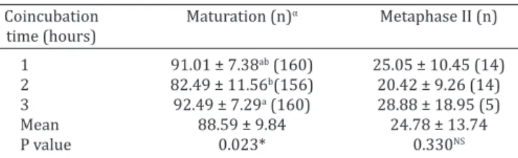
Regardless of the treatment used, the oocyte matura-tion rate was 88.59±9.84%. The maturamatura-tion rate was hi-gher (P=0.023) after coincubation time for 1 and 3 hours than it was for 2 hours (Fig.1). Out of all evaluated oocytes, 24.78% reached the stage of metaphase II, but were not penetrated; no differences were found (P=0.330) between the groups for this variable (Table 1). The percentage of de-generated oocytes was higher (P=0.023) in the ova of T2
oocytes (those exposed to spermatozoa for 2 hours) with mean of 17.65% (n=30 degenerated oocytes of the total examined), compared to 8.67% in T1 oocytes (n=15 of 173)
and 8.48% in T3 oocytes (n=14 of 165), which did not differ from each other (P>0.05) (Fig.2).
The effects of different coincubation time on porcine oocytes IVF are shown in Table 2 and Figure 1. The pene-tration rate was higher (P<0.05) after two hours of coin-cubation time (75.20±10.42%) versus 1 or 3 hours (69.78 ± 11.45% and 66.57±22.68%, respectively). Furthermore, 68.60% of the ova coincubated with the spermatozoa for 2 hours were monospermic. The percentage of monospermi-cally fertilized ova after 1 and 3 hours of coincubation was 42.67% and 66.35%, respectively (Fig.1). Despite having a lower rate (P<0.05) of sperm penetration, the oocytes ex-posed to spermatozoa for 3 hours presented a similar rate of monospermic fertilization after 1 hour of incubation.
The percentage of polyspermy ranged from 29.49 to 57.33% in all three groups. The oocytes exposed to sper-matozoa for 1 hour (T1) presented a higher (P<0.01) rate of polyspermy than those in T2 and T3.
Fertilization performance (%) did not differ (P>0.05) between oocytes exposed to spermatozoa for 1 (T1) or 3 hours (T3). However, the ova coincubated with the sperma-tozoa for 2 hours (T2) presented the best (P=0.048) rate of
fertilization performance (50.16 ± 8.52%) (Fig.3).
Significant differences were not found (P>0.05) betwe -en the treatm-ents evaluated for either the number of p-ene- pene-trated sperm per oocyte or the male pronucleus formation. The average number of penetrated sperm per oocyte and the mean of male pronucleus formation regardless of the treatment were 1.75±0.70 and 91.79±11.72%, respectively.
DISCUSSION
The reduction of coincubation time from 3 to 2 hours
re-sulted in an efficient increase in sperm penetration, mono -Fig.1. Evolution of nuclear maturation, penetration and
monos-permy rate in oocytes coincubated with spermatozoa for 1, 2, and 3 hours.
Fig.2. Percentage of degenerated oocytes after 1, 2, and 3 hours of coincubation with spermatozoa. a,b Different letters denote
significant differences according to Tukey’s test (P=0.023).
Table 1. Values (mean ± SD – %) of maturation rate and porcine oocytes in metaphase II after 1, 2 and 3 hours of
coincubation with spermatozoa
Coincubation Maturation (n)α Metaphase II (n)
time (hours)
1 91.01 ± 7.38ab (160) 25.05 ± 10.45 (14)
2 82.49 ± 11.56b(156) 20.42 ± 9.26 (14)
3 92.49 ± 7.29a (160) 28.88 ± 18.95 (5)
Mean 88.59 ± 9.84 24.78 ± 13.74
P value 0.023* 0.330NS
α (n) =number of oocytes evaluated. a,b Different letters in the same column
denote significant differences by Tukey test (*P<0.05). NS Non significant.
spermic fertilizations, and, most importantly, fertilization performance. On the other hand, the increase in coincuba-tion time reduced the number of polyspermic fertilizacoincuba-tions. The percentage of penetration remained at about 70%, and monospermy was considerably higher with 2 and 3 hours of gamete coincubations. In the short coincubation time (1 hour), the rate of monospermic fertilization was low, even with a penetration rate similar to the long incubation time (3 hours). The number of penetrated sperm per oocyte and the male pronucleus formation after 1, 2, and 3 hours re-mained constant, with average values of 1.48 to 1.93 and 86.83 to 96.88%, respectively. With fewer than two hours
of coincubation, monospermy decreased significantly
(P<0.01), affecting fertilization performance. Thus, we demonstrated that penetration, monospermy, and fertiliza-tion performance have a positive relafertiliza-tion with spermatic coincubation time.
The modification of gamete coincubation time during
porcine IVF to decrease the rate of polyspermy in vitro has
been previously investigated (Cheng et al. 1986, Mattioli et al. 1989, Coy et al. 1993, Abeydeera & Day 1997, Gil et al. 2004, 2007, lmiñana et al. 2005).
According to Coy et al. (1993), the longer the coincuba-tion time, the greater the number of collisions between sper-matozoa and zona pellucida; thus, the greater the risk that polyspermy will occur. However, this was not observed in our study, as we obtained a higher rate of polyspermy when the oocytes were exposed to spermatozoa for one hour.
The penetration and monospermy rates obtained in this study differed from the data presented by Coy et al. (1993).
For all data, our study showed an average higher than those obtained by the authors mentioned above. This difference can be explained by the fact that, in our study, we used oo-cytes obtained from the ovaries of prepubertal gilts after slaughter. In the study of these authors, the oocytes were recovered from the oviducts of prepubertal gilts. Further-more, their medium of sperm capacitation and IVF was dif-ferent from what was used in our study.
Regarding the percentage of degenerated oocytes at the end of the IVM period, the results of our study were in agreement with other experiments reporting averages ranging from 8% to 23% (Illera et al. 1998, Suzuki et al. 2003, Kim et al. 2010, Oberlender et al. 2013a).
In the present study, regardless of the time of coincu-bation (oocytes exposed to spermatozoa), maturation rates were within the range reported by various studies (Kikuchi et al. 2009, Zhang et al. 2012, and Oberlender et al. 2013a); during IVM, approximately 10% to 30% of oocytes did not reach metaphase II of the second meiotic division. In our study, approximately 7.51% to 17.57% of IVM failure was due to oocyte degeneration.
The numbers of penetrated sperm per oocyte obtained in our study were similar to those observed by Matás et al. (2011), Romar et al. (2012), and Oberlender et al. (2013a); in those studies, gamete coincubation was performed for one to three hours. Conversely, Coy et al. (2008b) obtained an average of 5.2 to 12.7 penetrated sperm per oocyte, val-ues higher than those found in our study (1.75±0.70). This result may have been due to time that the oocytes were ex-posed to spermatozoa, which was 4 hours in their study. Fig.3. In vitro fertilization (IVF) results. T1 = oocytes exposed to spermatozoa for 1 hour (polyspermic oocyte); T2 = oocytes exposed to
spermatozoa for 2 hours (monospermic oocyte), and T3 = oocytes exposed to spermatozoa for three hours (polyspermic oocyte). Dashed arrow = decondensed sperm head; straight arrow = sperm adhered to the zona pellucida and * = pronucleus formation.
Table 2. Data (mean ± SD) of IVF after 1, 2 and 3 hours of oocytes exposed to spermatozoa
Variable analyzed Coincubation time (hours) Mean CV (%) P value
1 (n)α 2 (n) 3 (n)
Penetration (%) 69.78 ± 11.45b(146) 75.20 ± 10.42a (142) 66.57 ± 22.68b (155) 70.54 ± 15.92 21.47 0.043* Monospermy (%) 42.67 ± 15.93b (102) 68.60 ± 17.44a (108) 66.35 ± 24.49a (99) 59.67 ± 22.46 27.53 <0.01# Polyspermy (%) 57.33 ± 15.93a (44) 31.41 ± 17.44b (34) 29.49 ± 25.68b (56) 38.90 ± 23.40 36.17 <0.01# Performance (%) 30.36 ± 9.52b (102) 50.16 ± 8.52a (108) 42.45 ± 16.68b (99) 33.00 ± 12.93 29.14 0.048*
NPSPOβ 1.93 ± 0.35 1.48 ± 0.25 1.85 ± 1.10 1.75 ± 0.70 37.50 0.248NS
MPF (%)γ 96.88 ± 7.05 (99) 92.08 ± 9.32 (103) 86.83 ± 15.48 (89) 91.79 ± 11.72 9.36 0.119NS
α (n) =number of oocytes evaluated; β NPSPO = Number of penetrated sperm per oocyte; γ MPF = Male pronucleus formation (%); a,b
Different letters in the same row, in each fertilization result, indicate significant differences by Tukey test (*P<0.05 and #P<0.01); NS
Regarding male pronucleus formation, results from our study were in agreement with those of Romar et al. (2012) and Oberlender et al. (2013a), who observed that male pro-nucleus formation was in excess of 85% of oocytes by 18 hours after fertilization. In our study, the rate of male pro-nucleus formation ranged from 86.83% to 96.88%.
CONCLUSIONS
Under these assay conditions, especially in relation to the sperm concentration used, gamete coincubation for a period of two hours appears to be optimal for monosper-my and fertilization performance. Thus, this is the optimal time period for obtaining a large number of pig embryos capable of normal development.
In addition, as described by other researchers, the ex-amination of other parameters, such as medium volume, sperm concentration, and physiological environment, may
further improve the efficiency of porcine IVF.
Acknowledgements.- To the staff of the slaughterhouse “ElPozo Alimen-tación”, Alhama de Murcia, Murcia, Spain for supplying the biological sam-ples. This work was supported by Federal Institute of Education, Science and Technology of South of Minas Gerais (IFSuldeMinas, Muzambinho Campus (“Instituto Federal de Educação, Ciência e Tecnologia do Sul de Mi-nas Gerais (IFSuldeMiMi-nas, Campus Muzambinho”) and in part by FAPEMIG (Research Support Foundation of the State of Minas Gerais), CNPq
(Natio-nal Council for Scientific and Technological Development) and CAPES (Co -ordination of Improvement of Higher Education Personnel), Brazil.
Conflict of interest statement.- The authors have no competing interests.
REFERENCES
Abeydeera L.R. & Day B.N. 1997. Fertilization and subsequent develop-ment in vitro of pig oocytes inseminated in a modified Tris-buffered medium with frozen–thawed ejaculated spermatozoa. Biol. Reprod. 57:729-734.
Abeydeera L.R., Wang W.H., Cantley T.C., Rieke A., Murphy C.N., Prather R.S. & Day B.N. 2000. Development and viability of pig oocytes matured in a protein-free medium containing epidermal growth factor. Theriogenol-ogy 54:787-797.
Algriany O., Bevers M., Schoevers E., Colenbrander B. & Dieleman S. 2004.
Follicle size-dependent effects of sow follicular fluid on in vitro cumulus expansion, nuclear maturation and blastocyst formation of sow cumu-lus oocytes complexes. Theriogenology 62:1483-1497.
Almiñana C., Gil M.A., Cuello C., Roca J., Vazquez J.M., Rodriguez-Martinez H. & Martinez E.A. 2005. Adjustments in IVF system for individual boars: value of additives and time of sperm-oocyte co-incubation. Theriogeno-logy 64:1783-1796.
Ballester L., Romero-Aguirregomezcorta J., Soriano-Úbeda C., Matás C.,
Romar R. & Coy P. 2014. Timing of oviductal fluid collection, steroid
concentrations, and sperm preservation method affect porcine in vitro
fertilization efficiency. Fertil. Steril. 102:1762-1768.
Bijttebier J., Van Soom A., Meyer E., Mateusen B. & Maes D. 2008.
Preovu-latory follicular fluid during in vitro maturation decreases polyspermic fertilization of cumulus-intact porcine oocytes in vitro maturation of porcine oocytes. Theriogenology 70:715-724.
Cheng W.T.K., Polge C. & Moor R.M. 1986. In vitro fertilization of pig and sheep oocytes. Theriogenology 25:146.
Coy P. & Romar R. 2002. In vitro production of pig embryos: a point of view. Reprod. Fertil. Dev. 14:275-286.
Coy P., Cánovas S., Mondéjar I., Saavedra M.D., Romar R., Grullón L., Matás C. & Avilés M. 2008a. Oviduct-specific glycoprotein and heparin modulate
sperm–zona pellucida interaction during fertilization and contribute to the control of polyspermy. Proc. Natl Acad. Sci. USA 105:15809-15814.
Coy P., Grullón L., Cánovas S., Romar R., Matás C. & Avilés M. 2008b. Hard -ening of the zona pellucida of unfertilized eggs can reduce polyspermic fertilization in the pig and cow. Reproduction 135:19-27.
Coy P., Lloyd R., Romar R., Satake N., Matás C., Gadea J. & Holt W.V. 2010.
Effects of porcine pre-ovulatory oviductal fluid on boar sperm function.
Theriogenology 74:632-642.
Coy P., Martinez E., Ruiz S., Vazquez J.M., Roca J., Matas C. & Pellicer M.T. 1993. In vitro fertilization of pig oocytes after different coincubation in-tervals. Theriogenology 39:1201-1208.
Funahashi H. & Day B.N. 1993. Effects of the duration of exposure to sup-plemental hormones on cytoplasmic maturation of pig oocytes in vitro. J. Reprod. Fertil. 98:179-185.
Funahashi H., Ekwall H. & Rodriguez-Martinez H. 2000. Zona reaction in porcine oocytes fertilized in vivo and in vitro as seen with scanning elec-tron microscopy. Biol. Reprod. 63:1437-1442.
Funahashi H., Mcintush E.W., Smith M.F. & Day B.N. 1999. The presence of Tissue Inhibitor of Matrix Metalloproteinase-1 (TIMP-1) during meiosis improves porcine ‘‘oocyte competence’’ as determined by early embry-onic development after in vitro fertilization. J. Reprod. Dev. 45:265-271. Gadea J. 2002. Manual básico del laboratorio de semen. Universidad de
Murcia, Murcia, España. 39p.
Gil M.A., Abeydeera L.R., Day B.N., Vazquez J.M., Roca J. & Martinez E.A. 2003. Effect of the volume of medium and number of oocytes during in vitro fertilization in embryo development in pigs. Theriogenology 60:767-776.
Gil M.A., Almiñana C., Cuello C., Parrilla I., Roca J., Vazquez J.M. & Martinez E.A. 2007. Brief coincubation of gametes in porcine in vitro fertilization: role of sperm:oocyte ratio and post-coincubation medium. Theriogenol-ogy 67:620-626.
Gil M.A., Ruiz M., Vazquez J.M., Roca J., Day B.N. & Martinez E.A. 2004. Effect of short periods of sperm-oocyte coincubation during in vitro fertiliza-tion on embryo development in pigs. Theriogenology 62:544-552.
Grupen C.G. & Nottle M.B. 2000. A simple modification of the in vitro fertil-ization procedure. Theriogenology 53:422.
Grupen C.G., Nagashima H. & Nottle M.B. 1997. Role of epidermal growth factor and insulin-like growth factor-I on porcine oocyte maturation and embryonic development in vitro. Reprod. Fertil. Dev. 9:571-575. Hancock J.L. & Howell G.J.R. 1959. The collection of boar semen. Vet.
Re-cord. 71:664-665.
Hong J.Y., Yong H.Y., Lee B.C., Hwang W.S., Lim J.M. & Lee E.S. 2004. Effects of amino acids on maturation, fertilization and embryo development of pig follicular oocytes in two IVM media. Theriogenology 62:1473-1482. Hunter R.H.F. 1973. Polyspermic fertilization in pig after tubal deposition
of excessive numbers of spermatozoa. J. Exp. Zool. 183:57-64.
Hunter R.H.F. 1991. Oviduct function in pigs, with particular reference to the pathological condition of polyspermy. Mol. Reprod. Dev. 29:385-391. Illera M.J., Lorenzo P.L., Illera J.C. & Petters R.M. 1998. Developmental
competence of immature pig oocytes under the influence of EGF, IGF-I, follicular fluid and gonadotropins during IVM-IVF processes. Int. J. Dev.
Biol. 42:1169-1172.
Iritani A., Niwa K. & Imai H. 1978. Sperm penetration in vivo of pig follicu-lar oocytes matured in culture. J. Reprod. Fertil. 54:379-383.
Kikuchi K., Somfai T., Nakai M. & Nagai T. 2009. Appearance, fate and uti-lization of abnormal porcine embryos produced by in vitro maturation and fertilization. Soc. Reprod. Fertil. Suppl. 66:135-147.
Kim J., You J., Hyun S.H., Lee G., Lim J. & Lee E. 2010. Developmental com-petence of morphologically poor oocytes in relation to follicular size and oocyte diameter in the pig. Mol. Reprod. Dev. 77:330-339.
Larsen R. 1986. Semen collection from the boar, p.969-972. In: Morrow D. (Ed.), Current Therapy in Theriogenology. W.B. Saunders, Philadel-phia.
Matás C., Coy P., Romar R., Marco M., Gadea J. & Ruiz S. 2003. Effect of sperm preparation method on in vitro fertilization in pigs. Reproduction 125:133-141.
Matás C., Martinez E., Vázquez J.M., Roca J. & Gadea J. 1996. In vitro pen-etration assay of boar sperm fertility: effect of various factors on the penetrability of immature pig oocytes. Theriogenology 46:503-513.
Matás C., Vieira L., García-Vázquez F.A., Avilés-López K., López-Úbeda R.,
Carvajal J.A. & Gadea J. 2011. Effects of centrifugation through three dif-ferent discontinuous Percoll gradients on boar sperm function. Anim. Reprod. Sci. 127:62-72.
Mattioli M., Bacci M.L., Galeati G. & Seren E. 1989. Developmental com-petence of pig oocytes matured and fertilized in vitro. Theriogenology 31:1201-1207.
Nguyen B.X., Kikuchi K., Uoc N.T., Dang-Nguyen T.Q., Linh N.V., Men N.T., Nguyen T.T. & Nagai T. 2015. Production of Ban miniature pig embryos by in vitro fertilization: a comparative study with Landrace. Anim. Sci. J. 86:487-493.
Oberlender G., Murgas L.D.S., Zangeronimo M.G., Pontelo T.P., Menezes T.A.
& Silva A.C. 2013b. Porcine follicular fluid concentration of free insu -lin-like growth factor-I collected from different diameter ovarian folli-cles. Pesq. Vet. Bras. 33:1269-1274.
Oberlender G., Murgas L.D.S., Zangeronimo M.G., Silva A.C., Menezes T.A., Pontelo T.P. & Pereira L.J. 2012. Aspectos relacionados à ocorrência da polispermia durante a fertilização in vitro (FIV) em suínos. Rev. Cient. Eletr. Med. Vet. 19:1-28.
Oberlender G., Murgas L.D.S., Zangeronimo M.G., Silva A.C., Menezes T.A., Pontelo T.P. & Vieira L.A. 2013a. Role of insulin-like growth factor-I and
follicular fluid from ovarian follicles with different diameters on porcine
oocyte maturation and fertilization in vitro. Theriogenology 80:319-327.
Ocampo M.B., Ocampo L.C., Mori T., Ueda J. & Kanagawa H. 1994. Timing
of sequential changes in chromosome configurations during the second
meiotic division and cytoplasmic events of pig oocytes matured and fer-tilized in vitro. Anim. Reprod. Sci. 34:281-288.
Park C.H., Lee S.G., Choi D.H. & Lee C.K. 2009. A modified swim-up method
reduces polyspermy during in vitro fertilization of porcine oocytes. Anim. Reprod. Sci. 115:169-181.
Parrish J.J., Krogenaes A. & Susko-Parrish J.L. 1995. Effect of bovine sperm
separation by either swim-up or Percoll method on success of in vitro fertilization and early embryonic development. Theriogenology 44:859-869.
Petters R.M. & Wells K.D. 1993. Culture of pig embryos. J. Reprod. Fertil. Suppl. 48:61-73.
Pursel V.G. & Johnson L.A. 1975. Freezing of boar spermatozoa; fertiliz-ing capacity with concentrated semen and a new thawfertiliz-ing procedure. J. Anim. Sci. 40:99-102.
Rath D., Long C.R., Dobrinsky J.R., Welch G.R., Schreier L.L. & Johnson L.A. 1999. In vitro production of sexed embryos for gender preselection: high-speed sorting of X-chromosome-bearing sperm to produce pigs after embryo transfer. J. Anim. Sci. 77:3346-3352.
Romar R., Coy P. & Rath D. 2012. Maturation conditions and boar affect timing of cortical reaction in porcine oocytes. Theriogenology 78:1126-1139.
Romar R., Funahashi H. & Coy P. 2016. In vitro fertilization in pigs: New molecules and protocols to consider in the forthcoming years. Theriog-enology 85:125-134.
Satake N., Elliott R.M., Watson P.F. & Holt W.V. 2006. Sperm selection and competition in pigs may be mediated by the differential motility acti-vation and suppression of sperm subpopulations within the oviduct. J. Exp. Biol. 209:1560-1572.
Suzuki H., Saito Y., Kagawa N. & Yang X. 2003. In vitro fertilization and poly-spermy in the pig: factors affecting fertilization rates and cytoskeletal reorganization of the oocyte. Microsc. Res. Tech. 61:327-334.
Tokeshi I., Yoshimoto T., Muto N., Nakamura S., Ashizawa K., Nakada T. & Tatemoto H. 2007. Antihyaluronidase action of ellagic acid effectively prevents polyspermy as a result of suppression of the acrosome reac-tion induced by sperm-zona interacreac-tion during in vitro fertilizareac-tion of porcine oocytes. J. Reprod. Dev. 53:755-764.
Wang W.H., Abeydeera L.R., Han Y., Prather R.S. & Day B.N. 1999.
Morpho-logic evaluation and actin filament distribution in porcine embryos pro -duced in vitro and in vivo. Biol. Reprod. 60:1020-1028.
Wang W.H., Day B.N. & Wu G.M. 2003. How does polyspermy happen in mammalian oocytes? Micr. Res. Tech. 61:335-341.