UNIVERSIDADE DA BEIRA INTERIOR
Ciências da Saúde
Melatonin as Metabolic Regulator of Sertoli Cells
Cátia Sofia Fernandes Rocha
Master Degree Thesis in Biomedical Science
Ciências Biomédicas
(2
ndCycle of Studies)
Supervisor: Prof. Pedro Fontes Oliveira, PhD
Co-Supervisor: Prof. Marco G. Alves, PhD
i O conteúdo do presente trabalho é da exclusiva responsabilidade do autor:
_____________________________________
iii
Dedicatória
v
Agradecimentos
A realização desta tese de mestrado só foi possível com importantes incentivos e apoios de várias entidades e instituições. Desejo agradecer a todos aqueles que direta, ou indiretamente, me ajudaram a terminar esta importante etapa académica da minha vida:
Ao meu orientador, Professor Doutor Pedro Fontes Oliveira, por todo tempo e conhecimento científico disponibilizado para me orientar e aconselhar. Por toda a paciência, correções e críticas durante todo o percurso que possibilitaram a melhoria deste trabalho. Sem a sua excelente orientação este trabalho não seria possível.
Ao meu co-orientador, Professor Doutor Marco G. Alves, por todas as correções, sugestões e críticas que foram essenciais para a elaboração desta tese. Agradeço todo o tempo e conselhos disponibilizados comigo.
Aos meus colegas de laboratório: Vanessa, Raquel, Ana Raquel, Tânia, Gonçalo, Tito, Ana Martins e especialmente ao Luís, por toda a ajuda, disponibilidade e acompanhamento no laboratório.
Às amizades efetuadas durante todo este percurso, em especial: Alexandra Soares, Elisabete Costa, Vanessa Conde e Raquel Nunes por todo o companheirismo, amizade e compreensão demonstrada durante todos estes anos. Foram essenciais para conseguir ultrapassar todos os obstáculos, sem o vosso apoio não seria possível.
Ao meu namorado, por todo o apoio, força e compreensão durante toda esta etapa. Pelo companheirismo, incentivo e partilha de todos os bons e maus momentos vividos durante esta etapa.
À minha família, em especial aos meus pais. Por estarem sempre presentes em todos os momentos da minha vida. Por acreditarem sempre em mim, que eu era capaz, pelo apoio e força incondicional, o incentivo e a paciência demonstrada. Pelo amor, carinho e compreensão, sem eles não seria possível ultrapassar todas as dificuldades e obstáculos.
vii
Resumo
A melatonina, juntamente com a insulina, cooperam na regulação da homeostase da glucose. Nos testículos, o metabolismo das células de Sertoli é essencial para a espermatogénese. Uma vez que os efeitos da melatonina na fisiologia reprodutiva masculina permanecem largamente desconhecidos, neste trabalho colocamos como hipótese que a melatonina pode afetar a espermatogénese através da modulação do metabolismo das células de Sertoli, interagindo com a insulina. Para testar esta hipótese foram feitas culturas de células de Sertoli na presença ou ausência de insulina ou melatonina (ou ambos) e a produção/consumo de metabolitos foi determinada através da técnica de ressonância magnética nuclear. Os níveis proteicos dos transportadores de glucose (GLUT1 e GLUT3), fosfofrutoquinase, lactato desidrogenase (LDH) e transportador monocarboxilado 4 foram determinados através da técnica de Western Blot. A atividade da LDH foi também avaliada. As células de Sertoli tratadas somente com melatonina revelaram um aumento no consumo de glucose através da modulação da expressão do GLUT1, mas diminui os níveis proteicos e a atividade da LDH, o que resulta numa baixa produção de lactato. Além disso, as células de Sertoli expostas somente à melatonina produziram e acumularam menos acetato do que as células expostas à insulina. É de salientar que o tratamento combinado da insulina com melatonina aumenta a produção de acetato pelas células de Sertoli, contudo, o conteúdo intracelular em acetato permanece mais baixo do que nas células expostas somente à insulina. Finalmente, o estado redox foi normalizado nas células de Sertoli tratadas com melatonina, conforme refletido pelo rácio lactato/alanina intracelular. É de notar que nas células de Sertoli expostas à insulina mais melatonina a produção de lactato aumenta e é normalizada a expressão de algumas enzimas, bem como de transportadores, envolvidos na glicólise. Estes resultados demonstram a relevância da melatonina na regulação do metabolismo das células de Sertoli, o que por sua vez pode afetar a espermatogénese. Uma vez que o lactato produzido pelas células de Sertoli fornece suporte nutricional e tem um efeito anti-apoptótico nas células germinativas, uma terapia com melatonina pode ser efetiva para indivíduos diabéticos com problemas relacionados com infertilidade.
viii
Palavras-chave
x
Resumo alargado
A melatonina é uma hormona produzida pela glândula pineal de todos os mamíferos. Ela é conhecida como a “hormona do sono” pelo facto de a sua produção ser mais elevada no período noturno. Os níveis de melatonina apresentam um ciclo diário permitindo o ritmo circadiano de várias funções biológicas. O seu papel na regulação do sistema reprodutor feminino e masculino é de grande relevância. Relativamente ao sistema reprodutor masculino, a melatonina para além de desempenhar uma função endócrina, apresenta também uma função parácrina, uma vez que a sua síntese já foi demonstrada em órgãos do aparelho reprodutor masculino.
A regulação da homeostase da glucose nos testículos é um fator essencial para a manutenção da espermatogénese. As células de Sertoli são células somáticas essenciais para o desenvolvimento das células germinativas do túbulo seminífero, apresentando importantes funções, de entre as quais sustentação, proteção e nutrição das mesmas. Como tal, o metabolismo das células de Sertoli é fundamental para o correto desenvolvimento da espermatogénese. A melatonina, juntamente com a insulina, cooperam na regulação do metabolismo da glucose. Contudo, os efeitos da melatonina na fisiologia reprodutiva masculina, principalmente nas células de Sertoli, permanecem amplamente desconhecidos. Assim, neste trabalho, colocamos como hipótese que a melatonina pode regular a espermatogénese, interagindo com a insulina, através da modulação do metabolismo das células de Sertoli.
Para testar esta hipótese foram efetuadas culturas primárias de células de Sertoli na presença ou ausência de melatonina e insulina ou com ambos. Foram avaliados a produção e o consumo de diversos metabolitos importantes no metabolismo das células de Sertoli (glucose, lactato, acetato e alanina) através da técnica de ressonância magnética nuclear. Os níveis proteicos dos transportadores de glucose (GLUT1 e GLUT3), fosfofrutoquinase, lactato desidrogenase (LDH) e transportador monocarboxilado 4 (MCT4) foram determinados através da técnica de Western Blot. A atividade da LDH foi também avaliada. É de salientar que as células germinativas utilizam o lactato como fonte principal de energia, daí a necessidade de avaliar os níveis proteicos de todos estes intervenientes da via glicolítica, visto que a LDH é responsável pela conversão do piruvato a lactato e o MCT4 é responsável pelo transporte do lactato para as células germinativas.
Os nossos resultados mostraram que as células de Sertoli tratadas somente com melatonina revelaram um aumento no consumo de glucose. Este aumento foi acompanhado pela modulação da expressão do GLUT1, contudo houve uma diminuição dos níveis proteicos
xi e da atividade da LDH. Note-se que apesar de ocorrer um aumento do consumo de glucose este não resulta num aumento da produção de lactato. Em relação ao acetato, que é um metabolito intermediário para a síntese de ácidos gordos e colesterol, as células de Sertoli expostas somente à melatonina produziram e acumularam menos acetato do que as células expostas à insulina. Relativamente ao tratamento combinado da insulina com melatonina, as células de Sertoli aumentaram a produção de acetato, apesar de o acetato intracelular permanece mais baixo do que as células expostas somente à insulina. De notar que o estado redox foi normalizado nas células de Sertoli tratadas com melatonina ou melatonina juntamente com insulina. Este parâmetro foi avaliado através do rácio lactato/alanina intracelular uma vez que a conversão do piruvato a lactato ou a sua conversão a alanina está associada com a reoxidação do NADH a NAD+. É de salientar ainda que as células de
Sertoli expostas à insulina mais melatonina, aumentam a produção de lactato, e ocorre uma normalização da expressão de algumas enzimas bem como de transportadores importantes envolvidos no processo glicolítico.
Em suma, os nossos resultados demonstraram que a melatonina tem um papel importante na regulação do metabolismo das células de Sertoli o que pode afetar a espermatogénese. Estudos têm demonstrado que a melatonina melhora a homeostase da glucose em ratos diabéticos. A diabetes mellitus é um problema que tem afetado cada vez mais jovens em todo o mundo e pode interferir com a sua capacidade reprodutiva. Os nossos resultados sugerem que uma terapia com melatonina poderá melhorar o potencial reprodutivo em indivíduos diabéticos. De facto, o lactato produzido pelas células de Sertoli fornece suporte nutricional e tem um afeito anti-apoptótico nas células germinativas. Concluindo, uma terapia com melatonina pode ser efetiva para indivíduos diabéticos com problemas de fertilidade, contudo mais estudos são necessários para garantir a eficácia e segurança desta terapia.
xiii
Abstract
Melatonin cooperates with insulin in the regulation of glucose homeostasis. Within the testis, glucose metabolism in the somatic Sertoli cell (SC) is pivotal for spermatogenesis. Since the effects of melatonin on male reproductive physiology remain largely unknown, we hypothesized that melatonin may affect spermatogenesis by modulating SCs metabolism, interacting with insulin. To test our hypothesis SCs were cultured in the absence or presence of insulin or melatonin (or both) and metabolite production/consumption was determined by proton nuclear magnetic resonance (1H-NMR).
Protein levels of glucose transporters (GLUT1 and GLUT3), phosphofructokinase 1, lactate dehydrogenase (LDH) and monocarboxylate transporter 4 were determined by Western blot. LDH activity was also assessed. Melatonin-only treated SCs showed increased glucose consumption via modulation of GLUT1 expression, but decreased LDH protein expression and activity, which resulted in lower lactate production. Moreover, melatonin-only exposed SCs produced and accumulated less acetate than insulin-exposed cells. Notably, the combined treatment (insulin plus melatonin) increased acetate production by SCs, but intracellular acetate content remained lower than in insulin-only exposed cells. Finally, intracellular redox state was normalized in SCs by melatonin exposure, as reflected by the intracellular lactate/alanine ratio. Interestingly, SCs exposed to insulin plus melatonin produced more lactate and normalized the expression of some glycolysis-related enzymes and transporters. These findings illustrate that melatonin regulates SCs metabolism and thus, may affect spermatogenesis. Since lactate produced by SCs provides nutritional support and has an anti-apoptotic effect in developing germ cells, melatonin supplementation may be an effective therapy for diabetic male individuals facing subfertility/infertility.
Keywords
xv
Table of Contents
Dedicatória ... iii Agradecimentos ... v Resumo ...vii Palavras-chave ... viii Resumo alargado ... x Abstract ... xiii Keywords ... xiii I. Introduction ... 1 1. General aspects... 22. Spermatogenesis and hormonal regulation ... 3
3. Sertoli cell: structure and morphology ... 7
4. Sertoli cell: functions ... 8
5. Sertoli cell: metabolism ... 10
6. Melatonin ... 14
7. Melatonin and male reproductive function ... 15
8. Melatonin and glucose metabolism ... 20
II. Aim of project ... 22
III. Methods ... 24
1. Chemicals ... 25
xvi
3. Sertoli cell culture... 25
4. RT-PCR ... 26
5. Quantitative real-time PCR ... 27
6. Experimental groups ... 27
7. Intracellular metabolite extraction and 1 H-NMR analysis... 28
8. Western blot ... 29
9. Lactate dehydrogenase activity assay ... 29
10. Statistical analysis ... 30
IV. Results ... 31
1. Rat SCs express the melatonin receptors 1A and 1B ... 32
2. Melatonin increases GLUT1 expression and glucose consumption in rat SCs ... 33
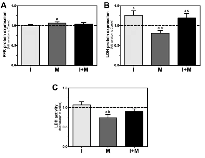
3. Melatonin decreases LDH protein levels and activity in rat SCs ... 34
4. Melatonin decreases lactate production but its combination with insulin stimulates lactate production by SCs ... 36
5. Melatonin increases acetate production and accumulation in SCs and decreases alanine production ... 37
V. Discussion ... 41
VI. Conclusion ... 46
VII. References ... 48
xviii
List of Figures
Figure 1: Schematic representation of the male reproductive tract. 2 Figure 2: Schematic representation of spermatogenesis. 5 Figure 3: Hormonal regulation of spermatogenesis. 6
Figure 4: Metabolic cooperation in testis. 13
Figure 5: The biosynthesis’ pathway of melatonin. 15
Figure 6: Illustration of main melatonin functions in male reproductive health. 19
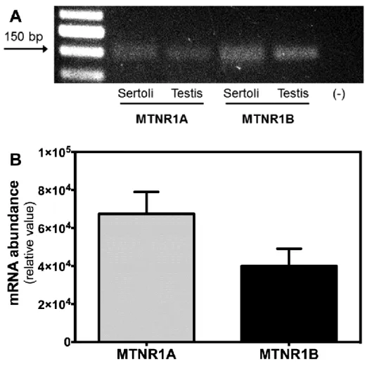
Figure 7: Melatonin receptors in rat Sertoli cells (SCs). Identification and quantification
of mRNA for the two subtypes of melatonin receptors (MTNR1A and MTNR1B) in rat SCs. 32
Figure 8: Glucose transporter 1 (GLUT1) and glucose transporter 3 (GLUT3) protein
expressions, glucose consumption and intracellular glucose in rat Sertoli cells (SCs).
34
Figure 9: Phosphofructokinase 1 (PFK1) protein expression, lactate dehydrogenase (LDH)
protein expression and LDH activity in rat Sertoli cells (SCs). 32 35
Figure 10: Monocarboxylate transporter 4 (MCT4) protein expression, lactate production
and intracellular lactate content in rat Sertoli cells (SCs). 34 37
Figure 11: Alanine production, intracellular alanine content and intracellular
lactate/alanine ratio in rat Sertoli cells (SCs). 35 39
xx
Abbreviations
1H-NMR - Proton nuclear magnetic resonance
AA-NAT - Arylalkylamine N-acetyltransferase
ABP - Androgen binding protein
AR - Androgen receptor
ATP - Adenosine triphosphate
bFGF - Fibroblast growth factor
BSA - Bovine serum albumin
BTB - Blood-testis barrier
CAT – Catalase
cDNA – complementary DNA
DAB - 3,3’ diaminobenzidine hydrochloride
DHT – Dihydrotestosterone
DM - Diabetes mellitus
DMEM:Ham’s F12 - Dulbecco’s modified Eagle’s medium/Ham’s nutrient mixture F12
DNA - Deoxyribonucleic acid
EDTA - Ethylene diaminetetraacetic acid
ER – Estrogen receptor
FBS - Fetal bovine serum
FSH - Follicle-stimulating hormone
xxi
GnRH - Gonadotropin-releasing hormone
GPx - Glutathione peroxidase
HBSS - Hank’s balanced salts solution
HH - Hypobaric hypoxia HI - Intermittent hypoxia HIOMT - Hydroxyindole-O-methyltransferase I/R - Ischemia/reperfusion LDH - Lactate dehydrogenase LDHC – LDH type C LH - Luteinizing hormone
M-MLV RT - Moloney Murine Leukemia Virus Reverse Transcriptase
MCT - Monocarboxylate transporter
MDA - Malondialdehyde
Mel1A - Melatonin receptor 1A
Mel1B - Melatonin receptor 1B
Mel1C – Melatonin receptor 1C
mRNA - Messenger RNA
MT1 - Melatonin receptor 1A
MT2 - Melatonin receptor 1B
MTNR1A - Melatonin receptor 1A
MTNR1B - Melatonin receptor 1B
xxii
NADH - Reduced nicotinamide adenine dinucleotide
NAS - N-acetylserotonin
NE - Norepinephrine
NO - Nitric oxide
PBS - Phosphate buffered saline
PCR - Polymerase Chain Reaction
PFK - Phosphofructokinase
PI3K - Phosphoinositide 3-kinase
PKB - Protein kinase B
PMSF- phenylmethylsulfonyl fluoride
qRT-PCR - Quantitative real-time PCR
RIPA – Radio-Immunoprecipitation Assay
RNAt - Total ribonucleic acid
RNS - Reactive nitrogen species
ROS - Reactive oxygen species
RT-PCR - Reverse-Transcriptase PCR
SC - Sertoli cell
SCN - Suprachiasmatic nuclei
SOD - Superoxide dismutase
STF - Seminiferous tubules fluid
STZ - Streptozotocin
xxiii
1
2
1. General aspects
The success of reproductive events is a fundamental condition for all species. However, in last decades, the fertility rates have been declining. Although a huge knowledge in human reproduction biochemistry and physiology has been acquire, it is not entirely clear the causes and processes that lead to infertility (for review (Hamada, A. et al., 2012; Ray, A. et al., 2012)). Factors like age, diet, life habits, environment, genetic conditions and some diseases may result in alteration of fertility (for review (Barazani, Y. et al., 2014; Shah, K. et al., 2003)). It is estimated that about 15% of couples worldwide are unable to get pregnant after 1 year of unprotected intercourse (Rowe, P. J. & F. H. Comhaire, 2000). Moreover, 8% of men in reproductive age seek for medical assistance with issues related to infertility problems (for review (Esteves, S. C., 2013)). Thus, it is crucial to unravel the molecular mechanisms that control human reproduction.
Figure 1: Schematic representation of the male reproductive tract. The male reproductive tract is constituted by diverse organs and structures that allow the maintenance of reproductive function. It is constituted by the gonads (testes), genital tract (epididymis, vas deferens and urethra), external organs (penis and scrotum) and accessory glands (seminal vesicle, prostate and bulbourethral gland).
The mammalian testes are paired ovoid organs suspended by the spermatic cords and protected from outside through the scrotum (Setchell, B. P., 1978)) (Figure 1). The testis is divided in many compartments, hundreds of coiled tubules named seminiferous tubules. Spermatogenesis occurs in the seminiferous epithelium that is outlined by the seminiferous tubules. Besides germ cells in different stages of differentiation, the seminiferous epithelium also contains the somatic Sertoli cells (SCs) that embrace the germ cells and are essential for
3 their development (De Gendt, K. et al., 2004). In the basement membrane of the seminiferous tubules are located the immature spermatogonia that undergo to a mature spermatozoa. This process starts in the periphery of seminiferous tubules and is progressed to the tubular lumen. Noteworthy, spermatogenic lineage development occurs in an orderly manner, referred as the spermatogenic cycle (for review (Clermont, Y., 1972)). Actually, in one seminiferous tubule, several spermatogenic cycles occur at the same time in a synchronized way, which means that germ cells within each layers changes in synchrony with the other layers over time, producing the sequence of stages. Thus, sequential stages occur with repetition along the length of the tubules, in a wave of the seminiferous epithelium from the rete testis to the center of the seminiferous tubule (Perey, B. et al., 1961). Also, the cycle of germinal epithelium is different between species. In fact, it was described that there are 14 stages in rat spermiogenesis (Leblond, C. & Y. Clermont, 1952) and 12 stages in mouse (Oakberg, E. F., 1956a, 1956b). Besides, the complete spermatogenic cycle lasts around 40-50 days in rodents (depending upon species), and about 65–75 days in humans (for review (Sikka, S. C. & R. Wang, 2008)). The seminiferous tubules converge into the rete testis at the hilum, and the channels that constitute the rete testis connect to produce the tubules of the efferent ducts that transport sperm to the epididymis (Setchell, B. P., 1978)(Figure 1). Between the seminiferous tubules are the interstitials Leydig cells that secrete testosterone to circulation.
The main functions of testes are the synthesis of steroid hormones and the occurrence of spermatogenesis (Luboshitzky, R. et al., 2002; Rato, L. et al., 2010). Noteworthy, these two functions occur in different compartments. The synthesis of steroid hormones takes place in the interstitial space that is a vascularized area; and spermatogenesis occurs in an avascular area that is located in seminiferous tubules. Also, the cooperation between testicular cells (germ, SC and Leydig cells) is very important to the regulation and control of spermatogenesis (Gnessi, L. et al., 2000; Walker, W. H. & J. Cheng, 2005).
2. Spermatogenesis and hormonal regulation
Spermatogenesis is a complex process that involves a network of regulatory systems which include the hypothalamic-pituitary gonadal axis, the male reproductive organs and endocrine and paracrine factors (Kaur, G. et al., 2014; Rato, L. et al., 2012b; Rochira, V. et al., 2006).The seminiferous tubules epithelium is composed by developing germ cells in different stages. In there, an immature germ cell through meiosis and differentiation becomes a mature germ cell. During this process, it is necessary the cooperation between
4 developing germ cells and the somatic SCs (for review (Mruk, D. D. & C. Y. Cheng, 2004; Rato, L. et al., 2010)).
Spermatogenesis can be divided in four stages: mitosis, meiosis, spermiogenesis and spermiation (for review (Cheng, C. Y. et al., 2010)). Next to the wall of the seminiferous tubule, in touch with the SCs, are the immature germ cells (spermatogonia - diploid) that undergo consecutive mitosis in order to expand the germ cell population, providing the sufficient number of cells required for fertility (Orth, J. M. et al., 1988)). Next, spermatogonia proliferate and/or differentiate to primary spermatocytes that undergo to meiosis phase (Figure 2). In meiosis, deoxyribonucleic acid (DNA) is duplicated and the genetic information is recombined in the primary spermatocytes. They divide to form the secondary spermatocytes, which quickly divide again without DNA duplication, so, the germ cells emerge from meiosis as haploid spermatids (Figure 2) (for review (Cheng, C. Y. & D. D. Mruk, 2010a; Cheng, C. Y. et al., 2010)). Then, spermatids undergo a metamorphosis process where occurs the maturation and differentiation to a mature spermatozoon (for review (Mruk, D. D. & C. Y. Cheng, 2004)) (Figure 2). This process is characterized by alterations in nuclear proteins, where the nuclei turns spherical and in cellular size and shape. At this point the spermatozoon is an elongated flagellated cell with potential for movement (Chapin, R. E. et al., 2001; Sofikitis, N. et al., 2008). During spermiation, the mature spermatids are released from the seminiferous epithelium into the tubule lumen and proceed through the rete testis and efferent ducts until reach the epididymis (Alves, M. G. et al., 2013c; Chapin, R. E. et al., 2001).
5
Figure 2: Schematic representation of spermatogenesis. Spermatogenesis is a complex event that is divided in several stages. This process begins with a diploid spermatogonial stem cell that leads to a diploid spermatogonium which in turn leads to diploid primary spermatocyte by several mitotic divisions. Next, the first meiotic division occurs, and the primary spermatocytes undergo to a haploid secondary spermatocyte. Then, after second meiotic division, haploids spermatids are formed. Spermatids undergo through a process called spermiogenesis and at the end there are four haploids sperm cells resulted from one spermatogonium (adapted from (Cheng, C. Y. & D. D. Mruk, 2010a)).
Hormonal regulation is essential for the correct development of germ cells and can result from gonadotropins released from the hypothalamic-pituitary axis or by gonadal organs.
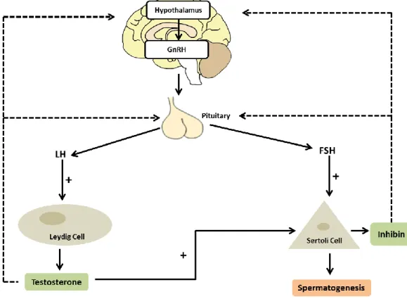
The main hormones that control spermatogenesis are the gonadotropin follicle-stimulating hormone (FSH) and luteinizing hormone (LH). They are released by the anterior pituitary gland that is stimulated by the pulsatile gonadotropin releasing hormone (GnRH) and acts on the testes to regulate the spermatogenic process (for review (Cheng, C. Y. & D. D. Mruk, 2010b; Sokol, R. Z., 2009)) (Figure 3). The hormones FSH and LH act in different places on the testes. FSH promotes SC proliferation and SCs possess receptors for this hormone (for review (McLachlan, R. I. et al., 2002)) (Figure 3). LH acts on Leydig cells and stimulates testosterone production (Costa, R. R. et al., 2011). Noteworthy, there is a relation between these two cells. The testosterone produced by Leydig cell is diffused to the seminiferous
6 tubules where it can regulate SC function and thus, spermatogenesis (for review (Mruk, D. D. & C. Y. Cheng, 2004; Walker, W. H. & J. Cheng, 2005)) (Figure 3). Testicular hormones also have an unquestionable role in spermatogenesis regulation. These hormones exert some negative feedback in the release of gonadotropins. Hormones produced by SC like inhibin, activin, and follistatin regulate FSH secretion. Inhibin and follistatin decrease FSH levels whereas activin stimulates FSH secretion (for review (Ying, S.-Y., 1988)). Besides, testosterone inhibits LH secretion via several mechanisms (Marifke, J. & J. Sandlow, 2013)), in a process essential for the maintenance of spermatogenesis. Testosterone can also be converted to estrogen by aromatase enzyme (Carani, C. et al., 1997; Nathan, L. et al., 2001). This step in steroidogenesis pathway is pivotal for the irreversible conversion of androgens to estrogens in many tissues. Although estrogen is considered a ‘female’ hormone, it has an important role in the development of spermatogenesis (Carreau, S. et al., 2011; Lubahn, D. B. et al., 1993; Wu, C. et al., 2001). Indeed, estrogens and estrogen receptors (ERs) expression has been detected in testis and it has been reported that estrogens can stimulate the proliferation of undifferentiated spermatogonia (Carreau, S. et al., 2011; Chieffi, P. et al., 2002; Fietz, D. et al., 2014; Lubahn, D. B. et al., 1993; Wu, C. et al., 2001).
Figure 3: Hormonal regulation of spermatogenesis. The hypothalamus produces gonadotropin releasing hormone (GnRH) which stimulates the pituitary gland to produce the gonadotropins luteinizing hormone (LH) and follicle-stimulating hormone (FSH). Then, they regulate the spermatogenic cycle by acting in Leydig cell and Sertoli cell (SC), respectively. Leydig cells produce the well-known androgen testosterone which in turn act on SC and/or act as negative signal to hypothalamus and/or in pituitary gland. The somatic SC secretes many substances that influence spermatogenesis. Inhibin is one of the hormones produced by SC that have a negative feed-back on hypothalamus as well as in pituitary gland.
7
3. Sertoli cell: structure and morphology
The SC is a somatic cell that plays a crucial role in development and regulation of spermatogenesis (for review (Petersen, C. & O. Soder, 2006)). Besides, the SCs are also essential in fetal gonadal development. They play a role in the functional development of the testes such as in the formation of the seminiferous cords and thus, in the expression of the male phenotype (for review (Mackay, S., 2000)). There is a current established dogma which states that SCs stop their proliferation in puberty in most species and they just duplicate during fetal and early postnatal life (Cortes, D. et al., 1987). So, the size and maturational status of SCs is established during fetal and early postnatal development, becoming determined around puberty, and the number of SCs that men can have for the rest of their life is determined in puberty (Sharpe, R. M. et al., 2003; Steinberger, A. & E. Steinberger, 1971). This is a maturational process where SC goes through morphological and physiological changes from their immature proliferative state to a mature non proliferative state. These changes are associated with the formation of the blood-testis barrier (BTB) and thus, with the support function of SCs to germ cells (for review (Sharpe, R. M. et al., 2003)). Furthermore, there is a correlation between SC number and sperm output and testicular size (Orth, J. M. et al., 1988). However, some reports have been contesting this dogma (for review (Tarulli, G. A. et al., 2012)). Actually, in nonmammalian species like spiny dogfish (Squalus acanthias) and in the fruitfly (Drosophila melanogaster), the equivalent of the SC proliferates throughout adult life (McClusky, L. M., 2005). In mammals, other reports contest the fact that SCs are able to regenerate only until adulthood. In fact, in vitro studies with SCs isolated from adult mice and human adult testis demonstrate the ability of SCs to repair DNA damage and suggest that SCs are more like arrested proliferating cells than terminally differentiated somatic cells (Ahmed, E. A. et al., 2009). Moreover, in vivo studies with hamsters demonstrated that adult SCs can recover their proliferative ability in part by the action of FSH (Tarulli, G. A. et al., 2008; Tarulli, G. A. et al., 2006). These arguments illustrate that SCs can retain proliferative potential when appropriately stimulated.
The SCs are located within the seminiferous tubules. They adhere to the basement membrane of the seminiferous tubules, and their apical ends are directed to the lumen of seminiferous tubules. The SC has a clear cytoplasm with elements of germline and its nucleus is well defined with an oval shape and presents a big prominent nucleolus which is usually near the cell base (Bawa, S., 1963). Besides, SCs have a big extension area, and embrace a large number of germ cells (for review (Mruk, D. D. & C. Y. Cheng, 2011)).
Adjacent SCs establish tight junctions forming the BTB. This barrier creates a unique environment where spermatogenesis occurs (Dym, M. & D. W. Fawcett, 1970; Mital, P. et al., 2011). It physically divides the seminiferous epithelium in two compartments: the basal (constituted by spermatogonia and primary spermatocytes in the beginning of the meiotic
8 prophase) and the adluminal compartment (constituted by primary spermatocytes in different stages of meiotic spermatocytes, secondary spermatocytes and round and elongated spermatids) (Dym, M. & D. W. Fawcett, 1970). The BTB also allows the formation of a specific intratubular fluid that is tightly controlled by SCs. This barrier regulates the flow of nutrients, growth factors and wastes, both in and out of the seminiferous epithelium, controlling the development of germ cells (for review (Mital, P. et al., 2011)). Noteworthy, BTB also works as an immunological barrier that protects germ cells from harmful agents and from the host’s own immune system, regulating the cytokines levels in the seminiferous epithelium (Mital, P. et al., 2011; Pérez, C. V. et al., 2012). Likewise, BTB prevents the flow of some molecules and substances for the adluminal compartment of the seminiferous tubules. Another important physiological function of this barrier is that they present dynamic channels and transporters in the apical and basolateral membrane that allow the establishment of a specialized and essential environment for the correct development of spermatogenesis (for review (Alves, M. G. & P. F. Oliveira, 2013; Bernardino, R. L. et al., 2013a; Martins, A. D. et al., 2014)). Moreover, this barrier must allow the migration of the germ cells in development through the seminiferous epithelium. Thus, BTB suffers a restructuration to facilitate the transit of germ cells in specifics stages of spermatogenesis, as the movement of preleptotene to leptotene spermatocytes during the stages VIII to XI (Russell, L., 1977). Noteworthy, the event of postmeiotic germ cell development and spermiation occurs behind the BTB in the apical compartment. In fact, SCs, which are the main components of BTB, can alter their morphology to allow the correct development of spermatogenesis. These alterations are due to adjustments in their apical and lateral portions (Wang, F. et al., 2008). Likewise, the microfilaments and a structure rich in actin are essential for formation of junctions between SCs or SC–germ cells and thus for SCs plasticity (Maekawa, M. et al., 1991; Mruk, D. D. & C. Y. Cheng, 2004). Disruption of microfilaments results in a misdirection of spermatids and separation of elongating spermatids from SCs (Russell, L. D. et al., 1988). Also, the water transport from the interstitial space into the lumen serves as vehicle for moving sperm from the testis to the epididymis and SCs are responsible for these fluid movements (Setchell, B. P. et al., 1969). However the exact mechanisms that allow this restructuration are not entirely understood. Noteworthy, SCs can only support a fix number of germ cells. The ratio SC: germ cell is about 1:50 in adult rat testis (Weber, J. E. et al., 1983).
4. Sertoli cell: functions
As discussed, the appropriate development of a SC population is crucial for the spermatogenic capacity during adult life (Orth, J. M., 1982). One important function of SCs is the control of the seminiferous tubules fluid (STF) that is responsible for the transport of spermatozoa to the epididymis. This fluid contributes to the establishment of the correct
9 microenvironment for the occurrence of spermatogenesis (for review (Rato, L. et al., 2010)). The STF has a different composition from the interstitial fluid which is maintained due to the action of the BTB. The STF is constituted by Na+, Cl-, a high concentration of K+ (Clulow, J. &
R. C. Jones, 2004) and a very strict pH control. In fact, SCs express many types of ion membrane transporters that are involved in the movement of particles (for review (Bernardino, R. L. et al., 2013a; Jesus, T. T. et al., 2014; Martins, A. D. et al., 2014)). These transporters control the osmolality and the pH of luminal fluid (for review (Rato, L. et al., 2010). Likewise, ion channels have been identified in SCs (for review (Rato, L. et al., 2010)) and allow the passive diffusion through the cells.
Another important function of SCs is its influence in hormonal regulation by the secretion of substances and proteins and by the secretion of products that nourish the germ cells. Several proteins and other substances are secreted by SCs such as androgen binding protein (ABP), aromatase, androgen receptor (AR) transferrin and glycoproteins (Fritz, I. B. et al., 1976; Karzai, A. W. & W. W. Wright, 1992; O'Brien, D. A. et al., 1993; Petersen, C. & O. Soder, 2006; Skinner, M. K. & M. D. Griswold, 1983). SCs also secrete anti-mullerian hormone, inhibin, as well as metabolic intermediates like acetate (Alves, M. G. et al., 2012) and lactate (Robinson, R. & I. B. Frittz, 1981). Moreover, SCs can respond to the testosterone produced by Leydig cells and convert it to estrogen. Simultaneously, they can control germ cell number by inducing apoptosis and phagocyting apoptotic germ cells (Xiong, W. et al., 2009).
The secretion of these paracrine agents by SCs is highly dependent of hormonal regulation. One of the most important hormones that regulate SC function is the gonadotropin FSH, since, within the testis, FSH receptors are exclusively present in SCs (for review (McLachlan, R. I. et al., 2002)). In fact, FSH controls SCs proliferation during the perinatal and pubertal period, which in turns contributes to determine the spermatogenic capacity during adulthood. In response to FSH, SCs produce inhibin which is usually used as biomarker of the spermatogenesis functional state since inhibin levels are highly dependent of germ cells (for review (De Kretser, D. M. et al., 2004)). In fact, it provides a signal to the pituitary about the capacity of sperm production, since it is generally considered the feedback hormone for FSH in the male. In a study with men, it was found a correlation between blood levels of inhibin B and sperm concentration in the ejaculate (Jensen, T. K. et al., 1997). Also, a study with men with normal sperm concentrations and others with testicular dysfunctions showed that inhibin B and FSH levels were inversely correlated and there was a positive correlation between inhibin B and sperm concentrations (Hipler, U. C. et al., 2001). Others also confirmed that lower inhibin B levels in blood from men with abnormalities in spermatogenesis (mainly SC only tubules) (Bohring, C. & W. Krause, 1999; Foresta, C. et al., 1999). It is established that sperm number is related to SC number. Thus, there is a relation between inhibin B, sperm number and SCs functions (Johnson, L. et al., 1984). Moreover, a study with primates, where germ cells were diminished by testicular irradiation, also showed
10 a rapidly decrease in inhibin B levels. However, when there was a recovery of sperm cells, the levels of inhibin B rise (Foppiani, L. et al., 1999; Schlatt, S. et al., 2002). Thus, Inhibin B may be a reliable marker of testicular function since it can indicate the integrity and activity of the testicular stem cell system and provide an endocrine signal for the number of functional testicular stem cells offering an improved diagnosis of testicular dysfunction.
Likewise, mice lacking FSH receptor by homologous recombination, in spite of being fertile manifest small testes and alterations in spermatogenesis (Dierich, A. et al., 1998). In concordance with this is the study with males homozygous for an inactivating FSH receptor mutation that also show spermatogenic failures but not azoospermia or absolute infertility (Tapanainen, J. S. et al., 1997). These facts illustrate the role of FSH to assure an adequate viability and motility of the sperm.
Androgens are crucial for the control of SCs functions. These cells express ARs (Lyon, M. F. et al., 1975) that play an important role in BTB functioning and also in their maturation (Buzek, S. W. & B. M. Sanborn, 1988). Moreover, the inexistence of ARs in SCs leads to spermatogenesis arrest in meiosis (De Gendt, K. et al., 2004; Holdcraft, R. W. & R. E. Braun, 2004). It should be noted, however, that the androgens responsible for the regulation of SCs function are mainly testosterone metabolites like dihydrotestosterone (DHT). In addition, the role of estrogens in spermatogenesis is a very debatable subject. It is known that estrogens are produced by SCs via aromatase (Rommerts, F. F. et al., 1982). In fact, there is an elevated concentration of estrogens in rete testis fluid (Free, M. J. & R. A. Jaffe, 1979). They also inhibit testosterone production and germ cells apoptosis altering the proliferation and differentiation of spermatogonia. ERs have also been identified in SCs (Fietz, D. et al., 2014; Lucas, T. F. et al., 2014; Oliveira, P. F. et al., 2014a), illustrating that they can play an important role in SCs function, though it is not well understood.
5. Sertoli cell: metabolism
SC glucose metabolism regulation is crucial for normal spermatogenesis. This process is highly regulated, particularly by hormones, in such a way that it has been proposed that the hormonal control of SC metabolism regulates spermatogenesis (for review (Alves, M. G. et al., 2013c)).
It is known that SC metabolism presents some unique characteristics (Riera, M. F. et al., 2002) since the majority of glucose is converted to lactate and not oxidized via the Krebs cycle (Robinson, R. & I. B. Frittz, 1981). This mechanism is essential for developing germ cells that prefer lactate for their energy metabolism (Boussouar, F. & M. Benahmed, 2004; Jutte, N. H. et al., 1981; Riera, M. F. et al., 2002). In fact, developing germ cells are incapable of
11 metabolize glucose and use the lactate produced by SCs to fulfill their energetic demands. Moreover, isolated spermatids exposed only to glucose, without another energy source, are unable to maintain the necessary rate of adenosine triphosphate (ATP) (Grootegoed, J. & P. Den Boer, 1990)).
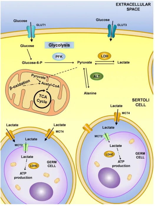
The SCs take up glucose from the interstitial fluid, and it passes through the BTB via specific glucose transporters present in SCs. There are four isoforms of glucose transporters (GLUT1, GLUT2, GLUT3 and GLUT8) (Carosa, E. et al., 2005) identified in SCs. However, GLUT8 has not yet been identified in the plasmatic membrane (Piroli, G. G. et al., 2002) and thus, it is expected that it does not have a significant role in extracellular glucose transport mechanisms. Indeed, it has been reported that GLUT1 and GLUT3 are responsible for the maintenance of glucose uptake in a synergistic way (Alves, M. G. et al., 2013c; Galardo, M. N. et al., 2008). Although glucides are polar molecules that can cross the lipid bilayers by diffusion, they do it in a very inefficient way, so, their passage is mediated trough carriers. After glucose is up taken by SCs, it follows the glycolytic pathway, where it is converted to glucose-6-phosphate and then into pyruvate by the action of several enzymes (Figure 4). In SCs, pyruvate is then converted to lactate via lactate dehydrogenase (LDH) (for review (Alves, M. G. et al., 2013c)) with the concomitant oxidation of reduced nicotinamide adenine dinucleotide (NADH) to oxidized nicotinamide adenine dinucleotide (NAD+) that is essential for
the continued production of ATP by glycolysis (for review (Kreisberg, R. A., 1980)) (Figure 4). The lactate produced is exported through the plasma membrane of SCs by specific monocarboxylate transporters (MCTs) to the intratubular compartment where developing germ cells are located (Rato, L. et al., 2012a) (Figure 4). Actually, 14 members of the MCT family have been described in several cells and tissues (Bonen, A. et al., 2006), however SCs mainly express MCT1 and MCT4 (Galardo, M. N. et al., 2007). It is documented that MCT1 has a higher affinity and a major role in lactate import from the extracellular milieu, while MCT4, which has a lower lactate affinity, is primarily a lactate exporter (Bonen, A., 2001). Although the glycolytic process is highly conserved between species during the evolutionary process, there are glycolytic enzymes that show specific isoforms in testes. Germ cells specifically express a unique type of isoenzyme, the LDH type C (LDHC) (Odet, F. et al., 2008). Interestingly, the selective disruption of Ldhc gene results in male infertility because spermatic motility and hyperactivation pattern, which are necessary for fertilization, are decreased as well as ATP levels in sperm (Goldberg, E. et al., 2010; Odet, F. et al., 2008). Therefore, lactate production by SCs plays a crucial role in spermatogenesis that goes far beyond the metabolic regulation of germ cells. In fact, it has been reported that lactate can regulate the germ cells survival (Jutte, N. H. et al., 1982) and lactate infusions in adult rat testes with cryptorchidism improved spermatogenesis (Courtens, J. L. & L. Ploen, 1999).
Many factors can regulate these metabolic processes but, the hormonal regulation of SCs metabolism, has been highlighted in the last years (for review (Alves, M. G. et al.,
12 2013c)). FSH can stimulate lactate production and LDH activity through phosphoinositide 3-kinase (PI3K) / protein 3-kinase B (PKB) in SCs (Meroni, S. et al., 2002). Many other hormones have been reported to control glucose metabolism in SCs. For instance, in vitro exposure of SCs to DHT increases glucose consumption but does not alter lactate production, illustrating that DHT may reverse these cells metabolism from glycolysis to the Krebs cycle (Rato, L. et al., 2012a). Although stimulation of Krebs cycle is associated with a more efficient way to produce ATP from glucose when lactate production by these cells is compromised, the spermatogenesis may be arrested.
However, although lactate is the main source of energy to developing germ cells, glucose is also pivotal for other processes such as sperm capacitation (Cappello, A. R. et al., 2012), illustrating the importance of glucose and its metabolism for male reproductive function.
Interesting, SCs are mainly known as lactate producers but they also play a key role in the conversion of essential fatty acids (Retterstol, K. et al., 2001). Indeed, SCs can also use lipids to produce energy via fatty acids β-oxidation (Xiong, W. et al., 2009). This mechanism allows them to recycle lipids phagocytized from degraded apoptotic germ cell (Xiong, W. et al., 2009). Thus, besides glucose, SCs can metabolize various metabolites as energy sources including palmitate, ketone bodies and fatty acids (Jutte, N. et al., 1985). Recently has been reported that human SCs produce high amounts of acetate (Alves, M. G. et al., 2012). Acetate is described as a crossroad metabolite and is the most common intermediate for the synthesis of fatty acids and cholesterol (Yoshimoto, M. et al., 2001). Although the exact role for this acetate, produced by SCs and exported, remains unclear, it has been suggested that it may be essential to maintain a high rate of lipid synthesis needed to produce germ cells. Noteworthy, acetate production is under hormonal control, particularly insulin and sex steroid hormones (Alves, M. G. et al., 2012).
Another important metabolic characteristic of SCs is their metabolic plasticity. For instance, in glucose or insulin deprivation conditions, these cells adjust their metabolism to maintain the adequate lactate production for the developing germ cells (Oliveira, P. F. et al., 2012; Riera, M. F. et al., 2009). Glycogen has also been suggested to be important for SCs metabolism since the presence of glycogen and glycogen phosphorylase has been reported in these cells (Leiderman, B. & R. E. Mancini, 1969). Thyroid hormones also regulate SCs metabolism. In fact, in vitro exposure to triiodothyronine (T3) stimulates protein synthesis and lactate production by SCs (Palmero, S. et al., 1995). However, the mechanisms responsible for these effects remain largely unknown though there was a stimulation of GLUT1 synthesis and MCTs are reported to possess affinity to thyroid hormones (Carosa, E. et al., 2005). Growth factors and autocrine and paracrine mediators are also regulators of germ cell and SC metabolism. However, the specific mechanisms are not entirely understood. For
13 instance SC express receptors for basic fibroblast growth factor (bFGF), and it modulates the secretion of estrogens, transferrin and lactate, as well as glucose uptake and LDH activity (Han, I. S. et al., 1993).
Figure 4: Metabolic cooperation in testis. Glucose is taken up by Sertoli cells from the interstitial fluid through the action of glucose transporters (GLUT1 and GLUT3). Glucose is then metabolized to pyruvate through a series of reactions mediated by enzymes including the phosphofructokinase (PFK). The pyruvate can have three distinct fates: it can be converted into acetyl-CoA and enter the Krebs cycle, be converted to lactate by lactate dehydrogenase (LDH) or be converted to alanine by alanine aminotransferase (ALT). These metabolically active cells produce lactate at high rates that is then exported to the intratubular fluid by monocarboxylate transporters (mainly MCT4) (Alves, M. G. et al., 2013a).
14
6. Melatonin
Melatonin is a hormone produced in the pineal gland of all mammals. It has been extensively studied over the last decades due to its physiological properties and clinical relevance. It was first isolated in 1956 and its chemical structure was determined after being extracted from lyophilized bovine pineal glands (Lerner, A. B. et al., 1959). Several studies showed that melatonin is synthetized by other species like bacteria (Tilden, A. R. et al., 1997), unicellular protists (Hardeland, R. et al., 1995) and plants (Dubbels, R. et al., 1995).
Melatonin (N-acetil-5-metoxitriptamina) is synthetized from the amino acid tryptophan that is picked up from the bloodstream to pineal gland – specifically by the pinealocytes – where it is converted into 5-hydroxytryptophan by the enzyme tryptophan hydroxylase. Subsequently, 5-hydroxytryptophan is converted into 5-hydroxytryptamine (or serotonin) by the enzyme 5-hydroxytryptophan decarboxylase and thereafter into N-acetylserotonin (NAS), through acetylation by arylalkylamine N-acetyltransferase (AA-NAT). Finally, NAS produces melatonin through O-methylation by the hydroxyindole-O-methyltransferase (HIOMT) (Axelrod, J. & H. Weissbach, 1960; Barrenetxe, J. et al., 2004) (Figure 5).
Melatonin acts as a neuroendocrine mediator of the photoperiod and, in humans and other diurnal mammals, its production occurs during the dark phase. Melatonin is secreted in a circadian mode, and its secretion rhythm is controlled by an internal clock, named suprachiasmatic nuclei (SCN), located in the hypothalamus (for review (Reiter, R. J., 1993)). During the night period, the SCN sends electrical signals to the pineal gland, causing the release of norepinephrine (NE) from post-ganglionic sympathetic nerve endings on pinealocytes, which initiate and sustain an elevated melatonin production (for review (Axelrod, J., 1974; Maronde, E. & J. H. Stehle, 2007)). Interestingly, melatonin is secreted with a marked circadian rhythm that is distinct between subjects and, thus, it is vital to scrutinize melatonin mechanisms of action.
15
Figure 5: Biosynthesis’ pathway of melatonin. Melatonin is produced from the amino acid tryptophan that is picked up from bloodstream and is then converted in 5-hydroxytryptophan by the enzyme tryptophan hydroxylase. Then, the synthesis of serotonin from hydroxytryptophan by the 5-hydroxytryptophan-descarboxylase enzyme occurs. Through the action of the enzyme arylalkylamine N-acetyltransferase, serotonin is converted to N-acetylserotonin, which in turns leads to melatonin synthesis through O-methylation.
7. Melatonin and male reproductive function
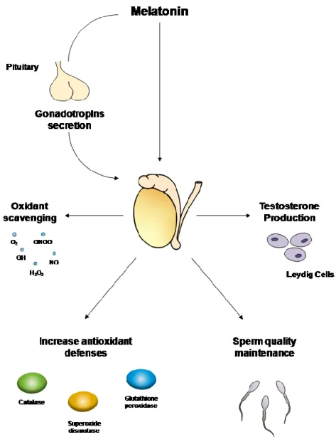
Since 1954, when Kitay and Altchule demonstrated that pineal gland has influence on reproductive function (Kitay, J. I. & M. D. Altschule, 1954)), it has been shown that melatonin has a vast range of effects in the regulation and maintenance of male and female reproductive function. In seasonal breeders, exposure to photoperiod and thus melatonin is critical for the establishment of the reproductive competence (Bittman, E. L. et al., 1983; Malpaux, B. et al., 1996; Reiter, R. J. et al., 2009b). In humans, the effect of seasonal photoperiod length in the reproductive capability is not inexistent, but tends to be significantly smaller. Yet, melatonin has multiple effects on the release of gonadotropins by the anterior pituitary (melatonin receptors have been described in hypothalamic neurons, which regulate the release of pituitary gonadotrophs), on gonads (testes and ovaries) and gonadal adnexa (particularly, prostate and breast) (Dillon, D. C. et al., 2002; Frungieri, M. B. et al., 2005; Gilad, E. et al., 1998; Woo, M. M. et al., 2001; Wu, Y.-H. et al., 2006) (Figure 6). In a study performed with young boys, plasma concentrations of melatonin tended to be16 lower with the advancing of sexual development, illustrating that melatonin may play a key role in male puberty (Cohen, H. N. et al., 1982; Silman, R. et al., 1979; Waldhauser, F. et al., 1984). It has also been reported the existence of a correlation between abnormal levels of melatonin in blood and hypothalamus-pituitary-gonads axis activity disorders, both in males and females (Kadva, A. et al., 1998; Kumanov, P. et al., 2005). Several reports suggest that melatonin directly influences the testicular function. For instance, melatonin receptors have been identified in testicular cells, particularly in Leydig cells (Izzo, G. et al., 2010; Ng, T. B. & L. L. Lo, 1988; Valenti, S. et al., 1997), and melatonin can regulate not only testicular growth but also testosterone production by Leydig cells (Frungieri, M. B. et al., 2005; Olivares, A. et al., 1989; Valenti, S. et al., 1995) (Figure 6).
In male hamsters, an increase in testicular weight and altered spermatogenesis was reported after pinealectomy (Hagen, S. C. & J. H. Asher, Jr., 1983). In fact, a significant increase in the testicular weight has been reported after only 40 days of pinealectomy (for review (Erlich, S. S. & M. L. Apuzzo, 1985)). Further studies showed that melatonin infusions during the night can stimulate gonadal growth only if provided on a daily basis, since it was ineffective if melatonin was intermittently administrated (Prendergast, B. J. & J. L. Hugenberger, 1999). Interestingly, long-duration infusions of melatonin induced gonadal regression (Elliott, J. A. et al., 1989). Noteworthy, melatonin interferes with male reproductive system morphology. Its administration in male gonads of hamsters caused a reduction in the seminiferous tubules diameter, inhibiting spermatogenesis. It also caused several morphological alterations in Leydig cells such as decreased size, reduced cytoplasm and angular nuclei (Ooi, V. E. & T. B. Ng, 1989). Besides, daily afternoon injections of melatonin to male hamsters resulted in testicular atrophy and a reduction in pituitary LH concentration (Reiter, R. J. et al., 1977). Although some of those reports indicated that melatonin exerts its function on the testes primarily through the hypothalamic-pituitary axis and not directly at the gonadal level, recent studies in male hamsters showed that melatonin may act as a local inhibitor of androgen production via down-regulation of the expression of steroidogenic acute regulatory protein and key steroidogenic enzymes (Niedziela, M. et al., 1995; Rossi, S. P. et al., 2012). Data indicates that the effect of melatonin on steroidogenesis involves the interaction between the melatonergic system and the local corticotropin-releasing hormone system, with the subsequent inhibitory action on androgen production by Leydig cells (Frungieri, M. B. et al., 2005; Rossi, S. P. et al., 2012).
In humans, these effects are difficult to study but the effect of melatonin on sperm quality has been reported. Indeed, melatonin receptors were described in the epididymis (Shiu, S. Y. et al., 2000), prostate cells (Shiu, S. Y. et al., 2003; Siu, S. W. et al., 2002) and in spermatozoa (Van Vuuren, R. J. et al., 1992). A study by Levine and collaborators (1990) showed that sperm concentration, count and motility were significantly lower in men during the summer, when the days are longer (Figure 6). When the offspring of those men was
17 analyzed, a significantly low birth rate was found to occur in the spring (Levine, R. J. et al., 1990).
In contrast to pineal melatonin, extra-pineally produced melatonin does not significantly contribute to circadian rhythms (Bubenik, G. A., 2002; Lewy, A. J. et al., 1980). Production of extra-pineal melatonin occurs in practically all cells of the body (Stefulj, J. et al., 2001), and local melatonin synthesis has been reported in testes (Stefulj, J. et al., 2001; Tijmes, M. et al., 1996). For instance, the presence of melatonin has been observed in human seminal fluid (Bornman, M. et al., 1989). It has been suggested that the melatonin produced by extra-pineal tissues has a crucial role in the protection against oxidative damage, taking advantage of the well-known antioxidant properties of melatonin (for review (Bubenik, G. A., 2002; Galano, A. et al., 2011; Hardeland, R. et al., 2009)).
Melatonin plays an important role on the development and/or outcomes of several pathologic situations associated with male reproductive system dysfunction. It is known that hypobaric hypoxia (HH) can alter the reproductive function of men working in high altitudes, affecting spermatogenesis (Okumura, A. et al., 2003; Shevantaeva, O. N. & Y. I. Kosyuga, 2006). The testicular damage resulted from these conditions, has been associated with the increase of reactive oxygen species (ROS) production, vasodilatation, increase of testicular temperature and angiogenesis (Farias, J. G. et al., 2005). Interestingly, it has been reported that melatonin protects the male reproductive function by sheltering sperm from hypoxia-related deleterious effects (Vargas, A. et al., 2011). In fact, melatonin treatment was able to decrease teratozoospermia (i.e. sperm with abnormal morphology) in HH-subjected rats, and decrease the sperm with unstable DNA and sperm lipoperoxidation in both, HH and intermittent hypoxia (HI)- subjected rats (Vargas, A. et al., 2011). Other studies also reported that melatonin counteracts/diminishes hypoxia-related testicular damage through its action as ROS scavenger (Bustos-Obregón, E. et al., 2010; Hartley, R. et al., 2009) (Figure 6).
Testicular torsion is a common pathology and is very frequent in infants and adolescents (Williamson, R. C., 1976). In this condition there is a cutting off of blood supply to the testicle, and ischemia occurs. The testicular damage associated with torsion/ischemia is often associated with ROS production during ischemia/reperfusion (I/R) (Anderson, J. B. & R. C. Williamson, 1986; Filho, D. et al., 2004). In a study using adult male rats, melatonin treatment prevented testicular damage resultant from I/R, which results in decreased germ cell apoptosis, increased expression of proliferating cell nuclear antigen (a marker of cell proliferation) and production of testosterone (Kanter, M., 2010). Moreover, pretreatment with melatonin in an I/R model prevents testicular mitochondrial degeneration and the enlargement of intercellular spaces between SCs and spermatic cells (Kanter, M., 2010). The mechanisms responsible for melatonin protective effects were proposed to be associated with its antioxidant activity. Indeed, melatonin is capable to reduce the total nitric oxide (NO)
18 levels and reverse histopathological changes in testicular tissue after I/R injury, decreasing sperm malondialdehyde (MDA) formation (a biomarker of oxidative stress) and the percentage of abnormalities in rat sperm (Ekici, S. et al., 2012; Koksal, M. et al., 2012; Kurcer, Z. et al., 2010; Yurtcu, M. et al., 2008). All these studies confirmed that melatonin beneficial effects are related with its antioxidant properties as a scavenger of free radicals that are formed during the testicular I/R condition (for review (Filho, D. et al., 2004)).
The increase of free radicals is correlated with decrease of sperm quality and function (for review (Piomboni, P. et al., 2012)). The seminal fluid, as well as sperm cells, possesses an effective machinery to counteract the oxidative stress when produced in normal conditions. An unbalance between oxidative stress and ROS scavenging leads to male infertility. Spermatozoa are very susceptible to oxidative stress since their plasma membrane contains large quantities of polyunsaturated fatty acids and their cytoplasm contains low concentrations of scavenging enzymes. Indeed, plasmatic antioxidants levels of infertile men are significantly lower than those of fertile individuals (for review (Agarwal, A. et al., 2003)). In mammals, spermatozoa must be capacitated and thereafter undergo an acrosome reaction and hyperactivation. ROS play an essential role not only in these sperm physiological functions (capacitation and hyperactivation) but also in sperm-oocyte fusion (Aitken, R. J. et al., 2004; Allamaneni, S. S. et al., 2004; Lamirande, E. et al., 1998). Melatonin is present in human seminal fluid (Bornman, M. et al., 1989) and melatonin membrane receptors have also been identified in spermatozoa (Van Vuuren, R. J. et al., 1992). These evidence reinforce that melatonin is essential for sperm function. Indeed, nighttime supplementation of melatonin resulted in hamster sperm hyperactivation and this response was inhibited by the administration of the melatonin receptor antagonist luzindole (Fujinoki, M., 2008). Nevertheless, the role of melatonin in modulation of sperm parameters and sperm hyperactivation remains largely unknown. Further studies are needed to clarify the role of melatonin in these relevant processes for the male reproductive potential in humans and other mammals.
As discussed, melatonin is an effective antioxidant molecule and melatonin receptors were not only identified in the epididymis, as low affinity melatonin-binding sites have also been reported in spermatozoa (Gwayi, N. & R. Bernard, 2002) (Figure 6). Thus, it has been proposed that melatonin can influence not only spermatozoa during its transit throughout the epididymis, but also during sperm storage. There are only few studies conducted to elucidate melatonin effect on in vitro spermatozoa storage. For instance, it has been reported that human spermatozoa storage in a medium containing melatonin reduced the number of non-viable spermatozoa and increased the progressive motile and rapid cells, particularly by decreasing the NO levels (Du Plessis, S. et al., 2010). Besides, melatonin can effectively prevent apoptosis of human spermatozoa, by protecting them against the generation of free radicals, denoting a potential protective effect of melatonin during in vitro storage (Espino,
19 J. et al., 2010; Espino, J. et al., 2011; Ortiz, A. et al., 2011). It has also been suggested that melatonin can exert its beneficial effect in sperm by decreasing apoptosis (Huang, F. et al., 2009) through modulation of the anti-apoptotic protein bcl-2 expression (Radogna, F. et al., 2008). Although there are an increasing number of studies focused on the effects of melatonin in the male reproductive health, most of the mechanisms remain unknown and should deserve special attention. Nevertheless, it is undoubtedly that melatonin can counteract the deleterious effects of several pathological conditions that result in diminished male fertility.
Figure 6: Illustration of main melatonin functions in male reproductive health. Melatonin acts in the hypothalamic-pituitary axis influencing the release of gonadotropins, which in turn regulate gonadal function (particularly spermatogenesis and androgen synthesis). Also, melatonin directly influences testosterone production by Leydig cells and sperm quality through its anti-apoptotic and scavenger properties. Melatonin’s antioxidant capability reduces testicular damage in pathogenic conditions characterized by an overproduction of reactive oxygen species. Melatonin has the ability to scavenger many types of radicals, hydroperoxide (H2O2), nitric oxide (NO∙), hydroxyl radical (∙OH), singlet oxygen
(1O
2) and peroxynitrite (ONOO-) and others. It also enhances antioxidant defenses, by regulating
20
8. Melatonin and glucose metabolism
Recent reports highlight the possible involvement of melatonin in glucose homeostasis (Cipolla-Neto, J. et al., 2014; Ha, E. et al., 2006). It is known that SCN regulates plasma glucose. In SCN-lesion studies, it was shown that glucose does not increase in the beginning of the active period (La Fleur, S. E. et al., 2001a; Reiter, R. J. et al., 2009a). In fact, glucose blood levels are modulated by a circadian rhythm and sleep under physiological conditions (La Fleur, S. E. et al., 2001a). Interestingly, metabolism-associated genes such as the genes encoding the glucagon receptor, glucokinase, glucagon, GLUT2, glucose-6-phosphate transport protein, pyruvate kinase, and pyruvate dehydrogenase (La Fleur, S. E. et al., 2001a; La Fleur, S. E. et al., 2001b; Panda, S. et al., 2002) show circadian oscillations in its expression. Other enzymes associated with cellular metabolism, such as glucose-6-phosphatase, acetyl-CoA carboxylase, LDH, fatty acid synthase, and glycogen phosphorylase also demonstrated rhythmic expression (for review (Froy, O., 2007)). In fact, rats plasma glucose concentrations display a daily rhythm generated by the hypothalamic biological clock (Ruiter, M. et al., 2003), which causes a decrease in glucose level in the dark period (La Fleur, S. E. et al., 2001b).
Several studies have demonstrated that melatonin decreases glucose levels, in spite of others did not report the same effects (Bojkova, B. et al., 2008; Rios-Lugo, M. J. et al., 2010; Terron, M. P. et al., 2013). It has been reported that increasing concentrations of melatonin stimulate glucose formation in renal rabbit tubules (Derlacz, R. A. et al., 2005). Moreover, it has been shown that melatonin stimulates glucose transport, via insulin receptor, in murine skeletal muscle cell (Ha, E. et al., 2006). Noteworthy, pinealectomy of rats causes a profound alteration in glucose homeostasis, such as hyperinsulinemia and acumulation of triglicerides in the liver (La Fleur, S. E. et al., 2001b; Nishida, S. et al., 2003). Also, melatonin has been shown to reduce insulin secretion by islets in response to glucose (Picinato, M. C. et al., 2002) and despite enhanced insulin secretion, the blood glucose level in rats was reported to be significantly elevated by melatonin (Fabis, M. et al., 2002). Likewise, in the liver of exercised rats melatonin induced an increase in lactate concentration accompanied by changes in carbohydrate and lipid metabolism (Mazepa, R. C. et al., 2000).
Indeed, melatonin has been reported to play a crucial role in metabolic disorders. Diabetes mellitus (DM) is a worldwide health problem that affects an alarming number of young male individuals (ADA, 2012). Few studies concerning melatonin effects have been performed in patients with DM, but most of them point towards an important role of melatonin on the overall health of those individuals. It has been reported that a variation in the melatonin receptor 1B (MTNR1B or Mel1B or MT2) gene expression is associated with an impairment of insulin response and with a deterioration of insulin secretion over time (Bonnefond, A. et al., 2012; Lyssenko, V. et al., 2009). Furthermore, individuals with type 2