Carla Isabel Pereira Magalhães
The role of the Restriction/Modification
system of Clostridium pasteurianum on its
electro-transformation
13 Carla Isabel P er eir a Magalhães The role of t he R es triction/Modification sys tem of Clostridium pasteurianum on its electro-transformationDissertação de Mestrado
Mestrado em Bioengenharia
Trabalho efetuado sob a orientação do
Professor Doutor Manuel José Magalhães Gomes Mota
e coorientação do
Doutor Leonardus Dorothea Kluskens
Carla Isabel Pereira Magalhães
The role of the Restriction/Modification
system of Clostridium pasteurianum on its
electro-transformation
DECLARAÇÃO
Nome: Carla Isabel Pereira Magalhães
Endereço eletrónico: [email protected]; [email protected]
Número do Bilhete de Identidade: 13808152
Título da dissertação:
The role of the Restriction/Modification system of Clostridium pasteurianum on its electro-transformation
Orientador:
Professor Doutor Manuel José Magalhães Gomes Mota
Co-Orientador:
Doutor Leonardus Dorothea Kluskens
Ano de conclusão: 2013
Designação Ramo de Conhecimento do Mestrado:
Ciências Biológicas
DE ACORDO COM A LEGISLAÇÃO EM VIGOR, NÃO É PERMITIDA A REPRODUÇÃO DE QUALQUER PARTE DESTA DISSERTAÇÃO DE MESTRADO
Universidade do Minho, ____ de outubro de 2013
Assinatura:
AGRADECIMENTOS
Gostaria de agradecer ao professor Manuel Mota pelo apoio demonstrado desde o início do trabalho, mesmo depois da reviravolta de tema que este sofreu.
À Doutora Lígia Rodrigues e ao Doutor Leon Kluskens, que foram das pessoas mais importantes para a motivação e desenvolvimento crítico ao longo de todo o trabalho. De forma particular gostava de agradecer à Doutora Lígia pela prontidão com que me recebeu no seu projeto e pela oportunidade de estudar um organismo com tanto carisma. Ao Doutor Leon por toda a atenção, disponibilidade e paciência que dedicou ao trabalho bem como à minha pessoa. A sua orientação atenta, sugestões e conselhos fizeram com que todos os contratempos fossem ultrapassados de forma mas tranquila. É bem mais fácil trabalhar quando se é autorizado a cometer erros, antes de mais porque somos humanos e depois porque é uma forma excelente de aprender. Um sincero obrigado a ambos.
À professora Madalena Alves e a todos os elementos do LBA, de forma especial à Sónia e à Raquel, que me fizeram sentir bem-vinda e apoiada em qualquer que fosse o meu dilema. Aos meninos da PBMS, Ilha2, onde a carga de trabalhos e as frustrações encontradas foram maiores, obrigada pelo bom ambiente e apoio mútuo, estes são sem dúvida fatores importantes no desenvolvimento da investigação científica, um trabalho com tantos altos e baixos. Um especial obrigado ao Franklin que apesar de estar sempre sobrecarregado com o próprio trabalho cede, sem qualquer reticência, algum do seu tempo para esclarecer qualquer tipo de dúvida que vá surgindo a pessoas menos experientes. Um outro obrigado sincero à Gracinha pela partilha das frustrações e vitórias.
Aos meninos do mestrado em Bioengenharia, especialmente aos “Só me apetece ganir” pelas magníficas pausas para o café, onde descarregámos as frustrações de quem está agora a começar e onde nos era permitido dizer as maiores barbaridades e que só nós compreendemos.
A todos os amigos que acompanharam esta viagem direta ou indiretamente e que me apoiaram incondicionalmente fosse qual fosse o estado de espírito. Um obrigado carinhoso à Carlinha que apesar de estar na mesma condição e com dilemas semelhantes não deixava de me pôr para cima e de me dizer que tudo ia correr bem.
À pessoa mais carinhosa do mundo, o Clip, obrigada por todo o apoio e valorização. Também és a pessoa que me mantém acima da linha de água.
Nada disto seria possível sem o apoio incondicional da família. Espero que todo o esforço da parte deles para que chegasse a este momento seja realmente recompensado. Eu sei que não foi fácil. À Patrícia, que apesar de viver comigo, nem sempre recebeu a merecida atenção da irmã.
Como não poderia deixar de ser, a todos aqueles que mesmo não mencionados fizeram desta minha jornada um fardo menos pesado.
ABSTRACT
Clostridium pasteurianum is a Gram-positive and anaerobic bacterium with a great biotechnological potential. It is one of the few microorganisms capable of hydrolyzing glycerol to produce solvents as ethanol and butanol, which have a wide applicability in the market as biofuels.
The development of a genetic system for this microorganism would increase its application opportunities since gene overexpression or inactivation could improve their solventogenic characteristics. Its genetic information is already known but this organism has a particular resistance to transformation. This resistance can be explained by a very efficient restriction system that does not allow the entrance of non-methylated DNA or DNA with a methylation pattern different from it. Therefore, foreign DNA must be correctly methylated prior to transformation. For this purpose, a specific methyltransferase is needed to transfer methyl groups to a certain nucleotide of a specific sequence.
The goal of this thesis was to create a genetic system in C. pasteurianum that allows genome modification and
foreign protein expression, ultimately improving C. pasteurianum DSM 525 transformation.
Preliminary simple electro-transformations in which the parameters to make competent cells and the electroporation conditions were altered, did not result in positive results.
Being aware of the possibility of a restriction system presence in this organism, experiments with M.MspI
methylated DNA were performed, however they demonstrated the inability of this methyltransferase to improve the microorganism transformation.
The presence of restriction enzymes was confirmed when a characterization of the restriction system of C.
pasteurianum was performed using MspI methylated and non-methylated DNA. The presence of a discrete digestion
pattern was detected, and M.MspI methylation could not protect the foreign DNA from C. pasteurianum restriction action.
The polyamine spermidine, with known affinity for negatively charged DNA, showed to be efficient against C.
pasteurianum crude extract digestion action, however not sufficiently to facilitate this microorganism electro-transformation.
By accessing the genome information, the Restriction/Modification (R/M) systems of this microorganism were analyzed. The GATC type IIP R/M system was chosen in order to verify the restriction and methylation enzymes activity with the same target sequence. Three genes, one REase (DpnII) and two MTases (Dam and MdpnII) were cloned in pETduet-1, followed by overproduction in BL21 (DE3).
The codon usage of the host and original organism were not compatible, and the protein production in tRNAs provider strains was tested. Protein production was detected, however was not possible to re-confirm their presence.
The common protein folding problems were analyzed using a disulfide bond enhancer strain. Nevertheless, the production problem may not be related to this, since no different protein over-production was detected.
Restriction reactions with the REase BstUI and C. pasteurianum crude extract, using DNA methylated by M.SssI
(m5CG), were developed and showed that the REase responsible for hindering foreign DNA entering C. pasteurianum
recognizes the sequence 5'-CGCG- 3'.
In a second analysis of the C. pasteurianum genome a methyltransferase-encoding gene was identified that may
be involved in methylating the sequence 5'-CGCG- 3'. The in silico analysis was performed and its codon usage was also
improved to be compatible with E. coli.
In this work, the reasons for C. pasteurianum’s recalcitrance to transformation were identified, the knowledge
RESUMO
Clostridium pasteurianum DSM 525 é uma bactéria Gram-positiva anaeróbia com um elevado potencial biotecnológico. Este é um dos poucos microrganismos capaz de hidrolisar glicerol para produzir solventes como etanol e butanol, que têm uma grande aplicabilidade no mercado.
O desenvolvimento de um sistema genético para este organismo permitiria aumentar as suas oportunidades de aplicação sendo que a sobre-expressão ou inativação de um determinado gene pode melhorar as suas características solventogénicas. A sua informação genética já é conhecida, mas este microrganismo apresenta uma particular resistência à transformação. Esta resistência pode ser explicada pela presença de um eficiente sistema de restrição que não permite a entrada de DNA não metilado ou DNA metilado de forma diferente da própria bactéria. Desta forma, o DNA estranho deve ser corretamente metilado antes da transformação. Para que isto seja possível é necessária a presença de uma metilase específica para transferir grupos metilo para um determinado nucleótido de uma sequência específica.
O objetivo desta tese foi criar um sistema genético em C. pasteurianum que permitisse modificações no genoma
e a expressão de proteínas heterólogas, ou seja, que permitisse melhorar a transformação de C. pasteurianum DSM 525.
Foram realizadas transformações preliminares simples com parâmetros que diferem na forma de obter células competentes e nas condições de eletroporação, contudo os resultados obtidos não foram positivos.
Tendo conhecimento da possibilidade da presença de um sistema de restrição neste organismo, foram realizadas
experiências com DNA metilado pela enzima M.MspI, sendo que estas demonstraram a incapacidade da metiltransferase
para melhorar a transformação deste microrganismo.
Foi confirmada a presença de enzimas de restrição aquando da caracterização do sistema de restrição de C.
pasteurianum usando DNA não metilado ou metilado pela enzima M.MspI. Foi detetada a presença de um padrão de
digestão distinto, verificando-se que a enzima M.MspI não consegue proteger o DNA estranho da ação de restrição de C.
pasteurianum.
A poliamina espermidina, com conhecida afinidade por DNA negativamente carregado, mostrou ser eficiente
contra a ação de digestão do extrato cru de C. pasteurianum, contudo não o suficiente para facilitar a
electro-transformação deste microrganismo.
Tendo acesso ao genoma, foi então analisado o sistema de Restrição e Modificação (R/M) deste microrganismo. Foi escolhido o sistema R/M tipo IIP GATC para verificar a atividade de enzimas de restrição e metilação com a mesma sequência de reconhecimento. Foram clonados três genes no vetor pETduet-1, uma enzima de restrição (REase – DpnII) e duas metiltransferases (MTases – Dam and Mdpn), seguindo-se a produção em BL21 (DE3). O conjunto de codões usados pelo hospedeiro e pelo organismo de origem não eram compatíveis, foi então testada a produção proteica em estirpes fornecedoras de tRNAs. Foi observada produção proteica contudo não foi possível re-avaliar a sua presença.
Foram analisados problemas de enrolamento (do inglês folding), usando uma estirpe que facilita a formação de pontes
dissulfito. No entanto, o problema na produção não deve estar associado ao enrolamento proteico sendo que não foi detectada produção proteica nestas condições.
Foram desenvolvidas reações de restrição com a REase BstUI e extrato cru de C. pasteurianum usando DNA
metilado pela enzima M.SssI (m5CG) e foi mostrado que a REase responsável pelo impedimento da entrada de DNA em C.
pasteurianum reconhece a sequência 5'-CGCG- 3'.
Numa segunda análise ao genoma de C. pasteurianum DSM 525 foi identificado um gene que codifica uma
metiltransferase que pode estar envolvida na metilação da sequência 5'-CGCG- 3'. Foi feita a análise in silico e o tipo de
codões usados foi melhorado para ser compatível com E. coli.
Neste trabalho, foram identificadas as razões para a resistência deste microrganismo à transformação, foi consolidado o conhecimento sobre o seu sistema de R/M e foi proposta uma metodologia para transformar de forma eficiente esta bactéria.
TABLE OF CONTENTS
AGRADECIMENTOS ... iii
ABSTRACT ... v
RESUMO ... vii
LIST OF FIGURES ... xii
LIST OF TABLES ... xiv
ABBREVIATIONS ... xv
1. LITERATURE REVIEW ... 3
1.1. Clostridium Genus ... 3
1.2. Clostridium pasteurianum in Biofuel Industry ... 4
1.3. Transformation in Clostridium ... 6
1.3.1. Bacterial transformation... 7
1.3.2. Electroporation ... 7
1.3.3. Clostridium transformation ... 9
1.3.4. R/M Systems ... 10
1.3.4.1. Types of restriction enzymes ... 10
1.3.4.2. Type II ... 12
1.3.5. Clostridium R/M systems ... 13
1.4. ClosTron system in Clostridium transformation ... 17
1.5. Aim ... 21
2. MATERIALS AND METHODS ... 25
2.1. Bacterial strains and plasmids ... 25
2.2. Genes ... 26
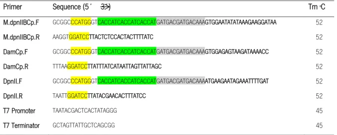
2.3. Primers ... 26
2.4. Culture media ... 27
2.5. Nucleic acids methods ... 27
2.5.1. Plasmid isolation of E. coli and DNA purification ... 27
2.5.2. Genomic DNA extraction from C. pasteurianum DSM 525 ... 28
2.5.3. DNA sample analysis by agarose gel electrophoresis and DNA concentration determination ... 28
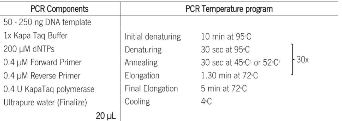
2.5.4. Gene Amplification (Polymerase Chain Reaction - PCR) ... 28
2.5.5. Colony PCR ... 29
2.5.6. DNA Sequencing... 30
2.6.1. Double digestion ... 30
2.6.2. Dephosphorylation of restriction fragments ... 31
2.6.3. Ligation of DNA fragments ... 31
2.6.4. Introduction and maintenance in a host organism ... 31
2.7. C. pasteurianum crude extract ... 32
2.8. DNA modification ... 32
2.8.1. Plasmid enzymatic cleavage... 32
2.8.2. DNA methylation ... 32
2.8.3. DNA condensation by spermidine ... 33
2.8.4. C. pasteurianum restriction endonuclease assay ... 33
2.9. Bacterial cell transformation ... 33
2.9.1. Electro-transformations of C. pasteurianum DSM 525 ... 33
2.9.1.1. C. pasteurianum electrocompetent cells ... 34
2.9.1.2. C. pasteurianum electro-transformation ... 34
2.9.2. E. coli transformation ... 35
2.9.2.1. Chemically competent E. coli cells ... 35
2.9.2.2. Heat shock transformation of E. coli ... 35
2.10. Protein Production and analysis ... 36
2.10.1. Protein Expression ... 36
2.10.2. Cell rupture ... 36
2.10.3. SDS-PAGE analysis ... 36
2.10.3.1. Coomassie Blue staining ... 37
2.10.3.2. Silver Staining ... 37
2.10.4. Protein purification ... 38
2.10.5. Western Blottting ... 39
3. RESULTS AND DISCUSSION ... 43
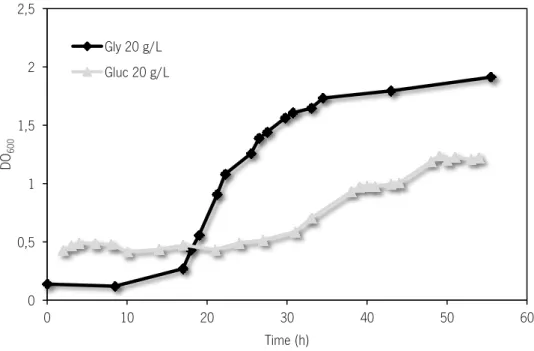
3.1. Glycerol as C. pasteurianum carbon source ... 43
3.2. Preliminary electro-transformations of C. pasteurianum ... 44
3.3. M.MspI role on C. pasteurianum DSM 525 transformation ... 45
3.4. Crude extract of C. pasteurianum exhibits restriction activity ... 46
3.5. DNA condensation by the polyamine spermidine ... 47
3.6. dpnII, dam and mdpn genesas a C. pasteurianum R/M System ... 49
3.7. Cloning ... 52
3.7.1. dpnII, dam and mdpn genes amplification ... 52
3.8. Protein production ... 56
3.8.1. BL21 (DE3) production ... 57
3.8.2. Improvement of the conditions for protein production ... 57
3.8.2.1. Low temperatures of protein production ... 57
3.8.2.2. Codon Usage Analysis ... 58
3.8.2.3. BL21 (DE3) RIL and Rosetta (DE3) pLysS production ... 59
3.8.2.4. Enzymatic experiments ... 61
3.8.2.5. Origami (DE3) production ... 61
3.9. dcm gene importance in C. pasteurianum DSM 525 R/M system ... 62
3.10. C. pasteurianum DSM 525 REase identification ... 62
4. CONCLUDING REMARKS AND FUTURE PERSPECTIVES ... 69
5. REFERENCES... 73
6. APPENDIX ... 83
I. Media and solutions recipe ... 83
II. Gene amplification sequence by PCR ... 84
III. SDS-PAGE protein production results ... 85
IV. Multiple sequence alignment ... 86
LIST OF FIGURES
Chapter 1Figure 1.1. US biodiesel production and its impact on crude glycerol prices. ... 5
Figure 1.2. Sequence of events occurring in the bilayer membrane when subjected to an electric field as electroporation. ... 8
Figure 1.3. Specific methylation ceases restriction endonucleases action. ... 14
Figure 1.4. Schematic representation of mutant generation using the ClosTron system. ... 19
Chapter 2 Figure 2.1. pET-24d+ (a) and pETduet (b) vector maps. ... 30
Figure 2.2. Simplified protein purification process using the His-PurTM Ni-NTA columns with the GFP protein. ... 38
Chapter 3 Figure 3.1. C. pasteurianum growth using glucose (20 g/L) and glycerol (20 g/L) as a carbon source. ... 43
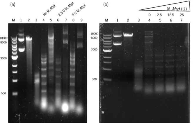
Figure 3.2. C. pasteurianum crude extract characterization.. ... 47
Figure 3.3. Spermidine condensation protects DNA of C. pasteurianum crude extract restriction. ... 48
Figure 3.4. Genomic organization of the genes of GATC R/M system of C. pasteurianum DSM 525 are presented as cluster on this microorganism genome.. ... 51
Figure 3.5. Gene amplification by PCR. ... 52
Figure 3.6. pET24d(+) enzyme digestions.. ... 53
Figure 3.7. Vectors used to replace pET24d(+)... 54
Figure 3.8. Colony PCR results from the transformation of JM109 E. coli with the vector pETduet-1 ligated to genes dam, dpnII or mdpn. ... 55
Figure 3.9. Length analysis of plasmids extracted from the cells transformed with the ligation pETduet-1 and the genes mdpn (pCM001), dpnII (pCM002) and dam (pCM003).. ... 56
Figure 3.10. SDS-PAGE analysis of the CFE after BL21 (DE3) production of the enzymes Dam (a) and DpnII (b) at 37oC. ... 57
Figure 3.11. SDS-PAGE analysis of BL21 (DE3) RIL CFE production of the enzymes Dam (a) DpnII (b) and Mdpn (c) at 37oC.. ... 59
Figure 3.12. SDS-PAGE gel of His-tag purified CFE of the BL21 (DE3) RIL DpnII (a) and Mdpn (b) 4.5 h sample production... 60
Figure 3.13. Western blotting result of the 4.5 h sample of Mdpn production in BL21 (DE3) RIL. ... 61
Figure 3.14. Restriction reaction of m5CG methylated and non-methylated pMTL007C-E2::Cpa-spo0A-666a with the enzymes DpnII, BstUI and C. pasteurianum crude extract.. ... 63
Figure 3.15. Restriction activity comparison between C. pasteurianum (Cp) crude extract (C. Ext) and the enzyme
BstUI. ... 64 Chapter 6
Figure 6.1. SDS-PAGE analysis of BL21 (DE3) CFE production of the enzymes Dam (a) Mdpn (b) and DpnII (c) at 16 oC.. ... 85
Figure 6.2. SDS-PAGE analysis of Rosetta (DE3) pLysS CFE and TE production of the enzymes Dam (a) DpnII (b) and Mdpn (c) at 37 oC. ... 85
Figure 6.3. SDS-PAGE analysis of Origami (DE3) CFE and TE production of the enzymes Dam (a) DpnII (b) and Mdpn (c) at 37 oC.. ... 85
Figure 6.4. Alignment of the dcm gene with codon usage optimized (opC) and the conventional dcm gene (dcm) from C. pasteurianum. ... 87
LIST OF TABLES
Chapter 1Table 1.1.General properties of the four restriction enzyme types. ... 13
Table 1.2. Clostridium REases identified. ... 13
Table 1.3. Type II R/M Systems present in Clostridium listed in REBASE ... 16
Chapter 2 Table 2.1. Bacterial Strains and plasmids. ... 25
Table 2.2. List of primers used. ... 26
Table 2.3. PCR componentes and temperature program conditions to gene amplification with Kapa HiFi Polymerase. ... 29
Table 2.4. PCR components and temperature program conditions to gene amplification with KapaTaq DNA polymerase. ... 29
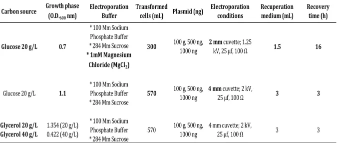
Table 2.5. C. pasteurianum electro-transformation parameters. ... 35
Chapter 3 Table 3.1. C. pasteurianum preliminary electrotransformation parameters. ... 45
Table 3.2. C. pasteurianum electrotransformation parameters using MspI methylated DNA. ... 46
Table 3.3. C. pasteurianum electrotransformation parameters using spermidine-condensed DNA. ... 49
Table 3.4. Genes and their corresponding proteins from C. pasteurianum DSM 525 genome related with type IIP R/M system. ... 50
Chapter 6 Table 6.1. Recipe for mRCM media... 83
Table 6.2. Recipe for T68 basal media. ... 83
Table 6.3. Acrylamide/Bis-acrylamide gels constitution.. ... 83
ABBREVIATIONS
ABE fermentation – Acetone - Butanol - Etanol fermentation
AdoMet – S-adenosyl-L-methionine AmpR – Ampicilin Resistance
APS – Ammonium persulfate ATP – Adenosine Triphosphate BSA – Bovine Serum Albumin
C. – Clostridium
oC – Degree Celsius
CaCl2 – Calcium Chloride
CAI – Codon Adaptation Index
CAGR – Compound Annual Growth Rate CFE – Cell Free Extract
CmR – Chloramphenicol / Thiamphenicol Resistance
Da – Dalton(s)
DNA – Deoxyribonucleic Acid DNP – DNA polymerase
dNTP – Deoxyribonucleotide triphosphate
E. – Escherichia
EDTA – Ethylenediaminetetraacetic acid EmR – Erythromycin Resistance
et al. – And others (et alii) F – Farad
FRT - FLP recognition target sites H2 – Hydrogen
His-Tag - Histidine tag (six histidines in a row) IEP – Intron Encoded Protein
IMAC – Immobilized metal affinity chromatograthy Inc. – Incorporation
IPTG - Isopropyl β-D-1-thiogalactopyranoside KanR – Kanamycin Resistance
L - Liter M – Molar m – Mili (10-3)
MCS – Multiple Cloning Site min – Minute (s)
mRCM – Modified Reinforced Clostridial Medium
ms – milliseconds MTase - Methyltransferase µ – micro (10-6)
n – Nano (10-9)
N2 – Nitrogen
NEB – New Englands Biolabs
NTA – Nickel-charged nitrilotriacetic acid O2 – Oxygen
OC – Open circular plasmid form O.D. – Optical Density
o/n - overnight Ω – Ohm
PCR – Polymerase Chain Reaction PDO – Propanediol
pg – pictograms RA – Restriction alleviation
RAM – Retrotransposition Activated Marker REase – Restriction Endonuclease RNA – Ribonucleic Acid
RNP – Ribonuclear Protein rpm – Rotations per minute rRNA – Ribossomal Ribonucleic Acid RT – Room Temperature
s – Second (s)
SAM – S-adenosyl-L-methionine
SDS-PAGE – Sodium dodecyl sulfate - Polyacrylamide gel electrophoresis
SOB – Super optimal broth
SOC – Super optimal broth for catabolite repression TAE – Tris-Acetate-EDTA Buffer
TBST – Tris-buffered saline with tween Tm – Melting temperature
V – Volts v – Volume
v/v – Volume per volume WT – Wild type
Chapter 1
1. LITERATURE REVIEW
1.1. Clostridium GenusThe genus Clostridium comprises a diverse group of bacteria that share several features; they are Gram-positive bacteria, strictly anaerobic, form endospores, present a rod-shaped morphology and are unable to reduce sulfate. The associated wide range of metabolic and physiological diversity makes it one of the largest group in the prokaryote kingdom with more than 200 known species (Dürre, 2007). The anaerobic bacteria became subject of scientific research when in 1861, the famous microbiologist Louis Pasteur, discovered that life is possible without oxygen. Although, some of the Clostridium genus on health effects were much earlier described by the Greek physician Hippocrates (460-377 BC) when he was analyzing the lockjaw and gas gangrene diseases.
The classification of microorganisms has long been based solely on physiological characteristics, which has led to some wrong classifications. Currently, 16S rRNA gene sequence analysis allows for a much more sophisticated taxonomic classification and has triggered the reclassification of some species, formerly classified as Clostridia. The phylum Firmicutes is structured into the class Clostridia, the order Clostridiales and several families, among them the Clostridiaceae. This family consists of 16 genera, one of them being Clostridium (Stackebrandt, 2004). The Clostridium designation was introduced in the literature by Trécul and Prazmowski between 1865 and 1880. It was the cells morphology that gave them the name, from the Greek kloster (κλωστήρ) or small spindle (Dürre, 2007).
Currently, the public reputation of clostridia is ill-famed; it is usually associated with severe diseases. However, only a few members of this genus are pathogens that cause debilitating and life-threatening intoxications, like botulism (Clostridium botulinum), tetanus (Clostridium tetani), gangrene (Clostridium perfringens), antibiotic associated diarrhea (Clostridium difficile) and food poisoning (C. perfringens). The vast majority of clostridia are entirely benign and some possess properties and attributes of great benefit that can have an important role in several research areas.
In bioremediation, some species are capable of dehalogenation of insecticides as tetrachloromethane or reduction of nitroaromatics such as trinitrotoluene (TNT) to the corresponding amino compounds (Clostridium
acetobutylicum, Clostridium pasteurianum, Clostridium bifermentans, Clostridium sordellii and Clostridium
sporogenes) (Dürre, 2007).
Within the medical field, namely in cancer therapy, a new and recent medical application uses recombinant clostridial spores to fight cancer (Umer et al., 2012), using the limitation of spores to only germinate in anaerobic or hypoxic environments such as the tumor microenvironment. Therefore, recombinant spores (where a gene product is able to convert a harmless pro-drug into a cytotoxic drug) will grow and provide specific targeting and multiplication at the tumor without any harmful effect to the neighboring healthy cells.
Clostridia, and more specifically the solventogenic species, are also applied in the industrial biofuels field. They are useful due to their capacity to produce organic solvents such as acetone (Liou et al., 2005), ethanol (NG et
al., 1981), and 1-butanol (Taconi et al., 2009), by fermentation. This application is increasing tremendously in an attempt to reduce the dependency of petroleum-based fuels and the environmental concerns associated with their use (Yazdani and Gonzalez, 2007).
In conclusion, Clostridium is one of the largest bacterial genera and belonging to this genus are important pathogens, but also organisms with impressive biotechnological and medical applications. Because of these huge capabilities they are considered industrial avant-garde microorganisms with a lot of metabolic potential that still awaits elucidation and commercial exploitation (Dürre, 2007).
1.2. Clostridium pasteurianum in Biofuel Industry
Owing to the enormous development of science and technology in the last two centuries, the worldwide population lifestyle has changed drastically, becoming much more comfortable and consumerist. The great amount of energy required, as well as other products, resulted in the exploitation of our fossil fuels (e.g., petroleum, coal and natural gases). However, as the third Newton’s law states, every action has a reaction. With the advantages of progress came also the environmental problems, such as air pollution, climate changes, global warming and global economic problems. The steadily decreasing availability of fossil fuels is also considered a concerning element. These issues have raised the incentive to paradigm-changing ideas and to create cleaner technologies with the same energy capacity (Sakuragi et al., 2011).
The introduction of biofuels as an alternative has been analyzed and proved to have great chances to reduce the dependence on fossil fuels, thus they are considered a promising approach in this area. Bioethanol (produced through the microbial fermentation of sugars derived from corn, sugarcane or sugar beet (Gray et al., 2006)), biodiesel (produced by the transesterification of vegetable oils or animal fats with an alcohol to produce esters (Marchetti et al., 2007)), and biobutanol (a fermentation product of anaerobic bacteria (Dürre, 2008)) are some examples of biofuels that can replace fossil fuels. Biobutanol has been gaining more attention than other biofuels and is expected to contribute significantly to the industrial needs in the very near future. Biobutanol is very interesting because it presents better properties than ethanol, such as lower corrosiveness, lower vapor pressure, lower water solubility, higher heating value and much higher energy content. Besides its application as a biofuel, butanol is also an important bulk chemical with a wide range of industrial uses. This solvent was, until the middle of the last century, mainly produced by ABE (Acetone-Butanol-Etanol) fermentation using anaerobic microorganisms of
Clostridium species. However, with the declining price of crude oil and the increase of carbon source prices, it
began to be produced almost exclusively by petrochemical processes. Nowadays, mainly due to the oil crisis, the interest in using biotechnology for butanol production has been renewed (Dürre, 2008). It is important to notice that even the fossil fuels based companies, such as BP or DuPont, have shown interest in developing alternative fermentative butanol processes (Lee et al., 2008).
Although it has been proved that these alternatives are secure, renewable and environmentally safe, the production of biofuels also presents some issues, such as low and nonspecific productivity, high cost, and low
solvent tolerance of microorganisms. Therefore, their economic viability is a concern (Sakuragi et al., 2011). The alternatives to continue the proliferation and co-integration with fossil fuels of biofuels are reducing the production cost of and increasing the revenues from industrial biofuels. To achieve these goals the use of biorefineries has been proposed (Kamm and Kamm, 2007). In this case the objective is to use the same feedstock for the co-production of a higher value, small-market chemical along with the biofuel(s). The revenues of both products will improve the economics of biofuel production. To economically improve this process, the biorefinery should consider the use of by-products or waste-streams of the biofuel as feedstock (Yazdani and Gonzalez, 2007). Since the higher investment of an industrial fermentation process is associated with feedstock, this is the principal target for cost reduction.
The biodiesel industry is facing a big expansion in the market. From this process about 10% (w/w) glycerol is obtained as waste-stream (Johnson and Taconi, 2007; da Silva et al., 2009; Yang et al., 2012). As biodiesel is produced in great amounts, more than the current needs, there is a stock accumulation of glycerol, resulting in a substantial fall in its price (Figure 1.1), (Yazdani and Gonzalez, 2007). In 2011, 1,995.5 kilo tons of glycerol were globally produced and in 2018 this value is expected to reach 3,060.4 kilo tons with a CAGR (Compound Annual Growth Rate) of 6.3% from 2012 to 2018 (http://www.transparencymarketresearch.com).
Figure 1.1. US biodiesel production and its impact on crude glycerol prices. In two years the glycerol price presented a 10-fold decrease. From (Yazdani and Gonzalez, 2007).
Crude glycerol as a by-product in biodiesel production is considered a hazardous waste (it contains other impurities such as methanol, ash/salts and residual fatty acids), even though it can be used to produce a large array of products such as ethanol (Jarvis et al., 1997), 1,3-propanediol (1,3-PDO) (Biebl et al., 1992; Yazdani and Gonzalez, 2007), succinic acid (Lee et al., 2001), hydrogen (Ito et al., 2005) and butanol (Biebl, 2001). Glycerol can be highly competitive with the common fermentation sugars, not only due to the lower price, but also because it contains highly reduced carbon atoms, enabling higher fuel yield and highly reduced chemicals (Yazdani and Gonzalez, 2007). Taconi and collaborators defended that glycerol will become a versatile building block chemical for
the production of high value compounds within an integrated biorefinery (Taconi et al., 2009). The advantages of crude glycerol recycling are huge: not only will the discard of a hazardous waste be hindered, but also a cheaper and good carbon source for many fermentation processes will be obtained, fomenting industrial biofuels production.
As mentioned before, a variety of microorganisms can metabolize glycerol, but in general, Clostridia are preferred. These particular anaerobic bacteria are generally not pathogenic, only need little vitamin supplementation and minimal nutrient supply to grow and produce a variety of products (Taconi et al., 2009) such as 1,3-PDO, lactate, acetate, butyrate, ethanol, butanol and even hydrogen (H2) (Dabrock et al., 1992). Great research and
investment efforts are currently being conducted regarding the potential use of solventogenic clostridia for the development of biotech processes towards the production of biofuels, with special attention to biobutanol production. The most studied butanol producing clostridia are C. acetobutylicum and Clostridium beijerinckii but these do not have the capacity to use glycerol as a sole carbon source, only metabolize glycerol in the presence of glucose (Vasconcelos et al., 1994). C. butyricum can ferment glycerol as sole carbon source but it does not express a functional butanol biosynthetic pathway (Heyndrickx et al., 1991). C. pasteurianum is a solventogenic species capable of metabolizing glycerol as a sole carbon source tolerating high concentrations (Biebl, 2001; Dabrock et al., 1992; Nakas et al., 1983) and produces higher amounts of PDO, butanol, and ethanol with just trace amounts of acetate and butyrate, compared to the former mentioned clostridia. The product yield obtained is extremely high, from 75% to 90%, with very small carbon loss by CO2, meaning that most of the substrate is converted to product.
Recent studies also demonstrated that this anaerobic bacterium can efficiently grow and obtain the same products when biodiesel-derived crude glycerol is used (Ahn et al., 2011; Jensen et al., 2012a; Taconi et al., 2009; Venkataramanan et al., 2012). The main disadvantage associated to C. pasteurianum crude glycerol bioconversion is the slow process kinetics (Khanna et al., 2012).
Therefore, the butanol production by C. pasteurianum using biodiesel waste streams containing glycerol as a carbon source is of particular interest, and has garnered special attention unlike the previous biobutanol workhorses
C. acetobutylicum and C. beijerinckii. The lack of knowledge on its genetics and metabolism explains the scarce
investment in the use of this species at an industrial level, until now. Since the C. pasteurianum DSM 525 genome has been recently published (Rappert et al., 2013), it will certainly be deeply investigated and it is expected that the microorganism’s capacities will be greatly enhanced.
1.3. Transformation in Clostridium
As previously mentioned, Clostridium is one of the largest bacterial genera with medical, biotechnological and environmental relevance. Despite the broad range of applications the basic biology of this genus is not well known when compared with their aerobic counterparts of the Bacillus genus(Heap et al., 2009). The better way to recognize and counterattack some pathogens action or exploit biotechnological or environmental properties for better applications, is to understand and thereafter modify essential biological processes at the molecular level. Thus, the need for tools that enable the genetic manipulation of this microorganism becomes clear.
1.3.1. Bacterial transformation
Genetic manipulation of microorganisms started with Frederick Griffith´s investigation (1928), when the development of a vaccine for pneumonia caused by Streptococcus pneumoniae led him to discover that biological material (“a transforming principle”) from a pathogenic Streptococcus strain could transform a non-pathogenic
Streptococcus strain, making it pathogenic (Griffith, 1928). The explanation was given by Avery and collaborators in
the 1940´s. They demonstrated that, after isolation of the biological material, DNA molecules from dead pathogenic strains could be transferred to non-pathogenic strains causing a heritable change (transformation). Two other natural processes of bacterial genetic modification were later discovered: the conjugation process, which is the transfer of DNA from one cell to another through direct contact, and the transduction process, defined as DNA transfection by bacteriophage infection (Prescott et al., 2005). Comparative analyses of prokaryotic genomes show that acquisition of genetic material through lateral gene transfer has been a major driving force in the evolution of these organisms (Johnsborg et al., 2007). Transformation is then defined as the uptake and expression of foreign DNA by cells. Specifically, transformation of bacteria involves DNA adsorption to the cell surface followed by uptake across the wall-membrane complex into the cytoplasm (Prescott et al., 2005).
Even though gene acquisition through natural transformation has contributed significantly to the adaptation and ecological diversification of several bacterial species, it is still a rare event for most bacteria. In modern bacterial genetics, these natural processes are of limited value, because ideally, DNA should be easily transferable into and stably maintained in any given bacterial species (Aune and Aachmann, 2010). Therefore, it was necessary to develop efficient artificial systems to transfer DNA, and currently, artificial transformation of many organisms is commonly done in many laboratories. The first report on the introduction of exogenous DNA into Escherichia coli
was demonstrated by Mandel and Higa in 1970. They treated a suspension of E. coli cells (which are not naturally competent) and bacteriophage λ DNA with a solution of CaCl2 at 0oC and observed cell transformation without using
the phage particle. Since then, many other processes were developed for transformation of bacteria but, with the two main methods for introduction of exogenous DNA into bacterial cells being chemical transformation and electroporation (Aune and Aachmann, 2010).
1.3.2. Electroporation
Electroporation or electropermeabilisation is the term applied to describe the appearance of pores in artificial or cellular membranes due to an elevated transmembrane voltage caused by an applied electric field (Neumann et al., 1982). When the transmembrane voltage exceeds a certain threshold (normally 0.2 to 1 V), the rearrangement of the molecular structure of the membrane occurs, leading to the formation of pores that allow the entry of a range of molecules. These can be drugs, antibodies, DNA and plasmids among others. This process can be divided in six steps.
1. Electric pulses are applied (on a microsecond to millisecond time) that produce the elevated transmembrane potential resulting in an electric field across the membrane around 108 V/m, for a 5 nm thickness
membrane.
2. The membrane is charged due to the ion flow.
3. A rapid and localized rearrangement of the molecular structure of the membrane occurs. 4. The pore formation takes place; these are then coated with water molecules (hydrophilic pores). 5. The transport of ions and molecules through these pores is increased.
6. After the electric field removal, the cell membrane recovers and returns to its normal state (Chen et al., 2006).
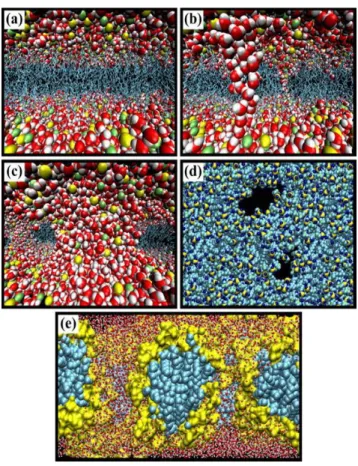
Figure 1.2 illustrates the sequence of events occurring at the inter-membrane level when an electric field, as electroporation, is applied.
Figure 1.2. Sequence of events occurring in the bilayer membrane when subjected to an electric field as electroporation. (a) Bilayer at equilibrium. (b) Formation of water wires at the initial stage of the electroporation process when the bilayer is subjected to a transverse electric field. (c) Formation at a later stage of large pores stabilized by lipid headgroups. Topology of water pores. (d) Top view (e) side view. In the first three panels, water molecules (O, red; H, white), lipid phosphate (yellow), and nitrogen (green) atoms are represented by van der Waals radii, and the acyl chains (cyan) by stick representation. In the last panel, the hydrophilic lipid head group (yellow) and the hydrophobic acyl chains (cyan) are represented by van der Waals spheres to underline the topology and nature of the water pores. Due to the use of perspective views, atoms in the front appear bigger than those in the back. From (Tarek, 2005).
This method has been used for a large number of bacteria, including previously recalcitrant strains, resistant to transformation. Today, the electroporation of whole cells is a common method practiced in many laboratories
because of its high efficiency and broad range of bacteria that can be transformed. However, this process is not evenly applied and does not have the same efficiency for all types of bacteria, which is due to differences in the bacterial membrane and cell wall structure. Hence, electroporation usually needs to be optimized for each specific cell type (Aune and Aachmann, 2010).
Since many bacterial strains used in laboratories are not naturally transformable, methods have been developed to allow DNA entrance. A transformation protocol usually follows three basic steps: the “preparation step”, where the cell is made competent, i.e. prepared to receive DNA; the “shock step”, where the cells are exposed to DNA by a non-lethal procedure, the artificial method per se that allows DNA to permeate into the cell; and the “recovery step”, where the cells are placed under optimal conditions (rich medium, optimal temperature) to allow restoring the damaged membrane and wall.
Bacterial transformation is a very inefficient procedure because it depends on many factors: bacterial strain, medium composition, growth phase, the competence procedure, and DNA size and concentration (Hanahan and Bloom, 1996). Actively growing cells in early exponential phase are more susceptible to transformation. The transformation process also differs between Gram-negative and Gram-positive bacteria (Chen and Dubnau, 2004). The former have an outer membrane and a low percentage of peptidoglycan (the component that confers rigidity and works as the scaffold of the cell wall), while the latter do not have an outer membrane, although they present a thicker peptidoglycan layer. Because of this higher percentage of peptidoglycan, the Gram-positive bacteria are generally more difficult to transform. In general, smaller molecules transform more efficiently than larger ones and with the increase of DNA concentration the dying rate of bacteria increases. The number of transformants rises until the saturation concentration is reached, decreasing slowly after that point (Szostková and Horáková, 1998). A poorly performed procedure can result in cells with reduced competence to take up DNA. The competency of a stock of competent cells is determined by calculating how many colonies (transformants) are produced per microgram of DNA added. An excellent preparation of competent cells will give approximately 108 colonies/ µg and a poor
preparation will yield about 104 colonies/ µg or less.
1.3.3. Clostridium transformation
The Clostridium genus’ importance, as previously discussed, led to the development of many studies at the
genetic and molecular level. To achieve this knowledge the introduction of recombinant DNA into viable clostridia cells is required. Regardless, as the Clostridium genus consists of strictly anaerobic Gram-positive bacteria (which makes the transformation process more laborious and time consuming), only a few successful protocols to transform Clostridia have been developed (Young et al., 1999). Processes such as protoplast transformation are being used in C. acetobutylicum (Lin and Blaschek, 1984; Reid et al., 1983). The conjugation process was used to insert DNA into C. difficile (Herbert et al., 2003; Purdy et al., 2002). Conjugative transposons, Tn916 or Tn1545, have been used to insert mutations in a wide range of bacteria, including some clostridia (Young et al., 1999). The electro-transformation is the most widely applied method to introduce foreign DNA into Clostridium cells: C.
acetobutylicum (González-Pajuelo et al., 2005; Kuit et al., 2012; Mermelstein et al., 1992; Nakotte et al., 1998; Oultram et al., 1988; Tyurin et al., 2000), C. cellulolyticum (Jennert et al., 2000; Tardif et al., 2001), C. beijerinckii
(Birrer et al., 1994), C. thermocellum (Olson and Lynd, 2012; Tyurin et al., 2004), C. perfringens (Chen et al., 1996), and these are just some examples of the use of this technique in Clostridium. Electroporation has been applied as an alternative to the previously mentioned methods, since these are technically cumbersome, time consuming and can present inconsistent results when compared to the quick and simple electroporation technique known to provide reproducible results.
1.3.4. R/M Systems
The way Clostridium is transformed, i.e. the shock step, although extremely important, is not the critical step on its transformation process, as these cells present a highly aggressive defense system against foreign DNA that protects the host cell from DNA not recognized as their own.
All the bacteria have naturally developed resistance mechanisms against invading nucleic acids (virus DNA or RNA) that hinder the cells infection and dead, preserving genetic isolation. Bacterial cells have developed resources to prevent bacteriophage (phage that only infects bacteria) adsorption and cell entry, to cut foreign DNA, and sometimes also commit “suicide” if it results in the elimination of the invader and prevention of the infection of the remaining cells (Labrie et al., 2010). The DNA cutting resources are very common.
The work of Bertani and Weigle (Bertani and Weigle, 1953), Luria and Human (Luria and Human, 1952) and Arber (Arber, 1965) brought evidence of the existence of bacterial enzymes that cleave foreign DNA. These enzymes, and others subsequently found, were grouped as Restriction systems, distinctive to each bacterium. Their main functions are to defend their host against extraneous DNA, the maintenance of species identity and allow speciation (Jeltsch, 2003), and modulate the frequency of genetic variation (Arber, 2000). These systems have been classified as an “immune system” seconded to eliminate invading DNA without adequate modification by methylation (Nikolajewa et al., 2005). A classical restriction system, constituted by restriction endonucleases (REases) is normally associated with methyltransferases (MTases), producing the Restriction and Modification (R/M) systems. REase recognize and cleave a specific DNA sequence, and MTase hinder the DNA cleavage, by modifying adenosyl or cytosyl residues through the transference of methyl groups from the donor S-adenosyl-L-methionine (SAM) within an also specific DNA sequence (Bickle and Krüger, 1993). These methylated bases can be 5-methylcytosine (m5C),
N4-methylcytosine (m4C) or N6-methyladenine (m6A). The methylation pattern of an organism works as a bar code that
represents the unique identity of each strain (Jeltsch, 2003). 1.3.4.1. Types of restriction enzymes
The REases are commonly distinguished into four types (I to IV) based on enzyme composition, requirements and mode of action (Pingoud et al., 2005; Roberts et al., 2003), as described below. The R/M system enzymes belong just to I, II and III type. Between these four types, type II is the most known, developed and with the higher
application, as will be further described. They are nominated according to the name of the bacterium of origin, with the first letter corresponding to the genus, the first and second epitope letters, and the first letter/number to the strain. As an example, the enzyme EcoRI was isolated from E. coli strain RY13 and was the first (I) enzyme of this strain to be analyzed. Restriction enzymes can be distinguished by adding the letter R to the name (R.EcoRI) and the methyltransferases adding the letter M (M.EcoRI). The genes are nominated, italicized, as ecoRIR and ecoRIM, respectively, to restriction enzymes and methyltransferases.
Type I R/M system is constituted by multi-subunit enzymes functioning as a single protein complex. This system generally comprises two HsdM subunits (hsd for host specificity for DNA), which modify DNA by methylation (m6A), two HsdR subunits, responsible for the restriction of the target sequence and one HsdS subunit, involved in
the recognition of the target sequence. When in contact with non-methylated DNA the entire complex acts as REase, requiring ATP and Mg2+ for activity. Usually, two asymmetrical bi-partite target sites are recognized, but the cleavage
site is away from the recognition sequences, approximately half-way between two sites. The complex acts as MTase when in contact with hemimethylated DNA (just one strand is methylated in a double stranded sequence as it happens after DNA replication of a completely methylated sequence). This complex requires the presence of the methyl group donor AdoMet (S-adenosyl-L-methionine) and is 100% active just with the HsdM and HsdS subunits. Some typical type I R/M system examples are the enzymes EcoAI, EcoR124I, and StySBLI, representing the subtypes IA, IB, IC and ID, respectively (Bickle and Krüger, 1993; Pingoud et al., 2005; Roberts et al., 2003).
Furthermore, it is believed that the type I R/M system is related with the restriction alleviation (RA) process responsible for the maintenance (non-cleavage by REases) of the own cell DNA when this is not yet methylated. Moreover, it was discovered that in case of damage, the restriction is alleviated, being a good opportunity for the entry of foreign DNA and for cell mutation (Keatch et al., 2004; Makovets et al., 2003).
Type III R/M system encompasses enzymes constituted by two subunits with modification and recognition activity (Mod), and two subunits responsible for restriction (Res). Cleavage (Res subunit) requires the presence of all the subunits, ATP hydrolysis, Mg2+ as cofactor and AdoMet as stimulator. Two asymmetrical sequences are
recognized in inverse orientation (head to head arranged) and the cleavage occurs close to one recognition site. The modification by methylation (Mod subunit) can occur independently of the Res subunit, creating an m6A base. Some
common examples are the enzymes EcoP1I and EcoP15I (Pingoud et al., 2005; Roberts et al., 2003).
Enzymes belonging to type IV R/M system only recognize and cleave non-specific, variable, and modified DNA (methylated, hydroxymethylated and glucosyl-hydroxymethylated bases) and have, unlike the other restriction systems, no methyltransferases. For successful cleavage REases require GTP and Mg2+. The recognition sequence is
not well defined, but for example, the McrBC enzyme, cleaves m5C-modified DNA. The McrB subunit is responsible for
1.3.4.2. Type II
The type II R/M system is a large and diverse R/M group, and due to its characteristics the enzymes are commonly used in genetic engineering processes. They are differentiated from the other R/M system enzymes by their simplified subunit organization, the generation of a defined restriction pattern, the recognition of specific targets and the cleavage within or close to the recognition sequence. REases can be monomers, dimers or tetramers, and normally require the presence of Mg2+ as cofactor. Their action is, saving some exceptions,
methyltransferase-dependent. MTases are monomers that require the presence of the methyl group donor AdoMet to directly transfer to the DNA double strand (m4C, m5C or m6A). The diversity of the type II R/M system has led to the development of
subdivisions based on the target sequence or on the enzyme structure. These may lead to an overlap of classifications, as one enzyme can be part of more than one subdivision (Pingoud et al., 2005; Roberts et al., 2003).
Type II-classified enzymes recognize asymmetrical sequences (type IIA), that cleave both sides of the recognition sequence (type IIB), enzymes that require just one polypeptide to have both cleavage and methylation activity (type IIC), that require two copies of the recognition sequence (types E and F), enzymes that require the presence of AdoMet as a stimulator to REase or MTase activity (type IIG), type II enzymes structurally similar to type I (type IIH), enzymes that only cleave methylated DNA (type IIM), homodimeric enzymes that cleave just one DNA strand shifted to the recognition sequence (type IIS), heterodimeric enzymes (type IIT) and the orthodox restriction enzymes (type IIP).
The most well-known type II subtype is the IIP. This contains the orthodox type II enzymes, homodimers recognizers of fixed 4 to 8 bp palindromic (symmetric) sequences, which cleave the DNA within the sequence or immediately adjacent to it in both strands, and producers of 3´-hydroxyl and 5´-phosphate ends. These enzymes generate either ‘blunt’ or ‘sticky’ ends. All these features together have led to an increase in type IIP enzymes-related research and development, and have increased their biotechnology and molecular biology applications. They are especially useful in gene analysis and cloning work. The existence of symmetric sequences is considered advantageous since it is energetically more economical for the cell. The speciation of two subunits is energetically more favorable, recognizing, each one, the half symmetrical sequence, than the speciation of a larger protein to recognize an entire sequence (Nikolajewa et al., 2005). A good example is the enzyme EcoRI that recognizes the sequence
5´-G|AATTC-3´ 3´-CTTAA|G-5´.
Table 1.1.General properties of the four restriction enzyme types.
R/M System Restriction Methylation
Machinery Cleavage site Machinery Cleavage site
Type I R2M2S Variable M2S m6A
Type II R Fixed M m6A, m5C, m4C
Type III R2M2 Variable M2 m6A
Type IV R Variable
Note: R- restriction subunit, M- methylation subunit, S- sequence recognition subunit. From (Suzuki, 2012).
In August 2013, 13,508 type II restriction enzymes, 16,716 type I restriction enzymes, 4,256 type III restriction enzymes, 4,747 type IV restriction enzymes, and 33,030 methyltransferases were listed on REBASE – The Restriction Enzyme Database (http://rebase.neb.com/rebase/rebms.html).
1.3.5. Clostridium R/M systems
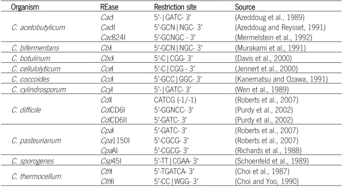
The highly aggressive defense system found in Clostridium consists of restriction endonucleases that digest foreign DNA, not methylated like their own. When the first attempts were made to insert exogenous DNA into microorganisms of this genus, no positive clones with extra DNA insertions were obtained (Mermelstein and Papoutsakis, 1993). Experiments using crude extract (cell content after membrane disruption) showed a specific digestion pattern of the DNA placed in contact with it. The existence of a pattern means that restriction endonucleases with a specific recognition sequence (type IIP) are present. Many Clostridium restriction enzymes have been identified and these are summarized in Table 1.2.
Table 1.2. Clostridium REases identified.
Organism REase Restriction site Source
C. acetobutylicum CacCacI II 5'-|GATC- 3' 5'-GCN|NGC- 3' (Azeddoug et al., 1989) (Azeddoug and Reysset, 1991)
Cac824I 5'-GCNGC - 3' (Mermelstein et al., 1992)
C. bifermentans CbiI 5'-GCN|NGC- 3' (Murakami et al., 1991)
C. botulinum CboI 5'-C|CGG- 3' (Davis et al., 2000)
C. cellulolyticum CceI 5'-C|CGG - 3' (Jennert et al., 2000)
C. coccoides CcoI 5'-GCC|GGC- 3' (Kanematsu and Ozawa, 1991)
C. cylindrosporum CcyI 5'-|GATC- 3' (Wen et al., 1989)
C. difficile CdiCdiI CD6I CATCG (-1/-1) 5'-GGNCC- 3' (Roberts et al., 2007) (Purdy et al., 2002)
CdiCD6II 5'-GATC- 3' (Purdy et al., 2002)
C. pasteurianum CpaCpaI 1150I 5'-GATC- 3' 5'-CGCG- 3' (Roberts et al., 2007) (Roberts et al., 2007)
CpaAI 5'-CGCG- 3' (Richards et al., 1988)
C. sporogenes Csp45I 5'-TT|CGAA- 3' (Schoenfeld et al., 1989)
C. thermocellum CthCthI II 5'-TGATCA- 3' 5'-CC|WGG- 3' (Choi et al., 1987) (Choi and Yoo, 1990)
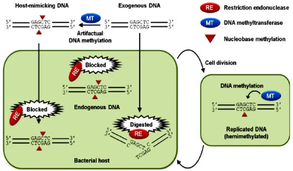
All this REases activity cease when methylated DNA was used. As Mermelstein stated “(…) if the nature of the restriction system is known, transforming DNA may be protected by methylation (…)” (Mermelstein and Papoutsakis, 1993). Cleavage can be completely hindered by the cognate methyltransferase if this mimics the cell intern methylation pattern. It can be affected if the methylation is not specific, i.e. if the REase is sensitive to other kind of methylation the cleavage is “impaired” (http://rebase.neb.com/rebase/rebms.html). However, the cleavage cannot be hindered at all if the REase is methylation-insensitive. Figure 1.3 represents a scheme of what occurs with methylated and non-methylated foreign DNA in a cell with an active restriction system.
Figure 1.3. Specific methylation ceases restriction endonucleases action. The transformation process is hindered in a cell with a very active restriction system since the cell REases (restriction endonucleases) cleave the DNA, not methylated as their own. If the foreign DNA is methylated as is their own cellular DNA (mimic), by one specific methyltransferase, the REases will not cleave this DNA and it will be inserted in the genetic information of this cell. Future generations will also have this extra information. From (Suzuki, 2012).
Positive clostridia transformations were achieved by in vitro or in vivo DNA methylation, i.e. respectively, using a purified MTase or one MTase gene inserted into a plasmid that in turn is inserted into a bacterium (e.g. the best known bacterial host - E. coli).
The most well-known in vivo strategy was developed by Mermelstein and collaborators in 1993, pAN1 and pAN2 plasmids were used to methylate the sequences 5'-Gm5CNGC - 3' and 5'-GGm5CC - 3', recognized and digested
by REases from C. acetobutylicum. These plasmids had the Φ3TI MTase gene (φ3tI) from Bacillus subtillis phage Φ3T inserted (Mermelstein and Papoutsakis, 1993). All the successful C. acetobutylicum transformations were performed using the plasmid pAN1 (φ3tI, cloramphenicol resistance) (Dong et al., 2010; González-Pajuelo et al., 2005; Mermelstein and Papoutsakis, 1993; Nakotte et al., 1998; Shao et al., 2007; Tyurin et al., 2000) or pAN2 (φ3tI, tetracycline resistance) (Dong et al., 2010; Kuit et al., 2012). In the in vivo system, the pAN1 (or similar vector) bacterial cell is transformed with the DNA intended to methylate and the modification occurs inside the cell.
After modification the DNA is extracted and transformed into the bacterium with the correspondent active restriction system.
The in vitro approach is simple and effective but limited because the commercial MTases usually lack each
cell’s specific methylation pattern. This process was used to transform Clostridium strains as C. cellulolyticum, where only the mixture of the DNA intended to methylate with the enzyme in the enzyme medium is needed, along with the optimal temperature requested(Jennert et al., 2000; Tardif et al., 2001). This need led to the identification, production and purification of these organism’s MTases to further use them as a modification strategy required to transform Clostridium cells (Davis et al., 2000).
At the moment (August, 2013) the REBASE lists 151 Clostridium strains. From these 34 have, identified or predicted, at least one type II R/M system with REase and the cognate(s) MTase(s) (presented in Table 1.3). The study of these R/M systems, specially the MTases will allow the transformation of recalcitrant microorganisms by specifically methylated DNA. Thus, their applications will be exploited and may be useful to biotechnological applications.
Table 1.3. Type II R/M Systems present in Clostridium listed in REBASE
Clostridium strain REase R/M Systems Name MTase Recognition site (5'-- 3')
C. acetobutylicum ATCC 824 Cac824I M.Cac824I 5'-GCNGC- 3'
C. acetobutylicum ABKn8 Cac8I M.Cac8I 5'-GCN(|)NGC- 3'
C. acetobutylicum DSM 1731 Cac1731ORF1526P M.Cac1731ORF1526P 5'-GCNGC- 3'
C. acetobutylicum EA 2018 Cac2018ORF1517P M.Cac2018ORF1517P 5'-GCNGC-3'
C. acidurici 9a Cac9aORF8000P M.Cac9aORF8000P 5'-GGNCC- 3'
C. botulinum CboI M.CboI 5'-C(|m5) CGG- 3'
C. botulinum A ATCC 19397 CboAORF2811P M.CboAORF2811P 5'-GCWGC- 3'
C. botulinum A Hall CboAHORF2744P M.CboAHORF2744P 5'-GCWGC- 3'
C. botulinum B Eklund 17B (NRP) Cbo17ORFCP M.Cbo17ORFCP 5'-GATC- 3'
C. botulinum B1 Okra CboB1ORF2548P M.CboB1ORF2548P 5'-CCGG- 3'
C. botulinum Bf CboBfORF47P M.CboBfORF47P 5'-GGWCC- 3'
C. botulinum CFSAN001628 Cbo16ORFCP M.Cbo16ORFCP 5'-CCGG- 3'
C. botulinum E1 'BoNT E Beluga' CboE1ORF1092P M.CboE1ORF1092P 5'-GCNGC- 3'
C. botulinum E3 Alaska E43 CboE3ORF2487P M.CboE3ORF2487P 5'-GRCGYC- 3'
C. botulinum F 230613 CboF2ORF2114P M.CboF2ORF2114P 5'-GCSGC- 3'
C. butyricum 5521 Cbu5521ORF1956P M.Cbu5521ORF1956P 5'-GATC- 3'
C. cellulolyticum H10 CceCceI ORF2549BP M.CceI 5'- CCGG- 3'
CceORF2549AP M.CceORF2549P 5'-GCGC- 3'
C. cf. saccharolyticum K10 Ccf10ORF12590P M.Ccf10ORF12590P 5'-GATC- 3'
C. difficile CD3 CdiI M.CdiI 5'-CATC|G (-1/-1)- 3'
5'-TGGCCA-3'
C. difficile CD6 CdiCdiCD6I CD6II M.M.CdiCdiCD6I CD6II 5'-GGNCC- 3' 5'-G(m6)ATC- 3'
C. hathewayi WAL-18680 Cha18680ORF3698P M.Cha18680ORF3698P 5'-GCWGC- 3'
C. kluyveri DSM 555 CklORF2671P M.CklORF2671P 5'-CCGG- 3'
C. kluyveri NBRC 12016 CklAORF2367P M.CklAORF2367P 5'-CCGG- 3'
C.leptum DSM 753 CleCleDORF2452P DORF3217P M.M.CleCleDORF2452P DORF3217P 5'- GGATG- 3' 5'-GGNCC- 3'
C. nexile DSM 1787 Cne1787ORF858P M.Cne1787ORF858P 5'-GGNCC- 3'
C. pasteurianum DSM 525 Cpa525ORF11401P M1.M2.CpaCpa525ORF11401P 525ORF11401P 5'-GATC- 3'
Cpa525ORF2340P M.Cpa525ORF2340P 5'-CGCG- 3'
C. perfringens ATCC 13124 CpeCpeAIIP AIIIP M.M.CpeCpeAII AIII 5'-GGW(5'-GAT(m5m5)C- 3' )CC- 3'
C. saccharolyticum WM1 CsaWM1ORF1186P M.CsaWM1ORF1186P 5'-CAATTG- 3'
C. saccharoperbutylacetonicum N1-4
(HMT) Csa1ORF40510P M1.M2.CsaCsa1ORF40510P 1ORF40510P 5'-GATC- 3'
C. tetani E88 CteEORF387P M.CteEORF387P 5'-GATC- 3'
C. thermocellum ATCC 27405
CthORF1513P M.CthORF1513P 5'-GATC- 3'
CthORF1749P M.CthV 5'-G(m5)CWGC- 3'
CthORF2320P M.CthVI 5'-GG(m5)CC- 3'
CthORF2470P M.CthORF2470P 5'-GATC- 3'
C. thermocellum LQ8 CthCthVORFCP VORF2275P M.M.CthCthVORFCP VORF2275P 5'-GATC- 3' 5'-GATC- 3'
C. thermocellum JW20 Cth20ORF2874P M.Cth20ORF2874P 5'-GATC- 3'
N- Any base, W- A or T, R- A or G, Y- C or T, S- G or C. (|) Cleavage site identified. (m5 or m6) methylation site identified. (|m5) cleavage and
1.4. ClosTron system in Clostridium transformation
To date only a few C. pasteurianum mutants were obtained, which have been obtained only by chemical mutagenesis (Daldal, 1985; Jensen et al., 2012b; Malaviya et al., 2012), i.e. no new DNA entered into cells, a random DNA change happened in the bacterium own DNA.
In order to be able to study the metabolic engineering (e.g. metabolic pathway manipulation), specific DNA alterations are required and for this efficient genetic engineering tools are needed.
The common practice to obtain mutants is by classical recombination-based procedures. Clostridia mutants have been mostly obtained by homologous recombination. Some clostridia mutated genes were reported in few species, notably in C. acetobutylicum, C. beijerinckii, C. perfringens and even in C. difficile (Heap et al., 2007), but they are inherently unstable and especially mutants from homologous recombination are difficult to isolate (Heap et al., 2010). Although gene transfer to the Clostridium genus is already possible, Young considered that mutated genes are most readily isolated and characterized if they are generated by insertional mutagenesis (Young et al., 1999). The normally applied genetic methodologies are the gene modification systems through “Knock-out” and “knock-in” (Dong et al., 2010; Heap et al., 2009; Kuehne et al., 2011).
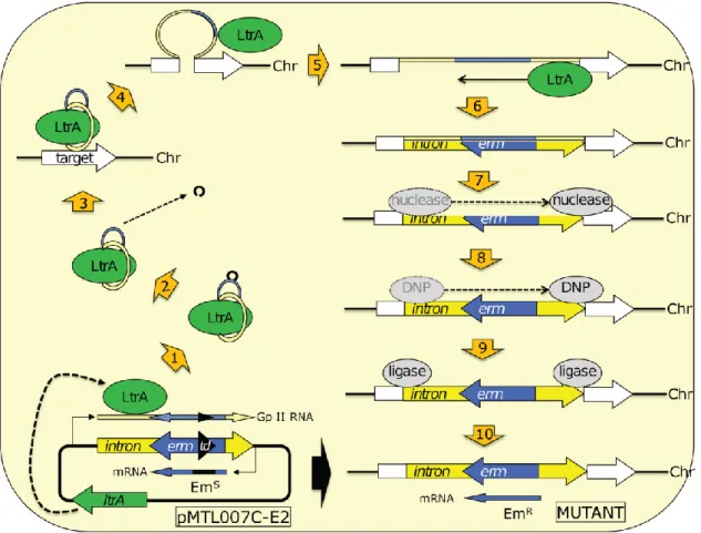
Based on the need for a reliable and efficient mutagenesis system, and given the existence of inadequate procedures for gene insertion/inactivation, Nigel Minton and collaborators developed the ClosTron (Heap et al., 2007). This is a mutagenesis system with molecular tools necessary to study and exploit the wide physiological range of the genus Clostridium. The construction system was based on the studies of the Lambowitz laboratory and on the use of group II intron from the ltrB gene of the bacterium Lactococcus lactis (Ll.ltrB) (Karberg et al., 2001). Bacterial group II introns are a newly characterized type of mobile elements, they are catalytic RNAs that excise themselves from RNA transcripts via a lariat intermediate and insert themselves into a new distal target site. They possess two important characteristics that enable their use for the directed construction of stable mutants. First, via an RNA-mediated “retrohoming” mechanism, these elements propagate to their specific site and establish connection mainly by base-pairing between the excised RNA intron (that can be rationally re-programmed) and the target DNA site. Second, the target specificity between target site and the excised RNA is dependent of the presence of a multifunctional intron encoded protein (IEP) which can be provided transiently during mutagenesis and subsequently removed to ensure the stability of the strain. The ltrA gene, that encodes the IEP, is normally within the Ll.LtrB intron to allow its permanent mobility. Knowing that, Lambowitz and coworkers, moved this gene from inside Ll.LtrB intron to another location in the plasmid and after the mutagenesis procedure, the plasmid will be lost and the excised RNA will not be moved again (Lambowitz & Zimmerly 2004). The retargeted introns resulting from this system were nominated “TargeTrons”.
Some mutagen processes were developed with the TargeTron, but it was lacking a selection method to obtain mutants with successful insertion, as only 10% of the population was correctly mutated. To overcome this limitation, Zhong and coworkers (Zhong et al., 2003) introduced into the group II intron an antibiotic resistance gene interrupted by a self-splicing group I intron, nominated retrotransposition-activated marker (RAM). All three
elements were organized in such way that just only after successful target insertion, the group I intron can splice out and the antibiotic resistance gene is restored. Therefore, the correct mutants will be resistant to the antibiotic resulting from the RAM gene (Heap et al., 2007).
As previously mentioned, the ClosTron system applied the TargeTron system as a specific tool to genetically modify genes in clostridia, and increased some potentialities, further explained. Therefore, the ClosTron plasmids pMTL007 were developed. The ClosTron RAM is based on the ermB gene (confers resistance to erythromycin, Em, following plating on thiamphenicol). The inactivation of the ermB gene is accomplished by the insertion of a small region of DNA encompassing a group I intron (from the td phage) inside the gene. This group I introns splice-out from mRNA, but this splicing is also orientation-dependent, the transcription to mRNA is not sufficient to allow splicing. Just when the opposite DNA strand encompassing the group II intron region is transcribed, the ltrA protein binds to the transcript, leading to the formation of a ribonuclear protein (RNP) complex. Now the group I intron has the correct orientation and will self-splice. Then the RNP recognizes the target DNA and binds to specific regions within the chromosome. As the ltrA protein has endonuclease and reverse transcriptase activity, nicks the DNA target, inserts the RNA and synthesizes the complementary DNA strand. Host nucleases degrade the inserted RNA, one DNA polymerase (DNP) synthesizes the opposite DNA strand, and host ligases seal the gaps, finishing the process. Now the ermB gene is present in the target gene and will have functional activity conferring Em resistance (Kuehne and Minton, 2012). An illustration of the ClosTron process is provided in Figure 1.4.