Ciências da Saúde
Study of the etiopathogenesis of Chronic
Rhinosinusitis with Nasal Polyps:
focus on the host-environment interaction
Rafaela da Cruz Vieira Veloso Teles
Thesis submitted for the degree of Doctor of Philosophy in
Medicine
Primary Supervisor: Rosa Roque Farinha, MD, PhD
Associate Supervisor: Christian von Buchwald, MD, DMSc
UNIVERSIDADE DA BEIRA INTERIOR
Ciências da Saúde
Estudo da etiopatogenia da Rinossinusite Crónica
com Pólipos Nasais:
foco na interação ambiente-hospedeiro
Rafaela da Cruz Vieira Veloso Teles
Tese para obtenção do Grau de Doutor em
Medicina
(3º ciclo de estudos)
Orientador: Professora Doutora Rosa Roque Farinha
Co-orientador: Professor Doutor Christian von Buchwald
Rafaela Veloso-Teles v
Dedication
To my Grandparents, that have always inspired me and taught me how to guide by intuition. To my Parents, that always believed in me and to all the effort they made to turn everything possible.
To Rui, that showed me how it is delightful to share the wins but also to have a comfort during defeats. Thank you for being the mirror of my soul.
To Miguel, for giving another meaning to my life.
To all the family and friends, for the stimulus they gave me.
To Professor Rosa Roque Farinha, for accepting the challenge of being my supervisor, and for her optimism and driving force concerning this investigation.
To Professor Christian von Buchwald, I will be always grateful for his sharing of expertise in Rhinology, for the invaluable advices and for the wonderful time that I spent in his department.
To the memory of Professor Sousa Martins, that inspired different generations of medical students, for his devotion as a doctor and to his merits in divulging throughout Europe the unique therapeutic properties of the Serra da Estrela air in diseases of the respiratory tract.
Rafaela Veloso-Teles vii
Publications
The investigation done for this Thesis lead to the publication of
the following papers in indexed and peer-reviewed journals:
Veloso-Teles R, Cerejeira R. Endoscopic sinus surgery for chronic rhinosinusitis with nasal polyps: clinical outcome and predictive factors of recurrence. Am J Rhinol Allergy. 2017 Jan 1;
31(1):56-62. (Appendix 1)
Veloso-Teles R, Cerejeira R, Roque-Farinha R, von Buchwald C. Higher prevalence of nasal polyposis among textile workers: an endoscopic based and controlled study. Rhinology. 2018
Jan 1; 56(2):99-105. (Appendix 2)
Veloso-Teles R, Cerejeira R, Rodrigues D, Roque-Farinha R, von Buchwald C. Food specific IgE and IgG antibodies in patients with chronic rhinosinusitis with nasal polyps: a case-control study. Ear Nose Throat J. 2019 Sep 23:145561319867668. doi: 10.1177/0145561319867668.
[Epub ahead of print] (Appendix 3)
Veloso-Teles R, Cerejeira R, Roque-Farinha R, von Buchwald C. Systemic immune profile in patients with chronic rhinosinusitis with nasal polyps. Ear Nose Throat J. 2019 Dec
Rafaela Veloso-Teles ix
Presentations
The investigation done for this Thesis lead to the following
Conference made at scientific events:
Veloso-Teles R, Cerejeira R. Epidemiologia da RSCcPN em Portugal: estudos baseados na endoscopia. (Epidemiology of CRSwNP in Portugal: endoscopic based studies). 65th Annual
Meeting of the Portuguese Society of Otorhinolaryngology–Head and Neck Surgery. Aveiro,
2018 (Appendix 5)
The investigation done for this Thesis lead to the following oral
presentation made at scientific events:
Veloso-Teles R, Cerejeira R, Rodrigues D, Roque-Farinha R, von Buchwald C. Food specific IgE and IgG-antibodies levels in patients with chronic rhinosinusitis with nasal polyps: a case-control study. 5th Congress of the Confederation of European Otorhinolaryngology–Head and
Neck Surgery (CEORL-HNS). Brussels, 2019 (Appendix 6)
The investigation done for this Thesis lead to the following
poster presentation made at scientific events:
Veloso-Teles R, Cerejeira R, Roque-Farinha R, von Buchwald C. Systemic immune profile in patients with chronic rhinosinusitis with nasal polyps: a case-control study. 5th Congress of the
Confederation of European Otorhinolaryngology–Head and Neck Surgery (CEORL-HNS).
Rafaela Veloso-Teles xi
Prizes
The investigation done for this Thesis received the following
prize:
“Clinical Research Prize ERS 2018”
for the best clinical investigation with the manuscript entitled “Higher prevalence of nasal polyposis among textile workers: an endoscopic based and controlled study”, assigned by the European Rhinologic Society, 2018 (Appendix 8)Rafaela Veloso-Teles xiii
Resumo alargado
Estudo da etiopatogenia da Rinossinusite Crónica com Pólipos
Nasais: foco na interação ambiente-hospedeiro
Introdução
A rinossinusite crónica (RSC) é uma doença inflamatória crónica do nariz e seios perinasais, que engloba dois fenótipos clínicos: a Rinossinusite Crónica sem Pólipos Nasais (RSCsPN) e a Rinossinusite Crónica com Pólipos Nasais (RSCcPN). Esta última destaca-se pela presença de formações polipoides, hiperplásicas, pedunculadas e edematosas nas cavidades nasais e seios perinasais, geralmente de forma bilateral. A RSCcPN é uma entidade clínica comum, com elevada morbilidade e cronicidade, já descrita no tempo do Antigo Egipto (2000 a.C.) e sobre a qual a investigação científica tem incidido de forma intensa nas últimas duas décadas. No entanto, apesar de toda a pesquisa realizada, a RSCcPN continua a ser um enigma na história da Medicina, permanecendo como uma doença idiopática de prevalência desconhecida, cuja fisiopatologia é em grande parte oculta. Estas incertezas refletem-se na eficácia limitada dos tratamentos disponíveis e explicarão, em parte, a elevada refratariedade da doença ao tratamento médico e cirúrgico. As principais limitações no estudo desta patologia têm sido: a sua sintomatologia inespecífica que dificulta o diagnóstico diferencial com outras patologias nasossinusais; os estudos epidemiológicos pouco fiáveis que estimam a prevalência da doença e avaliam os seus fatores de risco baseando-se em questionários sobre sintomas; a frequente ausência de diferenciação entre os tipos de RSC (RSCsPN e RSCcPN) em diferentes estudos e a inclusão de subtipos de doença (ex. RSCcPN no contexto de Fibrose Quística, Discinésias Ciliares primárias, Vasculites), que sendo casos raros e com mecanismos fisiopatológicos particulares, deverão ser alvo de estudos individualizados. Tendo em conta o consenso atual de que a RSC resultará de uma interação disfuncional entre hospedeiro-ambiente, pretende-se estudar fatores de risco exógenos e endógenos que possam estar na origem e perpetuação da inflamação da mucosa nasal que caracteriza a RSCcPN.
Este trabalho tem como objetivos:
1) Avaliar a eficácia da cirurgia endoscópica nasossinusal (CENS) no tratamento da RSCcPN e estabelecer fatores prognósticos de recidiva da doença;
2) Comparar a prevalência da polipose nasal (PN) num grupo de trabalhadores com e sem exposição ocupacional a poeiras;
3) Caracterizar e comparar alterações imunológicas sistémicas dos doentes com RSCcPN
xiv Rafaela Veloso-Teles 4) Clarificar o papel da alergia alimentar na RSCcPN, comparando os níveis séricos de
anticorpos IgE e IgG específicos contra antigénios alimentares em casos e controlos.
Material e Métodos
1) Estudo observacional retrospetivo de 85 doentes submetidos a CENS e com um follow-up mínimo de 9 meses. Os dados demográficos, a exposição ocfollow-upacional, as comorbilidades, a história cirúrgica prévia, os sintomas pré e pós-operatórios, os dados do exame ORL, resultados da TC e a informação sobre o tratamento médico e cirúrgico foram obtidos através da revisão dos processos clínicos. A análise estatística foi efetuada com recurso ao SPSS v.23. A estatística descritiva foi utilizada na caracterização da amostra. Utilizou-se o teste de McNemar na comparação dos sintomas pré e pós-operatórios. Os doentes com e sem recidiva de RSCcPN foram divididos em dois grupos independentes e foram comparados para múltiplos fatores: na avaliação da associação entre a recidiva e variáveis categóricas utilizou-se o teste de Qui-Quadrado (ou o teste exato de Fisher quando não se verificavam as assunções necessárias à execução do teste anterior); no estudo de associação entre a recidiva e variáveis quantitativas utilizou-se o teste de Mann-Whitney. Realizou-se uma regressão logística multivariada para avaliar a existência de fatores preditivos independentes na recidiva da polipose nasal. O teste de razão de verossimilhança, o teste de Hosmer e Lemeshow, a área sob a curva ROC foram realizados/calculados para avaliação do modelo criado, e procedeu-se à determinação do coeficiente de Nagelkerke’s. O teste de Wald’s e o teste de score foram obtidos para cada variável independente, assim como o Odds Ratio e seu intervalo de confiança a 95%.
2) Estudo epidemiológico transversal numa amostra randomizada de trabalhadores têxteis (n=215) e de trabalhadores de venda a retalho (n=101). Realizou-se uma entrevista clínica sistematizada, que incluiu os questionários RhinoQOL-pv e CATTM, e
uma avaliação endoscópica com ótica rígida 0º, com determinação do score endoscópico de Lund-Kennedy em cada participante. A análise estatística foi efetuada com recurso ao SPSS v.23. Utilizou-se a estatística descritiva na caracterização dos dois grupos de trabalhadores e procedeu-se à sua comparação. Na comparação de variáveis categóricas entre os grupos, utilizou-se o teste de Qui-Quadrado (ou teste exato de Fisher, quando adequado) e na comparação de variáveis quantitativas utilizou-se o teste de Mann-Whitney. O teste Binomial foi utilizado na comparação da prevalência de PN no grupo de trabalhadores têxteis com a prevalência de outros estudos publicados na literatura.
3) Estudo caso-controlo de 37 doentes com RSCcPN e 34 controlos sem RSC. Os dados clínicos foram obtidos por entrevista clínica e exame ORL. Realizaram-se TC do nariz e seios perinasais, teste cutâneo de Prick, espirometria; determinação dos parâmetros imunológicos no plasma (contagem diferencial de leucócitos, classes e
Rafaela Veloso-Teles xv subclasses de imunoglobulinas) e também dos níveis de 25-hidroxivitamina D (25-HOD), alfa-1-antitripsina (A1AT) e proteína C reativa (PCR). A análise estatística foi efetuada com recurso ao SPSS v.23. Utilizou-se a estatística descritiva na caracterização do grupo de casos e controlos. O teste de Mann-Whitney foi utilizado na comparação de variáveis contínuas entre os dois grupos e o teste de Qui-Quadrado ou o teste exato de Fisher na comparação de variáveis categóricas. Realizou-se uma subanálise com o teste de Kruskal-Wallis na comparação dos parâmetros analíticos entre 3 grupos (grupo controlo sem doenças respiratórias crónicas inferiores (DRCI), grupo RSCcPN sem DRCI e grupo RSCcPN com DRCI), seguido de comparações múltiplas interpares com o teste post-hoc de Dunn.
4) Estudo caso-controlo de 33 doentes com RSCcPN e 31 controlos sem RSC. Os dados clínicos foram obtidos por entrevista clínica, incluindo a aplicação do Questionário de Frequência Alimentar (QFA). Realizou-se o teste de ELISA com o kit OmegaDiagnostics® com 40 antigénios alimentares para determinação de anticorpos
IgG específicos e procedeu-se ao teste de imunoensaio usando o ImmunoCAP™ na avaliação de anticorpos IgE específicos contra 11 antigénios alimentares. Procedeu-se à análise estatística dos dados obtidos, com recurso ao SPSS v.23. Utilizou-se a estatística descritiva na caracterização dos dois grupos. O teste de Mann-Whitney foi utilizado na comparação de variáveis quantitativas entre os dois grupos, enquanto o teste de Qui-Quadrado ou o teste exato de Fisher foi utilizado na comparação de variáveis categóricas. O teste não-paramétrico de Spearman foi utilizado na avaliação de correlação entre variáveis quantitativas.
Resultados
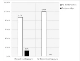
1) Houve uma melhoria significativa de todos os sintomas rinológicos após a CENS. As prevalências de complicações major e minor foram de 1,2 e 15,3%, respetivamente. A proporção de doentes com RSCcPN com recidiva da patologia após CENS foi de 31%, com 7% a necessitar de reintervenção cirúrgica. 60% dos doentes com RSCcPN reportaram exposição ocupacional a poeiras, das quais 90,4% correspondiam a poeiras de baixo peso molecular (BPM) (<5KDa). Os doentes com exposição ocupacional a poeiras apresentaram uma recorrência da doença significativamente superior ao grupo não exposto (48% vs 3%, p=5,5x10⁻⁶). A análise de regressão logística
multivariada identificou a exposição ocupacional a poeiras (p=0,001, OR=38,02, IC95%: [4,18; 345,69]) e a asma não-atópica (p=0,012, OR=8,65, IC95%: [1,62; 46,16]) como fatores preditivos independentes de recidiva da RSCcPN ao contrário das outras variáveis analisadas: idade, sexo, asma atópica, rinite alérgica, hábitos tabágicos, classificação endoscópica da polipose nasal, score de Lund-Mackay e uso pós-operatório de corticoide tópico. O modelo logístico apresentou área sob a curva ROC de 0,82 (p<0,001; IC95%: [0,73; 0,91]). O teste de razão de verossimilhança do
xvi Rafaela Veloso-Teles modelo criado obteve um p=1,2x10⁻⁷, o teste de Hosmer e Lemeshow demonstrou um
p=0,503 e o coeficiente de Nagelkerke’s foi de 0,44.
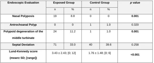
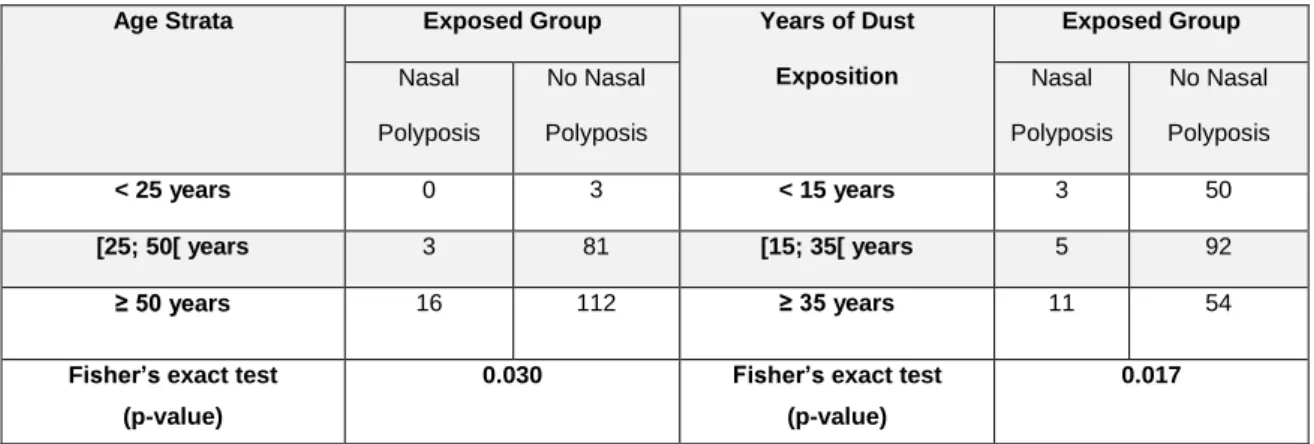
2) A PN foi diagnosticada em 19 participantes do grupo dos trabalhadores têxteis (8,8%) e em nenhum do grupo controlo (p=0,001). A prevalência da PN aumentou conforme o estrato etário (p=0,03) e dependendo do número de anos de exposição às poeiras (p=0,017). A degenerescência polipoide do corneto médio foi mais prevalente no grupo exposto (p=0,001), que também obteve um score de Lund-Kennedy mais elevado (p<0,001). No RhinoQOL-pv e no CATTM obtiveram-se scores
significativamente mais elevados entre os trabalhadores têxteis. A prevalência de PN nos trabalhadores têxteis (8.8%) foi significativamente superior às prevalências reportadas em estudos endoscópicos prévios (2,7% e 5,5%; p<0,001 e p=0,029, respetivamente).
3) No grupo dos doentes com RSCcPN a prevalência de doenças respiratórias crónicas inferiores (DRCI) foi significativamente superior ao grupo controlo (p<0,001), ao contrário da patologia atópica que não diferiu. Nos doentes com RSCcPN obteve-se uma contagem relativa de eosinófilos (p<0,001) e de basófilos (p=0,022) no plasma significativamente mais elevada do que no grupo controlo, ao contrário dos neutrófilos cuja contagem foi significativamente menor (p=0,013). Os doentes com RSCcPN apresentaram níveis mais elevados de IgG1 (p=0,022), mas mais reduzidos de IgG2 (p=0,014) e IgG3 (p=0,018) comparativamente aos controlos. Essas diferenças observadas foram mais evidentes nos doentes com RSCcPN e DRCI concomitante. Os níveis de IgG4, IgG total, IgA, IgM e IgE não diferiram entre os grupos, assim como a prevalência das deficiências de classes e subclasses de imunoglobulinas; os níveis de 25-HOD, A1AT e PCR também não diferiram de forma significativa.
4) No grupo com RSCcPN verificou-se uma concentração total de anticorpos alimentares do tipo IgG significativamente menor do que a do grupo controlo (p=0,012); esta diferença foi também observada para diferentes anticorpos IgG específicos (milho, soja, leguminosas, maçã e pera, frutos vermelhos, citrinos). No grupo controlo verificou-se uma correlação positiva entre os níveis de IgG1 séricos e a soma da concentração dos anticorpos IgG alimentares (p=0,049). Pelo contrário, no grupo com RSCcPN observou-se uma correlação negativa entra essas variáveis (p=0,048). Os níveis de IgG1 encontravam-se significativamente elevados no grupo com RSCcPN (p=0,041). Os níveis séricos de IgE específicas contra os diferentes alergénios alimentares avaliados, bem como a concentração total de IgE específicas alimentares, não diferiram de forma estatisticamente significativa entre os grupos.
Conclusões
Apesar da cirurgia endoscópica ser um tratamento eficaz na RSCcPN, com benefícios óbvios na resolução de sintomas no pós-operatório, a recorrência da doença é considerável. O
Rafaela Veloso-Teles xvii primeiro estudo deste trabalho demonstrou que a exposição ocupacional a poeiras e a asma não-atópica são fatores preditivos independentes de recidiva da doença. A identificação da exposição a partículas de BPM como principal exposição ocupacional reportada é também relevante. Estas partículas de BPM têm sido associadas ao risco de desenvolver asma ocupacional (não-atópica) e ao contrário das partículas de alto peso molecular (APM) que atuam por mecanismos IgE-mediados, não têm os seus mecanismos de ação bem estabelecidos. A distinção realizada entre asma atópica e não-atópica permitiu clarificar o impacto da asma nos resultados pós-operatórios, que era até então controverso.
O estudo epidemiológico realizado, baseado em endoscopia, foi pioneiro na avaliação do impacto da exposição ocupacional a poeiras na prevalência da PN e aponta para uma importante associação entre ambas, ao demonstrar uma prevalência da doença significativamente elevada nos trabalhadores têxteis. Os resultados deste trabalho alertam para um relevante problema de Saúde Pública, reforçando a necessidade de medidas de proteção dos trabalhadores expostos a poeiras (ex. uso de máscara com filtros apropriados) e a necessidade de controlo no funcionamento dos sistemas de exaustão de partículas e filtros de ar que garantam a qualidade do ar. Depreende-se assim que os doentes com RSCcPN devem sempre que possível trabalhar em ambientes livres de poeiras, de forma a reduzir o risco de recidiva e melhorar o seu prognóstico. Outros estudos epidemiológicos serão necessários na avaliação de outro tipo de exposições ocupacionais, tentando se possível comparar o impacto de exposição a poeiras de BPM e APM na prevalência de RSCcPN.
O estudo prospetivo, clínico-laboratorial, demonstrou ainda que os doentes com RSCcPN apresentam um perfil imune sistémico distinto dos controlos, com variações na contagem diferencial de leucócitos e um desvio IgG1 a nível das subclasses de IgG. Estas diferenças estão de acordo com o que tem sido reportado a nível local, nos pólipos nasais. De notar, que essas diferenças eram mais marcadas nos doentes com DRCI, o que reforça o conceito de “one airway, one disease”.
Relativamente ao estudo sobre o impacto da alergia alimentar na RSCcPN, esta não parece ter um papel relevante na sua etiopatogenia, seja esta resposta imune IgG ou IgE mediada. Além do mais, observou-se uma supressão de anticorpos IgG específicos contra antigénios alimentares nos doentes com RSCcPN, uma correlação negativa da sua soma com os níveis séricos de IgG1 e valores de IgG1 significativamente elevados nestes doentes. Esta supressão poderá estar relacionada com um desvio da resposta imune IgG-mediada contra outros agentes (p.ex. partículas inalantes) na RSCcPN e deverá ser investigada no futuro.
Palavras-chave
Rafaela Veloso-Teles xix
Abstract
Study of the etiopathogenesis of Chronic Rhinosinusitis with
Nasal Polyps: focus on host-environment interaction
Introduction
Chronic rhinosinusitis with nasal polyps (CRSwNP) is a common disease, with high morbidity and chronicity, but its exact etiology is still unclear and remains a difficult to treat condition. Considering the emerging consensus that chronic rhinosinusitis (CRS) results from a dysfunctional host-environment interaction, this study pretends to clarify exogenous and endogenous factors, which can contribute to the occurrence and perpetuation of sinonasal mucosa inflammation observed in CRSwNP.
The aims of this study are:
1) To evaluate endoscopic sinus surgery (ESS) efficacy in CRSwNP treatment and to establish prognostic factors for disease recurrence;
2) To compare the prevalence of nasal polyps (NP) in a group of workers with occupational dust exposure and in a control group;
3) To characterize systemic immunological alterations that occur in patients with CRSwNP compared to controls;
4) To clarify the role of food allergy in CRSwNP disease, comparing serum levels of food specific IgE and IgG antibodies in cases and controls.
Material and Methods
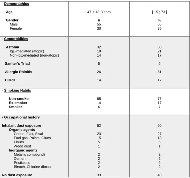
1) Retrospective observational study in 85 patients with CRSwNP submitted to ESS and a minimum follow-up of 9 months. Patients’ demographics, occupational exposure, comorbidities, previous nasal surgeries, pre and postoperative symptoms and ENT examination findings, CT results, medical and surgical treatment information were collected from medical records.
2) Cross-sectional study with a random sample of textile (n=215) and retail store employees (n=101). Clinical data was gathered through a systematic interview, which included RhinoQOL-pv and CATTM questionnaires. A systematic endoscopic nasal
examination was performed using a 0º rigid endoscope and Lund-Kennedy endoscopic score was determined for each participant.
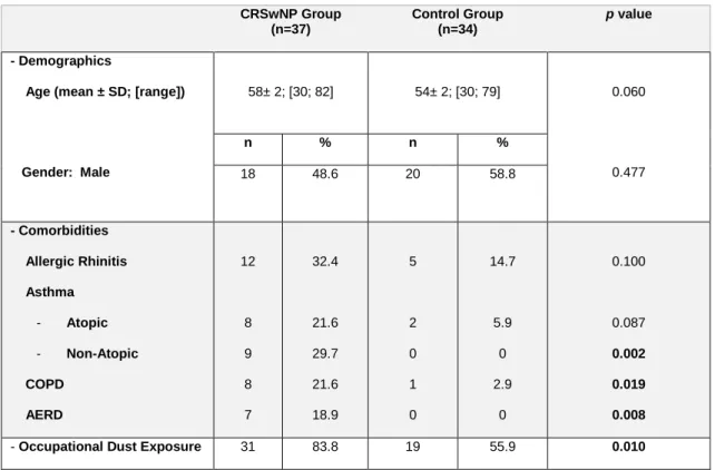
3) Case-control study with 37 CRSwNP patients and 34 controls without CRS. Clinical data was gathered through a systematic interview. CT scan, skin prick test, spirometry, immunological parameters (leukocyte differential count, immunoglobulin
xx Rafaela Veloso-Teles classes and IgG subclasses) and 25-hydroxyvitamin D (25-HOD), alpha1-antitrypsin (A1AT) and C-reactive protein (CRP) dosage in serum specimens were obtained. 4) Case-control study with 33 patients with CRSwNP and 31 controls without CRS.
Clinical data was gathered through a systematic interview (including the application of Food Frequency Questionnaire (QFA)). ELISA tests using OmegaDiagnostics® kit with 40 food allergens for detection of specific IgG antibodies were performed and food specific IgE antibodies were determined by immunoassay using ImmunoCAP™ against 11 food antigens.
Statistical analysis was performed using SPSS v.23.
Results
1) All rhinologic symptoms improved after ESS. The major and minor complications prevalences were 1.2% and 15.3%, respectively. Disease recurrence occurred in 31% of cases, but only 7% required surgical reintervention. Multivariate logistic regression analysis identified occupational dust exposure (p=0.001) and non-atopic asthma (p=0.012) as independent predictive variables in CRSwNP recurrence, unlike the other tested variables: age, sex, atopic asthma, allergic rhinitis, smoking habits, nasal polyps endoscopic grade, Lund-Mackay score and postoperative topical corticoid use. The adjusted logistic model had a ROC area under curve of 0.82 (p<0.001; CI95%: [0.73; 0.91]).
2) 316 participants were included in the study, i.e. 215 textile workers and 101 retail store workers. NP were found in 19 subjects (8.8%) among textile workers and none in the control group (p=0.001). The prevalence of NP increased by age strata (p=0.03) and by years of dust exposition (p=0.017). Polypoid degeneration of the middle turbinate was more prevalent in the exposed group (p=0.001) with Lund-Kennedy scoring also higher (p<0.001). RhinoQOL-pv and CATTM questionnaires had both
significantly higher scores among textile employees.
3) A significantly higher eosinophil (p<0.001) and basophil relative count (p=0.022) and a lower relative neutrophil count (p=0.013) were found among CRSwNP group. Patients with CRSwNP had higher IgG1 (p=0.022), but lower IgG2 (p=0.014) and IgG3 (p=0.018) serum levels compared to controls. IgG4, total IgG, IgA, IgM and IgE serum levels did not differ between groups, as well as the prevalence of immunoglobulin classes or IgG subclasses deficiency; 25-HOD, A1AT and CRP dosage had also no significant difference.
4) The overall sum of food IgG antibodies was significantly lower in CRSwNP compared to control group (p=0.012), and this difference was also observed for different specific IgG antibodies (corn, soya, grain legumes, pear and apple, berries, citric fruit). In controls a positive correlation between IgG1 and the sum of food IgG antibodies was
Rafaela Veloso-Teles xxi seen (p=0.049) but in CRSwNP group a negative correlation was found (p=0.048). Significant higher level of IgG1 was found among CRSwNP patients (p=0.041). Levels of serum specific IgE antibodies against the different studied food allergens, as well as the sum of food IgE antibodies, did not differ significantly between the groups.
Conclusions
Endoscopic sinus surgery proved to be an effective treatment in CRSwNP, but with a considerable disease recurrence. The first study of this investigation demonstrated that occupational dust exposure and non-atopic asthma are independent predictive factors of disease recurrence risk.
The epidemiologic study performed, based on endoscopy, was pioneer in the evaluation of occupational exposure to dust impact on NP prevalence and the results pointed to an important association between them by demonstrating a significantly higher prevalence of the disease among textile workers.
This investigation also showed a distinct systemic immunologic profile in CRSwNP patients compared to controls, and the variation observed in peripheral relative leukocyte count and the systemic IgG1 subclass shift are similar to what is known to happen in nasal polyp tissue.
Concerning food allergy, it does not seem to have an important role in CRSwNP etiopathogenesis, whether if it is IgG or IgE-mediated. Moreover, the observed suppression of specific IgG antibodies against food allergens, its negative correlation with IgG1 and the raised IgG1 serum levels in CRSwNP, can be related to deviated IgG responses against other targets (e.g. airborne particles) and warrants future investigation.
Keywords
Rafaela Veloso-Teles xxiii
Index
Chapter 1. Introduction 1
1.1 Chronic Rhinosinusitis with Nasal Polyps (CRSwNP): the scope of the problem 3
1.1.1 Definition 6
1.1.2 Epidemiology 7
1.1.3 Predisposing factors and associated comorbidities 8
1.1.4 Historical overview on CRSwNP etiopathogeny 15
Chapter 2. Aims of the Thesis 17
Chapter 3. Endoscopic Sinus Surgery for CRSwNP: clinical outcome and predictive factors of recurrence
21
3.1 Introduction 23
3.2 Materials and Methods 24
3.3 Results 26
3.4 Discussion and Conclusion 31
Chapter 4. Higher prevalence of nasal polyposis among textile workers: an endoscopic based and controlled study
35
4.1 Introduction 37
4.2 Materials and Methods 39
4.3 Results 41
4.4 Discussion and Conclusion 46
Chapter 5. Systemic immune profile in patients with CRSwNP 49
5.1 Introduction 51
5.2 Materials and Methods 53
5.3 Results 55
xxiv Rafaela Veloso-Teles Chapter 6. Food specific IgE and IgG antibodies in patients with CRSwNP 63
6.1 Introduction 65
6.2 Materials and Methods 67
6.3 Results 69
6.4 Discussion and Conclusion 73
Chapter 7. Final Discussion and Conclusion 77
8. References 83
Rafaela Veloso-Teles xxv
Acronym List
A1AT
Alpha-1-antitripsin
AAOHNS
American Academy of Otolaryngology, Head and Neck Surgery
ACOS
Asthma-COPD overlap syndrome
AERD
Aspirin-exacerbated respiratory disease
AFRS
Allergic fungal rhinosinusitis
ARS
Acute rhinosinusitis
BCE
Before Common Era
ANCA
Anti-neutrophil cytoplasmic antibodies
CAT
COPD Assessment Test
CEBM
Centre for Evidence Based Medicine
CFTR
Cystic fibrosis transmembrane conductance regulator
CLRD
Chronic lower respiratory diseases
COX-1
Cyclooxygenase-1
COX-2
Cyclooxygenase-2
COPD
Chronic obstructive pulmonary diseases
CRP
C-reactive protein
CRS
Chronic rhinosinusitis
CRSsNP
Chronic rhinosinusitis without nasal polyps
CRSwNP
Chronic rhinosinusitis with nasal polyps
CT
Computed Tomography
DBPCFC
Double-blind placebo-controlled food challenge
EAACI
European Academy of Allergy and Clinical Immunology
EAST
Enzyme allergosorbent test
EGPA
Eosinophilic granulomatous polyangiitis
ELISA
Enzyme-linked immunosorbent assay
ENT
Ear, nose and throat / otorhinolaryngology
EPOS
European position paper on rhinosinusitis and nasal polyps
ESS
Endoscopic sinus surgery
xxvi Rafaela Veloso-Teles
ICAR:RS
International Consensus Statement on Allergy and Rhinology:
Rhinosinusitis
Ig(s)
Imunoglobulin(s)
IgA(s)
Imunoglobulin(s) A
IgE(s)
Imunoglobulin(s) E
IgG(s)
Imunoglobulin(s) G
IgM(s)
Imunoglobulin(s) M
IL
Interleukine
ILC2s
Type 2 innate lymphoid cells
LGS
Leaky gut syndrome
LMW
Low molecular weight
MDI
Methylene diphenyl diisocyanate
mRNA
Messenger ribonucleic acid
NP
Nasal polyposis
NSAID
Nonsteroidal anti-inflammatory drugs
OSAS
Obstructive sleep apnea syndrome
PGE2
Prostaglandin E2
QFA
Questionário de Frequência Alimentar (Food Frequency
Questionnaire)
RAST
Radioallergosorbent test
RhinoQOL-pv Rhinosinusitis Quality of Life Survey Instrument, portuguese
version
ROC curve
Receiver operating characteristic curve
SDB
Sleep-disordered breathing
SERPINA1
Serpin family A member 1 gene
SPSS
®Statistical Package of the Social Sciences
SPT
Skin prick test
Th cell
T helper cell
VDR
Vitamin D receptor
Rafaela Veloso-Teles xxvii
Index of Tables and Graphics
TABLES
Chapter 3. Endoscopic Sinus Surgery for CRSwNP: clinical outcome and predictive factors of recurrence
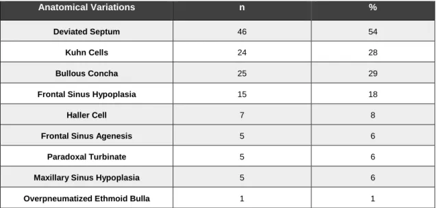
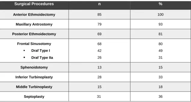
Table 1 Sample demographics, comorbidities, smoking habits and occupational history. 26 Table 2 Anatomical variations on CT scan and their absolute and relative frequencies. 27 Table 3 Surgical procedures and their absolute and relative frequencies. 28 Table 4 Independent predictive factors of recurrence in logistic regression analysis. 30
Chapter 4. Higher prevalence of nasal polyposis among textile workers: an endoscopic based and controlled study
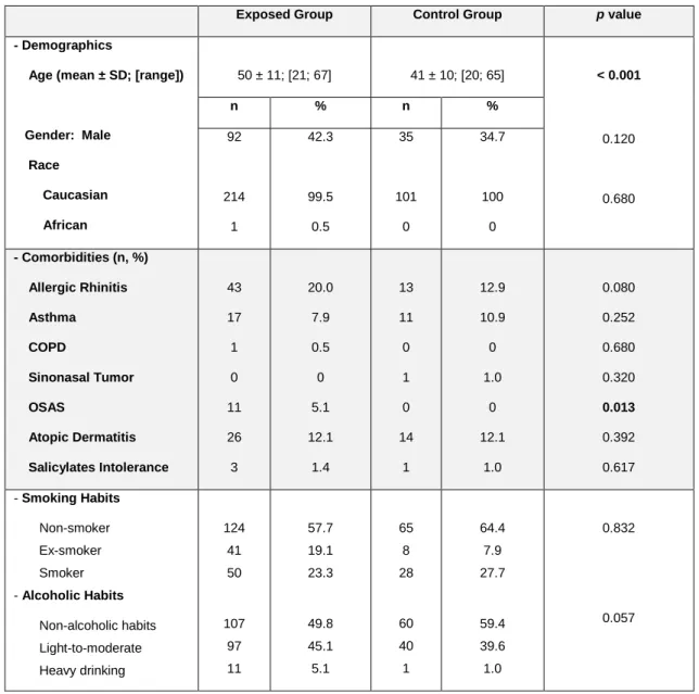
Table 5 Demographics, comorbidities, smoking and alcoholic habits in the exposed and control groups and their comparison.
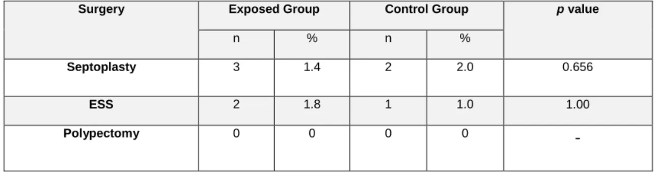
41 Table 6 History of nasal surgical procedures with absolute and relative frequencies. 42 Table 7 RhinoQOL-pv and CATTM mean scores in the exposed and control groups. 42
Table 8 Results of endoscopic evaluation, including Lund-Kennedy score, by group. 43 Table 9 Nasal polyps classification according to Lund criteria by group. 43 Table 10 Absolute prevalence of NP by age and years of dust exposition strata among
textile workers.
44 Table 11 Distribution of individuals with and without NP across working sectors in
textile industry.
45
Chapter 5. Systemic immune profile in patients with CRSwNP
Table 12 Demographics, comorbidities and occupational dust exposure in CRSwNP and control groups and their comparison.
55 Table 13 Comparison of systemic immunological parameters between CRSwNP
patients and controls.
56 Table 14 Prevalence of immunoglobulin classes and subclasses deficiency in CRSwNP
and control groups.
xxviii Rafaela Veloso-Teles Chapter 6. Food specific IgE and IgG antibodies in patients with CRSwNP
Table 15 Comparison of sample demographics, BMI, presence of CLRD, QFA score and subscores between patients with CRSwNP and controls.
69 Table 16 Comparison of food specific IgG antibodies levels and prevalence of test
positivity between patients with CRSwNP and controls.
71 Table 17 Correlation between the sum of food specific IgG concentrations and total
IgG and IgG subclasses in the serum among patients with CRSwNP and controls.
71
Table 18 Comparison between the sum of food specific IgE antibodies levels and the prevalence of test positivity between patients with CRSwNP and controls.
72
GRAPHICS
Chapter 3. Endoscopic Sinus Surgery for CRSwNP: clinical outcome and predictive factors of recurrence
Graphic 1 Relative and absolute frequencies of the preoperative and postoperative symptoms.
28 Graphic 2 Relative frequencies of CRSwNP recurrence in the group with and without
occupational dust exposure.
29 Graphic 3 Relative frequencies of surgical reintervention for CRSwNP in the group with
and without occupational dust exposure.
29
Chapter 5. Systemic immune profile in patients with CRSwNP
Graphic 4 Mean relative eosinophil count and serum concentrations of IgG1 in controls without CLRD, and in CRSwNP patients without and with CLRD.
57 Graphic 5 Mean relative neutrophil count and serum concentrations of IgG3 in controls
without CLRD, and in CRSwNP patients without and with CLRD.
57
Chapter 6. Food specific IgE and IgG antibodies in patients with CRSwNP
Graphic 6 Mean concentration values of specific IgG antibodies for different food allergens in patients with CRSwNP and controls.
1
Chapter 1.
INTRODUCTION
Rafaela Veloso-Teles 3
1. Introduction
1.1 Chronic Rhinosinusitis with Nasal Polyps (CRSwNP):
the scope of the problem
Chronic Rhinosinusitis (CRS) is an inflammatory disease of nasal and paranasal sinus mucosa, which includes two different clinical phenotypes: Chronic Rhinosinusitis without Nasal Polyps (CRSsNP) and Chronic Rhinosinusitis with Nasal Polyps (CRSwNP). CRSwNP distinguishes itself by the presence of polypoid, pedunculated, edematous and hyperplastic tissue masses in nasal cavity and paranasal sinus, most often bilaterally and generally arising from the middle meatus.[1–3] Nasal polyposis (NP) have been a medically recognised condition since the time of ancient Egypt, with literature records about it of approximately 2000 years BCE. Egypt civilization was known for its familiarity and dexterity in the nasal cavity because of their routinely removal of intracranial contents throughout the nose during the mummification process.[4] Later, Hippocrates (460-370 BCE), during the apex of Greek civilization, observed and described different otorhinolaryngology diseases, referring to the “nasal growths” as “polypus” due to their resemblance to the sea-polyp.[4] The word “polúpous” from the ancient Greek drives from the conjugation of “polús” (many) and “poús” (feet), which was later in the origin of the word “polypus” in Latin. Hippocrates also described a treatment modality for this disease with removal of nasal polyps with a snare (polypectomy), a method which persisted well into the second half of the 20th century.[4]
In the last two decades, a lot of investigation has been made on CRS, about its epidemiology, modifying factors, associated comorbidities, phenotypes and endotypes, disease biomarkers, prognosis and treatment. In 2005, the first European Position Paper on
Rhinosinusitis and Nasal Polyps (EP3OS)[5] was published, then it was actualized in 2007
(EPOS2007)[6], with the ultimate edition released in 2012 (EPOS2012).[1] This last consensus document of the European Rhinologic Society (ERS) was particularly exhaustive, trying to actualize the acquired knowledge about Rhinosinusitis and Nasal Polyps, to establish uniform definitions and diagnosis criteria and using CEBM (Centre for Evidence Based Medicine 1998) levels of evidence when making recommendations about diagnosis, treatment and disease approach. In 2007, the American Academy of Otolaryngology, Head and Neck Surgery (AAOHNS) also published a guideline for clinical practice concerning adult rhinosinusitis[7], that was recently actualized and reedited in 2015.[8] The rapid growth in number of publications in the last years led rhinologic experts from around the world, in an effort to both consolidate and critically appraise that information, to produce the International Consensus Statement on Allergy and Rhinology: Rhinosinusitis (ICAR:RS), published in 2016.[9]
4 Rafaela Veloso-Teles In spite of all the research that has been done, CRSwNP is still an enigma in Medicine. CRSwNP remains as an idiopathic disease, its true prevalence is unknown, its pathophysiology is unclear, it has an important impact in patient quality of life with high morbidity, and despite the advances in surgical and medical treatment, it has a considerable rate of refractoriness and recurrence. There are different factors that have hampered the investigation about this clinical entity. First of all, symptoms of CRSwNP are nonspecific, making it difficult to distinguish it from other sinonasal diseases. Unfortunately, most of epidemiological studies about CRS have based their diagnosis in symptoms questionnaires, turning the prevalences they found merely speculative. The second problem is that many of the investigation that has been carried out does not separate CRS in its two main phenotypic types (CRSwNP and CRSsNP), or even its subtypes (e.g. CRSwNP associated with Cystic Fibrosis, Vasculitis, Primary Ciliary Dyskinesias, Major Immunodeficiencies, etc). These subtypes of CRSwNP, comprise uncommon cases, that must be evaluated in specific studies, since it is expected the involvement of different physiopathological mechanisms. Moreover, the etiopathogenesis of CRSwNP can have origin in different individual and environmental factors that are difficult to define, creating controversy and uncertainty about it.
Many researches have been carried out around CRS endotyping, trying to reach a classification of CRS based on the cellular and molecular physiopathological mechanisms (e.g. eosinophil-based endotype, Th2-based endotype, IgE-based endotype, cysteinyl-based endotype).[10] Until now there is no single and precise classification for CRSwNP endotyping and endotype-based treatments are still not approved for routine clinical practice, being reserved only for clinical trial settings, in which results are awaited. The difficulties around endotyping have to do with the largely unknown pathophysiology behind CRSwNP.
In western countries, 85% of the CRSwNP disease reveals a type 2 inflammatory pattern. In the last 15 years, several randomized double-blind studies on monoclonal antibodies in CRSwNP were performed, namely anti-IL-5 (reslizumab, mepolizumab), anti-IL4 receptor alpha (dupilumab), and anti-IgE (omalizumab).[11] These biological agents target specific immune pathways, such as IL-5 orchestrating the survival of eosinophils, or IL-4 and IL-13 regulating the formation of IgE and the chemotaxis of eosinophils, among other effects. It was observed for the first time that biologics targeting type 2 immune reactions might be successful in nasal polyps but those studies were characterized by limited number of patients and heterogeneous populations.[11] Until now, however, no biomarkers have been identified to predict response to a specific biologic or to monitor treatment success. Phase-3 studies on monoclonal antibodies with larger populations are being conducted and only with their results it will be feasible to define treatment indications, patient selection and possible side effects. In June 2019, the Food and Drug Administration from United States of America has approved the first monoclonal antibody (dupilumab) for inadequately controlled CRSwNP.
Rafaela Veloso-Teles 5 Type-2-cell-mediated immunity, rich in eosinophils, basophils, mast cells, CD4(+) Th2 cells, ILC2s, and type 2 subset of natural killer T cells and macrophages play a role in chronic eosinophilic diseases, such as Western CRSwNP, asthma, and atopic dermatitis.[12] However, in Asian subjects a primarily neutrophilic process occurs in adult bilateral polyps. Studies done in Asian countries like China, Thailand, Singapore and Malaysia found that neutrophil-predominant polyps occur in a large percentage of patients (approximately 49%).[13] The etiology of nasal polyposis in Caucasians and Asians may be different and probably need to be managed differently.
To overcome the previously mentioned limitations on CRS research and taking advance of all the acquired knowledge in the field, future studies must use a rigorous methodology, defining the types and subtypes of the disease that they intend to evaluate. Only then, we can make studies comparable, trying to uncover the subjacent physiopathological mechanisms and finally allow the development of specific treatment options for different CRS subgroups.
6 Rafaela Veloso-Teles
1.1.1. Definition
1.1.1.1 Clinical definition of chronic rhinosinusitis
The European consensus document EPOS2012[1], clinically defines CRS (with or without nasal polyps) in the adult as an inflammation of the nose and the paranasal sinuses characterised by two or more symptoms, one of which should be either nasal blockage/obstruction/congestion or nasal discharge (anterior/posterior nasal drip), with or without other symptoms of facial pain/pressure, and reduction or loss of smell. To establish the diagnosis of CRS there must be either endoscopic signs of nasal polyps and/or mucopurulent discharge primarily from middle meatus and/or edema/mucosal obstruction primarily in middle meatus; and/or Computed Tomography (CT) signs of mucosal changes within the ostiomeatal complex and/or sinuses. To be considered as chronic, the symptoms must persist for 12 or more weeks. Questions on allergic symptoms (i.e. sneezing, watery rhinorrhea, nasal itching, and itchy watery eyes) should be included.[1] Chronic rhinosinusitis with nasal polyps (CRSwNP) is diagnosed if chronic rhinosinusitis as defined above is present and bilateral polyps are endoscopically detected in the middle meatus.[1]
1.1.1.2 Definition for use in General Practice and epidemiology studies
About the definition to use in General Practice or for epidemiological studies, EPOS2012[1] states that it should be based on symptomatology without the need of otorhinolaryngological or imagiological examination.1.1.1.3 Definition for research
For research purposes, EPOS2012[1] considers that CRS must be defined as per the clinical definition. For the purpose of a study, the differentiation between CRSsNP and CRSwNP must be based on endoscopy.
Rafaela Veloso-Teles 7
1.1.2 Epidemiology
CRSwNP is a common clinical entity, but the epidemiological studies about it are scarce, especially in Europe.[1] Another important issue is that most of the epidemiological studies are based on symptoms questionnaires[14–16] and do not allow the distinction between CRSwNP and CRSsNP. The use of a special definition for epidemiology studies as recommended by EPOS2012, based only in sinonasal symptoms, make it impossible to distinguish the main types of CRS, or even from other sinonasal diseases, which can origin an overestimation of CRS. Moreover, many of the studies have used email questionnaires and telephone interviews that reduces, even more, the acuity of diagnosis.
A previous study in Sweden[17] has already showed that data obtained with questionnaires about CRSwNP prevalence can be unreliable, since not all patients that claim to have NP have polyps on nasal endoscopy and asymptomatic polyps will be missing, meaning that cases of subclinical disease will be dropped out.
Despite not having yet changed its epidemiological definition for CRS, EPOS2012[1] alerts that nasal endoscopy is a prerequisite for an accurate estimate of the prevalence of NP and that in the light of epidemiologic research, a distinction needs to be made between clinically silent NP or preclinical cases, and symptomatic NP.
Having all these facts in consideration, we can assume that the ideal methodology to use for CRS epidemiological studies includes standardized and validated symptom questionnaires for the disease, associated with nasal endoscopy.
In Europe, only two endoscopic-based studies have evaluated NP prevalence: an in-vivo study done in Sweden[17] that found a prevalence of 2.7% and a cadaver study done in Portugal[18] that showed a prevalence of 5.5%.
8 Rafaela Veloso-Teles
1.1.3 Predisposing factors and associated comorbidities
1.1.3.1 Demography
According to published data, CRSwNP prevalence seems to change according to demographical parameters. It is a disease that occurs in all ethnicities, becomes more common with age and appears to have a preference for male gender.[1] The average age of onset is 42 years and is rare under the age of 20.[1]
In the population-based study done in Sweden[17], Johansson et al. verified through a multivariate regression logistic analysis that male gender (OR: 2.7 [95% CI: 1.33–5.5]) and seniors (>60 years; 5%) were more commonly affected by the disease.
Despite being a universal disease with similar clinical manifestations, the subjacent physiopathological mechanisms seem to diverge between Caucasian and Asiatic patients. Comparing nasal polyps tissue of Caucasian and Asiatic origin, the last ones have a minor eosinophilic inflammation.[19]
1.1.3.2 Atopic status
In the past, it was tempting to speculate that atopy could predispose to CRSwNP occurrence, and this was one of the first theories about CRSwNP etiopathogeny. However, many studies that tried to correlate allergic rhinitis (IgE-mediated) with CRSwNP did not find any raise of atopic prevalence in this patients.[1] Furthermore, it was also demonstrated that patients with allergic rhinitis had prevalences of CRSwNP between 0.5-4.5%, which are similar to the prevalences found in general population.[1] More recently, a prospective study that included 210 CRSwNP patients found that atopic status did not correlate with disease severity or with disease recurrence in postoperative follow-up.[20]
Moreover, Settipane et al. and Grigoreas et al. conducted two large epidemiological studies and both showed higher prevalences of CRSwNP in patients with non-allergic rhinitis (4.7% and 8.9%) than in patients with allergic rhinitis (1.5% and 1.7%), with statistically significant differences.[21,22]
1.1.3.3 Asthma and other Chronic Lower Respiratory Diseases (CLRD)
The extent of the interrelationship between CRSwNP and chronic lower respiratory diseases (CLRD) and the subjacent physiopathological mechanism behind it still have to be clarified, deserving a close collaboration between Otorhinolaryngologists, Pneumologists/ Respiratory Physicians and Immunoallergologists. Once more, the epidemiological studies that exist about this topic are mostly based on symptom questionnaires[23] and have only evaluated CRS in general.[24] Nevertheless, there is convincing evidence that these diseases are correlated, reinforcing the concept of the unified airway (“one airway, one disease”[25]). In a study conducted by Settipane et al.[21] that included 2228 patients with asthma, it was demonstrated that patients with non-atopic asthma (non-IgE-mediated asthma) had aRafaela Veloso-Teles 9 significantly higher prevalence of NP when compared to patients with atopic asthma (IgE-mediated asthma), with prevalences of 12.5% vs 5.0%, respectively (p<0.01). These findings were also corroborated by Grigoreas et al., that also found a prevalence of NP in patients with non-atopic asthma (13%) much superior than in patients with atopic asthma (2.4%), in a sample of 1877 asthmatic patients.[22] A Danish study, that evaluated 40 patients with CRSwNP referred for surgery, clinically and objectively through peak expiratory flow, spirometry and bronchodilation tests, found a very high prevalence of asthma (65%), that was frequently previously undiagnosed (25%). This value diverged in a statistical significant manner from the control group.[26] More recently, that investigation team also reported a prevalence of asthma in CRSwNP patients among the primary care setting of 44%.[27] Even though the prevalence of asthma was still high, it was significantly less prevalent than the one found in patients undergoing endoscopic sinus surgery (ESS). This suggests that patients with more severe upper airway disease have more frequently associated lower airway disease.[26]
The impact of asthma in ESS postoperative results for CRSwNP has generated a lot of controversy among researchers, with ones finding a negative impact of this comorbidity[28,29] and others not finding any influence at all.[30,31] An important methodological limitation of those studies is the absence of differentiation between atopic and non-atopic asthma, beside the non-differentiation of CRS types, that can definitely contribute to the divergence of the results that have been found.
Concerning chronic obstructive pulmonary disease (COPD), its relationship with CRS is even more poorly defined. A study made in Norway in 2015, with patients with COPD, asthma and controls submitted to magnetic resonance (MR), showed that the probability of paranasal sinus opacification was 6 times higher in COPD and two fold higher in asthmatic patients compared to the control group.[32] In addition, there are clinical research studies showing a high prevalence of sinonasal symptoms in COPD patients (75%) [33], and an inverse correlation between nasal patency evaluated by rhinomanometry and pulmonary airflow obstruction (FEV1%), and therefore to COPD disease severity.[34] It would be of value to determine with an endoscopic based study the prevalence of CRSsNP and CRSwNP in a COPD group of patients.
As discussed above, CRSwNP has a higher prevalence among elderly and it is important to be aware that in this age strata it is sometimes difficult to distinguish asthma from COPD, a fact that has inclusively introduced a new but still not well defined clinical entity, the asthma-COPD overlap syndrome (ACOS).[35] More investigation is needed to study the association of CRSwNP and these chronic lower respiratory diseases (CLRD).
10 Rafaela Veloso-Teles
1.1.3.4 Aspirin exacerbated respiratory disease (AERD)
Aspirin-exacerbated respiratory disease (AERD) is a condition characterized by CRSwNP, asthma and sensitivity to aspirin and other nonsteroidal anti-inflammatory drugs (NSAID), a triad called as Samter’s triad, in behalf of Samter and Beers[36] that made it well known in scientific community. In these patients, aspirin and other NSAID that inhibit cyclooxygenase-1 (COX-1) induce unique non–IgE-mediated reactions that include a spectrum of respiratory reactions, including rhinitis, flushing, congestion, laryngospasm, and asthma exacerbations.[37] The prevalence of nasal polyposis in aspirin-sensitive asthmatics may be as high as 60-70%, as compared to less than 10% in the population of aspirin-tolerant asthmatics.[1] CRS patients with AERD tend to suffer from more extensive sinus disease and they benefit from ESS to a lesser extent than patients without AERD, with more predisposition for disease recurrence.[1] Among patients with CRSwNP, AERD represents 9.7% of the patients.[38] Patients with aspirin sensitivity, asthma and NP are usually non-atopic and the prevalence increases over the age of 40 years. It has a predominance for male gender and also seems to have an hereditary predisposition.[1,37]
1.1.3.5 Ciliary dyskinesia
Primary ciliary dyskinesia (PCD) is an autosomal recessive genetic disease, characterized by ciliary mobility disfunction with improper airway mucociliary clearance. There are currently 33 known genes associated with PCD, with mutation at DNAI1 and DNAH5, which encode for components of the outer dynein arm complex in cilia, being the two most common genes associated with PCD.[39] About half of these patients presents with situs
inversus.[39] In 1933, Kartagener recognized the combination of situs inversus, bronchiectasis
and chronic rhinosinusitis, that was designated as Kartagener Syndrome.[40] In 1977, Eliasson et al. first coined the term “immotile cilia syndrome” for Kartagener Syndrome to associate infertility with chronic sinopulmonary infections.[40] In the literature, it has been published different cases of patients with Kartagener Syndrome presenting the CRSwNP phenotype.[40– 42].
CRS affects ≥ 70% of patients with PCD and nasal polyps are prevalent in 15% to 30% of these patients, a much higher value compared to 3-4% in the general population.[43] Alanin MC et al. studied 24 PCD patients and found a significant improvement in CRS-related symptoms 12 months after ESS and adjuvant therapy (saline irrigation, topical nasal steroids and 2 weeks of systemic antibiotics plus instigations of local colistin if Pseudomonas
aeruginosa was cultured at ESS). Moreover, a trend toward better lung function was observed
after ESS. In this cohort nasal polyps were observed in 42% of the patients, including polyps in 2 children. Sinus hypoplasia was also a common trait in more than 50% with PCD.[43]
Secondary ciliary dyskinesia (SCD) can occur after epithelium injuries (e.g. viral airway infections, exposition to pollutants) and is commonly temporary. Mucostasis, hypoxia, microbial products, and mediators and toxic proteins generated during chronic inflammation probably all contribute to diminished mucociliary function. These factors decrease
Rafaela Veloso-Teles 11 mucociliary function by direct toxic effects on cilia, ciliary loss, other ultrastructural alterations in the epithelium and changes in the viscoelastic properties of the mucus.[44] In the majority of CRS patients, dysfunction in mucociliary function seems to be more a secondary than primary event, and is probably reversible, although restoration takes some time.[1]
1.1.3.6 Cystic Fibrosis
Cystic Fibrosis (CF) is caused by a defect in the protein responsible for chloride and bicarbonate transport (cystic fibrosis transmembrane conductance regulator protein, CFTR).[10] In patients with CF, the inability of the cilia to transport the viscous mucus cause mucociliary malfunction and consequently CRS. NP are present in about 40% of patients with CF and are generally more neutrophilic.[1] Nasal Polyps are rare in children and it has been reported that the majority of children with CRSwNP has CF[45], making it mandatory to exclude CF in every child presenting with nasal polyposis.
Annaes K et al. followed a cohort of 106 CF patients submitted to ESS plus adjuvant antibiotic treatment (two weeks of intravenous and 6 months of topical antibiotics) founding that the frequency of pulmonary samples with CF pathogens was reduced one-year after surgery, and the symptoms of CRS and quality of life significantly improved.[46] In CF patients a marked association exists between upper and lower airway cultures, with paranasal sinuses often being colonised by CF-lung-pathogenic Gram-negative bacteria. The combined treatment of CRS (ESS plus systemic and topical antibiotic treatment) seems to be of benefit in upper and lower airway disease in CF patients.[46]
1.1.3.7 Vasculitis
Eosinophil granulomatosis with polyangiitis (EGPA), formerly known as Churg-Strauss syndrome, is a rare systemic disease characterized by necrotising, granulomatous inflammation involving small to medium vessels, associated to severe asthma, peripheral and tissue eosinophilia and CRS. CRSwNP is present in about 60% of patients with EGPA[47], and there are many case reports about this association.[48–50] Following the criteria of the American College of Rheumatology, the diagnosis can be confirmed with high specificity and sensitivity if 4 of 6 of the following manifestations are present: bronchial asthma; eosinophilia >10%; sinusitis (acute or chronic; alternatively also opacity in imaging); (if applicable transient) pulmonary infiltrates; histologically confirmed vasculitis with detection of extravascular eosinophils; mononeuritis multiplex or polyneuropathy.[51] EGPA is considered as a ANCA (anti-neutrophil cytoplasmic antibodies) associated vasculitis, but less than 50% of patients have ANCA positivity.[50] When present they correspond to IgG class antibodies with a predominant perinuclear pattern (p-ANCA).(37, 40)
12 Rafaela Veloso-Teles
1.1.3.8 Immunodeficiency
Congenital immunodeficiencies typically manifest during infancy and childhood as frequent, chronic or opportunistic infections, with symptoms appearing early in life, commonly during infancy and childhood.[1] However, depending on the deficiency and dysfunction of the immune system, it can manifest later in life and present with CRS.[1] Therefore, EPOS2012 considers that immunological testing should be an integral part of the diagnostic pathway of patients with CRS.[1]
In 2015, a systematic literature review and meta-analysis has alerted that humoral immunodeficiency could be a frequent condition in patients with CRS.(46) This study revealed a immunodeficiency of total IgG, IgA and/or IgM in 13% of patients with recurrent CRS (not controlled by conservative management) and in 23% of patients with difficult-to-treat CRS (not controlled despite successful ESS and appropriate conservative treatment for at least one year).(46) The prevalence of IgG subclasses and specific antibody deficiency in CRS patients was 5 to 50% and 8 to 34%, respectively.(46) This study concluded that immunoglobulin deficiency is a frequent condition in CRS patients.(46) However, this study presented limitations that are inherent to the analysed studies: most of them were not controlled, had incomplete information about possible secondary causes of hypogammaglobulinemia (such as systemic corticoid use), did not separate CRS with and without nasal polyps, and used different methodologies and study designs.
Concerning immunological testing specifically in CRSwNP, the published literature is very scarce. In 2014, a non-controlled study in 161 patients with CRSwNP found a prevalence of IgG subclass deficiency of 13.7%, and they found no correlation between the presence of humoral deficiency and either symptom evolution after medical and surgical treatment or the dose of corticosteroids needed to control disease, after a follow-up of 5 years.[53] The authors concluded that a link between IgG subclass deficiency and nasal polyposis seemed unlikely. The lack of reference data on general population prevalence of IgG subclass deficiency and the absence of a control group, made it impossible to find any association between IgG subclass deficiency and NP.[53]
1.1.3.9 Environmental factors
The role of environmental factors in the development of CRSwNP remains undervalued and only a few studies have payed attention to it. In 1995, a Portuguese clinical-pathological study in 92 cases of CRSwNP, demonstrated with appropriate histochemical techniques that 69 out of 92 nasal polyps (75%) had inclusions of an exogenous material in the interior of the polyps (namely, particles of iron, wood, cement, cork, paper, glass, tobacco, textile fibres and chalk), that could be either localized inside the macrophages or free and sparse in the polyp tissue.[54] In that study, it was also interesting to observe that in 36 patients that abandoned their occupational exposure, only two (5.5%) demonstrated disease recurrence after a follow-up of 12 years.[54] In 2002, Kim J and Hanley J, suggested in a case control study with 55 CRSwNP patients and 55 controls, that the use of woodstove as a
Rafaela Veloso-Teles 13 principal source of heating or the occupational exposure to noxious inhalant compounds (other than tobacco smoke) constitute risk factors for NP development.[55] In 2012, Hox et al. also found that occupational exposures can be a risk factor for the occurrence of CRS and for its recurrence or persistence.[56] In 467 patients submitted to ESS for CRS, 25% had a relevant occupational exposure (reported on questionnaires sent by mail) and the prevalence of those exposures increased linearly and significantly with the number of ESS procedures.[56] Lastly, a multicentric study based on self-administered questionnaires conducted in China, that included 10,633 subjects, found a prevalence of CRS of 8.0% and found that some occupational and environmental exposures were strongly associated with CRS (namely, having a clearance-related job, occupational exposure to dust, occupational exposure to poisonous gas, a pet at home or carpet at home or at the workplace).[16] As discussed above, on epidemiology subchapter (see 1.1.2), studies as this last one based on self-administered questionnaires, are not truly reliable and do not allow to distinguish between CRSwNP and CRSsNP.
About cigarette smoking, there is controversial evidence about its role on CRSwNP. There are studies demonstrating that the prevalence of CRSwNP is reduced in smokers compared to the general population[57,58], while others reported smoking as a risk factor for CRSwNP recurrence.[59,60]
1.1.3.10 Anatomic Variations
Some authors have mentioned certain anatomic variations such as concha bullosa, deviated nasal septum or Haller’s cells (infraorbital ethmoid cells), as potential risk factors for CRS development.[61,62] However, the studies that have made these assertations often establish CRS diagnosis by mucosal thickening on CT scan, when it has already been demonstrated that incidental mucosal thickening can occur in almost a third of asymptomatic population.[63] The majority of these studies are old, do not take in consideration the clinical definition of the disease and do not differentiate between acute rhinosinusitis (ARS), CRSsNP or CRSwNP.[9] Moreover, there are already published studies showing that the prevalence of anatomic variations in CRS patients is not superior to the ones found in the general population.[63] The International Consensus Statement on Allergy and Rhinology: Rhinosinusitis published in 2016 (ICAR:RS 2016) considers that CRSwNP patient populations have rarely been independently studied to determine the influence of anatomic variation on this disease and the few studies that independently evaluated this group of patients suggested a minimal influence on pathophysiology and instead favoured a systemic inflammatory process leading to sinonasal disease.[9] Later, a study in CRSwNP patients and controls found a higher prevalence of some anatomic variations in CRSwNP patients (i.e. deviated septum, concha bullosa, agger nasi cells, frontal hypoplasia and accessory ostium in maxillary sinus).[64] In this study, the sample selection for control group is not well explicit and only some kind of septum deviations were considered. As already reported on PCD
14 Rafaela Veloso-Teles subheading, sinus hypoplasia seems to be a common trait in these patients that are also prone to develop nasal polyposis.[43]
Further research is needed about this topic. The role of anatomic variations on CRSwNP pathogenesis remains controversial, meanwhile it is valuable to recognize them for a good surgical planning of these patients.
1.1.3.11 Other factors
Concerning alpha-1-antitrypsin (A1AT), there are two published articles about SERPINA1 gene polymorphisms in CRSwNP patients. One of these case-control studies was realized in Canada and demonstrated an association between single nucleotide polymorphisms (SNPs) of the SERPINA1 gene (rs1243168 e rs4900229) with clinically severe CRSwNP.[65] The other study took place in Germany and reported a significantly higher prevalence of genetic A1AT polymorphisms (PI-MS and MZ) among CRSwNP patients compared to controls.[66] It is a topic with scarce data about it, but the existing results seem to be consistent and deserve to be replicated in other populations.
The role of vitamin D in CRSwNP etiopathogeny has been ambiguous. Vitamin D has been shown to be a potent immunomodulatory steroid hormone involved in the regulation of epithelial cells, dendritic cells, monocytes, macrophages and T-cells functions.[9] In 2016, a systematic review suggested a correlation between low vitamin D and polypoid CRS phenotypes.[67] However, this review included only seven articles, three of them with retrospective character, the majority without a control group, and with heterogeneous methods for vitamin D dosage, for reporting the outcomes and for analysis of the confounding variables. More studies with rigorous methodology must be done to clarify this possible association.