Searching for biocompounds in algae and
seagrasses with potential use in the
treatment of Alzheimer’s disease
UNIVERSIDADE DO ALGARVE
Faculdade de Ciências e Tecnologia
Searching for biocompounds in algae and
seagrasses with potential use in the
treatment of Alzheimer’s disease
Mestrado Integrado em Engenharia Biológica
Dissertação realizada sob a orientação de:
Prof. Dr. João Carlos Serafim Varela
Dra. Luísa Margarida Batista Custódio
UNIVERSIDADE DO ALGARVE
Faculdade de Ciências e Tecnologia
Centro de Ciências do Mar – Grupo MarBiotech
potential use in the treatment of Alzheimer’s disease
Declaração de autoria de trabalho
Declaro ser a autora deste trabalho, que é original e inédito. Autores e trabalhos consultados estão devidamente citados no texto e constam da listagem de referências incluída.
____________________________________ Águeda Laura dos Santos Silvestre
© Águeda Laura dos Santos Silvestre
A Universidade do Algarve reserva para si o direito, em conformidade com o disposto no Código do Direito de Autor e dos Direitos Conexos, de arquivar, reproduzir e publicar a obra, independentemente do meio utilizado, bem como de a divulgar através de repositórios científicos e de admitir a sua cópia e distribuição para fins meramente educacionais ou de investigação e não comerciais, conquanto seja dado o devido crédito ao autor e editor respetivos.
Agradecimentos
Dra. Luísa Custódio, pela amizade, convívio, ensinamentos, orientação, partilha, rigor, sinceridade, estímulos e entusiasmo. Levo-os comigo até sempre! Não é fácil encontrar forma justa de agradecer…
Professor Dr. João Varela, pela cordialidade e permissão que me concedeu, quando mostrei interesse em aprender com o grupo MarBiotech e quando manifestei a intenção de, finalmente, terminar o curso. Agradeço igualmente toda a ajuda e interesse para que eu pudesse participar no XVII Congresso Nacional de Bioquímica, em 2010, na cidade do Porto. Não esqueço!
Professora Dra. Luísa Barreira, pelas explicações, maior ajuda e disponibilidade no momento do tratamento estatístico, essencial para a elaboração deste trabalho. Assim como, a simpatia e o incentivo com que me brindou, nos momentos em que nos cruzámos ultimamente, para que eu finalizasse esta tese, mesmo volvido tanto tempo. Colegas de laboratório, pelo companheirismo e por me terem proporcionado o melhor ambiente de trabalho que poderia ter tido, durante o período de prática desta dissertação.
Catarina e Ivone, por terem transformado numa viagem tão divertida, a desafiadora vida de estudante.
Família do Grupo Muzenza de Capoeira, por sempre serem capazes de me oferecer momentos felizes e demonstrar o vosso afeto e orgulho em mim. Em especial, à Dora, pelo encorajamento; à Sol, pela ajuda no Inglês; e à Daiana, por mostrar tanto interesse na leitura deste trabalho, assim como as suas observações e críticas sempre construtivas!
Mãe, pai, Ana Teresa, Pena e meu filho por serem a minha força motriz, maior exemplo e inspiração. Toda a preocupação e fé nas minhas capacidades fortaleceram-me e permitiram o fortaleceram-meu regresso.
Reconheço, agradeço e deixo registado o meu vínculo e comprometimento para com todos que me ajudaram ao longo do percurso que percorri na Universidade do Algarve e me possibilitaram o processo de elaboração e finalização da presente tese.
Part of this thesis was published in the following scientific papers:
Custódio L, Justo T, Silvestre L, Barradas A, Vizetto C, Pereira H, Barreira L, Rauter AP, Alberício F and Varela J. (2012). Microalgae of different phyla display antioxidant, metal chelating and acetylcholinesterase inhibitory activities. Food Chemistry, 131, 134–140.
Custódio L, Ferreira C, Pereira H, Silvestre L, Duarte C, Barreira L, Rauter AP, Alberício F and Varela J. (2012). The marine halophytes Carpobrotus edulis L. and
Arthrocnemum macrostachyum L. are a potential source of nutritionally important
PUFAs and biocompounds with antioxidant, metal chelating and anticholinesterase inhibitory activities. Botanica Marina, 55, 281–288.
Custódio L, Silvestre L, Rocha MI, Rodrigues MJ, Vizetto-Duarte C, Barreira L and Varela J. (2016). Methanol extracts from Cystoseira tamariscifolia and Cystoseira nodicaulis are able to inhibit cholinesterases and protect a human dopaminergic cell line from hydrogen peroxide induced cytotoxicity. Pharmaceutical Biology, 5, 1–10.
Table of Contents
Page List of tables v List of figures vi Abbreviations viii Abstract ix Resumo xi 1. Introduction 11.1. Alzheimer’s disease (AD) 2
1.1.1. Symptoms 2
1.1.2. Causes of AD 3
1.1.3. Risk factors for AD 6
1.1.3.1. Advanced age 6
1.1.3.2. Family history 6
1.1.3.3. Apolipoprotein E-ε4 (APOE-ε4) 7 1.1.3.4. Cardiovascular disease risk factors 7
1.1.4. Cholinergic theory 7
1.1.5. Oxidative stress and metal accumulation 9
1.1.5.1. Iron and AD 10
1.1.5.2. Copper and AD 12
1.1.6. Treatment 12
1.1.6.1. ChE inhibitors 13
1.1.6.2. Metal chelation as a neuroprotective strategy 14
1.2. Marine natural products 16
1.2.1. Macroalgae 18
1.2.2. Microalgae 19
1.2.3. Seagrasses 21
1.2.4. Halophytes 22
1.3. Objective 24
2. Materials and Methods 25
2.3. Metal chelating activity 30
2.4. AChE and BChE inhibitory activity 32
2.5. Statistical analysis 33
3. Results 34
3.1. Iron and copper chelating activity 35
3.1.1 Fe2+-chelating activity 35 3.1.1.1 Macroalgae 36 3.1.1.2 Microalgae 38 3.1.1.3 Seagrasses 38 3.1.1.4 Halophytes 39 3.1.2 Cu2+ chelating activity 40 3.1.2.1 Macroalgae 41 3.1.2.2 Microalgae 43 3.1.2.3 Seagrasses 43 3.1.2.4 Halophytes 44
3.2. ChE inhibitory activity 46
3.2.1 AChE inhibitory activity 46
3.2.1.1 Macroalgae 46
3.2.1.2 Microalgae 48
3.2.1.3 Seagrasses 48
3.2.1.4 Halophytes 49
3.2.2 BChE inhibitory activity 50
4. Discussion 51
4.1. Iron and copper chelating activity 52
4.2. AChE and BChE inhibitory activity 54
5. Conclusion and future perspectives 58
List of tables
Page
Table 2.1. List of the species included in this work. 27
Table 3.1. AChE inhibitory activity (%) of methanol extracts of microalgae
species. 48
Table 3.2. AChE inhibitory activity (%) of methanol extracts of seagrasses
List of figures
Page
Figure 1.1 Neuronal network in a healthy brain. 4
Figure 1.2 Formation of β-amyloid plaques. 5
Figure 1.3 General aspect of a healthy neuron and a diseased neuron,
exhibiting the formation of tau tangles.
6
Figure 1.4 Actuation of acetylcholinesterase in a neurotransmission signaled
by acetylcholine
8
Figure 1.5 Oxidative stress and mitochondria. 10
Figure 1.6 Galanthamine. 14
Figure 1.7 Kelp forest. 16
Figure 1.8 Spores from brown macroalgae Haplospora globosa; root system
of the seagrass Cymodocea nodosa.
17
Figure 1.9 Biodiesel production by microalgae – closed system bioreactor. 21
Figure 2.1 General aspects of some macroalgae species included in this work.
Cystoseira tamariscifolia (brown); Plocamium cartilagineum (red); Codium fragile (green).
28
Figure 2.2 General aspect of the seagrasses included in this work. Zostera
noltei and Cymodocea nodosa.
28
Figure 2.3 General aspects of some microalgae species included in this work.
Microalgae Botryococcus braunii and Isochrysis galbana.
29
Figure 2.4 Ria Formosa lagoon, Algarve – Portugal and some species of the
halophytes species included in this work.Salicornia ramossisima,Sarcocornia
List of figures (cont.)
Page
Figure 3.1 Example of 96-well microplates with an iron chelating assay
employing methanol extracts with low Fe2+ chelating activity.
35
Figure 3.2 Fe2+ chelating activity (%) of methanol extracts of brown, red and green macroalgae species.
37
Figure 3.3 Fe2+ chelating activity (%) of methanol extracts of microalgae species.
38
Figure 3.4 Fe2+ chelating activity (%) of methanol extracts of C. nodosa and Z. noltei species.
39
Figure 3.5 Fe2+ chelating activity (%) of methanol extracts of halophytes species.
40
Figure 3.6 General aspect of a copper chelation assay made in 96-well
microplates.
41
Figure 3.7 Cu2+ chelating activity (%) of methanol extracts of brown, red and green macroalgae species.
42
Figure 3.8 Cu2+ chelating activity (%) of methanol extracts of microalgae species.
43
Figure 3.9 Cu2+ chelating activity (%) of methanol extracts of C. nodosa and Z. noltei species. species.
44
Figure 3.10 Cu2+ chelating activity (%) of methanol extracts of halophytes species.
45
Figure 3.11 AChE inhibitory activity (%) of methanol extracts of brown, red
and green macroalgae species.
47
Figure 3.12 AChE inhibitory activity (%) of methanol extracts of halophytes
species.
49
Figure 3.13 BChE inhibitory activity (%) of methanol extracts of 4 brown
macroalgae, 1 red macroalgae and two halophytes.
Abbreviations
ACh Acetylcholine
AChE Acetylcholinesterase AChI Acetylthiocholine iodide
AD Alzheimer’s disease
APP Amyloid precursor protein BChE Butyrylcholinesterase BChI Butyrithiocoline iodide CNS Central nervous system
ChE Cholinesterase
DFO Desferrioxamine
DTNB 5, 5-Dithio-bis (2-nitrobenzoic) acid FDA US Food and Drug Administration
OS Oxidative stress
PD Parkinson’s disease
PV Pirocatechol violet
ROS Reactive oxygen species
Abstract
The number of older people at risk of developing dementia is growing rapidly worldwide, and Alzheimer’s disease (AD) represents the most common cause of dementia in the elderly. The principal characteristics of AD include the presence of amyloid plaques, neurofibrillary tangles, brain atrophy in specific brain areas and loss of the neurotransmitter acetylcholine (ACh), which is hydrolysed by the cholinesterases (ChE) acetylcholinesterase (AChE) and secondly by butyrilcholinesterase (BChE).
Pharmacological treatments currently used to alleviate AD symptoms include ChE inhibitors, but they exhibit bioavailability problems and side effects like hepatotoxicity and gastrointestinal disorders. Thus, there is a high interest in finding better ChE inhibitors from natural sources.
Due to the high oxygen consumption and lipid content, the central nervous system (CNS) is more sensitive to oxidative stress compared to other parts of our body. Thus, special interest has been assigned in nutritional antioxidants and metal chelation therapy as viable neuroprotective approaches for neurodegenerative disorders.
Marine organisms are recognized as rich sources of novel biologically active compounds. However, its application in the treatment of neurological disorders is still scarcely explored. In this context, the main goal of this study was to evaluate the AChE and BChE inhibitory activity of methanol extracts made from different species of macro- and microalgae, seagrasses and halophytes, as well as evaluate their chelating activity on iron (Fe2+) and copper (Cu2+) ions.
The most active species against both enzymes were the brown macroalgae Cystoseira
compressa, C. nodicaulis and C. tamariscifolia and the halophytes Carpobrotus edulis
The chelating activity was higher for Cu2+ than for Fe2+ in the majority of the species tested, being the most active the red macroalgae Plocamium cartilageneum. These species are thus promising candidates for more detailed in vitro and in vivo studies aiming their use as sources of innovative products with neuroprotective aplications.
Keywords: Alzheimer’s disease; metal chelation; acetylcholinesterase; butyrylcholinesterase; marine natural resources; neuroprotection.
Resumo
O número de pessoas idosas em risco de desenvolver demência cresce rapidamente em todo o mundo e o Alzheimer representa a causa mais comum desta forma de distúrbio. Esta é uma doença que se caracteriza pela presença de microplacas senis, emaranhados neurofibrilares, atrofia do cérebro em áreas específicas e perda do neurotransmissor acetilcolina (ACh), que é hidrolisado principalmente pela acetilcolinesterase (AChE) e, numa segunda instância pela butirilcolinesterase (BChE). Os tratamentos farmacológicos mais usados actualmente, para o alívio dos sintomas do Alzheimer, incluem inibidores de AChE e BChE, mas devido a problemas de disponibilidade biológica e efeitos colaterais como hepatotoxicidade e desordens gastrointestinais dos medicamentos comummente utilizados existe um grande interesse em encontrar inibidores destas enzimas a partir de recursos naturais.
Devido ao consumo de oxigénio e ao alto teor de lípidos, o sistema nervoso central (SNC) é mais propício a sofrer stress oxidativo em comparação com outras partes do corpo. Assim, enquanto potenciais métodos de neuroproteção, especial interesse tem sido atribuído a antioxidantes nutricionais e terapia de quelação de metais de transição, como o ferro e o cobre, formando complexos inactivos e evitando a formação dos prejudiciais radicais livres.
Os organismos marinhos têm provado, devido à produção de metabolitos secundários, ser fontes riquíssimas de novos compostos biologicamente activos e sem efeitos colaterais. No entanto, a sua aplicação no tratamento de distúrbios neurológicos ainda é pouco explorada e, portanto, o principal propósito deste estudo foi testar a capacidade inibidora da AChE e BChE de extratos metanólicos feitos a partir de diferentes algas, ervas marinhas e plantas halófitas disponíveis na costa sul do Algarve (Portugal), bem como avaliar o seu poder quelante em iões ferro (Fe2+) e cobre (Cu2+).
As espécies mais activas na inibição de ambas as enzimas foram as macroalgas castanhas Cystoseira compressa, C. nodicaulis e C. tamariscifolia e as halófitas
Carpobrotus edulis e Frankenia laevis. A actividade quelante foi maior para o Cu2+ do
que para o Fe2+ na maioria dos casos, sendo a espécie mais activa a macroalga vermelha, Plocamium cartilageneum. Estas espécies são, portanto, promissoras candidatas a novos e mais detalhados estudos in vitro e in vivo, visando o seu uso como fonte de inovadores produtos com aplicações neuroprotetoras.
Palavras-chave: Alzheimer; actividade quelante; acetilcolinesterase; butirilcolinesterase; recursos naturais marinhos; neuroproteção.
1.
INTRODUCTION
This section begins with a definition of Alzheimer’s disease and its characteristics, followed by information on cholinergic theory and chelating activity. Is also contains some information about the marine natural resources used in this study, such as main components and uses. Finally, the purpose and motivation for conducting this thesis is presented.
1.1.
Alzheimer’s disease (AD)1.1. Alzheimer’s disease (AD)
Dementia (in Latin: irrationality) is a group of symptoms that may accompany neurological disorders or conditions, appears usually in the elderly and affects important daily living skills such as memory, thinking, comprehension, calculation and language (Holden & Kelly, 2002). It affects approximately 47 million people worldwide, the maiority of them in developing countries and it is estimated that this number could increase to 131 million by 2050 (Alzheimer’s Disease International, 2015). The commonest causes of dementia are Alzheimer’s disease (AD), Parkinson disease (PD), Dementia with Lewy Bodies and Myasthenia gravis (Holden & Kelly, 2002).
AD was first identified in 1906 by Alois Alzheimer, a German psychiatrist and neuropathologist, but only in the last 40 years research has revealed more about its causes and possible treatments (Zarotsky et al., 2003). However, despite the research in recent years, its exact cause(s) still remains to be clarified (Zarotsky et al., 2003). Nowadays, AD is considered the fourth leading cause of death in developed nations (after heart disease, cancer and strokes) and is characterized by the presence of amyloid plaques, neurofibrillary tangles, neuronal and synaptic loss and brain atrophy in specific brain areas (Natarajan et al., 2009).
1.1.1. Symptoms
The difficulty to process and remember new information is perhaps the most common symptom in patients with AD, due to the fact this is a disease begins affecting areas of the brain responsible for forming new memories. As the disease progresses from mild to moderate and severe, the damage increases and other cognitive and functional capacities are affected, such as: memory loss that disrupts daily life; difficulties in planning or solving daily problems; difficulty in the completion of familiar tasks at home, work or at leisure; confusion with time or place; trouble understanding
retrace steps; decreased or poor judgment; withdrawal from work or social activities; changes in mood and personality; aphasia (problems with words in speaking or writing); apraxia (loss of the ability to execute or carry out learned purposeful movements) and agnosia (failure to recognize objects).
In very advanced stages of the disease, people need more care in their day living, becoming increasingly dependent and losing the ability to communicate and recognize relatives. Because of its fragility, the patient becomes increasingly vulnerable to infections, including pneumonia and this can cause ultimately, death.
1.1.2. Causes of AD
Although the exact cause of AD is still unknown, it is believed that this disease develops due to a number of factors, including complex alterations in chemical and electrical processes that take place within the brain.
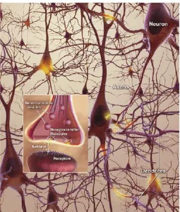
A normal healthy brain contains billions of neurons, constantly receiveing information from each other in the form of electrical charges travelling down the axon to the end of the neuron – neurotransmitters, which move across microscopic gaps, or synapses, between neurons (Rodgers, 2008). They bind to receptor sites on the dendrites of the next neuron (Fig. 1.1) and AD is the responsible for the disruption of the neuronal network, causing the destruction of memory and thinking skills over time (Rodgers, 2008).
Figure 1.1 Neuronal network in a healthy brain (Rodgers, 2008).
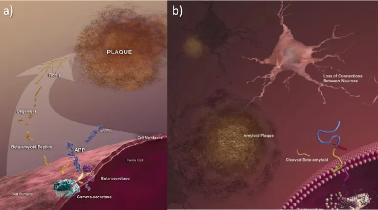
AD is associated with inflammatory processes (Stuchbury and Munch, 2005). Abnormal structures called β-amyloid plaques (outside the neurons) and neurofibrillary tangles (inside the neurons) are classic biological hallmarks of the disease and can induce inflammation (Ferreira et al., 2006):
Plaques form with the extracellular β-amyloid peptides accumulation as well as dystrophic neurites, reactive astrocytes, phagocytic cells and protein fragments derived from degenerating cells or liberated from neurons (Jalbert et al., 2008). amyloid are fragments, result of amyloid precursor protein (APP) cleavage by β-secretase, α-secretase and γ-secretase (Brewer, 2007). These fragments increase in size and become insoluble and consequently toxic, contributing to cell death (Fig. 1.2) (Brewer, 2007).
Neurofibrillary tangles are made when tau protein separates from microtubules, which are used in a normal brain to stabilize critical structures to the cell's internal
nutrients transport system, causing neurotransmitter deficits and neuronal cell death (Jalbert et al., 2008). Tau strands aggregate inside the neurons, forming tangles and, therefore, disable the transport system and destroy the cell (Fig. 1.3) (Brewer, 2007).
Figure 1.3 General aspect of a healthy neuron (a) and a diseased neuron, exhibiting the formation of tau tangles (b) (Rodgers, 2008).
Although many events that happen in the brain of patients with AD are well known, there are still many unclear factors such as what other changes are taking place in the aging brain and its cells and what influence do other diseases, genetics, and lifestyle factors have on the risk of developing AD as the brain and body age (Rodgers, 2008).
1.1.3. Risk Factors for AD
1.1.3.1. Advanced age
People younger than 65 years can develop AD, but the risk increases after this age (Geldmacher, 2010).
1.1.3.2. Family history
Individuals who do not have a first-degree relative with AD are less likely to develop it than those who have and the risk becomes higher when the individual has more than one first-degree relative with AD (Huang et al., 2004).
1.1.3.3. Apolipoprotein E-ε4 (APOE-ε4)
A genetic factor in late-onset Alzheimer’s disease is APOE-ε4, which is one of three common forms (ε2, ε3 and ε4) of the APOE gene, responsible for providing the blueprint for a protein that carries cholesterol in the bloodstream (Jalbert et al., 2008). Everyone inherits one form of the APOE gene from each parent, but those who inherit the form APOE-ε4 have increased risk of developing AD in an earlier age (Jalbert et al., 2008). The risk can also increase if the individual inherits two APOE-ε4 genes, but this does not guarantee the development of AD (Jalbert et al., 2008).
On the other hand, APOE-ε4 which has an arginine at position 112 rather than a cysteine, while the other apolipoprotein alleles have cysteine at this position (Reynolds, 1997), may be involved in copper binding and to the diminished antioxidant effect of the E-4 allele (Brewer, 2007).
1.1.3.4. Cardiovascular Disease Risk Factors
The cardiovascular diseases that offer a major risk in developing AD are associated with high cholesterol (especially in midlife), Type 2 diabetes, high blood pressure (especially in midlife), physical inactivity, smoking and obesity (Geldmacher, 2010). Thus, remaining mentally and physically active and consuming a diet low in saturated fats and rich in vegetables, may support heart and brain health (Geldmacher, 2010).
1.1.4. The Cholinergic theory
The cholinergic system is composed by a set of cells that produce and/or are stimulated by the neurotransmitter acetylcholine (ACh) and control the central nervous system (CNS) functions (Gibbs, 2010). ACh is released to travel across the synaptic cleft and the two types of receptors, muscarinic and nicotinic (post-synaptic terminal), respond to ACh, facilitating intracellular communication, memory processing and higher cognitive functions (Fig. 1.4) (Gibbs, 2010). Acetylcholinesterase (AChE) is
the main enzyme that hydrolyzes ACh into a choline and an acetyl groups (Ferreira et
al., 2006).
Figure 1.4 Actuation of acetylcholinesterase in a neurotransmission signaled by acetylcholine – After signalling, acetylcholine is released from receptors and broken down by AChE to be recycled in a continuous process. Source: www.vrp.com/brain-health.
The basal forebrain is the brain area where declines in the level of ACh in AD patients can be found and the projection to the hippocampus and neocortex lead to impairments in memory and cognitive functions (Zarotsky et al., 2003; Natarajan et al., 2009).
Acetyltransferase is needed for the synthesis of acetylcholine, and its activity can be reduced up to 90%, as a result of the loss of cholinergic neurons (Zarotsky et al., 2003).
Treatment strategies have therefore focused on replacing the level of ACh, enhancing cholinergic activity in the affected regions of the brain, or inhibiting AChE action, avoiding its degradation (Zarotsky et al., 2003; Natarajan et al., 2009). However this current treatment does not halt the progression of AD, but contribute to modest improvements in memory, thinking and reasoning skills (Zarotsky et al., 2003; Natarajan et al., 2009).
The first organophosphate AChE inhibitor was synthesized in the 1850s and in the 1930s synthetic cholinesterase inhibitors, such as neostigmine, began to be used to treat autonomic nervous system manifestations (Taylor, 1998).
AChE inhibitors not only increase the level of ACh but also prevent the formation of β-amyloid plaques, activating secretase, which acts on amyloid precursor protein thereby preventing neuronal death due to inflammation in AD (Natarajan et
al., 2009).
The mammalian brain contains two cholinesterases, AChE and butyrylcholinesterase (BChE) (Kuhl et al., 2006). Butylcholines are not physiological substrates in the brain, and, although their function remains unclear, it has been found to increase progressively in patients with AD. In the human brain, BChE is found in neurons and glial cells, as well as in plaques and neurofibrillary tangles in patients with AD (Zarotsky et al., 2003). So, it is believed that after AChE, BChE plays an important role in the inhibition of ACh (Orhan et al., 2004). Until now, there are no drugs that inhibit BChE as strongly as AChE (Orhan et al., 2004).
1.1.5. Oxidative stress and metal accumulation
Oxidative stress (OS) and the hypothesis that brain metal dysregulation, resulting in reactive oxygen species (ROS) generation from H2O2 and inflammatory processes, plays a pivotal role in different clinical disorders, such as neurodegeneration, diabetes, hypothyroidism, liver failure, atherosclerosis, ischemia reperfusion injury, cancer and cardiovascular diseases (e.g. stroke or thalassemia) (Conforti et al., 2009;Weinreb et al., 2009). OS triggers a cascade of events leading to apoptotic/necrotic cell death in neurodegenerative disorders, such as AD, PD, Huntington’s disease and amyotrophic lateral sclerosis (Zecca et al., 2004; Tirosh et al., 2007; Ebrahimzadeh et al., 2008). Moreover, due to the high oxygen consumption and
lipid content, the CNS is more sensitive to oxidative stress compared to other parts of our body (Pangestuti & Kim, 2011).
Free radicals, which are generated in mitochondria (Fig. 1.5) can help cells to fight infections, but can also damage the neuron’s cell membrane or DNA, because they are very reactive and easily react with other molecules (Rodgers, 2008).
Figure 1.5 Oxidative Stress and mitochondria – The arrows indicate the movement of free radicals, which can spread easily from damaged mitochondria to other parts of the cell (Rodgers, 2008).
1.1.5.1. Iron and AD
Iron is involved in several processes (El & Karakaya, 2004; Zecca et al., 2004), such as: transport, storage and activation of oxygen (as a central element of the heme
molecule, which is a critical part of haemoglobin); respiration;
activity of several enzymes, including cytochromes, which acts in electron transport or tyrosine hydroxylase, which is required for dopamine synthesis; synthesis of steroid hormones and bile acids;
detoxification of external substances in the liver and signal controlling in some neurotransmitters, like dopamine and serotonin systems in the brain.
However, when iron homeostasis is not well regulated it can degrade lipids, proteins and cells as for example astrocytes, microglia or neurons, due to unwanted oxidative reactions (Rival et al., 2001; Zecca et al., 2004; El & Karakaya, 2004). The excess iron, derived from the breakdown of transfused red blood cells, is deposited as hemosiderin and ferritin in the liver, spleen, endocrine organs and myocardium (Ebrahimzadeh et al., 2008).
Consequently, there is increasing evidence that iron accumulation in the brain with age can cause, as mentioned above, a vast range of disorders to the CNS (Zecca et al., 2004).
There are two classes of iron-related neurodegenerative disorders (Zecca et al., 2004):
- Those resulting from iron accumulation in specific brain regions;
- Those resulting from deficiency in iron metabolism and/or homeostasis.
In these disorders are usually involved protein modification, misfolding and aggregation, causing the formation of the intracellular inclusion bodies that are the postmortem characteristics of many neurodegenerative diseases, like AD and PD (Zecca et al., 2004).
Iron might also have a direct impact on plaque formation, due to its action as a modulator of α-secretase in APP cleavage (Huang et al. 2000; Rodgers et al., 2002; Kawahara, 2003; Zecca et al., 2004), although some authors argue that by binding iron, β-amyloid might, in fact, protect the surrounding neurons from OS (Zecca et al., 2004).
Iron is also involved in the formation of ROS. ROS are formed when iron (Fe2+) reacts with H2O2 to form •OH (hydroxyl radicals), which are very unstable and reactive,
via the Fenton reaction (Eq. 1 and 2; Koschnick & Haller, 2006),initiating the processes
of OS and the inflammatory cascade, that result in the production of cytotoxic cytokines in the microglia and surrounding neurons and activation of transcription factors (Rival et al., 2001; El & Karakaya, 2004;Weinreb et al., 2009):
1.1.5.2. Copper and AD
Copper is an essential component of several enzymes and proteins and is critical for numerous reactions vital for life, as for example, antioxidant defense, neuropeptide synthesis and immune function (Brewer et al., 2006; Brewer, 2007). The deficiency on this element leads to anemia and bone marrow suppression, followed by a neurologic syndrome called a myelopathy (Hedera et al., 2003).
However, like iron, copper also participates in the generation of ROS through Fenton chemistry and can produce oxidative damage in much the same manner (Brewer et al., 2007).
At this time the evidences are conflicting whether too much copper is involved in the pathogenesis of AD as well as others neurodegeneratives diseases. Some authors claim that copper is directly related with the onset of neurological diseases (Sayre et al., 2000; Cherny et al., 2001; Nakano et al., 2004; Angeletti et al., 2005; Nelson & Alkon, 2005; Soragni et al., 2008; Kong et al., 2008). However, some authors such as Phinney et al. (2003) and Bayer et al. (2003) reports in vivo studies where amplification of a copper transporter improved brain copper and reduced β-amyloid formation, increasing longevity.
1.1.6. Treatment
The pharmacological treatments currently used to alleviate AD symptoms include antioxidant therapy, the use of ChE inhibitors, nicotinic and muscarinic agonists, estrogen, nerve growth factor (NGF), low molecular lipophilic compounds that can activate neurotrophic factor signaling pathway, nonsteroidal antiinflammatory drugs such as ibuprofen and COX-2 inhibitors, drugs that interfere with β-amilose formation and deposition, and also drugs that attenuate toxicity induced by β-amilose (Park & Kim, 2002).
The changes in the brain of AD patients may begin near 10 years before patients experience symptoms such as memory loss, and this is considered as the ideal period in which the future drugs should be administered (Jalbert et al., 2008).
1.1.6.1. ChE inhibitors
Scientists argue that the inhibition of AChE and BChE represents an effective therapy for AD management (Grossberg, 2003;Darreh-Shori & Soininen, 2010).
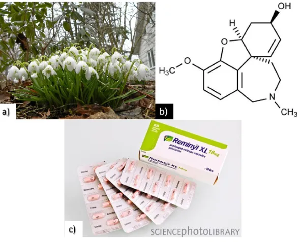
However, and despite the high demand for ChE inhibitors for AD and other neurological disorders, only synthetic AChE inhibitors, such as tacrine and donepezil, and the natural products rivastigmine and most recently galanthamine have been approved by FDA (Zarotsky et al., 2003). The latter have been also approved in Europe by the European Registration Bureau and is commercially available as Reminyl® (Fig. 1.6c) (Zarotsky et al., 2003).
Studies have also revealed the relevance of BChE due to its presence in some cholinergic neurons in which AChE is absent, as well as its capability to induce neurotoxicity of some plaques (Greig et al, 2005; Oboh et al., 2015). But only rivastigmine can inhibit both AChE and BChE (Colovic et al., 2013; Pohanka 2014).
The above mentioned drugs exhibit some side effects, as for example, hepatotoxicity, gastrointestinal disorder, anxiety, nervousness, drowsiness, mouth dryness or tiredness and also bioavailability problems, making necessary to find alternative ChE inhibitors from natural sources (Pangestuti and Kim 2011). As examples, Ginkgo biloba (Ginkgoaceae) and Huperzia serrata (Pteridophyta) have been extensively investigated as natural therapeutic agents for AD patients (Park & Kim, 2002; Conforti et al., 2009).
Galanthaminehydrobromide is a tertiary alkaloid (Fig. 1.6b) that was originally isolated from the Galanthus worownii (snowdrop plant) (Fig. 1.6a) (Willis et al., 2009) and is now synthesized for use in the treatment of mild to moderate AD (Zarotsky et
al., 2003; Butler, 2005).
Prior to its use in patients with AD, galanthamine was available in Eastern Europe as a curare-reversal agent in anesthesia and as a treatment for neurologic conditions, such as myasthenia gravis (Zarotsky et al., 2003). The interest in this
reach the brain, penetrating the blood barrier and affect cholinergic transmission (Zarotsky et al., 2003).
Figure 1.6 a) General aspect of Galanthus worownii
(Source: http://art-nature-garden-passion-bensimon.blogspot.com/);
b) chemical structure of galanthamine (4aS,6R,8aS)-4a,5,9,10,11,12-hexahydro-3-methoxy-11-methyl-6H-benzofuro[3a,3,2ef][2]benzazepin-6-ol hydrobromide
(Source: http://dailymed.nlm.nih.gov/dailymed/); c) Reminyl®, galanthamine Alzheimer's drug
(Source: http://www.sciencephoto.com/media/412296/enlarge).
1.1.6.2. Metal chelation as a neuroprotective strategy
Chelation of the metal ions is considered the main strategy to avoid ROS generation (Ebrahimzadeh et al., 2008).
Antioxidant and other supportive therapies can scavenge ROS and can also attenuate inflammation pathways, protecting red blood cells against oxidant damage (Ferreira et al., 2006; Ebrahimzadeh et al. 2008).
Iron chelators are used to form soluble and stable complexes with iron, which are then excreted in the feces and/or urine (Ebrahimzadeh et al., 2008). This iron chelation may result in the improvement of life quality and overall survival (Ebrahimzadeh et al., 2008).
Tirosh et al. (2007) reported that the antibiotic iron chelator Clioquinol and the continued intramuscular administration of the drug DFO (desferrioxamine) could prevent neurotoxicity in mice and slow the clinical progression of AD. However,
Clioquinol is highly toxic and DFO is poorly passed through the blood–brain barrier
(Tirosh et al., 2007).
In this sense, special interest has been assigned in nutritional antioxidants and metal chelation agents as viable neuroprotective alternative approaches for neurodegenerative disorders (Tirosh et al., 2007; Weinreb et al., 2009). These may have compounds that can chelate metal ions, such as iron and copper to form inactive complexes and prevent the generation of potentially damaging free radicals (Weinreb
1.2. Marine Natural Resources
The oceans contain about 90% of the world’s living biomass, which represents approximately half of the total global biodiversity (Pangestuti & Kim, 2013). This wide diversity of organisms is recognized as an important reservoir of potent and innovative molecules responsible for helping them to survive in the hostile environment characterized by a competition for space, maintenance of unfouled surfaces, deterrence of predation and the ability to successfully reproduce and exhibiting strong pharmacological potential (Salvador et al., 2007).
Among marine organisms, marine algae found attached to rocks in the intertidal zone and washed up on the beach in giant underwater forests (Fig. 1.7) have been identified as an under-exploited resource for bioactive molecules (Natarajan et
al., 2009;Pangestuti & Kim, 2011).
Figure 1.7 Kelp forest at Catalina Island, California, USA (Source: http://www.uwphotographyguide.com/giant-kelp-forests).
Marine algae has been used in traditional medicine in China (Folmer et al., 2010) and as a subsidiary food (Natarajan et al., 2009) for more than 2000 years. Marine algae has also been widely used in Ancient Egyptian and Ayurvedic medicine (Folmer et al., 2010). Specifically, Hamed et al., (2015) postulates that the low incidence of neurodegenerative diseases in East Asia can be related to their high fish
and marine algae consumption. However, there is not enough data currently from clinical trials (Cole et al., 2009).
In Western medicine, the first record of the medicinal use of algae dates back to the 1960’s in Italy, in the treatment of breast cancer (Folmer et al., 2010).
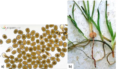
In contrast to seagrasses, that have a true root system (Fig. 1.8b), seeds and fruit and veins that carry molecules around the plant, algae have holdfasts and spores (Fig. 1.8a) (Coles et al., 2004).
Figure 1.8 Spores from brown macroalgae Haplospora globosa (a) (Source: www.algaebase.org); root system of the seagrass Cymodocea nodosa (b) (Source: www.terra.es).
Besides their use as antifouling, stabilizers, gelling agents or emulsifiers in food industries, a wide number of seaweeds species exhibit important biomedical applications, such as the treatment of tuberculosis, arthritis, cold, influenza, worm infestations, as a cholesterol lowering drug, ovarian cysts, breast lumps, lymph node swellings and lymphomas (Natarajan et al., 2009), antibacterial, antifungal, antiviral and/or antitumor (Moreau et al., 2006; Kong et al., 2008; Folmer et al., 2010; Vo & Kim, 2010), anticoagulant (Athukorala et al., 2007), antioxidant (Rupérez et al., 2002;El & Karakaya, 2004; Lim et al., 2006;Ganesan et al., 2007), anti-allergic (Li et al., 2008), anti-inflammatory (Kim et al., 2009) or anti-obesity (Maeda et al., 2007;Tsukui et al.,
2007; Kong et al., 2010). Apart from all these capabilities, algae can have also interest as neuroprotectants (Natarajan et al., 2009).
1.2.1. Macroalgae
Macroalgae can be classified into three classes based on their pigmentation, namely brown, red and green algae, which are referred to as Ochrophyta, Rhodophyta, and Chlorophyta, respectively (Khan et al., 2010).
Brown macroalgae can contain polysaccharides and diterpenoids (Moreau et
al., 2006). These diterpenoids display antitumor effects and may include cyclic
diterpenes from Dictyotaceae species (Gedaraa et al., 2003) or meroditerpenes from
Cystoseira usneoides (Moreau et al., 2006; Zubia et al., 2009;Taskin et al., 2010) and
Sargassum tortile (Moreau et al., 2006). In addition, a linear diterpene,
12-(R)-hydroxygeranylgeraniol, isolated from Bifurcaria bifurcata (Culioli et al., 2004) has been reported for its cytotoxicity against cultured human tumor cell lines (Moreau et
al., 2006).
Macroalgae polysaccharides has been widely used in the food industry and in medicine (Zvyagintseva et al., 1999) and since the 1940s, its production has attained commercial significance through their application as thickening and gelling agents for several industrial applications (Burtin et al., 2003).
The main polysaccharides of brown macroalgae are fucoidans, laminarans, and alginic acids. In particular, fucoidans and laminarans contents vary from 20 to 50% of defatted alga dry weight (Zvyagintseva et al., 1999).
Fucoidans are nontoxic polyelectrolytes and possess various pharmacological activities, as for example, antioxidant, antibacterial, antiviral, antitumor, immunosuppressive, antipeptic, antilipemic, antigemostatic and anticoagulant (Zvyagintseva et al., 1999). On the other hand, alginic acids can be used for heavy metal binding and as immunostimulators (Zvyagintseva et al., 1999; Chandini et al., 2007; Ye et al., 2008; Koz et al., 2009; Zubia et al., 2009; Chiheb et al., 2009).
Macroalgae and its biocompounds can, thus, exhibit numerous remarkable properties on biological systems, namely antioxidant (Sathya et al. 2013), anti-inflammatory (Sugiura et al., 2013), anti-allergic (Sugiura et al., 2009), antimicrobial (Eom et al., 2012), anticancer (Lee et al., 2012), antidiabetic (Lee & Jeon, 2013) and neuroprotective activities (Barbosa et al., 2014).
1.2.2. Microalgae
Microalgae are constituted by a vast array of novel compounds, such as: nutrients (including proteins, vitamins, minerals, fatty acids); carotenoid pigments, such as xanthophylls and carotenes; as well as phenolic acids and tocopherols which are known to exhibit antioxidant properties (Cha et al., 2008; Raposo et al., 2013). Thus, because of the presence of several primary and secondary metabolites in algal cells, microalgal biotechnology has received much interest, with its application in the energy, food, pharmaceutical and cosmetic industries (Olasehinde et al., 2017).
Recently, microalgal biotechnology has proved that it is now possible to produce some carotenoids commercially through aquaculture (Cha et al., 2008). These include, for instance, β-carotene from Dunaliella, astaxanthin from Haematococcus, and lutein from Chlorophycean strains (Cha et al., 2008). Carotenoids are highly bioactive and are reported as potent free radical quenchers, singlet oxygen scavengers, and lipid antioxidants, thereby acting as photoprotectants under conditions of excessive light (Cha et al., 2008). Some carotenoids such as β-carotene and lycopene may reduce the risk of cardiovascular diseases and certain cancers, whereas lutein and zeaxanthin may reduce the risk of eye disorders (Cha et al., 2008). Other carotenoid, such as xanthophylls, extracted from Chlorella ellipsoidea, might be also useful as functional ingredients in the prevention of human cancers, since studies have shown that these species have antiproliferative effects, including induction of apoptosis in vitro cellular models (Cha et al., 2008;Gardeva et al., 2009). Astaxanthin, another carotenoid, produced by the microalga Haematococcus pluvialis, has several
essential biological functions, including antioxidant activity and protection against lipid-membrane peroxidation of essential polyunsaturated fatty acids and proteins, DNA damage, and ultraviolet light effects (Cerón et al., 2007). Studies have also shown that they may act through other mechanisms such as gap junction communication, cell growth regulation and modulation of gene expression (Cerón et al., 2007; Cha et al., 2008).
Cosmetics and nutraceuticals industries may also benefit from the use of microalgae biomass as a low cost renewable source of phytosterols and metal chelators compounds, due to its high unsaponifiable content (Gangadhar et al., 2016).
There is also a growing interest in polyunsaturated fatty acids (PUFAs), due to their involvement in human health (Alonso et al., 1998; Barreira et al., 2015). Microalgae are potential sources of these long-chain PUFAs, especially for Eicosapentaenoic acid (EPA, 5,8,11,14,17-cis-eicosapentaenoic acid), which have beneficial effects in the prevention and treatment of certain medical conditions including coronary heart disease, blood platelet aggregation and several carcinomas (Belarbi et al., 2000).
Oher reports have also revealed the anti-inflammatory (Guzman et al., 2001), hypocholesterolemic (Dvir et al., 2015) and antiviral (Huleihel et al., 2002) activities of microalgal-derived extracts and compounds.
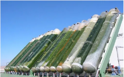
Another developing area in the use of microalgae is biodiesel production (Chisti, 2007; Damiani et al., 2010; Pereira et al., 2013a, 2013b) (Fig. 1.9), since they have a fast growth rate and high photosynthesis efficiency, allowing them to be industrially cultivated (Lu et al., 2009). However, since rapid-growing cells contain less oil, biodiesel production from microalgae is not economically feasible yet (Lu et al., 2009). To overcome these biological and technical challenges, Lu et al. (2009) did an approach to biodiesel production by a heterotrophic fermentation process with C.
protothecoides, which produces maximum amounts of algal biomass rich in oil, mainly
composed by more than 90% of fatty acids (Lu et al., 2009).
Pereira et al. (2015), for instance, not only proved that microalgae can be a rich source of fatty acids, but also showed that microalgae contain molecules with relevant
bioactivities, including antioxidant, inhibition of BChE and tyrosines, cytotoxic and antileishmanial activities.
Figure 1.9 Biodiesel production by microalgae – closed system bioreactor. Source: http://www.global-greenhouse-warming.com/biodiesel-from-algae.html).
1.2.3. Seagrasses
Seagrasses are a group of flowering plants (angiosperms) with roots, leaves and rhizomes (Coles et al., 2004). They are the only flowering plants that can live underwater and are less primitive than algae, whereby there are only about 60 species of seagrasses around the world (Coles et al., 2004).
Seagrasses occur in protected bays and lagoons and also in deeper waters and the depth at them occurs is limited by water clarity, since most species require high levels of light (Coles et al., 2004).
food or as raw material for the production of compounds with nutritional interest in Russia (Achamlale et al., 2009). For example, Achamlale et al. (2009) showed that a bioactive pectin from the genus Zostera, zosterin, can decrease the toxicity of antitumour drugs and eliminate heavy metals from human organisms.
Others seagrasses such as Cymodocea nodosa, used in this work, has an important ecological role in the marine ecosystem, despite that, knowledge of its chemical content is limited (Kontiza et al., 2008).
It has also been described other properties of seagrasses, such as antidepressant activity in humans, due especially to the biologically active compounds, the cyclitols (Kumar et al., 2008; Nuissier et al., 2008) and antiviral, including HIV-1, antioxidant, anti-inflammatory, anticarcinogenic, anti-allergenic and antithrombotic, due to the presence of rosmarinic acid (Achamlale et al., 2009; Custódio et al., 2016).
According to Kumar et al. (2008) the use of the roots of the seagrass Enhalus
acoroides as a remedy against stings of different kinds of rays and scorpion is very
popular in India; Cymodocea spp. is used as a tranquillizer for babies, as soothing help during pregnancy and against cough and malaria; Halophila spp. is a strong medicine against malaria and skin diseases and found to be very effective in early stages of leprosy.
1.2.4. Halophytes
Although halophytes represent a small fraction of the overall plant population (aprox. 2%), they display important roles in the environment, such as desalinization and prevention of soil erosion, loss of biodiversity and bioproductivity (Gago et al., 2011). These plants can survive in different environments, such as salt marshes and estuaries, cliffs and dunes near the ocean, and some are adapted for near-desert environments where water supplies may be limited and highly saline (Gago et al., 2011).
Halophytes are considered as good sources of food, fibre and bioenergy (Gago
et al., 2011). Some halophytes, such as Salicornia ssp., Aster tripolium, Atriplex ssp. or Inula crithmoides are consumed in Europe as fresh or cooked gourmet foods, for
example, in salads as a substitute of salt (Gago et al., 2011; Ventura et al., 2011). They have a high nutritional content, which includes proteins, carbohydrates, fiber, calcium, potassium, magnesium, iron, manganese, copper, vitamin C and β-carotene (Gago et
al., 2011).
It has been reported some important therapeutic applications of different halophyte species, as for example, Salicornia spp., including immunomodulation, antioxidant and antitumor proporties (Chung et al., 2005).
Recently, Medini et al. (2015) suggested the strong potential of the halophyte
Limonium densiflorum as a source of phenolic compounds, marked as having great
potencial in the food and pharmaceutical industry. In turn, Arthrocnemum
macrostachyum is rich in phenolics and flavonoids and is also a potential source of
antioxidants (Custódio et al., 2012 (a); Rodrigues et al., 2014). These secondary metabolites play different roles in the physiology and cellular mechanisms of plants, including pigmentation as well as resistance to pests, predators and oxidative stress (Barreira et al., 2017). The stress conditions to which the halophytes are exposed (high salinities and UV radiation), usually trigger to a production of ROS and hence often have antioxidant capacity, attributed to their phenolic compounds (Barreira et al., 2017; Ksouri et al., 2008). This is the case of H. italicum subsp. picardii flowers, with similar or even higher antioxidant potential than the commercial green and herbal red teas, which has also showed moderate anti-diabetic potential and low toxicity in in
vitro models (Pereira et al., 2017a). In another study, Pereira et al. (2017b) also tested
the leaves and the flowers infusions of the halophyte species Crithmum maritimum L. and observed its high antioxidant potencial.
Regarding to ChE inhibitors, Rodrigues et al. (2017) has identified a bioactive compound from Juncus acutus, junconol, and proved its capacity to inhibit the enzyme AChE on neuronal and glial cells in vitro.
1.3. Objective
The number of older people at risk of developing dementia is growing rapidly worldwide, and AD represents the most common cause of dementia in the elderly (Olasehinde, 2017).
The search for novel anticholinesterases compounds from natural resources as therapeutics agents for AD and other CNS disorders is based on the need for agents targeted to brain areas affected, with reduced toxicity and side-effects.
Marine natural products are considered as important sources of novel biologically active compounds, but its application in the treatment of neurological disorders is still rather unexplored. In this context, the objective of this study was to screen for the AChE and BChE inhibitory activity of some commonly available macro- and microalgae, seagrasses and halophytes species of the southern coast of Algarve (Portugal), as well as evaluate their chelating activity on iron and copper ions.
2.
MATERIALS AND
METHODS
In this chapter it is described the processes used in the evaluation of the algae, seagrasses and halophytes, as potential new sources of bioactive compounds with neuroprotective activity.
2.1.
Plant material2.2.
Extraction2.3.
Metal chelating activity2.4.
AChE and BChE inhibitory activity2.1. Plant material
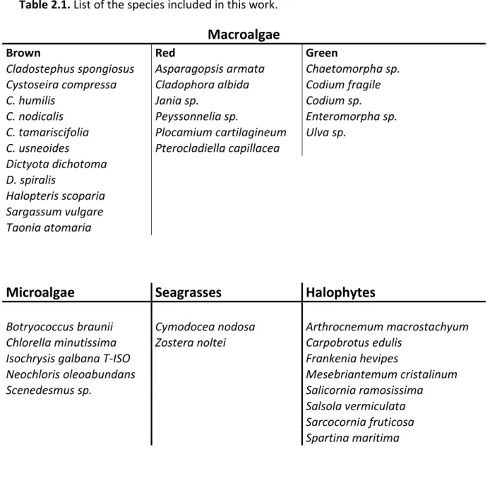
The list of the species included in this work is presented in Table 2.1.
Macroalgae (Fig. 2.1) and seagrasses (Fig. 2.2) samples were collected on the Algarve coast in July-November 2009. Species identification was made by Dr. Aschwin Engelen (Centre of Marine Sciences, University of Algarve, Portugal). Samples were washed in seawater, kept cold until arrival to the laboratory, washed with tap water, freeze dried, ground with a coffee grinder and stored at -20°C.
Microalgae (Fig. 2.3) samples were provided by NECTON S.A. as a solid dark green frozen paste and were stored at -20°C.
The halophytes samples were collected in the Ria Formosa lagoon (Fig. 2.4), in July-August 2010, which is an area that extends 60 km along the southern coast of Portugal (Algarve), covering approximately 18,400 hectares (Gago et al., 2011). Aerial parts were washed with tap water, dried at 40°C for 48h, ground with a coffee grinder and stored at room temperature. The taxonomical classification was performed by the botanist Dr. Manuel J. Pinto (National Museum of Natural History, University of Lisbon, Botanical Garden, Portugal).
Table 2.1. List of the species included in this work.
Macroalgae
Brown Red Green
Cladostephus spongiosus Asparagopsis armata Chaetomorpha sp.
Cystoseira compressa Cladophora albida Codium fragile
C. humilis Jania sp. Codium sp.
C. nodicalis Peyssonnelia sp. Enteromorpha sp.
C. tamariscifolia Plocamium cartilagineum Ulva sp.
C. usneoides Pterocladiella capillacea
Dictyota dichotoma D. spiralis
Halopteris scoparia Sargassum vulgare Taonia atomaria
Microalgae
Seagrasses
Halophytes
Botryococcus braunii Cymodocea nodosa Arthrocnemum macrostachyum
Chlorella minutissima Zostera noltei Carpobrotus edulis
Isochrysis galbana T-ISO Frankenia hevipes
Neochloris oleoabundans Mesebriantemum cristalinum
Scenedesmus sp. Salicornia ramosissima
Salsola vermiculata Sarcocornia fruticosa Spartina maritima
Figure 2.1 General aspects of some macroalgae species included in this work.
Cystoseira tamariscifolia (a) (brown), Plocamium cartilagineum (b) (red) and Codium fragile (c) (green). Source: www.algaebase.org
Figure 2.2 General aspect of the seagrasses included in this work. Zostera noltei (a) and
Figure 2.3 General aspects of some microalgae species included in this work.
Botryococcus braunii (a) (Source: www.algaebase.org) and Isochrysis galbana (b)
(Source: www.sciencephoto.com)
Figure 2.4 Ria Formosa lagoon, Algarve (a) (Source: http://algarvecom.blogspot.com) and some species of the halophytes species included in this work: Salicornia
ramossisima (b) (Source: http://www.flickr.com), Sarcocornia fruticosa (c) (Source:
2.2. Extraction
Aliquots (1 g) of milled samples were mixed with 20 mL of methanol and homogenized in an Ultra Turrax T25 (IKA Labortechnik Basic) in order to disrupt cells (1900 rpm in two cycles of 1 minute each). Then, the volume was made up to 40 mL with methanol, and samples were extracted for 16h at room temperature (RT, approximately 20oC) with stirring.
The extracts were then centrifuged (3000 rpm, 15 min, 20°C, BECKMAN COULTER ALLEGRA 6R CENTRIFUGE), the upper layer carefully removed, filtered with Whatmann nº 4 filters and dried in a vacuum evaporator (temperature below 50°C, ROTAVAPOR R-114). Dried extracts were weight, resuspended in methanol at the concentration of 10mg/mL and stored at -20°C. Each extraction was repeated 3 times.
2.3. Metal chelating activity
The Fe2+-chelating activity was determined by measuring the formation of the Fe2+-ferrozine complex according to Custódio et al. (2012, b).
Ferrozine can quantitatively form complexes with Fe2+. However, in the presence of chelating agents, the complex formation is disrupted with the result that the purple colour of the complex is decreased. Measurement of colour reduction, therefore, allows for the estimation of the chelating activity of the coexisting chelator (Ebrahimzadeh et al. 2008).
In 96-well microplates, 30 µL of the extracts were added to 200 µL of distilled water and then immediately mixed with 30 µL of FeCl2 (0.1 mg/ml water).
After 30 minutes, 12.5 µL of ferrozine solution (40 mM in water) was added. Samples were then incubated at RT for 10 min. and the absorbance was measured in a microplate reader (BioTek Synergy 4) at 562 nm.
The Cu2+-chelating activity was determined using pirocatechol violet (PV – indicator for metal titration) according to Saiga et al. (2003) and Megías et al. (2009).
In 96-well microplates, 30 µL of the extracts were mixed with 200 µL of Na acetate buffer (50 mM, pH 6), 100 µL of CuSO45H2O (5×10⁻⁵ g/mL) and 6 µL of PV (4 mM in Na acetate buffer).
The complex of PV with CuSO4 is blue and the colour changes to yellow when PV dissociates from Cu2+ ion in the presence of chelating agents. The absorbance was measured in a microplate reader (BioTek Synergy 4) at 632 nm.
Due to the color of the extracts, it was necessary to use a colour control, which absorbance was subtracted to the absorbance of the samples, thus only the values related to the complex ferrozine-Fe2+ or to the complex PV-CuSO4, in iron and copper chelating activity, respectively. In the first case, the sample was added to 242.5 µL of distilled water, which volume is the same comprised by the distilled water, FeCl2 and ferrozine. In the second case, the sample was added to 306 µL of buffer, which volume is the same comprised by buffer, CuSO45H2Oand PV.
The extracts were evaluated at the concentrations of 1, 5 and 10 mg/mL and results were expressed as percentage of chelating activity relative to a negative control containing methanol in place of the sample. A solution of the synthetic metal chelator ethylenediaminetetraacetic acid (EDTA) at the concentration of 1 mg/mL was used as positive control.
The percentage of chelating activity of Fe2+ and Cu2+ was determined by using the next formula:
Where Acontrol is the absorbance of the negative control and Asample is the absorbance of the sample, which was obtained by subtracting the absorbance of the
2.4. AChE and BChE inhibitory activity
The AChE and BChE inhibitory activities were assessed by the Ellman’s colorimetric assay (Ellman et al., 1961), according to previously described methods (Orhan et al. 2006, 2009; Custódio et al., 2012 (b)).
Briefly, 140 µL of 0.1 mM sodium phosphate buffer (pH 8.0), 20 µL of the extracts at the concentrations of 1, 5 and 10 mg/mL and 20 µL of AChE or BChE (0.28 U/mL) solution were mixed in 96-well microplates and incubated at RT for 15 min. Then, 10 µL of the substract acetylthiocholine iodide (AChI) or butyrithiocoline iodide (BChI) (4 mg/mL) were added to initiate the reaction, together with 20 µL of a solution of the dye 5,5-Dithio-bis (2-nitrobenzoic) acid (DTNB) at the concentration of 1.2 mg/mL.
The hydrolysis of AChI and BChI was monitored by the formation of the yellow 5-thio-2-nitrobenzoato anion as a result of the reaction of DTNB with thiocholines, catalyzed by enzymes at a wavelength of 412 nm using 96-well microplate reader (BioTek Synergy 4).
Results were expressed as percentage of inhibitory activity relative to a negative control containing methanol in place of the sample. Galanthamine was used as reference at the concentration of 1 mg/mL.
The percentage of inhibition of AChE and BChE was determined by using the formula:
Where Acontrol is the absorbance of the negative control and Asample is the absorbance of the samples obtained by subtracting the absorbance of the colour control.
2.5. Statistical analysis
All the experiments were carried at least in triplicate and the results were expressed as mean ± standard deview (SD).
One-way analysis of variance (ANOVA) was used to compare the mean values of each method, using STATISTICA for Windows (release 7, STATISTICA INC).
A significant difference between the means of parameters was determined by using Kruskal-Wallis multiple comparison tests. A p-value of less than 0.05 was considered significant.
3.
RESULTS
This chapter is divided in two sections, one for the analysis of chelating activity on iron and copper and other for the analysis of inhibitory activity on AChE and BChE.
3.1. Iron and copper chelating activity
In this work we applied the classification suggested by Vinutha et al. (2007) for the AChE inhibitory activity to the metal chelating activity, to make easier the interpretation of the results. In this sense, metal chelating activity was classified as: potent (>50% activity), moderate (30–50% activity), low (<30% activity) or nil (<5% activity).
3.1.1. Fe
2+chelating activity
In the used method, the higher the chelation of ions by the sample, the smaller the number of ions available for reaction with ferrozine, so the reaction does not get the purple colour characteristic of the complex Fe2+-ferrozine. Thus, the lower the absorbance of the reaction mixture, the higher the Fe2+-chelating ability (Fig. 3.1).
Figure 3.1 Example of 96-well microplates with an iron chelating assay employing methanol extracts with low Fe2+ chelating activity. The concentrations of the extracts are increasing from top to bottom. The darker the reaction, the lower the chelating activity of the tested extract.
3.1.1.1. Macroalgae
The chelating activity varied between groups of macroalgae and significant differences were found between them (Fig. 3.2).
The best results were obtained in the group of red macroalgae, followed by the brown macroalgae and for last, the green macroalgae, where no relevant activities were detected (Fig. 3.2).
Regarding brown macroalgae, the species D. dichotoma displayed the highest iron chelating potential, with a value of 52.7% at 10 mg/mL (Fig. 3.2). The species P.
cartilagineum (red) also had a potent chelating activity (83.4% at 10 mg/mL), while for
the group of green macroalgae, the maximum value was obtained with the species
Enteromorpha sp (30.5%; Fig. 3.2) at the highest concentration tested.
Figure 3.2 Fe2+ chelating activity (%) of methanol extracts of brown (A), red (B) and green (C) macroalgae species. For the same group (brown, red or green), bars labelled with different letters are significantly different at p<0.05 (Kruskal-Wallis multiple comparison test).
Brown macroalgae C. spo ngiosu s C. com pressa C. hum ilis C. nodic aulis C. tam arasci folia C. usn eoides D. dich otoma D. spi ralis H. sco paria S. vul gare T. ato maria Fe 2+ che la ting a ct iv ity (% ) 0 20 40 60 80 100 Red macroalgae A. arm ata C. albida Jania sp . Peysso nnelia sp P. cart ilagine um P. cap illacea Green macroalgae Chaet omorp ha sp. C. fra gile Codiu m sp Entero morph a sp. Ulva sp . 1 mg/ml 5 mg/ml 10 mg/ml A B C Fe 2+ che la ting a ct iv ity (% ) b a bc a c c a a a a bc ab a c c b ab bc b ab bc bc a b b bc b b b a a a b b ab ab ab ab b b b b a a a a a b b b bc bc bc bc bc c ab ab bc bc bc b b b b bc
3.1.1.2. Microalgae
The species I. galbana was the most active followed by B. braunii with maximum values of 64.4% and 59.7% at the concentration of 10 mg/mL, respectively (Fig. 3.3). Microalgae B. bra unii C. min utissima I. galb ana clo ne N. ole oabund ans Scenede smus sp. Fe 2+ che la ti ng act iv ity (%) 0 20 40 60 80 100 1 mg/ml 5 mg/ml 10 mg/ml a a ab b bc c b b ab ab bc c b bc bc
Figure 3.3 Fe2+ chelating activity (%) of methanol extracts of microalgae species. Bars labelled with different letters are significantly different at p<0.05 (Kruskal-Wallis multiple comparison test).
3.1.1.3. Seagrasses
Seagrasses C. nodosa Z. noltii Fe 2+ ch el ati ng a cti vi ty (%) 0 20 40 60 80 100 1 mg/ml 5 mg/ml 10 mg/ml b a a a a a
Figure 3.4 Fe2+ chelating activity (%) of methanol extracts of C. nodosa and Z. noltei species. Bars labelled with different letters are significantly different at p<0.05 (Kruskal-Wallis multiple comparison test).
3.1.1.4. Halophytes
Two species had moderate capacity to chelate iron at 10 mg/mL, namely M.