UNIVERSIDADE DE LISBOA
FACULDADE DE CIÊNCIAS
DEPARTAMENTO DE BIOLOGIA ANIMAL
Identification and expression analysis
of genes putatively involved in pathogenicity
of Hemileia vastatrix to Coffea arabica
Ana Cristina Magalhães Vieira
UNIVERSIDADE DE LISBOA
FACULDADE DE CIÊNCIAS
DEPARTAMENTO DE BIOLOGIA ANIMAL
Identification and expression analysis
of genes putatively involved in pathogenicity
of Hemileia vastatrix to Coffea arabica
Ana Cristina Magalhães Vieira
Dissertação orientada por:
Prof. Doutor Octávio Fernando de Sousa Salgueiro Godinho Paulo
Doutora Helena Manuela Gil Azinheira
Mestrado em Biologia Humana e Ambiente
Agradecimentos
Em primeiro lugar, gostaria de agradecer ao Professor Doutor Octávio Paulo e a Doutora Helena Azinheira, por me terem orientado durante este longo percurso. Ao Professor Octávio, pelo seu constante optimismo, inspiração e curiosidade científica que sempre me despertaram e cativaram. À Doutora Helena, pela sua constante disponibilidade, compreensão, ajuda e paciência nos períodos mais difíceis, em que tudo parecia correr mal. E por fim mas não menos importante, ao Doutor Pedro, pela sua distinta co-orientação, disponibilidade, interesse, motivação e optimismo que contribuíram muito para ter chegado até aqui.
Agradeço a toda a equipa do CIFC pelo bom ambiente de trabalho, disponibilidade e atenção. Em especial à Doutora Maria do Céu, pelos seus preciosos concelhos e motivação. À Doutora Andreia, o meu muito obrigado pela sua constante disponibilidade, boa disposição e amizade. À Doutora Dora, pelo seu optimismo e entusiasmo que sem duvida cativam e motivam todos a sua volta. E por fim, à Paula Leandro, por toda a sua ajuda e disponibilidade na parte da citologia.
Agradeço também a todo o grupo do CoBiG2, pelo fantástico ambiente de trabalho que me proporcionaram. Em especial à Joana Costa, por me ter ensinado os meus primeiros passos na biologia molecular e pela sua disponibilidade constante para ajudar.
Gostaria também de agradecer ao Diogo Silva, por ter sido o meu pilar ao longo deste ano. Sem ti, os nossos momentos, as nossas piadas e a nossa cumplicidade teria sido impossível ter chegado até aqui. Mas, finalmente podemos dizer: Conseguimos!
Gostaria também de agradecer ao Sr. Mek (mekie) ao Tiago (Lagartinha) e ao Pimpão, pela vossa maravilhosa amizade, principalmente nos maus momentos. Ao Sr. Mek, por ser absolutamente insubstituível, pelas piadas secas, pelo ombro amigo, e por tudo aquilo que só ele sabe ser. Ao Tiago, pela paciência, pelo carinho, amizade e principalmente pelos brainstorning sobre Real time e toda a sua problemática. Ao Pimpão, por ser o meu melhor amigo, o meu braço direito e ombro amigo. A ele tenho apenas a dizer, apesar de longe estaremos sempre perto e és e serás sempre….O Pimpão!
E por fim, mas não menos importante à minha família. Em primeiro lugar aos meus irmão Sandra e Ricardo, por tudo que desde sempre fizeram por mim. Bem ou mal, ajudaram a construir muito daquilo que sou hoje. Ao Hugo e a Joana, por me terem “adoptado” como uma irmã mais nova e por vezes “filha” mais velha, o vosso amor e carinho são absolutamente insubstituíveis. À Mamy e ao Cotinha, por todo o carinho, apoio, miminhos que sempre me deram e principalmente por me terem fornecido a hipótese de concretizar os meus sonhos. Em suma, obrigado a todos pelo apoio, carinho
Este trabalho contou com o apoio financeiro da Fundação para a Ciência e a Tecnologia (FCT), Portugal (PTDC/AGR-AAM/71866/2006) e do Ministère des Affaires Étrangères et Européennes, França, e FCT (Partenariat Hubert Curien PHC-Pessoa 14700TF).
Nota prévia
A escrita desta tese de mestrado encontra-se na língua Inglesa uma vez que esta é a língua científica universal. Por esta razão, o conhecimento e treino da sua escrita e gramática revestem-se de uma importância acrescida para quem tenciona seguir uma carreira em investigação científica em Biologia. A escrita da presente tese nesta língua representa assim um exercício apropriado que poder-se-á revelar proveitoso no futuro.
No decorrer deste mestrado foram reunidas as condições para a escrita de artigos científicos a submeter a revistas internacionais, razão pela qual a presente tese foi escrita sob a forma de duas publicações científicas de acordo com o Artº 4 das Regras e Recomendações Para a Elaboração de Dissertações de 2º Ciclo (Mestrado) do Departamento de Biologia Animal. Desta forma visa-se acelerar o processo de elaboração dos manuscritos e suas subsequentes publicações. Cada um dos manuscritos foi escrito de acordo com as instruções para autores das respectivas revistas científicas a que se pretende submeter. Especificamente, o 1º artigo segue as directrizes da revista “Journal of Microbiological Methods” e o 2º artigo segue as directrizes da revista “Fungal Genetics and Biology”. No entanto, para facilitar a leitura, e seguindo as normas do Artº 4 previamente referido, as figuras e tabelas foram incluídas ao longo do texto.
As referências bibliográficas da Introdução Geral foram elaboradas segundo os parâmetros da revista científica internacional, Nature. Esta é uma das revistas mais relevantes na área da Biologia e possui um sistema de citações cómodo para a leitura de textos de revisão científica. Adicionando o seu elevado factor de impacto na sociedade científica, pareceu apropriada a escolha desta revista como referência para a apresentação da bibliografia.
Resumo
O café é considerado o segundo produto mais importante no mercado internacional a seguir ao petróleo, representando uma importante fonte de receitas e divisas para mais de 60 países cafeicultores, muitos dos quais classificados como países em desenvolvimento. A sua importância a nível económico e social é inquestionável, visto que centenas de milhões de pessoas dependem, directa ou indirectamente, da sua indústria (cultura, processamento, comercialização, entre outros) para sobreviver. Actualmente, o mercado do café é considerado rentável e encontra-se em crescimento. Porém, pequenas perdas na produção e qualidade deste produto, como aquelas causadas por agentes patogénicos, têm consequências socio-económicas nefastas para a maioria dos países cafeicultores. Em casos extremos, as populações destes países são mesmo privadas dos cuidados básicos (tais como alimentos, medicamentos) e vivem em condições de vida bastante precárias. Deste modo, é vital implementar estratégias que visem o aumento da produtividade/qualidade do café no mercado internacional e, ao mesmo tempo, protejam a sua cultura de doenças epidémicas. Actualmente, várias destas doenças atacam o cafeeiro, sendo de destacar a ferrugem alaranjada, causada por Hemileia vastatrix Berkley and Broome, e a antracnose dos frutos verdes, causada por Colletotrichum kahawae Waller and Bridge pela sua importância económica. A ferrugem alaranjada afecta países cafeicultores por todo o mundo, gerando perdas de 30% se nenhuma medida de controlo for aplicada. Por outro lado, a antracnose dos frutos verdes encontra-se confinada ao continente Africano mas pode gerar perdas de produção mais severas, de até 80%. Neste trabalho, será apenas abordada a problemática da ferrugem alaranjada.
H. vastatrix é um fungo biotrófico que depende das células vivas do hospedeiro para
completar o seu ciclo de vida. A biotrofia caracteriza-se por um elevado grau de especialização por parte do agente patogénico, uma limitada actividade secretora, a formação de um interface rico em hidratos de carbono e proteínas entre as membranas celulares do fungo e da planta, a supressão das defesas da planta e a indução de genes do hospedeiro específicos para o estabelecimento da biotrofia, bem como a diferenciação de haustórios. Os haustórios são hifas especializadas, responsáveis não só pela nutrição do fungo, mas também pela emissão/recepção de sinais entre a planta e o agente patogénico. A necessidade de compreender toda esta dinâmica levou à realização de vários estudos moleculares e bioquímicos em diferentes organismos biotróficos tais como
Cronartium quercuum, Melampsora lini, M. larici-populina, Phakopsora pachyrhizi, Uromyces fabae, U. vignae, e Puccinia spp. Contudo, até à data nenhum estudo semelhante foi realizado em H. vastatrix.
genes potencialmente envolvidos no estabelecimento e manutenção da complexa interacção H.
vastatrix – C. arabica. Para atingir este objectivo, foi ainda efectuada uma validação prévia de
genes de referência que possam ser utilizados na normalização dos dados de expressão génica, os quais poderão constituir uma base para estudos análogos em H. vastatrix.
Actualmente várias técnicas podem ser utilizadas na quantificação da expressão génica. O PCR quantitativo em Tempo Real é a técnica de excelência nesta área, devido à sua simplicidade, sensibilidade e eficiência. No entanto, para que os valores de expressão sejam fidedignos e se possam realizar inferências dos resultados com segurança, é essencial uma correcta normalização dos genes de estudo. Antes de aplicar esta técnica, é preciso atender ao facto que H. vastatrix é um organismo biológico complexo, com certas particularidades, tais como a impossibilidade de crescer em cultura artificial (in vitro), o que pode dificultar muito uma correcta validação dos genes de referência. Por isso, foi necessário aplicar um passo adicional de normalização que permitisse estimar a quantidade do fungo presente nas folhas, durante o processo de infecção. O perfil de expressão de oito genes, descritos na literatura como genes de referência, foi analisado e a sua estabilidade determinada, com recurso a programas informáticos de vanguarda (GeNorm e NormFinder), em três conjuntos de dados distintos: in vitro (uredosporos germinados e apressórios);
in planta (estruturas de infecção pós penetração); in vitro+ in planta (conjunto de todas as
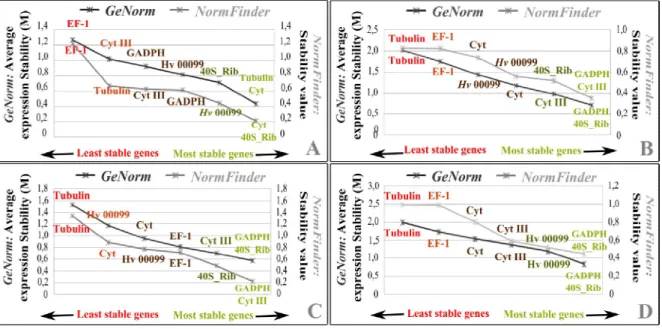
amostras). Apesar dos dois programas terem apresentado os mesmos resultados para cada conjunto de dados (in vitro, in planta, in vitro+ in planta), a combinação de genes mais estáveis diferia entre cada um deles. Assim, Cyt b, 40S_Rib e Hv00099 foram os genes mais estáveis para as amostras in
vitro, enquanto 40S_Rib, GADPH e Cyt III foram os mais estáveis para as amostras in planta. Para
o conjunto das amostras (in vitro+in planta) os genes mais estáveis foram 40S_Rib, GADPH e
Hv00099. Apesar destas diferenças, quando a expressão da proteína Gα foi analisada utilizando os
factores de normalização específicos ou o factor de normalização global (o mesmo para todas as amostras), foi obtido um perfil génico semelhante com apenas algumas diferenças pontuais. Concluiu-se assim que ambas as estratégias permitiram obter uma normalização adequada, estabelecendo-se assim um conjunto de genes de referência para os estudos subsequentes de expressão génica, que constituem o objectivo principal deste trabalho.
O perfil génico de onze genes potencialmente envolvidos no estabelecimento e manutenção da biotrofia foi obtido e a sua função inferida com base em estudos funcionais realizados noutras ferrugens. De acordo com a literatura, estes genes estavam envolvidos nos seguintes processos: i) modificação da parede celular do fungo para que esta não possa ser reconhecido pela planta (quitina desacetilases-CH1 e CH2); ii) vias de sinalização (proteina Gα e proteínas cinase mitogenicamente
de açúcares (manitol desidrogenase-MADp1, transportador de hexose-HXT1p e invertase-INV); v) interacção com a planta (proteína de ferrugem transferida-RTP1p).
A grande maioria destes genes revelou um perfil de expressão muito semelhante ao previamente descrito para outros fungos patogénicos, como por exemplo U. fabae, embora diferenças inesperadas tenham sido observadas para a quitina desacetilase, cujo aumento de expressão nas fases finais de infecção nunca tinha sido observado. MAPK também apresenta um perfil de expressão diferente do observado em outras ferrugens, uma vez que apenas é activada após a invasão do hospedeiro e não durante a formação dos apressorios, como foi previamente descrito. Este resultado sugere que este gene possa estar especificamente envolvido no processo de estabelecimento e manutenção da biotrofia, no entanto estudos funcionais são necessários para uma melhor compreensão do verdadeiro papel desta enzima. Por outro lado, os genes envolvidos na nutrição do fungo, tanto no transporte de aminoácidos como no transporte e metabolismo de açúcares, têm um perfil de expressão semelhante ao de U. fabae, o que claramente sugere uma elevada conservação nos genes responsáveis pela nutrição do fungo, durante o processo de biotrofia. Finalmente, como esperado, a RTP1 só foi detectada nas folhas infectadas, o que claramente sugere que também em H. vastatrix esta enzima pode estar envolvida na manutenção da biotrofia.
Os resultados apresentados neste trabalho permitiram pela primeira vez a quantificação relativa da biomassa de fungo presente nas folhas infectadas durante o processo de infecção. Visto que tal estratégia nunca foi aplicada, foi aberto um novo caminho para estudos de expressão não só em H. vastatrix, mas também para outros agentes patogénicos de plantas com as mesmas limitações e dificuldades. Adicionalmente, desvendamos pela primeira vez alguns aspectos do processo molecular envolvido na complexa interacção C. arabica - H. vastatrix. Este conhecimento poderá contribuir para o desenvolvimento de estratégias de controlo de doenças bastante mais eficazes do que as aplicadas actualmente.
Abstract
Hemileia vastatrix, the causal agent of coffee leaf rust, is a biotrophic fungus with a worldwide
distribution, causing serious social and economic problems in coffee growing countries. Expression studies may have a key role in unraveling the transcriptomics of this pathogen as well as its complex gene-for-gene-based interaction with the host. However, a proper normalization is required to obtain reliable results. In this way the expression stability of eight reference genes (GADPH, EF-1, β-Tubulin, Cyt III, Cyt b, Hv00099, Ubi, 40S_Rib) was analyzed and two strategies were defined as fit for normalization. The first part of this work allowed the correct validation of reference genes to H.
vastatrix. In the second part of this work, the gene expression profile of eleven H. vastatrix genes were
analyzed (CH1, CH2, Gα-protein, MAP Kinase, AAT1_2, AAT3, INV, HXT1p, MAD1p, Asp_AT and RTP1p) for being putatively involved in the establishment and maintenance of the biotrophy. The expression profile of chitin deacetylases (CH1 and CH2) and Gα-protein revealed that these enzymes are involved in the establishment of the biotrophy during the early stages of infection. However, up regulation of CH1 in late infection stages in H. vastatrix is unparalleled in other rusts. The MAP kinase gene was only activated after host invasion and not during appressoria formation, unlike its homologues in other fungi, suggesting that this gene could be specifically involved in the establishment of the biotrophy. Amino acid transporters (AAT1_2 and AAT3), Invertase (INV), Hexose transporter (HXT1p) and Manitol desidrogenase (MAD1p) present similar expression profiles to Uromyces fabae, suggesting a fairly conserved process among rust fungi. Finally, the RTP1p seems to be highly expressed in planta, but not in the in vitro phases, suggesting a similar function to U. fabae. Overall, our results have provided valuable insights at the molecular level to the current understanding of the biotrophic interaction between H. vastatrix – C. arabica.
Table of Contents
Agradecimentos ... i
Nota prévia ... iii
Resumo ...iv
Abstract ... vii
Part I
General Introduction ... 11.The Host Coffea ... 2
1.1 Economic importance ... 3
1.2 Taxonomy ... 3
1.3 General characteristics ... 4
1.4 Origin and dissemination ... 4
2. The pathogen Hemileia vastatrix ... 5
2.1 Economic relevance ... 5 2.2 Taxonomy ... 5 2.3 Origin ... 6 2.4 Genetic variability ... 6 2.5 Life Cycle ... 6 2.6 Dispersion ... 7 2.7 Infection Process ... 7 2.8 Symptoms ... 8 2.9 Control ... 9 3. Plant-pathogen interactions ... 10 3.1 Gene-for-gene theory ... 10 3.2 Biotrophyc lifestyle ... 11
3.3 Gene putatively involved in the establishment and maintenance of the biotrophy .... 12
3.3.1 Suppression of host defense ... 12
3.3.2.Nutrient uptake ... 13
3.3.3 Reprogramming the host’s metabolic flow ... 14
Part II
1. Research article: Validation of RT-qPCR reference genes for in planta expression studies in the obligate biotrophic pathogen Hemileia vastatrix, the causal agent of coffee leaf rust
Abstract ... 16
Introduction ... 17
Materials and Methods ... 19
Results and Discussion ... 24
Conclusions ... 29
2. Research article:
Expression analysis of genes involved in signaling, establishement
of biotrophy, transport and metabolism in Hemileia vastatrix infecting coffee leaves
Abstract ... 35Introduction ... 35
Materials and Methods ... 37
Results and Discussion ... 40
Part III
Final considerations ... 53Introduction
Coffee was one of the first tropical crops to become a truly global commodity. By the early eighteenth century, coffee industry spanned the globe, linking consumers in Europe (which subsequently increased in North America) with producers in Africa, Asia, and Latin America. This agricultural commodity has an estimated retail value of 70 billion dollars and its industry is perceived as prosperous. But, although the coffee business is booming in consuming developed countries, recurrent price decreases cause immense hardship to countries where coffee production is a key economic activity, as well as to the farmers who produce it. As a consequence, during such periods of lower prices, life quality decreases considerably in developing countries that rely to a greater or lesser extent on its foreign exchange earnings for financing essential imports and services.
An additional problem for coffee producing countries is the occurrence of major epidemic diseases such as Coffee Leaf Rust (or orange rust), caused by Hemileia vastatrix Berkeley and Broome, since it limits the coffee production and increase the plight of farmworkers. Therefore, disease management has revealed as one of the most important steps in the coffee production system. Over the last decades there has been a marked shift away from the reliance on pesticides for disease control, towards a more integrated approach using various methods, including the use of resistant varieties. However, an understanding of the biology of these pathogens is essential and increasingly regarded as one of the foundations for the selection of sources of long-lasting resistance.
Focused on the genetic basis of H. vastatrix and its complex gene for gene interaction with the host, the present work intends to identify some of the genes involved in the infection process of H. vastatrix as well as to characterize their expression profiles, leading to a more informed deployment of resistant varieties.
Before presenting this work, a brief general introduction was made on the most relevant aspects of the host, the pathogen, H. vastatrix, and the plant-pathogen interaction. Genes were selected in the literature for their relevant role in the life and pathogenesis (or as reference genes) of other rust fungi, and homologues were searched in H. vastatrix using degenerated PCR primers derived from the comparison of such gene sequences. This preliminary task is reported in the supplemental material of both articles (genes used as reference genes are reported in the first article, while those involved in life and pathogenicity are reported in the second article). In the first article, an accurate normalization for
Real Time Quantitative PCR is presented and the best reference genes selected. The second article uses the previously validated reference genes to study the expression profile of genes involved in the establishment and maintenance of a H. vastatrix – C. arabica compatible interactions.
1. The Host Coffea
1.1 Economic importance
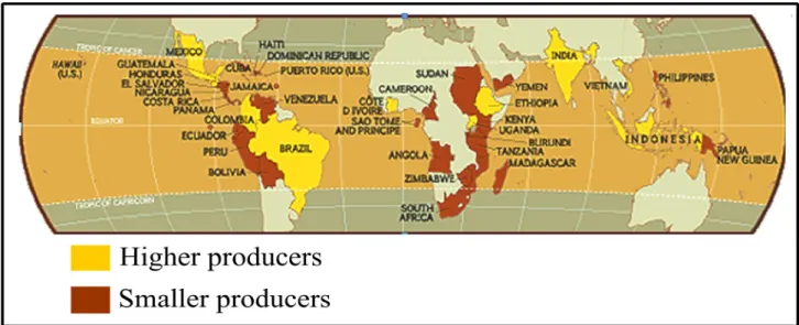
Coffee is the most important agricultural commodity, second only to oil in world trade1,2, and became crucial for the economy of more than 60 developing countries in Africa, Asia and Latin America (Figure1)3,4,5. Currently, its cultivation, processing, trading, transportation and marketing provides employment for hundreds of millions people worldwide4,5. In Ethiopia, for example, more than 700,000 families are involved in coffee production and more than 25 million people depend on the coffee industry, directly or indirectly, to survive4. Thus, even small losses in yield and quality of coffee production have high socio-economic consequences for the coffee growing countries, which often rely on coffee revenues to sustainability.
According to the International Coffee Organization (ICO), coffee production in 2008/2009 was over 128.1 million bags (1 bag = 60 kg) with a retail value of 70 billion USD 4, being Brazil, Vietnam and Indonesia, responsible for almost 60% of this value 4. From the total production, 75% comes from C.
arabica (mainly from Latin America and East Africa) and the remaining from C. canephora and
marginally from C. liberica (mainly from Africa and Asia). Regardless of cultivated species, Africa is responsible for 11% of the total world production, Asia and Oceania for the 26 % and Latin America for 63 %4.
Finally, it is important to note that coffee production represents one of the few solutions to fight poverty in developing countries and thus, the maintenance of acceptable production levels is primal for the life quality of millions of people.
1.2. Taxonomy
The first botanical description of the coffee plant was made in 1713 by Antoine-Laurent Jussieu who classified it as Jasminum arabicanum. Later, this classification was reviewed in 1731 by Linnaeus, which included it in the Coffea genus of the Rubiaceae family6. Cronquist, in 19887 made the more recently taxonomic classification of the Coffea genus7.
Kingdom: Plantae Division: Magnoliophyta Class: Magnoliopsida Order: Rubiales Family: Rubiaceae Genus: Coffea
At the intrageneric level, Chevalier (1947), based on the geographical distribution and fruit characteristics, established four sectors for the genus Coffea: Paracoffea, Argocoffea, Mascarocoffea and Eucoffea8. However, with the implementation of new tools (molecular, biochemical and cytogenetic) a great progress has been made on the Taxonomic level9 . Currently, the genus Coffea encompasses two subgenera: Coffea subgen Coffea and Coffea subgen Baracoffea and 103 described species10,11. However, only two species are economically important: C. arabica, also known as Arabica Coffee and C. canephora also known as Robusta Coffee11.
1.3. General characteristics
Coffea spp. grows in the tropical and sub-tropical countries (25º North-25º South), requiring very
specific environmental conditions for its production. Rainfall, sunlight, wind and soils are all important to produce a quality coffee, but the specific requirements vary according to the species grown1,12 C.
arabica is native to Ethiopian tropical forests and is adapted to the following conditions: Altitudes of
1600-2800m, little seasonal temperature fluctuations (20-28ºC), and a well distributed rainfall (1600-2000mm) with a dry season lasting 3-4 months12. This specie is responsible for the production of the best quality coffee, with lower caffeine content4.
C. canephora is native to the lowland forests of the Congo river basin, which extends up to lake
Victoria in Uganda, and therefore is adapted to the following conditions: Altitudes of up to 1200m, very little seasonal temperature fluctuations (24º-26ºC), and rainfall higher than 2000 mm distributed over a 9-10 month period12. This species is responsible for the production of a coffee with higher caffeine content, widely used in the production of soluble coffee4.
1.4. Origin and dissemination
The history of coffee cultivation is incompletely documented with regards to the domestication of coffee plants in Africa and their dispersal throughout the world by man 2. This event was characterized by successive reductions of diversity within the two subpopulations of wild coffee introduced from Ethiopia to Yemen (575 AD)2,13,14. Further reductions in diversity were produced at the beginning of the 18th century. Firstly, the Dutch East Indian Company stated to establish the coffee planting industry in Indonesia. Later, in 1710 a single plant from Java was introduced to the Amsterdam botanical gardens and some of the young seedlings obtained from this tree were taken to Suriname, onwards to French Guiana and from thence to Brazil. Subsequently, the coffee crop rapidly dispersed throughout most of the suitable locations in Latin America, not only from sources within the continent but also from Reunion island. In the African continent, despite being the place of origin of Arabica coffee, most of the current commercial plantations have resulted from re-introductions mainly from South America and Reunion2,13,14 Therefore, the history of domestication of coffee crops is recent and is characterized by several bottleneck events, which strongly narrowed down the genetic diversity of plants in cultivation throughout the world, with obvious consequences on the emergence and spread of diseases.
2
. The pathogen Hemileia vastatrix 2.1. Economic RelevanceCoffee Leaf Rust (CLR), caused by H. vastatrix, is the most important disease of C. arabica, the most economically relevant coffee species15,16,17,18. This disease may cause up to 30% yield losses in coffee crops, if no control measures are applied18,19, which leads to economic losses of US$ 1-2 billion per year5,20. Considering that most of the coffee producing countries are still in development, these losses can be particularly burdensome and create severe economical distress. In this way, Coffee Leaf Rust has been classified as one of the seven most important diseases and pests of tropical crops20.
One of the most dramatic examples of its devastating potential occurred the nineteenth century in Sri Lanka, where coffee plantations were ravaged, and in less than 28 years all coffee exports had stopped. The losses were so devastating that the annual coffee production of Sri Lanka decreased from 42 million to 3 million kg. As a result, in 1890 almost every coffee plantation was abandoned21,22. Currently, tea instead of coffee is produced in Sri Lanka and, the cup of tea, rather than coffee, has become a familiar part of the British culture.
2.2. Taxonomy
H. vastatrix was first described by Berkeley and Broome from C. arabica leaves in Sri Lanka. H. vastatrix has been classified in the Index Fungorum (www.indexfungorum.org) as:
Kingdom: Fungi
Phylum: Basidiomycota Class: Pucciniomycetes
Order: Pucciniales
Family: Incertae sedis Genus: Hemileia
2.3. Origin
Despite being native of Ethiopia, H. vastatrix was first recorded in 1861 by a British explorer in the Lake Vitoria (East Africa) 5,19,20,21. However, only in 1869 (Sri Lanka) it caused the first great epidemic. Since then, it has spread to most of the coffee growing countries worldwide in three waves. Between 1870 and 1920, it spread through the coffee zones of the Indian Ocean Basin and the Pacific. In the 1950 and 1960, it broke out in the burgeoning coffee farms of Africa Atlantic countries. Finally, it crossed the Atlantic Ocean during 1970 and 1980, and spread throughout the coffee zones of South America 520,21.
2.4. Genetic variability
The first race differentiation of H. vastatrix was carried out by Mayne (1932) in India, who differentiated the local rust samples into four physiologic races1923. No other studies were made on the physiological specialization of H. vastatrix until D’Oliveira initiated a world survey of coffee rust races in 1952 in Portugal19. The work carried out at the Coffee Rusts Research Center (CIFC) enabled the characterization of about 45 rust races, which have been inferred based on Flor's gene-to-gene concept. Currently, 9 virulence genes (v1-v9) of H. vastatrix were inferred based on nine genes (SH1-SH9)19,23.
2.5. Life cycle
In nature, H. vastatrix produces only the uredinal, telial and basidial stages19,20 The dycariotic uredospores (asexual stage) germinate and infect the coffee leaves, being responsible for the disease19,20,24. The teliospores are seldom in the coffee leaves and, like uredospores, could to be present in the pustules (spores release). This structure differentiates into basidiospores (sexual phase) after a meiosis, but seems to be unable to infect the coffee leaves. The basidiospores of H. vastatrix germinate on the surface of coffee leaves, but afterwards they are not able to invade the host. In this way, the function of teliospores and basidiospores in the life cycle of H. vastatrix remains unclear20,24.
Due to the inability of teliospores and basidiospors to invade the host, H. vastatrix is generally considered heteroecious (requires two host to complete its life cycle). However, the alternate host as well as the aecial and pycnial stages of the pathogen remains unknown 20,24.
2.6. Dispersion
The dissemination process of H. vastatrix uredospores is deeply dependent on the wind and rain and involves the following processes: removal from the host, transport through the air, and deposition on a new host2526. Events such as winds and storms are responsible for the dissemination of spores at great distances, while the rain, mainly the rain splash, is responsible for the spores dispersion at short distances 21 20. The spread of spores by the rain splash has additional advantages, such as spore protection from dissection and loss of viability in dry weather. Insects, birds, human and others animals, may also come in contact with the infected leaves and become a disease vector. Special attention must be paid for men, since it is one of the most important vectors on the dissemination of the disease throughout coffee crops worldwide 20,21,26. Currently, the dissemination from one continent to another has been attributed to wind currents and transport of contaminated coffee seeds and other plant material 20,21,25,26.
2.7. Infection process
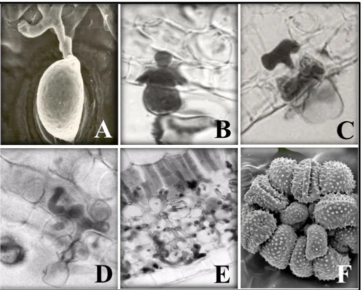
The infection process of H. vastatrix begins with the host-surface recognition in which, the uredospores recognize the chemical and physical properties of the plant surface and germinate, differentiating an appressoria over the stomata (Figure 2B)19. During germination, an optimal temperature of 24 ºC (20-30ºC) and the presence of water are crucial, otherwise, the entire process is aborted 20.
After the appressoria formation, the fungus penetrates the host through the stomata forming a penetration hypha that grows into the substomatal chamber (Figure 2C) 15,19,20. This hypha produces two thick lateral branches; each hypha with its branches resembles an anchor (Figure 2D). Each lateral branch of the anchor differentiate into an haustorial mother cell (HMC) that gives rise to an haustorium, which primarily infects the stomatal subsidiary cells (Figure 2E)15,19,27. The fungus continues it growth with formation of more intercellular hypha, including HMCs, and a large number of haustoria in the cells of the spongy and palisade parenchyma and even of the upper epidermis (Figure 2F)15,19,27. From 21 days after infection, a dense mycellium is observed below the penetration area and a uredosporic sorus protrudes like a “bouquet” through the stoma (Figure 2G)19. After all this process, the spores spread to new coffee leaves and the process restart once favorable conditions are met.
2.8. Symptoms
In susceptible plants, the first symptom are small chlorotic spots on the lower surface of the infected leaves, followed by sorus release and premature fall of the leaves (Figure3.A and 3.B)5,21. This premature shedding weakens coffee trees and affects, year by year, the production of wood needed to bear the future crop. Usually, CLR does not kill the tree, but progressively weakens it, resulting in severe die-back, since this process deprives the coffee plant from the vital nutrient obtained through the leaves via photosynthesis. The subsequent infection of new leaves after the fall of the older leaves are
Figure 2. A) Appressoria differentiation over the stomata; B)Penetration hypha; C) Anchor; D) Haustorial mother cell with haustoria E) Highly colonization tissues; F) Sorus like a “bouquet”
very common and sometimes cause a severe reduction on the yield of coffee berries5,21.
2.9. Control
Chemical control, mainly using cupric fungicides, is the most widely used method to control CLR28. However, it has revealed some disadvantages when implemented, namely the economic costs directly associated with fungicide purchase and application19,23,29. Moreover, in the rainy seasons, the chemicals are washed by the rain, losing their ability to suppress the host invasion 3031 and forcing the farmers to apply more fungicides. Thus, during low yield years, chemical control has to be carried out 2-3 times per year, while during the high yield years this application increase to 4-6 times for year28. However, most of smallholders, who produce the majority of the Arabica coffee, are usually unable to carry these spray programs completely, decreasing the success of this control measure28.
As compared to the chemical control, the cultivation of resistant varieties is a more sustainable disease control strategy. Breeding programs have been developed on research centers worldwide, mostly supported by resistant plant material selected and characterized at CIFC. This strategy has great advantages such as lower costs for farmers, environmental sustainability, and theoretically a long-term solution to coffee leaf rust19,23. Currently, several cultivars with high yield, good organoleptic characteristics, and a notable resistance to H. vastatrix have been developed. The Híbrido de Timor (HDT, a natural hybrid between C. arabica and C. canephora) was the progenitor of varieties such as Catimor (Caturra x HDT), Sarchimor (Villa Sarchi x HDT) and Cavimor (Catuai x HDT) are successful
Figure 3. Coffee leaf rust symptoms A) Small chlorotic spots on the lower surface of the infected
examples of these breeding programs22.
However, the coffee plant resistance may be overcame by the evolving pathogen, and thus developmental studies with the aiming of better understand the pathogenicity of this harmful fungus and its interaction with the host are crucial, in order to improve the breeding programs and leading to a more sustainable agriculture.
3. Plant-pathogen interactions
Plants in nature are generally resistant to most pathogens.29. Plants have the ability to recognize potential invading pathogens and have developed various elaborate defense mechanisms to ward off pathogen attack. At the same time, pathogens have developed strategies to compromise plant resistance mechanisms, in what must have been an evolutionary game of “ping-pong”19,29. Some of these mechanisms are constitutively expressed and provide physical and chemical barriers while others are induced only after pathogen attack19. The idea that successful pathogens suppress host defenses, and in some instance redirect cellular processes to create an environment that is favorable for their proliferation is accepted and very common in biotrophic fungi like H. vastatrix29. However, for some
pathogens, the ability to suppress host defenses depends on the pathotype, which can be explained by gene-for-gene theory.
3.1.Gene-for-gene theory
Half a century ago, Harold Flor's historical landmark publication revealed that the interaction between flax and flax rust (Melampsora lini) is governed by single resistance (R) genes in the plant and complementary avirulence (avr) genes in the pathogen32,33,34,35,36,37. Based on this strictly genetic model of gene-for-gene resistance, many scientists envisaged R proteins as receptors that specifically bind to a matching avr ligand, enabling recognition of the pathogen and subsequent elicitation of an array of plant defense responses that eventually lead to resistance32. In this case, the host and the pathogen establish a incompatible interaction. By contrast, pathogen can evolve in order to avoid plant recognition, leading to a late activation of plant defense mechanisms 33,37,38,39. In this case, pathogen colonizes the host tissues, establishing an compatible interaction with the host.
Currently, several avirulence genes were already identified and characterized in other pathosystems such as Tomato – Cladosporium fulvum, Peronospora parasitica and
Arabidopsis-Pseudomonas syringae, Flax- Melampsora lini38,67. For other pathogens such as H. vastatrix, despite
strong evidence of the presence of avirulence genes (classical genetics), their identification and characterization at molecular level has not been possible.
3.2. Biotrophic lifestyle
Rust fungi, such as H. vastatrix, establish a biotropic interaction with the host 40 41. In this kind of parasitism, pathogen feeds from living host cells, and the host as a whole suffers only minor damage over a longer period of time. Further, the pathogen is dependent on the living host plant to complete its life cycle42. The advantage of this kind of parasitism is the unlimited access, through altered plant translocation patterns, to the plant nutrients43. However, current evidence suggests that biotrophy requires an impressive degree of cellular interaction between plant and parasite, perhaps explaining why so few fungal pathogens exploit this type of parasitism43. Several studies conducted in order to better understand this type of parasitism establish the following properties as hallmarks of biotrophy: 1) Obligate biotrophs are not culturable in vitro (although research in some rust fungi has enabled in vitro cultivation in some extent); 2) They form highly differentiated infection structures (; 3) They have limited secretory activity; 4) They establish a narrow contact zone separating fungal and plant plasma membranes; 5) They engage in a long-term suppression of host defense responses; 6) They form haustoria (specialized hyphae that penetrates host cells) 19,40,41,42.
The haustoria, due to its role in the establishment and maintenance of the biotrophy, have been studied for plant pathologists, since their first description by Zanardini about 150 years ago41,44. Already in naming these structures [fr. L. haurine (haurio, hausi, haustum): to drink, to draw] de Bary (1863) propose one of the possible functions for the haustoria - the uptake of nutrients from the host. However, knowledge about this key element of the obligate biotrophic lifestyle is still fairly scarce 40,41,44. The main reasons for this are the preserving lack of functional transformation system for haustoria forming fungi and the fact that these organs are not formed in culture44. Molecular biology, however, opened a new dimension to investigate the role(s) of haustoria41. In 1997, Hahn & Mendgen45 developed techniques for isolating haustoria from host leaves, allowing the identification of genes preferentially expressed in the haustoria of Uromyces fabae (first haustorium-specific cDNA libray) 45. Currently, haustoria have been documented as primary in the fungal survival, since they are responsible for nutrient uptake and information exchange between the host and pathogen40,41,42,44. Additionally, it must
to modify the host metabolism to ensure total access to its nutrients41. Finally, it is important to note that haustoria are not truly intracellular: they breach the cell wall only and a newly formed host plasma membrane (the extrahaustorial membrane) surrounds them, resulting in a close association of fungal and plant membranes, separated only by a thin fungal wall and an extrahaustorial matrix. The extrahaustorial matrix is the site for translocation of nutrients and exchange of information, providing the optimal conditions for the maintenance of the biotrophy40,41,44,46.
3.3 Genes putatively involved in the establishment and maintenance of the biotrophy
As previously stated, biotrophy requires an impressive degree of cellular interaction between plant and parasite. In this way, rusts need to suppress host defense reactions, assimilate nutrients from the plant and reprogram the host's metabolic flow, in order to survive41. Several studies in rust fungi, mainly in
Uromyces fabae, began to unravel some genes involved in these processes. However, a long road still
needs to be traveled in order to understand all the strategies developed by the rust to survive.
3.3.1 Suppression of host defense
Rust fungi, developed several mechanisms in order to avoid plant recognition and tolerate various stress factors induced by the plant during the early stages of the infection process. Currently, several genes such as PIG 11 (Metallothionein), PIG 16 (cytochrome P-450 monooxygenases (P-450s)), chitin deacetylase, G-Protein and MAP Kinase, were described as deeply involved in these processes. Metallothionein (PIG11), protects plant pathogen cells against toxic concentrations of heavy metals as well as free oxygen radicals produced by the plant during the hypersensitive response47,48. P-450s catalyze a variety of oxygenation reactions on a large spectrum of substrates. While some of them are involved in the biosynthesis of secondary metabolites, such as hormones and toxins, the majority of P-450s are involved in detoxification of the pathogen when exposed to toxic plant metabolites45. Chitin deacethylases, seems to be responsible for the conversion of chitin (cell wall component of pathogen able to be recognized by the plant) to chitosan. This strategy during the intracellular growth may protect hyphae of plant pathogenic fungi from being lyzed by extracellular plant chitinases, since the chitosan is a poor substrate for plant chitinases49,50,51. The fungal G protein, mainly Gα – protein, not only plays a pivotal role in the regulation of several signaling pathways crucial to fungal survival, but is also involved in fungal development and pathogenicity52,53,54,55. The MAP kinase pathway is one of the most important signaling cascades regulated by Gα - protein and has been associated, by several
studies, with fungal pathogenicity53,54. The critical importance of these enzymes was proved in studies with Cryptococcus neoformans, Magnaporthe grisea, Cochliobolus heterostrophus and Fusarium
oxysporum, in which a single a mutation in Gα – protein or MAPK led to an incorrect appressoria
formation and a decrease/total loss of fungal pathogenicity56,57,58.
3.3.2 Nutrient uptake
As previously described, biotrophic fungi are entirely dependent on plant living cells to their growth and reproduction. In this way, nutrient uptake (sugar uptake and amino acid transport) is one of the most vital process for these organisms and involves several genes such as Amino acid transport (AAT1; AAT2; AAT3), Hexose transporter (HXTP1), Invertase (INV), Manitol desidrogenase (MAD1p), Beta-glucosidases (BGL) and Glucokinase (GLK)41.
Since 1997, three putative secondary transporters of amino acids were identified and characterized in
Uromyces fabae. AAT1p, was recently characterized as a broad specificity amino acid secondary active
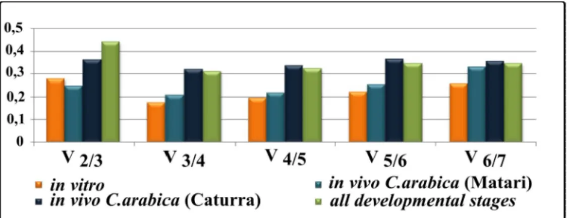
transporter with a main specificity for L-histidine and L-lysine. This amino acid transporter was expressed at all developmental phases of Uromyces fabae, preferentially in the haustoria59. AAT3p, exhibits a substrate preference for L-leucine and the sulphur containing amino acids L-methionine and L-cysteine60. As AAT1 the expression of AAT3 does not seem to be restricted to haustoria60. Finally, a specific subtract has not been identified for AAT2, but it is only expressed in haustoria61. Taken together it seems that amino acid uptake in U. fabae is not limited to hasutoria, but preferentially occurs in this structure41.
The sugar uptake is a complex process that aims to nourish the fungus by the plant. In this way, biotrophic fungi must be able to convert sucrose, the most abundant carbohydrate source produced by the plant, in their own nutrient source. Studies in U. fabae began to unravel some of these processes as well as the enzymes and sugars involved41. Since biotrophic fungi is unable to directly use sucrose as a nutrient source, the primary function of INV is the cleavage of sucrose into D-glucose and D-frutose62. An additional source, at least for the D-glucose, could be a Beta- glucosidases, since this enzyme has the capacity to use cellobiose, a breakdown product of cellulose, as a substrate, and consequently, could use degradation products of the plant cell wall available for its nutrition63. Subsequently, this monosaccharides (D-glucose and D-frutose) will be transfer into the haustoria by the HXT1p53. Once inside the haustoria, the monosaccharides will be converted into a nutrient source that can be used by
the pathogen. GLK1 and MAD1p have a crucial role in this process, since the first one converts D-glucose to D-glucose-6-phosphate, the direct source of energy, and the second one converts the D-frutose to manitol, the fungal storage compound41,64,65. Moreover, MAD1p and INV, seem to have an additional role in the fungal survival and maintenance of biotrophy. In this way, MAD1p is also responsible for the nutrient mobilization in germinating uredospores, allowing for fungal nutrition before the establishment of the biotrophy (haustoria formation) and INV is responsible for the additional sucrose supply at the infected leaves during all the developmental phases of the pathogen62,64 and it was also suggested that fungal invertases present in the apoplast are able to limit export of carbohydrates from the infected tissue via the phloem, conditioning the plant tissue for a conversion from source to sink62.
3.3.3 Reprogramming the host’s metabolic flow
Several studies, in Melampsora lini and Uromyces fabae, showed evidences that avirulence proteins from the rust fungi are recognized inside the plant cell. These proteins are also likely to be synthesized and secreted specifically by the fungal haustorium and must be transported from the extracellular matrix across the host plasma membrane. The first gene detected in the cytoplasm and nucleus of host cells was detected in U. fabae66. RTP1 interferes with plant gene expression, but the molecular basis of this interference remains unclear41. To understand the exact biochemical function of RTP1p in plant cells, complementary structural and molecular insights from the identified functional core of RTP1p are essential. It is therefore crucial to determine the 3D structure of RTP1p functional domain41. Regardless the validation of these specific functions the role of RTP1p in the maintenance of the biotrophy is unquestionable.
Validation of RT-qPCR reference genes for in planta expression studies in the obligate biotrophic pathogen Hemileia vastatrix, the causal agent of coffee leaf rust
Ana Vieiraa,b, Pedro Talhinhasa*, Andreia Loureiroa, Sébastien Duplessisc, Diana Fernandezc, Maria do Céu Silvaa, Octávio S. Paulob, Helena Gil Azinheiraa
a
Centro de Investigação das Ferrugens do Cafeeiro/Instituto de Investigação Científica Tropical, Oeiras, Portugal;
2
Computational Biology and Population Genetics Group, Centro de Biologia Ambiental, Faculdade de Ciências da Universidade de Lisboa, Lisboa, Portugal;
3
Institut National de la Recherche Agronomique, Nancy, France; 4
Institut de Recherche pour le Développement, Montpellier, France.
*corresponding author: [email protected]
Abstract
Hemileia vastatrix, the causal agent of coffee leaf rust, is a biotrophic fungus with a worldwide
distribution, causing serious social and economic problems in coffee growing countries. Expression studies may have a key role unraveling the transcriptomics of this pathogen as well as its complex gene-for-gene-based interaction with the host. Real time Quantitative PCR is the golden standard for gene expression analysis, although an accurate normalization is essential for adequate conclusions. Reference genes are often used for this purpose, but the stability of their expression levels requires validation under experimental conditions. Further, biotrophic fungi undergo important biomass variations along its infection process in planta, which raises the need for an adequate method to normalize the proportion of fungal cDNA in the total plant+fungus cDNA pool. In this work, the expression profiles of eight reference genes (GADPH, EF-1, β-Tubulin, Cyt III, Cyt b, Hv00099, Ubi, 40S_Rib) were analyzed across 28 samples, obtained in vitro (germinated uredospores and appressoria) and in planta (post-penetration fungal growth phases), to determine the most stable reference genes. Gene stability was assessed using the statistical algorithms incorporated in GeNorm and NormFinder software. Cyt b, 40S_Rib and Hv00099 were the most stable gene for in vitro dataset, while 40S_Rib, GADPH and Cyt III were the most stable in planta. For the combined datasets (in vitro+in planta), 40S_Rib, GADPH and Hv00099 were selected as the most stable. Subsequent tests on the Gα - protein expression showed that the reference genes selected for the combined dataset do not differ significantly from those selected specifically for the in vitro and in planta datasets. Our study provides tools for correct validation of reference genes in obligate biotrophic plant pathogens, as well as the basis for RT-qPCR studies in H. vastatrix.
Keywords: normalization factor; housekeeping genes; basidiomycete fungal plant pathogen; Coffea
1. Introduction
Gene expression analysis plays an important role in enhancing our understanding of the signaling and metabolic pathways which underlie cellular and developmental processes (Hu et al., 2009). Although several methods have been used to quantify gene expression, including Northern blotting, RNase protection assay, in situ hybridization and cDNA microarray technology, real-time quantitative PCR (RT-qPCR) is considered the gold standard for quantifying gene expression, thanks to its sensitivity, specificity, dynamic range and high throughput capacity (Bustin et al., 2005; Fang et al., 2006; Freeman et al, 1999; Udvardi et al., 2008; Walker et al., 2009). This technique can detect very low quantities of a target transcript even if only a few copies are present in the sample (Wong and Medrano, 2005). However an accurate normalization is required to obtain a reliable quantification of the transcript (Derveaux et al., 2010; Huggett et al. 2005; Thellin et al., 1999).
Until now, several procedures have been proposed to normalize RT-qPCR data such as sample size, RNA total quantification, genomic DNA and artificial molecules (Cruz et al., 2009; Huggett et al. 2005; Teste et al., 2009). However, none of them is appropriate for this technique, since they do not take into account sample variations, such as differences in the quantity and quality of RNA, and efficiencies of reverse transcription or PCR (Cruz et al., 2009; Huggett et al. 2005; Teste et al., 2009; Walker et al., 2009). On the other hand, internal control genes (references genes) are the most commonly used to normalize RT-qPCR data, though the success of this procedure relies on the appropriate choice of control genes (Dheda et al., 2005; Olsvik et al., 2005; Thellin et al., 1999). Typically, reference genes need to exhibit two major properties: firstly they should be essential for the maintenance of cellular function and viability, and therefore should be constitutively expressed in all tissues; secondly, their transcription should not be affected by the conditions understudy (Derveaux et al., 2010; Dheda et al., 2005; Paolacci et al., 2009; Stürzenbaum et al., 2001; Thellin et al., 1999; Wong and Medrano, 2005). However, some evidences show that all genes seem to be regulated under some conditions and there are always some variations in transcript levels, so that none of the commonly exploited genes can be viewed as a universal reference gene (Dheda et al., 2005; Stürzenbaum et al., 2001; Thellin et al., 1999). In many cases, the use of, a single reference gene is inadequate, and can produce wrong biological conclusions (Dheda et al., 2005; Hu et al., 2009; Stürzenbaum et al., 2001; Teste et al., 2009). Additionally, gene expression studies as well as reference gene validations are mainly limited to human or classical laboratory organisms and non-model species often suffer from lack of background information available (Axtner and Sommer, 2009). For all these reasons, it is essential a careful evaluation of one or even more suitable reference genes prior to the determination of the gene profiles, especially in non-model species. In this way, several programs like geNorm (Vandesompele et al., 2002) and NormFinder (Andersen et al., 2004) are often used. These methods are based on different statistical algorithms aimed at choosing the most stable reference genes, in order to correctly normalize the RT-qPCR results (Andersen et al., 2004; Pfaffl et al., 2004; Vandesompele et al., 2002). However, choosing the most suitable statistical method can be confusing, since each program has the potential to deliver different results. Thus, a combined analysis may be the best strategy to follow.
Actin, Glyceraldehyde-3-phosphate dehydrogenase (GADPH), Ribosomal genes (28S; 18S), Elongation factor (ef1) and β-tubulin are frequently used as reference genes for RT-qPCR without a proper validation, which may jeopardize the reliably of data (Bohle et al., 2007; Fang et al., 2006; Kim et al., 2003; Nicot et al., 2005).
Metarhizium anisopliae (Fang et al., 2006), Saccharomyces cerevisiae (Teste et al., 2009) and Phytophthora parasitica (Yan and Liou, 2006). As expected, different reference genes were selected for
each species: Actin, Secretion associated GTP-binding protein (sarA) and Cytochrome c oxidase (Cox5) were selected as the most stable genes in Aspergillus niger; in Metarhizium anisopliae, Elongation factor (EF-1), GADPH and Tryptophan biosynthesis enzyme (tryp) were the most stable; the ubiquitin-conjugating enzyme (Ubc), β – tubulin (Tub-b) and 40S ribosomal protein (WS21) were the most stable in Phytophthora parasitica; in Saccharomyces cerevisiae, the most stable genes were the Mannosyltransferase activity (ALG9), RNA Pol II transcription factor activity (TAF10), RNA Pol III transcription factor activity (TFC1) and Ubiquitin-protein ligase activity (UBC6). However, to our knowledge, no such studies are published concerning rust fungi.
Rust fungi represent a large group of plant pathogens causing diseases on a variety of plants, including several important crops such as cereals, legumes and coffee (Kemen et al., 2005). Hemileia
vastatrix is responsible for coffee leaf rust, a disease that can lead to yield losses of 30% if no control
measures are applied (Azinheira et al., 2010; Mendgen, 2000; Nunes et al., 2009; Silva et al. 2006; Talhinhas et al., 2010). This pathogen establishes a biotrophic interaction with its host and is completely dependent of plant living cells to grow and reproduce (Mendgen, 2000; Silva et al. 2006; Voegele and Mendgen, 2003). Unlike other rust fungi, H. vastatrix is amenable for in vitro assays only in the early stages of its differentiation (corresponding to uredospore germination and appressoria formation), while further differentiation must be obtained in planta for experimental purposes (Azinheira et al., 2001). In coffee leaves, appressoria formed over stomata differentiate an infection hypha which invades the substomatic cavity, from which the fungus grows colonizing the leaf tissues inter- and intra-cellularly, feeding from living coffee cells by specialized structures named haustoria. The infection cycle is completed with the formation of sporogenic hyphae and the release of uredospores, which occurs from 21 days after the infection (Silva et al., 2006). Therefore, the proportion of H. vastatrix biomass in planta increases along the infection process. This particularity can be a problem when the aim of the study is the proper validation of reference genes. In this way, an additional normalization step is necessary in order to successfully validate H. vastatrix reference genes for in planta studies. It should be noted that, even though quantification of fungal pathogens is frequently carried out by qPCR (Atallah et al., 2007; Mideros et al., 2009), including rust fungi (Acevedo et al., 2010; Jackson et al., 2006), the employment of this strategy for the validation of RT-qPCR fungal reference genes “in host” is still a novelty, as it has been reported very recently (Hacquard, Duplessis et al., accepted for publication in Physiological and Molecular Plant Pathology).
The main focus of this work was selecting potential H. vastatrix reference genes suitable for the normalization of RT-qPCR data in the subsequent expression studies. For this, a methodology for normalization of H. vastatrix nucleic acid content in planta was established, and eight genes from different functional classes were selected for RT-qPCR primer design and validation as references to be used in H. vastatrix gene expression studies both in vitro and in planta.
2. Material and Methods
2.1. Fungal and plant material
In vitro growth: Fresh uredospores of the H. vastatrix isolate CIFC 1065 (race II) were spread in sterile
distilled water in petri dishes to germinate (GU - germinated uredospores sample) or inoculated on artificial oil-collodion membranes (Heath and Heath, 1976), to differentiate appressoria (A) (Azinheira et al., 2001) and incubated during 24h under darkness at 24ºC.
In planta growth: Both Caturra and Matari varieties of C. arabica were inoculated with fresh
uredospores of H. vastatrix isolate CIFC 1065, establishing compatible interactions. The inoculation was performed by spreading the uredospores (1 mg/pair of leaves) over the lower surface of young coffee leaves. After an incubation period of 24 h in a dark moist chamber, the plants were moved to greenhouse conditions. The leaves were collected at seven time-points after inoculation (18h, 24h, 48h, 72h, 7D, 14D and 21D) in order to obtain in vitro+in planta of H. vastatrix.
Controls: Control material were ungerminated H. vastatrix uredospores (U), uninoculated young coffee
leaves, and freeze-dried Lecanicillium sp. (formerly Verticillium sp.; an hyperparasite isolated from H.
vastatrix uredospores) mycelia obtained upon culture in liquid medium (Potato Dextrose Broth, Difco,
USA).
Fungal and plant material were immediately frozen by immersion in liquid nitrogen and stored at -80°C until RNA extraction. At each collection time, germination of spores, appressorium formation and fungal growth in host tissues were visualized by light microscope and evaluated as previously described (Silva et al., 1999).
2.2. Reference genes
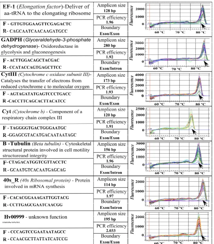
Candidate reference genes represent three different functional classes (metabolism related, structure related and secretion related), reducing the chance that they may be co-regulated. They were selected from the literature, along with a new candidate gene (a predicted H. vastatrix secreted protein), selected based on the stability of 454 pyrosequencing analysis between three datasets of H. vastatrix (Talhinhas et al., 2010). The genes are: Elongation Factor (EF1-a) (Fang et al., 2006); β-Tubulin (Tub) (Yan and Liou, 2006); Glyceraldehyde-3-Phosphate Dehydrogenase (GADPH) (Fang et al., 2006); Ubiquitin (Ubi); Cytochrome c Oxidase Subunit III (CytIII) (Bohle et al., 2007); Cytochrome b (Cyt) (Grasso et al., 2006); 40 S Ribosomal Protein (40S_R) (Yan and Liou, 2006) and a predicted H. vastatrix Secreted Protein (Hv00099). Sequences for these genes in H. vastatrix were obtained either from the employment of degenerated PCR primers designed from homologous genes (including cloning using the CloneJET PCR cloning kit (MBI Fermentas, Lithuania) in other rusts or from the H. vastatrix 454 datasets (Table 1).
Table 1. RT-qPCR primer sequences and amplicon characteristics for control genes, including melting
curve analysis (arrows denote results for negative controls) tested in H. vastatrix samples in planta. EF-1 and β-tubulin were amplified from H. vastatrix DNA based on the following degenerated primers
obtained from homologous genes in U. fabae and M. lini: EF-1 forward,
CBGARCGHGARCGTGGTATCAC; EF-1 reverse, TTVGADATACCRGCYTCRAATTCACC;
β-tubulin forward, ACHGGDGCTGGAATGGG; β-tubulin reverse,
2.3. RT-qPCR primer design and efficiency test
The primer design and choice of target sequence are essential steps to ensure a specific and efficient amplification of the desired products. Primers were designed using PerlPrimer v1.1.17 (Marshall, 2004) with melting temperatures of 60°C, lengths of 18-24 bp and GC content of 50–60%. Exon/intron boundaries were determined by aligning each cDNA sequence with its corresponding genomic sequence. Whenever possible (Table 1), primer were designed in the junction of two different exons, thus preventing amplification of a potential DNA contamination. However, this strategy was not possible to follow for four genes, where it would generate poor primer quality. Nevertheless, the presence of DNA in RNA samples was tested, as described under ‘Total RNA isolation and cDNA synthesis.
Additionally, precautions have to be taken when working with a biotrophic fungus. Since this implies that fungal cDNA will be mixed with plant cDNA for the in planta samples, primers have to be specific for the fungus and cannot amplify the plant cDNA. Besides this, H. vastatrix can be hyperparasited by ascomycete fungi from the genus Lecanicillium, which should neither be amplified.
To avoid these problems, a blast tool-based strategy was followed (in silico strategy). First, sequences of the candidate genes obtained from H. vastatrix transcriptome and Sanger sequencing (using degenerated primers), were used to search for homologous sequences in three data bases (NCBI (http://www.ncbi.nlm.nih.gov/); HarvEST Coffee database (http://harvest.ucr.edu/) and Verticillium
group database
(http://www.broadinstitute.org/annotation/genome/verticillium_dahliae/MultiHome.html)) with BlastN and BlastX tools. The homologous sequences obtained with the highest score were used as a template for the alignment of primers developed, in order to assess their similarity. Only primers with more than five bp differences were accepted.
The primer efficiency was experimentally tested with the LinRegPCR program developed by Ramakers et al. (2003) which uses a linear regression analysis of fluorescence data from the exponential phase of PCR amplification to determine amplification efficiency (E). LinRegPCR software utilizes an iterative algorithm (considering the number of data points, regression coefficient and slope of the regression line) for the selection of the exponential phase in each PCR amplification. We analyzed our fluorescence data (details of RT-qPCR experiments bellow) using this software and obtained E values for each reaction. Additionally, a combined analysis using an average E value (arithmetical mean of E values of all samples) was applied and that value was used in subsequent analyses (Cikos et al., 2007; Rutledge and Stewart, 2008).
2.4. Total RNA isolation and cDNA synthesis
Total RNA was extracted from frozen samples using the RNeasy Plant Mini kit (Qiagen, Germany), with addition of an in-solution DNase I digestion. Only RNA samples with 260 nm/280 nm wavelength ratio between 1.9 and 2.1 and 260 nm/230 nm wavelength ratio greater than 2.0 before and after DNase I digestion were used for cDNA synthesis. The quality of RNA samples was also assessed by electrophoresis on 1.2% agarose gels. RNA quantity was determined by means of
UV-VIS-spectrophotometry (Lambda EZ201, Perkin-Elmer, USA). A control PCR was run on extracted RNA samples to check the absence of genomic DNA with the cytochrome b primers (Table 1). First-strand cDNAs were synthesized from 1 µg of total RNA in 20 µl final volume, using Omniscript RT kit (Qiagen) and oligo(dT)18 primer (MBI Fermentas) following the manufacturer’s instructions. For each sample, a final volume of 500 µl of cDNA was obtained and stored at -20ºC.
2.5. Real-time Quantitative PCR
The RT-qPCRs were performed using an iQ5 real-time thermalcycler (BioRad, USA), based on EvaGreen® Supermix (BioRad). Each 15 µl reaction comprised 6 µl template, 7.5 µl EvaGreen Supermix, 0.4 µl of each primer (10 µM) and 0.7 µl of sterile distilled water. The reactions were subjected to an initial denaturation step at 95°C during 10 min followed by 45 cycles at 95°C for 15s, 60°C for 20s and 75ºC 15s. A melting curve analysis was performed at the end of the PCR run over the range 60-95°C, increasing the temperature in a stepwise fashion by 0.5°C every 10s. Baseline and quantification cycle (Cq) were automatically determined using the iQ5 Optical System Software. Three negative controls (1-no template; 2- cDNA of plant; 3- cDNA of Lecanicillium sp.) were run to validate the in silico strategy and ensure that only cDNA of H. vastatrix was amplified. Each PCR reaction was performed in duplicate and the specificity of the amplicons was checked by melting curve analysis and by 2.5% agarose gel electrophoresis.
2.6. Assessment of expression stability
2.6.1. Normalization of H. vastatrix genomic DNA
For normalizing the expression levels of H. vastatrix reference genes in the in planta samples accordingly to fungal biomass, genomic DNA was extracted from the same leaves used for the RNA assays, using the DNeasy Plant mini kit (Qiagen) following the manufacturer’s recommendations (namely including a RNase treatment). Genomic DNA concentrations were estimated using the Nanodrop 1000 (Thermo Scientific, USA), and samples diluted to 10ng/ul. Real-time quantitative PCR was performed on these samples at the same conditions previously described, using primers for Hv00099. The Cq results were used to normalize the Cq of reference genes using the following formula:
Cq normalized = Cq cDNA reference genes -Cq DNA Hv00099 (1)
2.6.2. Determination of reference gene expression stability
Cq values were obtained (as mentioned under ‘Real-time Quantitative PCR’) for each reference gene for all in vitro and in planta samples following the experimental design mentioned in ‘Fungal and plant