Francisco Duarte da Cunha Ventura
Characterization of gene or gene
clusters responsible for the
production of antimicrobial
compounds in Pseudoalteromonas
atlantica
Francisco Duar te da Cunha V enturaCharacterization of gene or gene clus
ter s responsible for t he production of antimicrobial compounds in Pseudoalter omonas atlantica
Dissertation for the M.Sc. degree in Biomedical
Engineering
Supervisors:
Maria João Vieira, PhD
Carina Almeida, PhD
Francisco Duarte da Cunha Ventura
Characterization of gene or gene
clusters responsible for the
production of antimicrobial
compounds in Pseudoalteromonas
atlantica
! ! Carina Almeida, for all the supervision throughout the entire work. You have gone way beyond your duty. For always pointing me in the right direction, for all the time I made you lose, for the great support in the laboratory. I can’t thank you enough! It’s been an honor and a privilege to work with you for the past year!!
has led to a rise of highly resistant bacteria. These microorganisms have been developing mechanisms that enable them to survive to the aggressions of classical antibiotics. Also, these self-defense mechanisms are easily transmitted between bacteria, which is a worrisome panorama.!
! It is necessary to get back to a proactive fight against these
microorganisms. Living organisms have long proven to be a rich source for antimicrobial compounds due to their need to fight for their place in ecological niches. The marine environment is known to be prolific with microbial communities and thus a great diversity of bioactive compounds was already discovered.!
! In this thesis work, I searched for novel antimicrobial compounds in a marine bacteria, Pseudoalteromonas atlantica. A genome mining approach was followed. A search for clusters coding for secondary metabolites with antimicrobial characteristics was done using softwares such as AntiSmash, ClustScan, HHpred or BLASTx. This process resulted in the finding of 17 putative clusters coding for polyketide synthase and also 1 putative cluster coding for a bacteriocin.!
! To assess if P. atlantica is, in fact, capable of producing antimicrobial compounds, and in the positive case, to further enhance the production of such
in the presence of a competitor (E. coli K12; Staphylococcus aureus;
Pseudomonas aeruginosa or Vibrio harveyi) has also resulted in the inhibition of Salmonella enteritidis.!
! These results indicate that the P. atlantica genome might be a source for
novel antimicrobial compounds. In fact, under the described conditions, P.
atlantica was capable of producing antimicrobial molecules with a narrow activity
spectrum.!
aparecimento de bactérias altamente resistentes. Estes microorganismos têm desenvolvido mecanismos que os permite sobreviver às agressões provocadas pelos antibióticos clássicos. Além disso, estes mecanismos de auto-defesa são facilmente transmitidos entre diferentes espécies de bactérias, o que é um cenário muito preocupante.!
! É então necessário voltar a uma atitude proactiva na luta contra estes microorganismos. Os microorganismos já há muito provaram ser uma fonte rica em compostos antimicrobianos, devido à sua necessidade de lutar por um lugar nos respectivos nichos ecológicos. O ambiente marinho é conhecido por ser fértil em comunidades microbianas e portanto uma grande diversidade de compostos bioactivos foram já descobertos.!
! Nesta tese procurei por compostos antimicrobianos numa bactéria
marinha, a Pseudoalteromonas atlantica. Seguiu-se uma estratégia de “genome mining”. Foi realizada uma procura de clusters de metabolitos secundários de cariz antimicrobiano, através do recurso a ferramentas como o AntiSmash, ClustScan, HHpred ou BLASTx. Todo este processo resultou na descoberta de 17 putativos clusters de poliquétidos sintetases e 1 putativo cluster de bacteriocina.!
Broth), a uma temperatura de 23ºC, um pH de 8 e uma agitação de 120 rpm. O meio de cultura gasto desprovido de bactérias provou inibir o crescimento de
Escherichia coli K12. Além disso, na presença de um competidor (E. coli K12; Staphylococcus aureus; Pseudomonas aeruginosa or Vibrio harveyi), o meio de
cultura de P. atlantica, também resultou na inibição de Salmonella enteritidis.! ! Estes resultados indicam que o genoma da P. atlantica pode ser uma fonte de compostos antimicrobianos inauditos. De facto, nas condições referidas no presente trabalho, a P. atlantica foi capaz de produzir moléculas antimicrobianas, com um espectro de actividade aparentemente muito específico.!
!
Abstract!
!
Chapter I - Introduction: Antibiotics!
1. Overview ! 2. A brief history!
3. Mechanisms of action!
4. The urgent need for new antimicrobials! 5. Nature: A source for novel antimicrobials!
5.1. Polyketides!
5.2. Nonribosomal peptides! 5.3. Bacteriocins!
6. Bioinformatic tools!
!
Chapter II - Bioinformatic research!
1. Overview!
2. Identification of interesting regions!
3. Clusters annotation and proteins identification! 4. Discussion iv!
!
!
3! 4! 6! 10! 13! 18! 23! 25! 27!!
!
33! 34! 42! 512.1.1. Effect of the growth medium! 2.1.2. Temperature, pH and agitation! 2.1.3. Addition of extracellular ATP! 2.1.4. Competitors!
2.2. Assessment of antimicrobial activity! 2.3. Determination of arbitrary units! 3. Results and discussion!
3.1. Effect of culture media on cell growth and antimicrobial activity!
3.2. Effect of temperature, pH and agitation on cell growth and antimicrobial activity!
3.3. Effect of addition of extracelullar ATP! 3.4. Effect of competitors!
!
Chapter IV - General conclusions and future work!
1. General conclusions and future work!
!
Chapter V - References and appendix!
1. References! 2. Appendix 59! 60! 61! 62! 64! 66! 67!
!
67!!
70! 75! 76!!
!
83!!
!
89! 96and Marshall B. (2004).Abstract!
!
Figure 1 - New antibacterial agents approved in the US per 5-year period
from 1983 to 2002. Adapted from Shlaes, D. et al (2004).!
!
Figure 2 - Compounds with new antibacterial templates divided into
development phases and their lead derivation source. Adapted from Butler M. and Cooper M. (2011).!
!
Table 2 - Bioactivity of Pseudoalteromonas species. Adapted from
Bowman (2007)!
!
Figure 3 - Example of type I PKS (lovastatin), type II PKS (doxorubicin)
and type III PKS (naringenin chalcone) From Hertweck (2009).!
!
Table 3 - Examples of Polyketides and their bioactivity. Adapted from
Pfeifer and Khosla (2001).!
!
Figure 4 - Repartition of six main biological activities displayed by curated
peptides in the Norine database (790 NRPs). From Caboche S. et al (2010).!
!
Figure 5 - Parameters selected in AntiSmash.!
!
Table 4 - Position of all clusters detected by AntiSmash!
!
Figure 6 - Screenshot of ClustScan workspace.!
14 20 21 24 34 35 36 6 11 13
!
Figure 8 - Screenshot of CLC Sequence Viewer workspace.!
!
Figure 9 - Screenshot of AntiSmash annotations.!
!
Figure 10 - Screenshot of InterPro search.!
!
Table 7 - PKS related ORFs detected near AntiSmash detection sites!
!
FIgure 11 - Schematic representation of a hypothetical Polyketide
Synthase in P. atlantica T6c composed by 4 distinct domains: AT - acyltransferase ; KS - ketosynthase ; KR - ketoreductase!
!
Figure 12 - Graphic of homologous gene clusters, for PKS #8 cluster -
AntiSmash.!
!
Figure 13 - Schematic representation of a hypothetical bacteriocin in P.
atlantica T6c.!
!
Figure 14 - Graphic of homologous gene clusters, for bacteriocin cluster -
AntiSmash.!
!
Figure 15 - Representation of the culture conditions used for the
competition assays.!
!
Table 8 - Effect of culture media in P. atlantica growth and the production
of inhibitory compounds.!
!
Figure 16 - Inhibition halos in an E.coli K12 lawn.!
42 43 44 45 47 48 49 50 62 68 69
!
Figure 17 - Inhibitory effect of the filtered sterilized spent MB of P. atlantica
in E. coli K12 lawns over time (A) - Arbitrary units over time (B) - Inhibition halos on E. coli K12 lawns, recorded after several dilutions of the spent medium.!
!
Table 12 - Inhibition of targets by bacteria-free spent MB for optimized
culture conditions.!
!
Table 13 - Effect of competing species (E. coli K12, S.aureus, P.
aeruginosa, V. harveyi) in dialysis membrane on production of antimicrobial compounds by P. atlantica.
72
73
74
!
!
!
!
!
!
!
Chapter I!
Introduction: Antibiotics
1. Overview!
!
! Antibiotics are compounds that can either kill (bactericidals), or inhibit the growth (bacteriostatics) of bacteria. This categorization between bactericidals and bacteriostatics isn’t as obvious as it may appear, since it depends on the drug type, concentration and on the bacterial species. They can be of natural source, when they are for instance produced by living organisms. Also, they can be produced by chemical synthesis or derive from a biological source.!
! Since their discovery, antibiotics have revolutionized medicine in many ways. Countless lives were saved by these compounds. Nevertheless, the use of antibiotics has been accompanied by a growing number of resistant microorganisms. It is feared that because of this, we may face a new era, like the preantibiotic one.!
! The study of antibiotics has had its ups and downs all over the years, but it has become of major interest in the last decade. It is important to understand how this all began, in order to understand where we stand nowadays.!
!
!
2. A brief history!
!
! Sir Alexander Fleming set the beginning of antibiotics timeline. In 1929 Sir Fleming was conducting experiments with Staphylococcus variants. Plates containing Staphylococcus were left in contact with air and, by chance, they became contaminated by a fungus. Sir Fleming noticed that the areas around the mold became transparent, Staphylococcus were undergoing lysis. He concluded that the contaminant was a Penicillum mold that was producing a powerful bactericidal, the penicillin [1].!
! In 1940, years before the use of penicillin as a therapy, a bacterial penicillinase was identified. After its mass production, several strains showed the capacity to resist to penicillin. Just then, experiments were conducted so that the penicillin could be modified chemically, in order to prevent cleavage by penicillinases (β-lactamases) [2].!
! By 1944 Waksman, Feldman and Hinshaw discovered the streptomycin
from Streptomyces griseus, a bacteria commonly found in the soil [2]. They found that streptomycin was effective against virulent human tubercle bacilli. This discovery led to a growing interest in soil bacteria, which later proved to be the main resource for the discovery of several antibiotics. Streptomycin was then used to cure tuberculosis, but in the mean time, strains of Mycobacterium
tuberculosis resistant to the antibiotic arose during patient treatment [2].!
! As in penicillin and streptomycin, many powerful antibiotics were
strains appeared through a process that remained unknown until the mid 1950s [3]. In Japan, a country devastated by war at the time, an epidemic of Shigella
dysenteriae turned to be particularly hard to irradiate, due to the growing number
of resistant strains. Sulfonamide was no longer effective in 80% of the cases [3]. Kitamoto wrote about S. dysenteriae strains that could resist to four different antibiotics. Later on, it was proved that the resistance of these strains could be transferred to other Enterobacteriaceae simply requiring a cell-to-cell contact, indicating that bacterial conjugation was involved in the process [3].!
! In the mid 80s, Michael Syvanen proved that the uniformity of the gene code among nature would allow organisms to use genes transposed from organisms of different species (Horizontal Gene Transfer). This revealed to be of particular interest when one tries to understand the rapid bursts in the evolution of organisms, also helping to understand the way which pathogens gained resistance to several antibiotics [2].!
! Since Sir Fleming found penicillin in 1929 till now, the development of antibiotics has come up through different eras. It is safe to say that the majority of the antibiotics used nowadays derive from the ones discovered during the so called golden era, which took place between the 50s and the mid 60s. From then on came the lean years, or the innovation gap. A gap that lasted until 2000, when a decrease of the discovery rate was observed. The main approach for the development of novel drugs has been the modification of the molecules previously known [2].!
3. Mechanisms of action!
!
! Antibiotics can be classified according to a different range of settings, such as their spectrum of activity, their chemical structure, but most commonly, antibiotics are categorized by their mechanism of action (Table 1). Each category, class or family aims to destroy or defunctionalize essential physiological or metabolic targets of the bacterial cell [4].!
!
!
!
Table 1 - Main antibiotic families and mechanisms of action. From Levy S. and Marshall B. (2004).
Mechanism of action Antibiotic families
Inhibition of cell wall synthesis Penicillins; cephalosporins; daptomycin; monobactams; glycopeptides
Inhibition of protein synthesis
Tetracyclines; aminoglycosides; oxazolidonones; streptogramins; ketolides;
macrolides; lincosamides Inhibition of DNA synthesis Fluoroquinolones
Competitive inhibition of folic acid synthesis Sulfonamides; trimethroprim
Inhibition of RNA synthesis Rifampin
• Inhibition of cell wall synthesis!
! Antibiotics that inhibit the cell wall synthesis act by interfering with the formation of the peptidoglycan wall present in bacteria. The process by which that occurs is what distinguishes the different families belonging to this mechanism of action. For example, β-Lactams like penicillins, carbapenems and cephalosporins act by inhibiting transpeptidase enzymes, whose function is to cross-link peptidoglycan chains that compose the bacterial cell wall, causing it to lyse [5].! ! Another way for an antibiotic to affect cell wall integrity is by binding with peptidoglycan units and by blocking transglycosylase (enzyme that adds disaccharide pentapeptides to extend the glycan strands of existing peptidoglycan molecules) and also blocking transpeptidase activity [5]. That is the case of a glycopeptides, such as vancomycin. Nevertheless, this family of antibiotics is ineffective against Gram-negative bacteria, due to their lower permeability, by contrast with β-Lactams, which are effective against both Gram-positive and Gram-negative bacteria. There are other ways to inhibit the cell wall synthesis or the cell wall integrity, besides the ones used by β-Lactams and glycopeptides, like affecting the transport of individual peptidoglycan (e.g. Bacitracin), for example [5].!
!
• Inhibition of protein synthesis!
! Translation is a crucial process in biological lifeforms. It is a series of processes by which information coded in RNA is used to create proteins constituted by aminoacids. One of these processes includes the presence of a very important organelle - the ribosome. This organelle is divided into two
subunits. In the case of prokaryotes, there is a small subunit (30s - responsible for reading the mRNA) and the big subunit (50s - responsible for joining amino acids to the growing peptide chain). Antibiotics that act through this mechanism (inhibition of protein synthesis) are molecules that act by compromising the function of one of these two subunits. Therefore, they can be divided into 30s inhibitors or 50s inhibitors [6].!
! The 30s inhibitors like the tetracyclines act by compromising the function of the small ribosome subunit. Tetracycline, for example, blocks the aminoacyl-tRNA to the ribosome and therefore stops protein synthesis [5].!
! Antibiotics belonging to macrolides, lincosamides or oxazolidonones
families are considered to be 50s inhibitors, since they act upon the big subunit of the bacterial ribosome. They stop protein synthesis by either blocking initiation or elongation of the translation process. Also, some act by hindering translocation of peptidyl-tRNA. Peptidyl is one of the binding sites in the ribosome for tRNA [5].!
!
• Inhibition of DNA synthesis!
! Deoxyribonucleic acid or just DNA is an essential molecule of all living organisms. It would be correct to assume that a living organism would be killed if its DNA synthesis stopped. That is just how quinolones, a family of synthetic broad-spectrum antibiotics, work. Fluoroquinolone (a quinolone with a fluorine atom attached to the central ring system) targets the function of DNA-topoisomerase and DNA-gyrase complexes [5]. Topoisomerases and gyrases are enzymes that participate in DNA replication. By canceling their function,
bactericidal. There are other families belonging to this mechanism of action and they target different components of the bacterial DNA machinery [5].!
!
• Competitive inhibition of folic acid synthesis!
! Unlike humans, bacteria do not acquire folic acid through diet. Instead they synthesize it. It is by affecting this mechanism that antibiotics like the sulfonamide work. All cells require folate cofactors for the biosynthesis of diverse cellular components, like the formation of the so important amino acid methionine, crucial for starting protein translation. Sulfone inhibitors work by being analog of para-aminobenzoic acid (pABA), which is a required intermediate of bacterial synthesis of folate. Sulfonamides act as alternative substrates for dehydropteroate synthase, an enzyme involved in the folate pathway. By doing so, they inhibit the growth of the target microorganism, therefore they are considered to be bacteriostatical antibiotics [7].!
!
• Inhibition of RNA synthesis!
! Like the DNA, ribonucleic acid or RNA plays an important role in expression of genes. Inhibition of RNA synthesis is a catastrophic event for prokaryotic nucleic acid metabolism. Drugs belonging to the rifamycin group inhibit or affect the normal behavior of RNA within bacterial cells. Rifampin, for example, hinders DNA transcription by inhibiting RNA polymerase, leading, in most cases, bacterial pathogens to death [5].!
4. The urgent need for new
antimicrobials!
!
! As stated by Hiroshi Nikaido in 2004, about 100 000 tons of antibiotics are produced per year [8]. Their intensive use has had a tremendous impact in worldwide microbial resistance. Because of this, many antibiotics that were effective many years ago, no longer produce a harmful effect in the same pathogens. Methicilin-resistant Staphylococcus aureus (MRSA) is a glaring example of a dangerous pathogen. It is resistant to methicilin and a wide range of antibiotics like aminoglycosides, macrolides, tetracycline, chloramphenicol, and lincosamides [8]. Since 2002 some strains of MRSA have also proved to be resistant to vancomicyn (the main strategy of eliminating MRSA until then) [9]. The rise of multidrug resistant bacteria, the scarcity of new classes of antibacterial drugs (Fig. 1) and the stagnation in antibiotics discovery is demanding new ways of controlling these very resistant pathogens [10, 11].!
! Alongside the heavy health burden of this problem, also comes an economic burden that hasn’t been correctly measured yet. As stated by WHO, “All costs for infections caused by resistant strains were consistently greater than those for infections caused by susceptible strains.” That is mainly due not only to the increase in the intensity of care needed by infected patients, but also to an increase in the length of stay of those patients in the hospitals. Moreover, as expected, infections with resistant strains are associated with worse clinical outcomes [11].!
! The laborious legal procedures to approve new antimicrobials, especially in the United States, have delayed the introduction of new antibiotics and large pharmaceutical companies are fending themselves from this area of investigation [10]. This obviously results in a declining number of new antimicrobials in development. Although all of this may appear to be a dantesque scenario, there are some good news as well. State of the art techniques, like genomics, proteomics, structure based design, high-throughput screening and combinatorial chemistry are paving their own path into new antimicrobial compounds [10].!
!
0 4 8 12 16 1998 - 2002 1993 - 1997 1988 - 1992 1983 - 1987Figure 1 - New antibacterial agents approved in the US per 5-year period from 1983 to 2002. Adapted from Shlaes, D. et al (2004).
5. Nature: A source for novel
antimicrobials!
!
! Today, there is an understanding that the method for finding new antibiotics used in the last 50 years is no longer effective. While 2nd, 3rd, etc. generations of antibiotics fail to deliver a continuous harm to pathogens along time, some researchers think that natural compounds might be the solution. Many antibiotics discovered until nowadays are secondary metabolites that some microorganisms naturally produce. Thinking that microorganisms have found their way against many pathogens long before they even become a known threat for humans, can be a successful contemplation [12].!
! Nowadays, efforts are being made in order to find novel antimicrobials from old sources, like Streptomyces [13]. Also, many are studying the capacity of other unstudied microorganisms (cyanobacteria or uncultured bacteria) to synthesize antimicrobials compounds. This is proving to be a successful strategy, since several novel scaffolds with antibiotic potential are being discovered. We can also think that many targets within pathogens haven’t been explored yet (like the fatty acid synthesis) and if novel scaffolds can act upon these targets, a new era may be arriving. Naturally occurring antibiotics can benefit from the fact that they have features which are not present in libraries of synthetic drugs molecules, like those found via high-throughput screening. It is not surprising that rather than chemically synthesized, the majority of antibiotics in the last stages of clinical trials derive from natural sources (Fig. 2) [13].!
!
! The marine environment is known to be proliferous with microbial
communities. Also, as many marine communities are highly specific to particular ecological niches (sponge or algae environments), there is a vast biodiversity among these microbial communities that inhabit seawater. This statement is useful when one tries to understand the implications of natural products discovery, such as naturally produced antibiotics [14].!
! It is estimated that about 0,5% to 6 % of the oceanic bacterioplancton belongs to the Pseudoalteromonas genus. Some species of this genus have
0 11 22
NDA Phase III Phase II Phase I
1 1 5 10 4 1 6 11 1 Syn-derived NP-derived Unknown
Figure 2 - Compounds with new antibacterial templates divided into development phases and their lead derivation source. Adapted from Butler M. and Cooper M. (2011).
proved to be efficient in producing bioactive compounds, such as antimicrobials, anti-fouling or algicidals compounds [15, 16].!
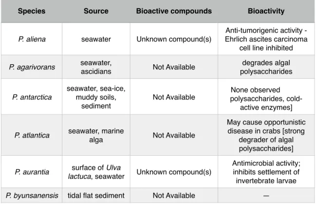
! Table 2 summarizes the bioactivity of compounds produced by
Pseudoalteromonas species. Although many bioactive compounds have already
been detected, the synthesis of such compounds are quorum-sensitive. Thus, the ecosystem in which these species grow, influence the biosynthesis of these compounds. As many aspects of ecological networks remain unexplored, there is room for the discovery of novel natural products [15].!
!
Table 2 - Bioactivity of Pseudoalteromonas species. Adapted from Bowman (2007).
Species Source Bioactive compounds Bioactivity
P. aliena seawater Unknown compound(s)
Anti-tumorigenic activity - Ehrlich ascites carcinoma
cell line inhibited
P. agarivorans seawater,
ascidians Not Available
degrades algal polysaccharides P. antarctica seawater, sea-ice, muddy soils, sediment
Not Available polysaccharides, cold-None observed active enzymes]
P. atlantica seawater, marine
alga Not Available
May cause opportunistic disease in crabs [strong
degrader of algal polysaccharides]
P. aurantia surface of Ulva
lactuca, seawater Unknown compound(s)
Antimicrobial activity; inhibits settlement of invertebrate larvae
P. carrageenovora
seawater, marine
alga Not Available
None observed degrader of algal polysaccharides] P. citrea seawater, mussels, ascidians, sponges Unknown compound(s) Inhibits settlement of invertebrate larvae; cytotoxic against sea urchin
[algal polysaccharide degradation]
P. denitrificans seawater
High molecular weight polyanionic substance; cycloprodigiosin HCl
Anti-tumorigenic activity; inhibits T-cell/lymphocyte proliferation; anti-malarial activity; induces settlement
of sea urchin Heliocidaris
erythrogramma P. distincta sponge Not Available
P. elyakovii mussels, marine
alga Not Available None observed
P. espejiana seawater Not Available None observed
P. flavipulchra seawater Not Available —
P. haloplanktis seawater Novel diketopiperazines
Probiotic benefits to shellfish; cold-active
enzymes
P. issachenkonii marine alga Isatin; unknown reddish- brown compound
Anti-fungal activity; hemolytic
P. luteoviolacea seawater, marine
alga Toxic antimicrobial protein; brominated pyrrole-containing compounds, 4- benzaldehyde; n-propyl- 4-hydroxybenzoate Antimicrobial activity; inhibits algal spore settlement; cytotoxic
against sea urchin
Strongylocentrotus intermedius; induces
settlement of sea urchin
Heliocidaris erythrogramma
P. maricoloris sponges Bromo-alterochromides A and B
Antibacterial activity; cytotoxicity against sea
urchins
P. marina tidal flat sediment Not Available —
P.
mariniglutinosa diatoms Not Available — P. nigrifaciens seawater, salted
foods, mussels Not Available —
P. paragorgicola sponge Not Available —
P. peptidolytica seawater Unknown compounds Antimicrobial activity, hemolytic
P. phenolica seawater 3,3’,5,5’-tetra-bromo-2,2-
biphenyldiol Antimicrobial activity
P. piscicida (and
related bacteria)
estuarine waters, fish samples
Toxic protein; possible yellow cyclic/acyclic brominated depsipeptide compounds; unknown anti-algal compound(s) Antibacterial; algicidal activity; possible cytotoxicity [opportunistic fish pathogen; thrombolytic
enzymes]
P. rubra seawater
High molecular weight polyanionic substance; cycloprodigiosin HCl;
rubrenoic acids
Antimicrobial activity; anti- tumorigenic activity; inhibits
T-cell/lymphocyte proliferation; anti-malarial;
bronchodilatatoric
P. ruthenica shellfish Unknown compounds Antimicrobial activity
P. spongiae sponge Not Available Strongly induces settlement of Hydroides elegans
P. tetraodonis puffer fish Tetrodotoxin Neurotoxic effects
P. translucida seawater Not Available —
P. tunicata marine alga,
tunicates
Unknown purple pigment; tambjamine-like
alkaloid YP1; toxic protein AlpP; other unknown sunstances
Anti-fungal, anti-algal, antimicrobial, inhibits settlement of invertebrate
larvae and algal spores; inhibits protists
!
! Because there are many strains belonging to the Pseudoalteromonas
genus, many of them live in the wild under different environmental conditions, which leaves room for a vast spectrum of different bioactive compounds produced by different strains. Although some of these compounds were already described and tested, there is a believe that there is an enormous potential for the discovery of novel compounds [15, 16].!
! Among the naturally occurring antibiotics, polyketides and non-ribossomal
peptides have gained their own status in antimicrobials research and production. Some of them are even known and widely used as antibiotics. Nevertheless, their synthase is yet to be fully understood. Besides, the ability for many microorganisms to produce these compounds is being tested in different species of bacteria and fungus [13].!
!
P. ulvae marine alga Unknown substances
Inhibits invertebrate larval settlement and algal spore germination and settlement
P. undina seawater, fish Not Available
hemolytic; [probiotic benefits; possible opportunistic fish pathogen]
5.1. Polyketides!
!
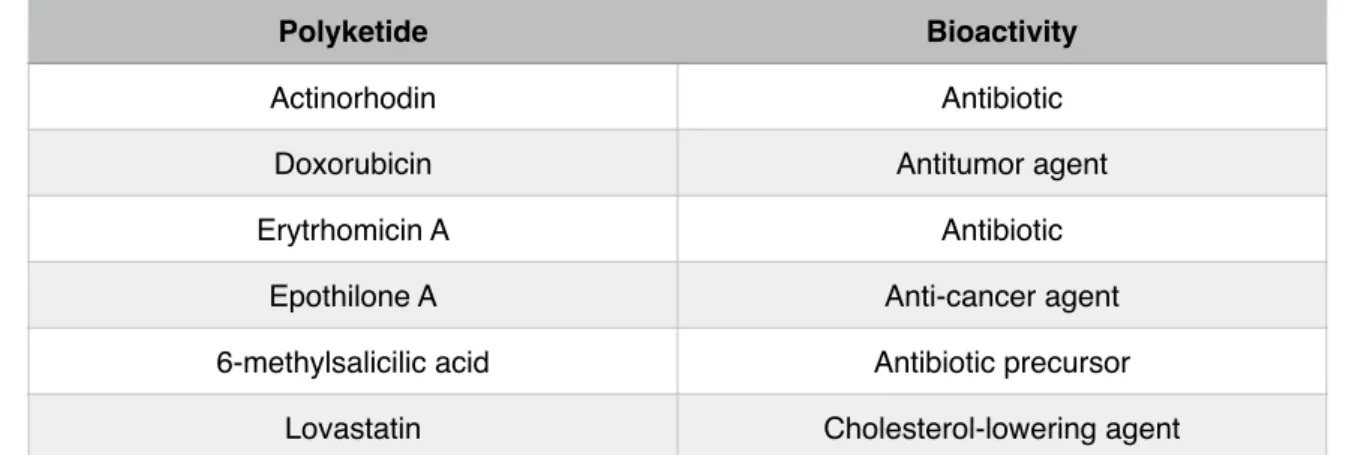
! Polyketides are small secondary metabolites that microorganisms produce. Their vast diversity in terms of structure and function is well known. They are produced by polyketide synthases, which are enzymatic assembly lines that determine the final structure of the produced polyketide. These metabolites can be of major interest in the clinical area (Table 3). For example, erythromycin A is a potent antibiotic used as a therapy against bacterial infections. Rapamycin is an immunosuppressant used in various surgery techniques. This is to say that polyketides may present very interesting characteristics. Some of them have already been useful, others may help us win the war against pathogens in the future. It is then important to try to understand how these powerful molecules are produced by some microorganisms [17].!
! Polyketides, are synthesized due to repetitive condensation reactions, in a process that is very similar to the synthesis of fatty acids. In these reactions that link carbon precursors, coenzyme A thioesters play an important role as they constitute the core of the molecule. However, polyketides are found in much more diverse structures than fatty acids. This diversity is of great usefulness since they also present different modes of action, thus can be used in different applications [18].!
! Polyketide synthases (PKS) are the enzymes of large dimension with specific catalytic domains that catalyze the referred condensations. Their core domains are ketosynthase (KS), acyltransferase (AT) and thiolation (T). PKS are
categorized into three classes (I, II and III) (Fig. 3) which differs slightly from fatty acid nomenclatures [18, 19].!
!
• Type I - best exemplified by the PKS responsible for building the backbone
of erythromycin ( 6-deoxyerythronolide B or 6-DEB). These PKS’s are constituted by multidomains resembling type I fatty acid synthases (FAS).!
!
• Type II - as one would imagine, this PKS resembles type II FAS. The growth
of the polyketide is iterative and KS, AT and T domains are re-used during polyketide synthesis.!
!
• Type III - They differ from other types by using an acyl-carrier-protein
independent mechanism. Besides, they typically lack multiple catalytic domains.!
!
Figure 3 - Example of type I PKS (lovastatin), type II PKS (doxorubicin) and type III PKS (naringenin chalcone) From Hertweck (2009).
!
! As polyketides are small metabolites hard to isolate, its correct characterization has been an hard quest for many researchers. Traditional methods used on the identification of polyketides usually involve phenotype screening, followed by the use of analytical chemistry techniques. For instance, Marinho and his colleagues [20] reported the presence of citreorosein (1), emodin (2), janthinone (3), citrinin (4), citrinin H1 (5) and dicitrinoln (6), six known polyketides produced by an endophytic fungi, Penicillum herquei. To evaluate the presence of these compounds, they have used classical methods of chromatography. They were identified by 1D and 2D Nuclear Magnetic Resonance spectroscopy (NMR) and Mass Spectrum analysis (MS), results were then compared to previous identifications of the referred compounds [20].!
! However, phenotype screening approaches are usually very time
consuming and depend on the availability of large libraries of organisms. Besides, the production of some metabolites might not be induced under the testing conditions [21] and important compounds might be lost. Actually, the
Table 3 - Examples of Polyketides and their bioactivity. Adapted from Pfeifer and Khosla (2001).
Polyketide Bioactivity
Actinorhodin Antibiotic
Doxorubicin Antitumor agent
Erytrhomicin A Antibiotic
Epothilone A Anti-cancer agent
6-methylsalicilic acid Antibiotic precursor
advances in genomics and genome sequencing have shown that the bacteria potential to produce molecules of pharmacological interest has been greatly underestimated. Nowadays, the development of bioinformatic tools and the increasing update in genome databases are being helpful to finally understand the steps behind the synthesis of these small metabolites. It is now possible to follow a genome mining approach to identify regions with potential interest before proceeding with further laboratorial testing [21].!
5.2. Nonribosomal peptides!
!
! Nonribosomal peptides are a class of potent antibiotics (like penicillin) and other important pharmaceuticals of great economic interest. These molecules are synthesized by a process that is independent of the ribosome and nucleic-acids, unlike the classical pathway for metabolites synthesis. They are assembled by nonribosomal peptide synthetases (NRPS), which are multimodular megaenzymes. Their biosynthesis relies not only in the 20 canonical amino acids, but also in some different building blocks, such as “d-configured and β-amino acids, methylated, glycosylated and phosphorylated residues, heterocyclic elements and even fatty acid building blocks” [22]. Due to this diversity of building blocks, there are generally a large number of active sites which are essential to the bioactive purposes of these compounds [22].!
! NRP synthetases are modularly organized enzymes, which comprise
multiple catalytic domains. Each module is responsible for adding one amino acid to the peptide and, therefore, their order in the chain influences the final product. The process of adding amino acids to the elongating chain continues until the final molecule is released by a thioesterase domain [23].!
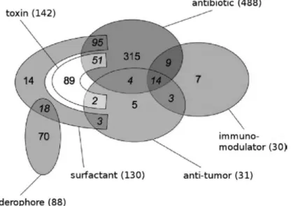
! Norine is a database entirely dedicated to NRPs, from where it is possible to perform analysis of NRP-related peptides, like predicting functions. Biological activities presented by NRPs mainly range from immunomodulating, iron chelating, antibiotics, toxins, surfactants to anti-tumor (Fig. 4) [24].!
!
!!
!
!
!
Figure 4 - Repartition of six main biological activities displayed by curated peptides in the Norine database (790 NRPs). From Caboche S. et al (2010).
5.3. Bacteriocins!
!
! Bacteriocins are antimicrobial peptides/proteins produced by bacteria that helps the producing bacteria to proliferate within an environment by eliminating other rival bacterial species that compete for the same environment. Unlike polyketides and non ribossomal peptides (NRP’s), which are synthesized by non ribossomal pathways, bacteriocins follow the classical ribosomal pathway. Because of this, bacteriocins are structurally different from either PKS and NRP’s, and thus some authors do not consider them as antibiotics. Also, unlike traditional antibiotics, bacteriocins usually restrict their activity to related species of the producing bacteria, and particularly to strains of the same species [25, 26].! ! As bacteriocins may exhibit significant potency against pathogens (some can even compromise antibiotic-resistant strains), they may be seen as a viable alternative to classic antibiotics and so help to solve the multi-resistant pathogens problem [27]. There are already many useful applications of these metabolites in some industries. For instance, bacteriocins are already used in the food industry to prevent the colonization by pathogens [25, 26]. On the other hand, as some pathogens produce their own bacteriocins that help them conquer unwanted places, such as the human nasopharynx (in the case of Streptococcus
pneumoniae), they may also play a negative role by competing with commensal
flora [26-28].
Bacteriocins include a very heterogeneous group of molecules, and so their classification is largely based on their molecular weight differences [29]. Their composition can consist of only 19 amino acids, while large bacteriocins
can have molecular weights up to 90 000 Da. Additionally, some small bacteriocins can also present some pos-translation modifications.!
! Generally, bacteriocins attack pathogens by compromising their
membrane. They act by binding to cell’s surface receptors that are recognized by that particular bacteriocin. In a microbial environment, there are usually three types of cells: the bacteriocinogenic (produce bacteriocins); the sensitive ones; the resistant ones. That diversity results in an ecological balance since each type has a stronger and weaker opponent. In spite of bacteriocin producers tend to kill strains belonging to their same species, there are some remarkable exceptions, for example, E. coli bacteriocins are proved to eliminate strains of the distant related Hafnia alvei [26].
As in polyketides, the growing number of genome databases is helping to improve the understanding of the genes or gene clusters involved in the production of bacteriocins, giving way for a regulation and functionalization of these powerful metabolites.!
6. Bioinformatic tools!
!
! Although bioinformatics is a cross-disciplinary field that began to emerge in the mid 60’s, it was only by the beginning of the last decade that it gained relevant advantage in microbiology. That is due to the massive amount of data generated by genomic research in the 90’s. Since then, bioinformatic has proved to be an indispensable field of knowledge in order to store and organize genomic sequences or predict metabolic behaviors, to refer some of the immense applications [30].!
! One of the most approaches to predict a gene or protein function is
data-mining or genome data-mining. It relies on the fact that different species may present a local similarity in some genes (closely related species present higher similarity). Using this technique, a researcher can search for patterns in databases of different species and if those patterns look alike with patterns that belong to a known sequence, this researcher finds evidence for what that sequence or gene might do [30]. Since 2000’s, genome sequencing of various microorganisms has allowed many researchers to identify new genes and pathways for the production of powerful antibiotics. Genome mining of actinomycetes has already resulted in the finding of new natural products, i.e., strambomycins A-D [31]. !
! The list below shortly describes some bioinformatic tools that can be useful for searching specific clusters/genes:!
• BLASTx: BLAST stands for Basic Local Alignment Search Tools, and particularly, BLASTx searches proteins giving a translated nucleotide query.!
!
• AntiSmash: It is a library with secondary metabolites produced by microbes which can be a powerful source of antibiotics and other pharmaceuticals. It uses a genome mining approach for biosynthetic clusters, to do so. Antismash can be used to find putative PKS, bacteriocins, lantibiotics, homo serine lactones or other types of secondary metabolites. AntiSmash is based on profile hidden Markov models (HMM) of genes that are specific for certain types of gene clusters. Profile HMM analysis complements standard pairwise search models used in BLAST, for example, for a better identification of gene clusters encoding secondary metabolites [32, 33].
!
• ClustScan: Or Cluster Scanner is a rapid, semi-automatic software for DNA sequences annotations. The main goal for the use of ClustScan is the discovery of novel biosynthetic gene clusters. ClustScan is particularly efficient in finding regions of a given genome that can possibly code interesting secondary metabolites, such as PKS, NRPS, immuno-supressants, etc. As in AntiSmash, ClustScan also uses HMM analysis to detect more accurately sequence alignments [34].
!
• CLC Sequence Viewer: Among a large amount of features, CLC sequence viewer was used as a genome browser. This software can be used to track
and retrieve sequences of the targets given by other tools, such as ClustScan.!
!
• HHpred: It is an interactive server for protein homology. It allows to search for protein homology within a wide choice of databases. Various single queries sequences (retrieved, for instance, from CLC sequence viewer or from NCBI) can be run one by one in HHpred, in order to see similarities between these queries and proteins coded by other organisms.!
!
• InterPro: It is a documentation resource for finding protein domains, families and functional sites of proteins. As each InterPro entry includes a description, annotations and relevant literature, it is a powerful tool for predicting a protein’s derivatives and its function. !
!
! As these tools help in the prediction and understanding of the biosynthesis of interesting metabolites, they are of major relevance when one aims to discover and try to induce the production of novel interesting compounds.!
!
!
!
!
!
!
Chapter II!
Bioinformatic research!
!
1. Overview!
!
! In order to evaluate if Pseudoalteromonas atlantica T6c possessed
clusters with potential antibiotic activity, a genome mining approach was required. First, P. atlantica T6c genome was downloaded in raw format from NCBI database. A search for polyketides synthases (PKS), nonribosomal peptides synthases (NRPS) and bacteriocins was done using AntiSmash (an antibiotics and secondary metabolites analyzer). In addition to AntiSmash, results were confirmed by using a second bioinformatic tool, ClustScan (a powerful tool for scanning clusters).!
! CLC Sequence Viewer was then used as a resource for clusters
annotation. First, each ORF within the genomic regions identified was copied and then pasted into BLASTx (Basic Local Alignment Search Tool), which searches proteins database using a translated nucleotide query. For each ORF within the interest region, its size, name, direction of translation and domains were captured.!
! Several software tools were used to search each protein’s catalytic domains - HHpred, Interpro and BLAST. From all the clusters annotated, only one seems to be a completed conserved PKS, with all the domains present in most of the PKS found in the literature.!
!
!
2. Identification of interesting
regions!
!
! A search for PKSs in the genome of P. atlantica (accession number: NC_008228) was done using AntiSmash (ANTIbiotics & Secondary Metabolite
Analysis SHell). Because PKSs regions are usually of a great diversity or
variability, in order to detect these clusters it is needed to select “Detect putative genes clusters based on PFAM domain probabilities” in search parameters. All the other parameters were set as default (Fig. 5).!
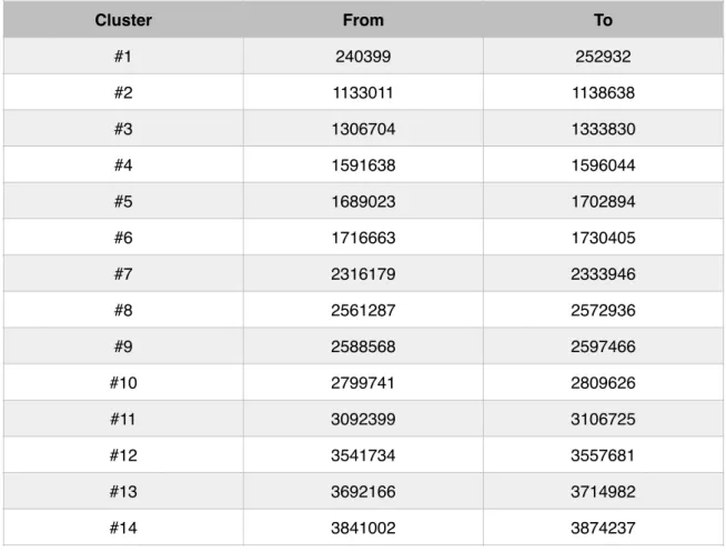
! A fasta format file of P. atlantica T6c genome was once again retrieved from NCBI database and then loaded to the “Nucleotide input” area. AntiSmash searches resulted in the finding in 17 putative PKS clusters and a cluster coding for a bacteriocin (Table 4). Unfortunately, this effort didn’t result in the finding of any NRP cluster.!
! To maintain explicitness, bacteriocin cluster will be analyzed in a section ahead.!
!
Table 4 - Position of all clusters detected by AntiSmash
Cluster From To #1 240399 252932 #2 1133011 1138638 #3 1306704 1333830 #4 1591638 1596044 #5 1689023 1702894 #6 1716663 1730405 #7 2316179 2333946 #8 2561287 2572936 #9 2588568 2597466 #10 2799741 2809626 #11 3092399 3106725 #12 3541734 3557681 #13 3692166 3714982 #14 3841002 3874237
!
! In order to confirm these results and to better identify interesting regions, a similar approach was done using ClustScan. The Pseudoalteromonas atlantica T6c genome was imported to ClustScan. Then, a search for PKSs and NRPSs was requested. This action resulted in dozens of putative interesting genes. These genes appear sorted by their position in the genome (Fig. 6).!
! Relevant information is obtained in a ClustScan search for these genomic
regions, such as the E-value (Expect value), which gives a good idea about the significance of a match. DNA and proteins coordinates, the size of the coded proteins and the sequence frame, are also some of the valuable information obtained from ClustScan. ClustScan was used in version 2.0.3.!
#15 3887354 3897050
#16 4408491 4423937
#17 4814303 4820705
DNA coordinates of genes given by this software were copied to lists of a python script (Fig. 7) that detects if coordinates of different genes are located within the same genomic area.!
!
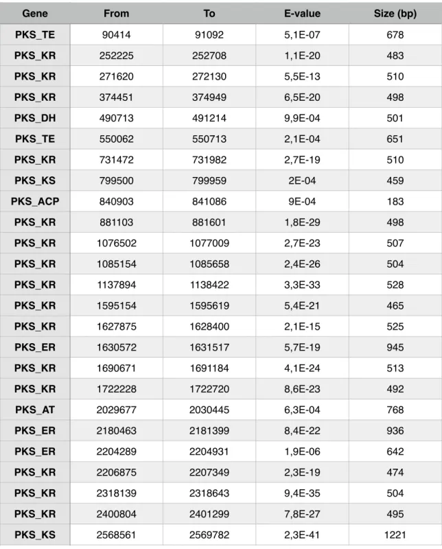
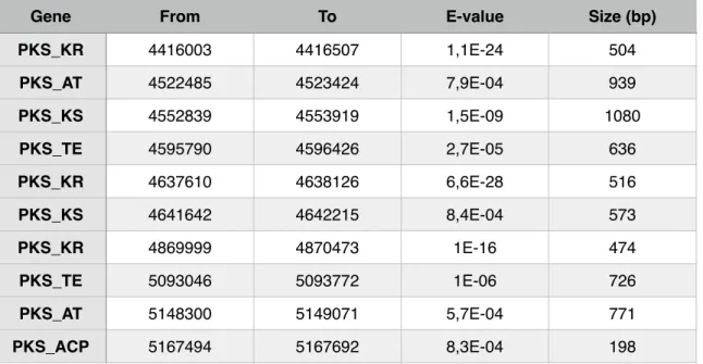
! This is important because two different genes don’t always mean two different clusters. It just means that these two genes may belong to the same cluster and therefore could participate together in the biosynthesis of a polyketide. Results from running the script showed the presence of 8 superposed regions and therefore resulting in a final count of 53 PKS clusters, as clustscan detected 62 genes putatively belonging to PKS’s (Table 5). These regions, where interesting genes are very close to one another are of major interest. Having the modular architecture of PKS’s into account, different genes detected by clustscan may represent different modules of the same PKS.
Table 5 - Position of all PKS genes and their main specifications given by ClustScan.
Gene From To E-value Size (bp)
PKS_TE 90414 91092 5,1E-07 678 PKS_KR 252225 252708 1,1E-20 483 PKS_KR 271620 272130 5,5E-13 510 PKS_KR 374451 374949 6,5E-20 498 PKS_DH 490713 491214 9,9E-04 501 PKS_TE 550062 550713 2,1E-04 651 PKS_KR 731472 731982 2,7E-19 510 PKS_KS 799500 799959 2E-04 459 PKS_ACP 840903 841086 9E-04 183 PKS_KR 881103 881601 1,8E-29 498 PKS_KR 1076502 1077009 2,7E-23 507 PKS_KR 1085154 1085658 2,4E-26 504 PKS_KR 1137894 1138422 3,3E-33 528 PKS_KR 1595154 1595619 5,4E-21 465 PKS_KR 1627875 1628400 2,1E-15 525 PKS_ER 1630572 1631517 5,7E-19 945 PKS_KR 1690671 1691184 4,1E-24 513 PKS_KR 1722228 1722720 8,6E-23 492 PKS_AT 2029677 2030445 6,3E-04 768 PKS_ER 2180463 2181399 8,4E-22 936 PKS_ER 2204289 2204931 1,9E-06 642 PKS_KR 2206875 2207349 2,3E-19 474 PKS_KR 2318139 2318643 9,4E-35 504 PKS_KR 2400804 2401299 7,8E-27 495 PKS_KS 2568561 2569782 2,3E-41 1221 Gene
PKS_ACP 2569899 2570100 5,7E-27 201 PKS_KR 2570526 2571018 4,1E-45 492 PKS_AT 2570967 2571972 5,5E-19 1005 PKS_KR 2588787 2589291 2,3E-33 504 PKS_KR 2589585 2590089 5,4E-39 504 PKS_TE 2601018 2601741 2,4E-05 723 PKS_ER 2615082 2616027 8,5E-20 945 PKS_KR 2638161 2638674 7,5E-23 513 PKS_AT 2745321 2746107 2,6E-04 786 PKS_KR 2828073 2828580 2,6E-32 507 PKS_TE 2950479 2951073 5E-04 594 PKS_KR 3033876 3034380 2E-10 504 PKS_KR 3095406 3095910 3,5E-28 504 PKS_DH 3180975 3181362 1,8E-04 387 PKS_KR 3182778 3183207 1,2E-10 429 PKS_ER 3199524 3200481 9,7E-15 957 PKS_KR 3312774 3313329 6,9E-24 555 PKS_ACP 3434229 3434406 9,5E-04 177 PKS_ER 3461127 3462114 3,9E-16 987 PKS_KR 3580353 3580860 6,4E-36 507 PKS_KR 3602340 3602886 4,6E-10 546 PKS_KR 3706116 3706611 2,9E-33 495 PKS_ACP 3707997 3708192 3,9E-06 195 PKS_ER 3905241 3905994 2,8E-04 753 PKS_KS 4057665 4058862 4,2E-30 1197 PKS_KR 4236369 4236873 1,6E-25 504 PKS_KR 4389030 4389540 6,1E-22 510
From To E-value Size (bp)
!
! From the 17 PKS clusters detected by AntiSmash, 11 were also detected
by ClustScan (Table 6). Due to the fact that these two softwares work in a different manner (while ClustScan finds genes and allows the user to build clusters based on that information, AntiSmash provides the entire clusters found in the genome), the discrepancy between these two results was expected.!
!
PKS_KR 4416003 4416507 1,1E-24 504 PKS_AT 4522485 4523424 7,9E-04 939 PKS_KS 4552839 4553919 1,5E-09 1080 PKS_TE 4595790 4596426 2,7E-05 636 PKS_KR 4637610 4638126 6,6E-28 516 PKS_KS 4641642 4642215 8,4E-04 573 PKS_KR 4869999 4870473 1E-16 474 PKS_TE 5093046 5093772 1E-06 726 PKS_AT 5148300 5149071 5,7E-04 771 PKS_ACP 5167494 5167692 8,3E-04 198From To E-value Size (bp)
Gene
Table 6 - Position of clusters detected in AntiSmash, ClustScan similarity and ClustScan E-value.
# DNA coordinates (From - To) Detected by ClustScan E-Value
1 240399 252932 ✔ 1,1E-20
2 1133011 1138638 ✔ 3,3E-33
3 1306704 1333830 ✘ NA*
!
To further evaluate the potential of the selected clusters, it is necessary to analyze and annotate each ORF within the different clusters. This analysis will allow to understand which core domains are present in a specific cluster and which clusters might be functional.!
(See appendix for more detailed information about the ORFs identified for each cluster.)!
!
!
6 1716663 1730405 ✔ 8,6E-23 7 2316179 2333946 ✔ 9,4E-35 8 2561287 2572936 ✔ 4,1E-45 9 2588568 2597466 ✔ 5,4E-39 10 2799741 2809626 ✘ NA 11 3092399 3106725 ✔ 3,5E-28 12 3541734 3557681 ✘ NA 13 3692166 3714982 ✘ 2,9E-33 14 3841002 3874237 ✘ NA 15 3887354 3897050 ✘ NA 16 4408491 4423937 ✔ 1,1E-24 17 4814303 4820705 ✘ NA3. Clusters annotation and
proteins identification!
!
! Positions of interesting regions given by AntiSmash were then used to build a map of the most interesting clusters. Since PKS clusters are usually very large, ORF’s within a ± 15 000 bp range of the target genes identified by AntiSmash and ClustScan were annotated. CLC Sequence Viewer (version 6.9) was used to locate these genes within P. atlantica genome (FIG. 8). To get to the target location, it is only needed to type the location desired (in bp) desired and then press the “find” button. This software also shows the direction of transcription of each ORF, and by using “selection” mode, one can click on the “arrows” and copy each ORF sequence for further analysis.!
! Sequences of each ORF within the range selected were copied to the blastx of NCBI. This blast (basic local alignment search tool) allows to “Search protein database using a translated nucleotide query”. Default parameters with BLOSUM 62 (BLOcks of Amino Acid SUbstitution Matrix) were used. This lagging action resulted in several hits or sequences producing significant alignments. Hits with the best scores were considered for further studying. For example, from position 2568566 bp to 2569804 bp in P. atlantica genome, there is a 1239 bp long sequence that codes a 412 aa enzyme which is identified as a 3-oxoacyl-ACP synthase [Pseudoalteromonas atlantica], also known as Beta-ketoacyl-acyl-carrier-protein synthase I, typically involved in polyketide synthesis. [19]!
! Besides this synergy between CLC Sequence Viewer and blastx,
AntiSmash is also of great utility when identifying proteins present in each cluster. By hovering the mouse over the arrows representing each ORF, a translated protein sequence appears with its identification as shown in a blastp (blast for proteins) of NCBI (Fig. 9). These annotations were confirmed with NCBI database.!
!
! In order to confirm if P. atlantica possesses the right means typically present in the biological machinery involved behind the production of compounds such as a polyketide or a bacteriocin, it is necessary to understand which role each of the identified proteins may be playing in the synthesis of these compounds. After the identification and annotation of all the proteins within the ranges of interest, an analysis was made using tools like InterPro and HHpred. InterPro is a tool that classifies proteins into families, predicts domains and identifies important sites (Fig. 10). HHpred detects protein homology in public databases and thus helps in the understanding of a protein function by presenting similar hits of known proteins.!
! Biosynthesis of polyketides is catalyzed by a series of enzymes (PKS) that accomplish sequential decarboxylative condensations and reductive reactions in order to produce vast polyketide products. PKSs are characterized by having core domains that contain active sites [35]. These core domains are: AT (acyltransferase), ACP (acyl carrier protein) and KS (ketosynthase). A starter unit (acetyl) is loaded by the AT domain onto the KS domain, through a process that is mediated by ACP. Other domains, like KR (ketoreductase), ER (enoylreductase), DH (dehydratase) have been characterized as modifying domains. When present, these domains perform a modification of the initial carbonyl group, and therefore play a role in the structure of the final product. TE (thioesterase) domain catalyzes the release of the final product when it reaches its full length [35].!
! Table 7 shows the most glaring PKS-related ORFs for each annotated cluster.!
Table 7 - PKS related ORFs detected near AntiSmash detection sites.
Cluster ORF Name Predicted function
#1
Patl_0202 3-ketoacyl-CoA thiolase Acyltransferase Patl_0209 short-chain dehydrogenase/reductase SDR Ketoreductase
#2 Patl_0952 3-ketoacyl-ACP reductase Ketoreductase
#3
Patl_1087 NAD-dependent epimerase/dehydratase Dehydratase
Patl_1093 acyltransferase 3 Acyltransferase
Patl_1095 acyltransferase 3 Acyltransferase
Patl_1096 acyltransferase 3 Acyltransferase
!
The majority of the regions only presents one ORF with a PKS-related function or domain. A functional region should have ORFs with multiple domains, or multiple ORFs with PKS-related domains. This situation appears to happen only in one particular case, from Patl_2120 to Patl_2125.!
!
#5 Patl_1046 3-oxoacyl-ACP reductase Ketoreductase
#6 Patl_1432 short-chain dehydrogenase/reductase SDR Ketoreductase #7 Patl_1914 short-chain dehydrogenase/reductase SDR Ketoreductase
#8
Patl_2120 beta-ketoacyl synthase Ketosynthase
Patl_2122 3-oxoacyl-(acyl-carrier-protein) reductase Ketoreductase Patl_2123 malonyl CoA-acyl carrier protein transacylase Acyltransferase Patl_2125 3-oxoacyl-(acyl-carrier-protein) synthase III Ketosynthase Patl_2125 fatty acid/phospholipid synthesis protein PlsX Acyltransferase #9 Patl_2139 short-chain dehydrogenase/reductase SDR Ketoreductase #11 Patl_2552 short-chain dehydrogenase/reductase SDR Ketoreductase
#12
Patl_2923 acetyl-CoA acetyltransferase Acyltransferase Patl_2935 TesB family acyl-CoA thioesterase Thioesterase #13 Patl_3071 short-chain dehydrogenase/reductase SDR Ketoreductase #16 Patl_3661 3-oxoacyl-[acyl-carrier protein] reductase Ketoreductase
#17 Patl_3994 aldo/keto reductase Ketoreductase
• PKS #8 CLUSTER!
Out of the 17 PKS clusters previously selected (Table 7 and appendix), one was identified as having all the core domains typically present in a PKS. From position 2568566 to 2574049 in the genome (Fig. 11), there is a transcripted cluster in reverse direction (complement) with possible two AT domains, one KS domains and one KR domain.!
!
!
! The first protein of the cluster is a fatty acid/phospholipid synthesis protein PlsX (ORF Patl_2125), which has a 332 aa region referred in NCBI as a putative acyltransferase. Besides the InterPro scan for this protein reports a molecular function “transferase activity, transferring acyl groups other than amino-acyl groups”.!
! The second protein in the chain is a beta-ketoacyl-ACP synthase.
Biosynthesis of PKS pressuposes that the ketoacyl and ACP modules work together in the catalyzation of the chain elongation. [35]!
FIgure 11- Schematic representation of a hypothetical Polyketide Synthase in P. atlantica T6c composed by 4 distinct domains: AT - acyltransferase ; KS - ketosynthase ; KR - ketoreductase
After the beta-ketoacyl-ACP synthase there is a malonyl CoA-acyl carrier protein transacylase. InterPro detects an acyltransferase domain and reports that these domains are involved PKS synthesis, as previous literature stated. [35]!
! The next protein in this cluster is a 3-oxoacyl-(acyl-carrier-protein) reductase (from NCBI blastx). This protein, also known as beta-Ketoacyl reductase. Although a small number of ketoreductases has been deeply studied, and the mechanistic basis of their function is poorly understood, KR domains are typically present in PKS, as they catalyze reduction of 2- methyl-3-ketoacyl-ACP substrates. [36]!
! The last protein of this cluster is again a beta-ketoacyl synthase, one of the core domains of a PKS.!
! This PKS cluster presents relative similarity with clusters in the genome of other species (Fig. 12). Blastp over proteins of this cluster detected that
Methylomonas methanica, Edwarsiella ictaluri and Xenorhabdus nematophila
presented homology with a sequence set of six ORFs of the entire cluster detected by AntiSmash.!
! Like in the case of P. atlantica there isn’t (yet) any reported proof that PKS
• BACTERIOCIN CLUSTER!
! Detected by AntiSmash, this cluster (Fig. 13) is 13 106 bp long, and is constituted by 12 different proteins, two of them have a biosynthetic function.!
!
! The first protein of the cluster has a TonB-dependent receptor, and a TonB-dependent plug domains, which have a transport-related function.!
! The fourth protein in this cluster is detected by AntiSmash as having a biosynthetic role in the production of the bacteriocin. Surprisingly, a search (HHpred, AntiSmash, blastp) for this protein detects a beta-lactamase (typically antibiotic-resistance related). Nevertheless, BioGraph, a web tool that searches functional paths between different biomedical entities [37], detects a relation in the metabolic pathway of bacteriocins and Beta-lactamase-type transpeptidase fold proteins, just like the one present in this cluster.!
! The sixth protein in the cluster chain, catalogued as a “hypothetical protein” in NCBI database is identified by InterPro as a Xylose isomerase-like, TIM barrel domain, with none molecular function predicted. Although AntiSmash detects this protein as participatory in the biosynthesis of bacteriocins, there is no
evidence in the literature that links this kind of protein with the synthesis of bacteriocins.!
! Once again, AntiSmash presents an homology graphic. However, for this bacteriocin cluster, Blastp detected a lower similarity (less ORFs in the same position) between this cluster and sequences of other species (Fig. 14).!
! Once again, evidences that these bacteria produce bacteriocins are not available in the literature.!
!
!
4. Discussion!
!
! The intense work over the analysis of the data retrieved by bioinformatic tools found in the literature is really impressive. The use of bioinformatic skills has led researchers to narrow genomic regions of interest and fully investigate over a smaller set of clusters.!
! Because Streptomyces is a genus known to be prolific in the production of
secondary metabolites, many have already tried to detect PKS and NRPS clusters in genomes of different strains. For example, bioinformatic research using ClustScan and also AntiSmash has also resulted in the finding of 4 modular PKS clusters and 6 NRPS clusters in Streptomyces tsukubaensis. In this work over the S. tsukubaensis genome, ClustScan detected 60 putative KS domains, of which 38 were assigned to the referred clusters [38]. Moreover, genome analysis of Streptomyces turgidiscabies resulted in the detection of 17 PKS/ NRPS clusters, using different bioinformatic tools (e.g. MiGAP - a microbial genome annotation software) [39].!
! Regarding Pseudoalteromonas species, the production of novel and
powerful bioactive compounds among Pseudoalteromonas genus has already been recognized. Previous studies have already identified putative type I PKS or hybrid PKS/NRPS clusters in other Pseudoalteromonas species [40].!
! Although genome mining generally relies on the identification of gene clusters of known compounds such as NRPS or PKS, other important molecules were also found using this technique. For example, works over the genome of
Pseudoalteromonas tunicata led to the discovery of a 3-Formyl-Tyrosine, via
searching for homologous ATP-grasp enzymes, which may possess antimicrobial properties [41].!
! AntiSmash has already been used to search clusters of antimicrobial interest. An AntiSmash analysis of the genome of Pseudoalteromonas
flavipulchra resulted in the finding of four bacteriocin-type gene clusters,
lantipeptide biosynthesis genes, four type I hybrid PKS/NRPS clusters and three NRPS clusters. Only one type I PKS gene was found in P. flavipulchra [42].!
! Although some of the state of the art techniques, like predicting compounds structure using bioinformatic tools, were not explored in this work, the number of antimicrobial clusters detected in P. atlantica T6c seems to be a sign that the genome of this strain might be prolific in regions responsible for antimicrobial compounds production [38, 43].!
! From the analysis of the results obtained here by bioinformatic research, it is presumable that if a PKS is really being translated through P. atlantica T6c genome, it must be type II PKS. That is because the PKS related proteins found in the genome, are monofunctional proteins, in contrast with the highly modular proteins typically present in type I PKSs. Also, an Acyl-Carrier-Protein (ACP) was found in various clusters, which eliminates the type III PKSs, since they are ACP independent. In spite of being shortly described, the presence of type II PKS clusters in marine bacteria has already been confirmed. However, no information is available for Pseudoalteromonas. BLAST searches over the genome
Streptomyces and Micromonospora strains isolated from the soft coral tissue in
the occurrence of bacteriocin clusters in marine bacteria was already verified, as several gene clusters were found in many cyanobacteria strains. The identification of such clusters was done by searching for the best hits for proteins’ sequences using BLASTp and Artemis (a genome browser and annotation tool), in a similar process used in this work [45]. Bacteriocin gene clusters normally comprise a region coding for an immunity protein against the bacteriocin itself [46], such protein was not detected through bioinformatic research in this work.!
! Regarding the most complete PKS cluster found (PKS #8 cluster),
although it possesses the core domains of a PKS, it lacks a TE domain, which is essential to facilitate the release of the molecule from the enzyme. Nevertheless, it is possible that a particular domain may be positioned relatively far away (a few ORFs from the center of the cluster) [38], and thus escaping from the thorough analysis of that particular region (over the center of the cluster).!