Nathalie de Sá Lopes
June 2014
Quantification of biofilm-associated
genes in S. epidermidis biofilms: its
impact in biofilm formation and 3D
structure
UMinho|20
14
N
athalie de Sá Lopes
Quantification of biofilm-associated genes in
S. epidermidis
biofilms: its impact in biofilm formation and 3D structure
Universidade do Minho
Escola de Ciências
Nathalie de Sá Lopes
June 2014
Master Thesis
Master Degree in Molecular Genetics
Quantification of biofilm-associated
genes in S. epidermidis biofilms: its
impact in biofilm formation and 3D
structure
Universidade do Minho
Escola de Ciências
Work conducted under the supervision of
Doutor Nuno Miguel Dias Cerca
and co-supervision of
iii
ACKNOWLEDGMENTS
É com muita satisfação que expresso neste pequeno espaço o mais profundo agradecimento a todas as pessoas que contribuíram direta ou indiretamente na realização da minha dissertação de Mestrado. Em primeiro lugar começo por agradecer ao meu orientador, Doutor Nuno Cerca, pela oportunidade que me deu para participar neste projeto, pelos conhecimentos transmitidos, acompanhamento e disponibilidade ao longo deste percurso. Quero também agradecer pela exigência continua, de forma a preparar me o melhor possível para qualquer obstáculo que possa surgir no meu percurso cientifico, tornando-me mais dinâmica, confiante, autónoma e autocritica.
Aos elementos pertencentes ao grupo do Nuno Cerca pela forma como me integraram, pela disponibilidade e ajuda prestada, pelo bom ambiente e convívio. Agradeço de uma forma especial ao Fernando Oliveira, pela disponibilidade em ajudar-me a iniciar o meu projeto, pelos conhecimentos transmitidos e pela confiança que depositou em mim, por toda a dedicação e troca de ideias ao longo do meu trabalho, e obviamente pelo companheirismo, paciência e momentos de descontração. À Ana Isabel pela partilha de conhecimentos, pelo auxílio e acompanhamento neste projeto, pela boa disposição e encorajamento, e principalmente pela oportunidade que me ofereceu para colaborar em outro projeto, contribuindo para a minha evolução cientifica. À Cármen Sousa que me deu a conhecer este projeto e o meu orientador, pelo apoio e ajuda prestada durante este percurso, pelos momentos de descontração e convivência. Aos restantes membros deste grupo, que fui conhecendo ao longo deste pequeno percurso, pela boa disposição, espirito de equipa e convívio.
A todos os colegas de laboratório pelo companheirismo, disponibilidade prestada, ótimo ambiente de trabalho e convívio.
Agradeço a todos os meus amigos, que apesar de estarem distantes, me acompanharam e apoiaram durante todo o meu Mestrado, e pelos momentos especiais que me proporcionaram. À Joana Guedes, a minha colega de quarto e amiga, pela paciência e apoio, pela integração no seu grupo de amigas e pelos momentos fantásticos que todas passamos ao longo destes dois anos de Mestrado.
Por fim, agradeço de forma muito especial aos meus Pais e Irmão por todo o apoio, incentivo, dedicação, carinho e confiança depositada em mim, dando me a oportunidade de seguir e alcançar os meus objetivos.
iv
This thesis was financed by the FCT Strategic Project PEst-OE/EQB/LA0023/2013 and the Project “BioHealth - Biotechnology and Bioengineering approaches to improve health quality", Ref. NORTE-07-0124-FEDER-000027, co-funded by the Programa Operacional Regional do Norte (ON.2 – O Novo Norte), QREN, FEDER. I also acknowledge the project “Consolidating Research Expertise and Resources on Cellular and Molecular Biotechnology at CEB/IBB”, Ref. FCOMP-01-0124-FEDER-027462 and the FCT grant SFRH/BD/66166/2009.
v
Quantification of biofilm-associated genes in Staphylococcus epidermidis biofilms: its impact in biofilm formation and 3D structure
ABSTRACT
Staphylococcus epidermidis is a common commensal coloniser of the human skin and is currently the most frequent cause of biomaterial associated infections. Several studies have attempted to identify the determinants that distinguish invasive from commensals S. epidermidis strains. Its pathogenesis is directly related to its ability to establish multi-layered and highly structured biofilms, resistant to antimicrobial agents. This bacteria expresses several protein factors that are responsible for the development of the biofilm, including the contribution of specific factors (icaA, aap and bhp genes) in the accumulation phase. In the last years, several research groups have been trying to understand the contribution of biofilm-associated genes involved in biofilm formation. Therefore, the aim of this thesis was to analyse the gene expression of icaA, aap and bhp and compare with the formation of the biofilm structure. Two S. epidermidis strains, one isolated from a hospital environment and another from the skin of a healthy person were characterized at the level of biofilm formation, at different times of incubation. According to our results, both strains demonstrated an increase of biomass production over time, revealing the importance to use screening assays with more than 24 h of incubation. A biofilm structure analysis was also performed to detect the presence of poly-N-acetylglucosamine (PNAG), the major component of S. epidermidis biofilm matrix. The results demonstrated a higher production of PNAG only after 48 h for SECOMO034.A1. Due to the low sensitivity of the method or low quantity of proteins produced, it was not possible to determine the concentration of proteins in the biofilm matrix. Finally, the gene expression at two different biofilm formation times were determined, confirming the importance of the icaA gene in the accumulation stage, explaining the high production of biomass and PNAG. On the other hand, the aap and bhp expression levels raised some questions about their role in the biofilm process.
vii
Quantificação dos genes associados à formação do biofilme de Staphylococcus epidermidis: o seu impacto na formação do biofilme e na estrutura 3D
RESUMO
Staphylococcus epidermidis é um colonizador comensal comum da pele humana e é atualmente a causa mais frequente de infeções associadas a biomateriais. Vários estudos têm tentado identificar os fatores determinantes que diferenciam as estirpes invasivas de S. epidermidis das comensais. A patogenicidade desta bactéria está diretamente relacionada com a sua capacidade de formar biofilmes altamente estruturados e resistentes a agentes antimicrobianos. Esta bactéria expressa diversos fatores que são responsáveis pelo desenvolvimento do biofilme, incluindo fatores específicos (genes icaA, aap e bhp) na fase de acumulação. Nos últimos anos, vários grupos de investigação têm tentado compreender a contribuição dos genes que estão envolvidos na formação do biofilme. O objetivo desta dissertação consistiu na análise da expressão dos genes icaA, aap e bhp e sua comparação com a formação e a estrutura do biofilme. Duas estirpes de S. epidermidis, uma isolada de um ambiente hospitalar e outra a partir de uma pessoa saudável, foram caracterizadas ao nível da formação de biofilme, a diferentes tempos de incubação. De acordo com os nossos resultados, ambas as estirpes demonstraram um aumento de produção de biomassa ao longo do tempo, revelando a importância de utilizar ensaios de rastreio com mais de 24 h de incubação. Uma análise da estrutura do biofilme também foi realizada para detetar a presença de poly-N-acetilglucosamina (PNAG), o componente principal da matriz de biofilmes de S. epidermidis. Os resultados demonstraram uma elevada produção de PNAG somente após 48 h, na estirpe SECOMO034.A1. Em conjunto, também se tentou visualizar as proteínas extra-celulares na matriz do biofilme. Contudo, não foi possível esta análise, provavelmente devido à baixa sensibilidade do método. Por fim, foi determinada a expressão dos genes a dois tempos de formação de biofilme diferentes, confirmando a importância do gene icaA na fase de acumulação e explicando a elevada produção de biomassa e PNAG. Por outro lado, os níveis de expressão dos genes aap e bhp não foram claramente associados à crescente acumulação de biofilme ao longo do tempo.
ix TABLE OF CONTENTS ACKNOWLEDGMENTS... iii ABSTRACT ...v RESUMO ... vii LIST OF ABREVIATIONS ... xi
LIST OF FIGURES ... xiii
LIST OF TABLES ... xiv
CHAPTER 1 – INTRODUCTION ... 1
1.1 Staphylococci... 3
1.2. CoNS role in skin colonization and nosocomial infections ... 3
1.3. S. epidermidis infections, pathogenesis and virulence factors ... 5
1.4. Biofilm Formation ... 7 1.4.1. Initial attachment ... 8 1.4.2. Biofilm accumulation ... 9 1.4.3. Maturation ... 13 1.4.4. Disassembly ... 14 1.5. Antimicrobial resistance ... 16 1.6. Aims ... 17
CHAPTER 2 – MATERIAL AND METHODS ... 19
2.1. S. epidermidis isolates ... 21
2.2. Planktonic growth ... 21
2.3. Biofilm formation ... 21
2.4. Biofilm Characterization ... 21
2.4.1. Biofilm quantification ... 21
2.5. Detection of biofilm-associated genes ... 22
2.6. Quantification of gene expression ... 23
2.6.1. RNA extraction ... 23
2.6.2. DNase treatment ... 24
2.6.3. RNA quantification ... 24
x
2.6.5. Real-Time PCR ... 25
2.7. Confocal laser scanning microscopy (CLSM) ... 26
2.8. Statistical Analysis ... 27
CHAPTER 3 – RESULTS AND DISCUSSION ... 29
3.1. Isolates characterization ... 31
3.2. Biofilm formation ... 33
3.3. Analysis of the biofilm structure by CLSM ... 34
3.4. Gene expression ... 38
CHAPTER 4 – MAIN CONCLUSIONS AND FUTURE PERSPECTIVES ... 43
REFERENCE LIST ... 47
xi
LIST OF ABREVIATIONS
Aae Adhesion protein
Aap Accumulation-associated protein
Agr Accessory gene regulator
AIP Autoinducing peptide
AMP Antimicrobial protein
Atle Autolysin protein
BAIs Biomaterial-associated infections
Bap Biofilm-associated protein
Bhp Bap homologue protein
CoNS Coagulase-negative Staphylococci
CVCs Central intravenous catheters
DEPC Diethylpyrocarbonate
DNA Deoxyribonucleic acid
eDNA Extracellular DNA
EDTA Ethylenediaminetetraacetic acid
FAME Fatty acid modifying enzyme
GlcNAc N-acetylglucosamine
HGSA Hospital Geral de Santo António
MSCRAMM Microbial surface components recognizing adhesive matrix molecules NaCl Sodium chloride
OD Optical density
PCR Polymerase chain reaction
xii
PIA Polysaccharide intercellular adhesion
PNAG Poly-N-acetylglucosamine
PSM Phenol Modulins
qPCR Quantitative real-time polymerase chain reaction
RNA Ribonucleic acid
SCCmec Staphylococcal cassete chromosome mec
TSA Tryptic soy agar
TSB Tryptic soy broth
TSBg Tryptic soy broth with 0.4% extra glucose
xiii
LIST OF FIGURES
Figure 1.1 Staphylococcus epidermidis biofilm formation stages (adapted from Otto, 2009)... 7
Figure 1.2 Polysaccharide intercellular adhesin synthesis: (a) icaADBC gene structure and role in PIA synthesis; (b) genetic organization of icaADBC gene and PIA regulator factors of Staphylococcus epidermidis (adapted from Otto, 2009). ... 11
Figure 1.3 Structure similarities between Bap and Bhp proteins in S. epidermidis strains (adapted from Tormo et al., 2005). ... 13
Figure 1.4 Mature biofilm structure representing the different layers (adapted from Habash and Reid,
1999). ... 14
Figure 1.5 Quorum sensing agr system (from Arciola et al., 2012). ... 16
Figure 3.1 Biofilm formation quantification determined by OD measurement of S. epidermidis PT12006 and SECOMO034.A1; * represents statistical significant differences between the strains at the different times of incubation. ... 33
Figure 3.2 Mean of z stack obtained at different focus depths, for each strain at two biofilm formation
stages. ... 34
Figure 3.3 Analysis of DAPI+WGA staining of biofilm formation at 12 h for both strains (A) PT12006
and (B) SECOMO034.A1. ... 36
Figure 3.4 Analysis of DAPI+WGA staining of biofilm formation at 48 h for both strains (A) PT12006
and (B) SECOMO034.A1. ... 37
Figure 3.5 Relative expression of PT12006 and SECOMO034.A1 for icaA, aap and bhp genes, in two different development stages of biofilm formation. The fold increase is related to the expression in planktonic cultures. ... 39
Figure 3.6 Normalized expression of the genes of interest at 12 and 54 h of incubation by qPCR, on
xiv
LIST OF TABLES
Table 1.1 Prevalence of CoNS and S. epidermidis in BAIs. ... 4
Table 1.2 Examples of nosocomial infections associated with S. epidermidis biofilms (adapted from Costerton et al., 1999) . ... 5
Table 1.3 Staphylococcus virulence factors and functions (adapted from Longauerova et al., 2006 and Otto, 2012). ... 6
Table 1.4 S. epidermidis factors involved in biofilm formation (adapted from McCann et al., 2008 and Otto, 2009). ... 9
Table 2.1 Information of the primers. ... 22
Table 2.2 Primers used in qPCR assays. ... 25
Table 2.3 Information of the stains used in CLSM. ... 26
CHAPTER 1
3
1.1 Staphylococci
Staphylococci represent a group of bacteria found in human normal microflora and other animals and are divided into the coagulase-positive (e.g. Staphylococcus aureus) and coagulase-negative species (Roth and James, 1988), according to the presence or absence of the coagulase enzyme, respectively (Wang et al., 2003). Kloos and Musselwhite (1975) defined through quantitative studies, that the staphylococci group comprises 50% of bacterial isolates from the head, nares and axillae and 10 to 70% isolates from legs and arms (Kloos and Musselwhite, 1975). Coagulase-negative staphylococci (CoNS) are a group of bacteria that commensally inhabit the human skin and mucous membrane (Roth and James, 1988). They belong to the genus Staphylococcus with approximately 39 species and 21 subspecies (Euzeby, 2013; Tille, 2013). The microorganisms that belong to this genus are Gram-positive cocci (0.5 to 1.5 µm of diameter), non-motile, non-spore forming and generally unencapsulated (Götz et al., 2006; Tille, 2013). Most species are also facultative anaerobes, catalase positive and oxidase negative (Götz et al., 2006). CoNS were divided into two groups according to their resistance or susceptibility to novobiocin. The novobiocin susceptible species include S. epidermidis, S. haemolyticus, S. hominis, S. lugdunensis and S. schleiferi while the novobiobin resistant species are S. saprophyticus and S. xylosus (von Eiff et al., 2002).
1.2. CoNS role in skin colonization and nosocomial infections
The human skin microflora is colonized by a large amount of microorganisms that live harmlessly as commensals on the skin surface. CoNS species make part of this group of organism (Roth and James, 1988) and skin colonization by CoNS may have a crucial role in normal human skin microflora maintenance, inhibiting the colonization and enhancing the killing of other pathogenic microorganism (Otto, 2009).
Initially CoNS were considered non-pathogenic being dismissed as culture contaminants (Eng et al., 1982). However, several years later CoNS were identified as the causative of various nosocomial infections (Dandalides et al., 1986; von Eiff et al., 2001). Subsequently the distinction among contaminating (commensal) and clinical significant CoNS isolates (invasive) became a major challenge (O'Gara and Humphreys, 2001). Therefore the development of effective methods to properly differentiate commensal from invasive isolates is critical (Gu et al., 2005). CoNS infections (including S. epidermidis) arise usually in immunecompromised host including human immunodeficiency virus
4
positive (HIV-positive) patients, premature newborns, intravenous drugs abusers and patients with malignant diseases or under immune-suppressive therapy (Longauerova, 2006). In situations of trauma, inoculation or implantation of medical devices, S. epidermidis can also influence healthy patients after penetrating the skin barrier or the mucous membranes (Otto, 2008). Clinical manifestations of CoNS infections generally are subtle and non-specific and can be considered subacute or even chronic. Different infection symptoms can arise depending on the device implanted, place of insertion and on the patients conditions (von Eiff et al., 2002).
Therefore, the high incidence of these infections is generally associated with the developments in medicine namely the implantation of biomedical devices. Consequently, the risks of developing serious diseases has raised, affecting the quality of life and increasing the mortality rate (Mack et al., 2013). CoNS, especially S. epidermidis, have been considered the most frequently microorganisms linked to nosocomial infections (Table 1.1) (Vuong and Otto, 2002). Among the nosocomial infections causes, biomaterial-associated infections (BAIs) can reach one million cases per year (Darouiche, 2004). S. epidermidis is responsible for approximately 90% of the infections related with artificial joints (Ehrlich et al., 2004).
Table 1.1 Prevalence of CoNS and S. epidermidis in BAIs.
INFECTIONS (PERCENTAGE) REFERENCE
CoNS
Endocartis
- Prosthetic valve infections (17%; 15%-40%) - Intracardiac devices (26%)
(Lalani et al., 2006; Wang et al., 2007; Murdoch et al., 2009) (Murdoch et al., 2009)
Catheter related infections (50-70%) (O'Gara and Humphreys, 2001) Joint replacement infections (20-50%) (O'Gara and Humphreys, 2001) Cardiac pacemaker infections (25%) (Rogers et al., 2009)
S. epidermidis
Bloodstream infections
- Cardiac assist devices (38%) - Intravascular devices (87%)
(Simon et al., 2005; Gordon et al., 2006) (Dufour et al., 2012)
Catheter-related infections (50-70%) (von Eiff et al., 2002) Prosthetic valve infection (82%) (Chu et al., 2009) Urinary tract infections (95%) (Dufour et al., 2012)
5
The medical devices related to the BAIs include intravascular, cardiovascular, neurosurgical, urological, orthopaedic and dental devices (von Eiff et al., 2005). There are different types of devices: intravascular devices, that interact with coagulation factors and circulating blood cells; and extravascular devices, which interact with the adjacent tissue, interstitial fluid and attracted phagocytes (Zimmerli and Trampuz, 2013). These devices may contain abiotic material, such as metals or polymers and some of them may also contain biological materials, e.g. blood vessels, allogenic or xenogeneic sources (Anderson et al., 2008; Anderson and McNally, 2011). The majority of the medical devices must be removed to treat the local infection, following a frequent surveillance after the new device implantation (Brooks et al., 2013).
1.3. S. epidermidis infections, pathogenesis and virulence factors
S. epidermidis is the most common organisms responsible for nosocomial infections, as mentioned above. The infections caused by this bacterium normally occur when the integrity of the skin barrier is disturbed by the insertion of medical devices. Its pathogenesis is mainly linked with the biofilm formation in the surface of medical devices (Vuong and Otto, 2002) such as central intravenous catheters (CVCs), prosthetic joints, cardiac pacemakers, heart valves and vascular grafts (Table 1.2) (Rogers et al., 2009). Besides biofilms, S. epidermidis pathogenesis is also related to the capacity of antibiotic resistance (Rogers et al., 2009) and to the ability to avoid the immune system response (Kong et al., 2006).
Table 1.2 Examples of nosocomial infections associated with S. epidermidis biofilms (adapted from Costerton et al., 1999) .
NOSOCOMIAL INFECTIONS BACTERIAL SPECIES
Arteriovenous shunts S. epidermidis and S. aureus Central venous catheters S. epidermidis and others Hickman catheters S. epidermidis and C. albicans Mechanical heart valves S. aureus and S. epidermidis Orthopedic devices S. aureus and S. epidermidis Penile prostheses S. aureus and S. epidermidis
The most studied S. epidermidis virulence factors are mainly related to the components involved in the biofilm formation process, namely factors contributing in the adhesion, intercellular aggregation and
6
dissembling (Otto, 2004). Although, certain factors are found on both clinical and commensal isolates thus demonstrating also a role in the commensal lifestyle and evidencing that CoNS are indeed “accidental pathogens” (Rohde et al., 2004; Kocianova et al., 2005). The majority of S. epidermidis virulent factors have a role during its commensal lifestyle such as poly-N-acetylglucosamine (PNAG), poly-γ-glutamic acid (PGA) and metalloprotease of S. epidermidis (SepA) protease, which are responsible for the protection of the bacteria from antimicrobial proteins (AMPs), where AMPs are a major determinant of host immune system (Vuong et al., 2004a; Kocianova et al., 2005; Lai et al., 2007). The presence of these factors on both lifestyles of S. epidermidis further complicates the distinction between commensal and invasive isolates. The production of lipases, proteases, toxins and other exoenzymes contribute to the persistence of S. epidermidis in the organism and possibly also in degradation of host tissue, thus being associated to bacterial virulence (Otto, 2004). In Table 1.3 are represented some of the virulence factors of S. epidermidis.
Table 1.3 Staphylococcus virulence factors and functions (adapted from Longauerova et al., 2006 and Otto, 2012).
VIRULENCE FACTOR GENE FUNCTION
Protective exopolymers
PNAG PGA
icaA, icaD, icaB and icaC capA, capB, capC and capD
Resistant to AMPs
SepA protease Aps system
sepA apsR, apsS and apsX
Exoenzymes Lipases Cystein proteases Serin protease FAME Metaloproteinase
gehC and gehD sspB sspA unknown
sepA
Persistence in fatty acid secretions Possibly tissue damage
Degradation of fibrinogen
Detoxification of bactericidal fatty acid
Involved in lipase maturation, AMP resistance and, potentially, tissue damage
Other factors
Staphyloferrins A and B SitA, SitB and SitC
Peptidoglycan/lipoteichoic acid
sfna locus sitA, sitB and sitC tagF, femA and others
Siderophores involved in iron acquisition Involved in iron uptake
7
1.4. Biofilm Formation
Bacterial species, until a few years ago, were characterized as an independent unicellular organism, without the capacity to communicate or interact with equal species in the vicinity (Shapiro, 1988). Further observations, revealed that bacterial species eventually lived within a dynamic structure, so-called biofilm, appearing to have a similar behaviour as multicellular organism (Costerton et al., 1999). Subsequently, the organization of bacterial species in biofilms was defined by Costerton et al. (1999) as protective mode of growth by forming a complex agglomerate of adherent multiple microbial species enclosed in a polymeric matrix.
The ability of bacteria to adhere to medical devices and form a complex biofilm are the main causes of the BAIs. However studies revealed that most of bacterial species (approximately 99%) live naturally organized in biofilms, indicating that biofilms are not always definitely linked to infections (Costerton et al., 1987; Dalton and March, 1998). The development of the biofilm matrix increases the antibiotic resistance when compared with the planktonic bacteria (free-floating bacteria) (Gilbert et al., 1990; Smith, 2005), as well as the capacity to escape the host immune response (Cerca et al., 2006). The poor efficiency of antimicrobial antibiotics can be explain due a notable adaptation to the biofilm growth by downregulation of basic cell processes and upregulation of numerous factors that are involved in resistance and defensive mechanisms (Yao et al., 2005). In addition, the planktonic bacteria have also a higher growth rate, as compared with the microorganism encased in the biofilm (Cerca et al., 2005). Since microorganism usually live in environments with low nutrients concentrations, therefore it is more advantageous to the bacteria to grow in biofilms (Cerca and Jefferson, 2012).
8
S. epidermidis biofilm formation follows four distinct main phases (Fig. 1.1). Initially the planktonic cells attach to the medical device surface and then the bacteria begin to accumulate, forming a biofilm with multiple layers, following the growth and maturation of the biofilm. The final stage of the biofilm cycle is the detachment of single cells or cells clusters which are responsible for the development of new biofilms (Rohde et al., 2006).
1.4.1. Initial attachment
Initial attachment of bacterial planktonic cells into the devices surface is the most crucial stage to induce the BAIs. The surface condition of the medical devices influences the biofilm formation, existing two types of adhesion. The adhesion can occur right after the implantation (early stage – adhesion to abiotic surfaces) or later, reaching even months after the implantation (late stage – adhesion to biotic surfaces) (Dunne, 2002). In later stages, the device surface has already suffered several alterations forming the conditioning film, composed with proteinaceous macromolecules components of body fluids (e.g. blood, urine, saliva or mucous). Each macromolecular component has a specific role that differs according to the organism involved and to the type of tissue cell (Gristina, 1987; Choong and Whitfield, 2000). The attachment of bacteria to abiotic surfaces occurs directly to native polymers surface and requires also the intervention of several physiochemical variables, such as hydrophobic and electrostatic interactions, Van der Waals forces, surface tension, stearic hindrance and temperature, to enhance the planktonic cells attachment (Dunne, 2002; Longauerova, 2006).
Among the two mechanisms that initiate the biofilm formation, the adhesion to biotic surfaces is more important. In order to reach an effective adhesion through this mechanism, a vast group of surface proteins, so called microbial surface components recognizing adhesive matrix molecules (MSCRAMMs) (Table 1.4), are associated and expressed by S. epidermidis (Otto, 2009).
The adhesion of S. epidermidis to abiotic surfaces is more specific and is normally mediated by hydrophobic interactions (Vacheethasanee et al., 1998). In case of abiotic surfaces, various bacterial surface structures and proteins are involved in the adhesion, including autolysin protein (atlE) (Heilmann et al., 1997), adhesion protein (aae) (Heilmann et al., 2003) and teichoic acids (Schoenfelder et al., 2010). Moreover the adhesion to biotic surfaces is mediated by cell wall-associated proteins, from S. epidermidis, that will interact with host extracellular matrix proteins such as collagen, fibrinogen, fibronectin and vitronectin (Rohde et al., 2010). The cell wall-associated proteins involved in this process are fibrinogen-binding protein (Fbe/SdrG) (Davis et al., 2001), SdrF
9
(Arrecubieta et al., 2007), fibronectin-binding protein (Embp) (Williams et al., 2002), GehD (Bowden et al., 2002) and other surface proteins of S. epidermidis.
Table 1.4 S. epidermidis factors involved in biofilm formation (adapted from McCann et al., 2008 and Otto, 2009).
FACTOR FUNCTION Initial attachment
Adhesion to abiotic surfaces AtlE/Aae* Autolysin and adhesion: binds to fibrinogen, fibronectin and vitronectin; promotes binding to polystyrene surfaces
Teichoic acids* Interacts with immobilized fibronectin Adhesion to biotic surfaces
(MSCRAMMs)
Fbe (SdrG)* Binds to fibrinogen SdrF* Binds to collagen Embp* Binds to fibronectin GehD* Binds to collagen
Accumulation
PIA* Forms a polysaccharide extracellular biofilm matrix Aap* Mediates PIA-independent biofilms
Bap/Bhp* Mediates PIA-independent biofilms SarA
Regulate PIA and ica gene SarZ
σB
LuxS
Detachment
Agr Quorum sensing system; controls production of enzymes and cell-cell communication
PSMs* α-type, β-type, δ-toxin and PSMδ; proinflamatory properties * - also considered as virulent factors
1.4.2. Biofilm accumulation
The accumulation stage is characterized by a significant increase of adhered bacterial cells into the devices surface, following proliferation and accumulation of bacterial species. An extensive network of multi-layered cellular clusters is developed, representing the extracellular matrix. The biofilm biomass is generally composed with 80 to 90% of matrix material (extracellular polymeric substance) and 10 to 20% of microbial cells (Kokare et al., 2009). The extracellular matrix usually comprises proteins, polysaccharides, extracellular DNA (eDNA) and apparently host factors (Izano et al., 2008; Boles and Horswill, 2011), providing a complete protection from any mechanism capable of interfering with its
10
development, like response of the immune system or the action of the antimicrobial agents (Mah and O'Toole, 2001; Qin et al., 2007). The conformation of the extracellular matrix can differ, depending on the staphylococcus strains and on the microenvironment conditions (Boles and Horswill, 2011). Normally strains that form more robust biofilms are composed mainly with polysaccharides while strains forming more weak biofilms are mostly composed with proteins and eDNA (Rohde et al., 2007; Izano et al., 2008; Boles and Horswill, 2011).
In S. epidermidis biofilms, the accumulation phase is accomplished by the production of factors that mediates the intercellular adhesion. The main factor regulating this process is the polysaccharide intercellular adhesion (PIA) (Mack et al., 1996) also recognized as poly-N-acetylglucosamine (PNAG) (Maira-Litran et al., 2002). PIA is a linear ß-1,6-linked glucosaminoglycan, possessing two charges: negative charges from O-succinylation and positive charges due to partial de-N-acetylation (Rohde et al., 2010). It is synthesized through enzymes encoded by the ica operon (Mack et al., 1996).
In the ica locus is represented the structural genes required for the PIA synthesis. This operon comprises four reading frames (icaA, icaB, icaC and icaD) and a fifth gene (icaR) located upstream of icaA (Fig. 1.2a and b), responsible for the regulation of icaADBC expression and also of S. epidermidis biofilm formation (Conlon et al., 2002). Each reading frame has a particular function on the PIA synthesis. IcaA and IcaD are membrane proteins whose function is to produce a chain of N-acetylglucosamine (GlcNAc) monomers from UDP-GlcNAc, where IcaA represents the catalytic enzyme and requires IcaD for full activity. IcaC is also a membrane protein and is responsible for the elongation and carriage of the chain through the cytoplasmatic membrane (Gerke et al., 1998). Once exported IcaB located in the cell surface, deacetylates the GlcNAc residues giving cationic charges that are essential for the surface binding of PIA supporting also biofilm formation, surface colonization and avoiding host immune response (Vuong et al., 2004a).
Several factors are involved on the regulation of ica gene expression, thus in PIA synthesis, highlighting SarA, sigmaB (σβ) (Handke et al., 2007), SarZ (Wang et al., 2008) and LuxS (Fig. 1.2b) (Xu et al., 2006). The quorum sensing system luxS is known to repress ica transcription and consequently decrease biofilm formation (Xu et al., 2006), while SarA and sigmaB regulatory proteins up-regulate ica transcription (Tormo et al., 2005a; Handke et al., 2007).
11
Figure 1.2 Polysaccharide intercellular adhesin synthesis: (a) icaADBC gene structure and role in PIA synthesis; (b) genetic organization of icaADBC gene and PIA regulator factors of Staphylococcus epidermidis (adapted from Otto, 2009).
Furthermore, the presence of environmental growth conditions such as sub-inhibitory concentrations of antibiotics, anaerobic growth conditions, high temperatures or osmolarity may enhance the PIA function in the biofilm formation (Boles and Horswill, 2011).
Besides ica gene, additional genes were considered to be involved in S. epidermidis biofilm formation. Studies demonstrated that some S. epidermidis strains are icaADBC-negative thus without the ability to produce PIA, but are able to developing PIA-independent biofilms. PIA-independent biofilms use alternative mechanism to mediate the accumulation process by the presence of additional intercellular adhesins such as accumulation associated protein (Aap) (Rohde et al., 2005) and biofilm associated protein (Bap/Bhp) (Tormo et al., 2005b).
Aap is a 220-kDa fibrillar protein anchored via a LPXTG motif, processed by both bacterial and host proteases and is implicated in S. epidermidis biofilms as a putative cell wall receptor for PIA (Hussain et al., 1997; Mack, 1999). A smaller length of Aap protein (≈140-kDa) is required to induce biofilm formation (Hussain et al., 1997). In addition Aap may also be exogenously activated by adding
12
granulocyte proteases (Rohde et al., 2005). The mechanism responsible for regulating Aap expression is not completely understood. Aap contain two domains: a) an N-terminal A domain, which may also have a lectin-like domain to mediate adherence to skin and b) a B domain, with a variable number of 128bp amino acid repeats (Bowden et al., 2005). Domain B repeats integrate G5 domains, which are zinc (Zn2+)-dependent, with the affinity to incorporate 2-3 Zn2+ ions (Conrady et al., 2008). The deletion
of Aap domain A results in domain B exposure, providing intercellular adhesion properties and, consequently, biofilm accumulation. This study enhances the role of domain B in intercellular adhesion and biofilm process demonstrating evidences that domain A is not specifically required to promote S. epidermidis biofilms formation (Rohde et al., 2005). The domain A seems to have role in skin colonization by mediating the adhesion to corneocytes (Macintosh et al., 2009), revealing that Aap is a bifunctional molecule important for both commensal and pathogenic S. epidermidis lifestyles. Since Aap has the capacity to mediate the intercellular adhesion, in a PIA-negative background, but also function as a putative cell wall receptor for PIA, leading to biofilm accumulation, it was attributed a bimodal role to this protein in S. epidermidis biofilm accumulation (Rohde et al., 2005).
Studies of Aap expression during different S. epidermidis biofilms stages have been performed. They revealed the role of this gene in early stages of biofilm formation, since in later stages a downregulation of the expression was observed (Vandecasteele et al., 2003), emphasising the idea that Aap is mainly essential in the accumulation phase rather than maintaining the biofilm development.
Another surface protein, Bap, is also thought to be involved in PIA-independent biofilms. It was initially found in bovine mastitis S. aureus isolates (Cucarella et al., 2001) and lately detected in the genome of S. epidermidis (Tormo et al., 2005b). The bap gene present in S. epidermidis has 8226bp, encoding a protein with 2742 amino acids residues and approximately 284.4 kDa of molecular mass. The protein contains an N-terminal signal sequence with two domains (A and B), for extracellular secretion and a C-domain, composed by 16 tandem repeat units with 86 amino acids each. The C-terminal sequence integrates a LPXTG motif (Cucarella et al., 2001; Tormo et al., 2005b). In S. epidermidis isolates from humans, a similar protein was discovered and named as Bap homologue protein (Bhp). Bhp composition is basically identical to Bap protein (Fig. 1.3) having also an N-terminal signal sequence and a putative carboxy-terminal segment with an LPXTG motif. In addition, it also contains a hydrophobic membrane-spanning domain and sequences of positively charged residues (Tormo et al., 2005b). However, further studies are necessary to gain access the role of Bhp protein in S. epidermidis biofilm formation, the mechanism that regulates its expression and also its pathogenicity towards BAIs.
13
Figure 1.3 Structure similarities between Bap and Bhp proteins in S. epidermidis strains (adapted from Tormo et al., 2005).
Among these two main proteins, linked with biofilm accumulation in PIA-independent isolates, Aap is encoded in about 90% of S. epidermidis isolates while Bhp is found in only 15 to 45% isolates, however these estimations may not be precise depending on the study performed (Rohde et al., 2007; Fey and Olson, 2010).
1.4.3. Maturation
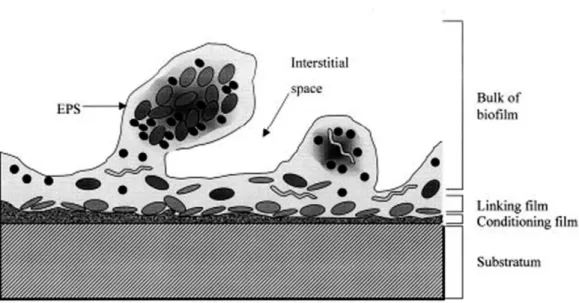
Once bacteria attach and accumulate on the devices surface, the biofilm begins to form a more dynamic and robust structure. The maturation consists on the generation of a slime glycocalyx to encase bacterial species linked in the surface, establishing a tree-dimensional structure with representative mushroom-like cells appearance nearby the fluid-filled channels (Dunne, 2002). The glycocalyx appears to increase the stability of the biofilm structure, thus influencing the BAIs treatment with antimicrobial agents and even blocking the host immune response (Vuong et al., 2004b; Patel et al., 2007). The channels represent an optimum hydrodynamic flow, intended to deliver all the conditions necessary to improve the growth potential, including nutrients, oxygen and also enable the removal of metabolic waste. Moreover, internal pH, carbon source and osmolarity likewise regulate the progression of biofilm maturation (Dunne, 2002). A mature biofilm is characterized by a set of layers: the main bulk, a linking film, a conditioning film and the surface where the bacterial species initially attached (Fig. 1.4) (Habash and Reid, 1999).
14
Figure 1.4 Mature biofilm structure representing the different layers (adapted from Habash and Reid,
1999).
Yao et al. (2005), and other authors, believed that the characteristics and behaviours of both planktonic cells and bacterial cells incorporated within the biofilm matrix were significantly distinct. Several factors evidenced this hypothesis: a) adjust their physiology to anaerobic metabolism, b) downregulate protein, and cell wall and DNA synthesis (Yao et al., 2005) and c) have spatial and temporal response according to their specific environment (Stewart and Franklin, 2008). In particular S. epidermidis cells living inside the biofilm bulk can be at four different physiological states (aerobic, anaerobic, dormant cells and dead cells) and these metabolic conditions may contribute for antibiotic resistant (Rani et al., 2007).
1.4.4. Disassembly
In the detachment stage, a dynamic equilibrium is achieved and individual cells or cells aggregates dissipate from the superior biofilm layer (bulk of biofilm) colonizing and inducing the establishment of a new biofilm in other sites or organs (Yao et al., 2005). Detachment of biofilm cells may implicate the degradation of biofilm extracellular matrix and some physiological modifications, to promote bacterial cells adaptation to the external conditions. In order to break through the biofilm matrix, S. epidermidis produce a group of extracellular enzymes, or surfactants, such as proteases and DNases (Boles and Horswill, 2011).
15
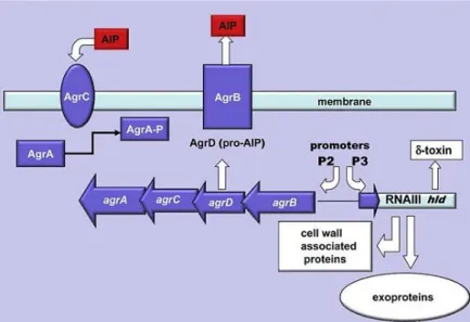
A regulatory system is necessary to control all the processes during the biofilm formation phases, including the production of enzymes, cell-cell communication and other mechanisms. The quorum sensing system is responsible for that regulation and is encoded by an accessory gene regulator (agr) system, which is activated during the transition from exponential to stationary phase, i.e., when the mature biofilm has achieved a state of equilibrium (Arciola et al., 2012). Agr gene contains two transcription units (RNAII and RNAIII) that are regulated by their specific promoters (P2 and P3) (Novick et al., 1993). The transcription unit RNAII comprises four genes: AgrA and AgrC, assembled forming a transmembrane transduction complex (Lina et al., 1998), AgrD, a pro-signalling peptide and AgrB, a membrane component, whose function consist on the exportation of post-translationally modified signalling peptide (Kong et al., 2006). RNAIII is also an effector molecule of agr system, controlling the transcription of target genes, such as virulence factors (Mayville et al., 1999; Thoendel et al., 2011). The activation of the quorum sensing agr system is obtained by a signalling molecule named autoinducing peptide (AIP), produced by AgrD, that is released from the biofilm to signalize the system (Kong et al., 2006). After reaching a critical threshold concentration, AIP activates a two-component signal transduction cascade (AgrC and then AgrA) promoting the production of virulent factors (Fig. 1.5) (Boles and Horswill, 2011).
The agr system in S. epidermidis enhances the biofilm detachment, spread and also contributes their virulence capacity by producing multiple-proteases and small forming toxins named as phenol modulins (PSMs) (Otto et al., 2004). PSMs are compounds of amphiphilic peptides, with inflammatory properties and are subdivided in different types: α-type peptides (≈20 amino acids; PSMα), β-type peptides (40-45 amino acids; PSMβ1, 2 and 3), δ-toxin (25 amino acids; PSMγ) and recently PSMδ (23 amino acids) (Otto et al., 2004; Otto, 2008). Curiously, under biofilm conditions, PSMβ expression is dominant in comparison with the other classes (Otto, 2008). PSMβ stimulates dissemination of cells by forming holes in biofilms. This will result in the modulation of the typical structure with cell towers and fluid-filled channels (Otto, 2008). Wang et al. (2011) also showed the role of PSMβ in S. epidermidis biofilm dissembling, in vitro, as well as the dissemination from colonized catheters, in vivo. Additionally, they tested the efficiency of antibodies against PSMβ to block bacterial dissemination from catheters, establishing a powerful tool to manipulate biofilms cells spread and subsequently reduce the incidence of BAIs (Wang et al., 2011). Relatively to δ-toxin, this PSM functions as a detergent that disrupts the biofilm polysaccharide matrix and has been hypothesized to have a role in necrotizing enterocolitis in neonates (Otto, 2009). δ-toxin also prevent hydrophobic interactions, among bacterial cells surfaces,
16
reducing the surface tension in biofilm interface, improving the cells detachment (McCann et al., 2008).
Figure 1.5 Quorum sensing agr system (adapted from Arciola et al., 2012).
1.5. Antimicrobial resistance
The resistance to antibiotics is the main challenge in infections associated with biofilm formation. The antibiotic resistance, acquired by bacteria, may be associated with some factors: a) the abuse of antibiotics, incorrect diagnosis or treatment and disobedience of the antibiotic therapy by patients (Otto, 2004); b) increase of immune-compromised patients, use of invasive procedures or devices, inappropriate disinfectants and default of practices to control diseases/infections in hospital environment (McCann et al., 2008). In the particular case of S. epidermidis, resistance to antimicrobial agents may be linked to several characteristics such as highly adaptive nature, inherit genetic variability, great recombination potential and the capacity to shift genetic material (Ziebuhr et al., 2006). These characteristics demonstrate its capacity to adapt new environments and to escape the antibiotic action, leading to advanced risks, inefficiency therapy and high mortality rates (Ziebuhr et al., 2006).
An important feature of biofilms is that the antimicrobial resistance is higher for bacterial cells within the biofilm, as compared with the planktonic population (Cerca et al., 2005). Antibiotics may eradicate the planktonic cells near surface or released from the biofilm, however no effect is obtained for biofilm
17
population regardless of the size of the antibiotic molecule (Cerca et al., 2005), allowing them to re-establish the biofilm and cause other infections. Subsequent treatments with antibiotics only reduce a minority of bacterial cells, enhancing these populations to be more resistant to antimicrobial agents (Ehrlich et al., 2004). Besides bacterial population and their diversity, additional factors can also contribute to high tolerance including: the diffusion barrier imparted by the exopolysaccharide matrix preventing some antibiotic penetration (Dufour et al., 2012), physiological conditions of biofilm-growing cells since growth conditions are different in the biofilm layers (Francolini and Donelli, 2010), induction of particular resistance mechanisms and/or the development of dormant persister cells (Dufour et al., 2012). Despite these tolerant factors, the antibiotic entrance may be facilitated through the disruption of one particular region of biofilm layers improving their efficiency against biofilm-mediated infections (Rani et al., 2007).
Many antibiotics are used to reduce the biofilms biomass as well as control S. epidermidis infections such as methicillin, rifamycin, quilonone, gentamycin, tetracycline, erythromycin, sulfonamides and glycopeptide antibiotics (Rogers et al., 2009). However, the most studied antimicrobial agent in nosocomial infections caused by S. epidermidis and other CoNS is methicillin. It is mediated by the mecA gene, located on a particular molecular vector, named as staphylococcal cassete chromosome mec (SCCmec), which encodes a penicillin-binding protein (Ziebuhr et al., 2006). Among most frequently isolated nosocomial pathogens 59.5% of S. epidermidis isolates are resistant to methicillin (Otto, 2008). Michelim et al. (2005) selected 98 S. epidermidis clinical isolates obtained from blood, catheters and other materials and evaluated their resistant to several antibiotics. The results revealed that 82.6% were resistant to gentamycin, 79.6% to erythromycin and 71.4% to ciprofloxaxin, excluding vancomycin that was vulnerable for all 98 isolates (Michelim et al., 2005). Oliveira and Cerca (2013) also assessed some antibiotic resistance in S. epidermidis (n=31) and other CoNS isolates (n=30). In particularly S. epidermidis demonstrated a higher rate of resistance for penicillin with 52%, following 48% to erythromycin, 42% gentamicin and 6% to ciprofloxacin. All isolates were also susceptible for vancomycin (Oliveira and Cerca, 2013).
1.6. Aims
S. epidermidis biofilms are involved on the majority of infections linked to implantation on medical devices. Among the different biofilm stages, biofilm attachment and accumulation are the most critical, allowing the production of a more stable and robust biofilm. While the role of the ica operon has been
18
extensively investigated, other biofilm forming factors are not fully explored. Therefore, the main objective of this work was to explore the role of three genes that have been considered the most important in S. epidermidis biofilm accumulation (icaA, aap and bhp). To determine the relative contribution of each gene in biofilm accumulation, biomass quantification and the 3D structure of the biofilms were related with the expression of those genes, overtime. Furthermore, to understand if the observed phenomenon was strictly present in clinical isolates, a commensal strain was used, in order to compare with the clinical isolate.
CHAPTER 2
21
2.1. S. epidermidis isolates
The S. epidermidis strains used in this study were either isolated from blood of patients with S. epidermidis infections (Laboratory of Microbiology of Santo Antonio General Hospital (HGSA), Oporto, Portugal) or from healthy volunteers (Oliveira, 2013).
2.2. Planktonic growth
Individual cells from each isolate were inoculated in a 10 mL tube with 1 mL of Tryptic Soy Broth (TSB, Liofilchem, Teramo, Italy) at 37 ºC with agitation at 120 rpm (ES-20 Shaker-Incubator, BioSan, Riga, Latvia) in order to obtain a starter culture. After reaching the exponential phase the pre-inoculums were diluted until the measured optical density (OD; 640 nm) was between 0,25 and 0,30 (approximately 2 x 10⁸ CFU/ml) (Cerca et al., 2004). The starter culture was diluted 1:100 in TSB supplemented with
0.4% (w/v) glucose (TSBG) in a 25 mL Erlenmeyer flask and incubated at 37º C with agitation at 120 rpm.
2.3. Biofilm formation
Bacterial biofilms were grown as previously described (Cerca et al., 2004). A starter culture was prepared and adjusted to the desired OD as described above. Subsequently, 10 µL of the started culture was inoculated in a 24-well microtiter plate (Orange Scientific, Braine-l’Alleud, Belgium) plus 990 µL of TSB, supplement with 0.4% of glucose (TSBG) to induce the biofilm formation. The cultures were grown for 12, 24, 36, 48, 60 and 72 h at 37 ºC on an orbital shaker at 120 rpm. At each 24 h, the growth medium was removed and exchanged by fresh TSBG. Biofilm formation assay was repeated no less than three times.
2.4. Biofilm Characterization 2.4.1. Biofilm quantification
Biofilm quantification for each time point was determined by OD measurement, as described before (Freitas et al., 2013). First the growth medium was removed and the biofilms were washed with 0.9% NaCl in order to remove all the detached cells. The biofilms were scraped and resuspended into 1 mL
22
of the same saline solution, following sonication at different time and amplitude: 10 seconds at 30%, 20 seconds at 30% and 40 seconds at 40%. After sonication, the biomass quantification of each biofilm was determined by measuring the OD at 640 nm. The cell suspensions were diluted with 0.9% of NaCl until the OD measurement was below 0.8 and the OD determination was calculated by multiplying the dilution factor by the OD measurement obtained.
2.5. Detection of biofilm-associated genes
The DNA extraction was performed by suspending five colonies of an overnight culture on Tryptic Soy Agar (TSA) plates, in 200 µL of ultrapure water. The suspension was heated at 100 ºC for 15 minutes in a thermal block, kept on ice for 5 minutes, in order to disrupt the cells, and then centrifuged at maximum speed (16100 g) for 10 minutes at 4 ºC. The supernatant was transferred to a new tube and stored at -20 ºC, until further use.
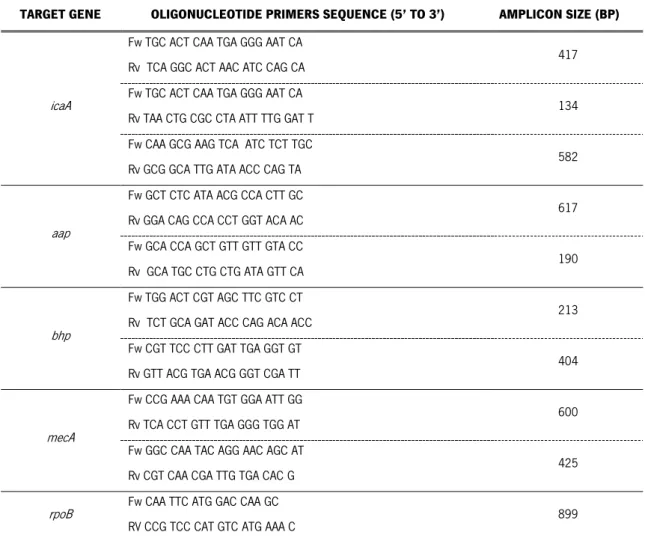
Table 2.1 Information of the primers.
TARGET GENE OLIGONUCLEOTIDE PRIMERS SEQUENCE (5’ TO 3’) AMPLICON SIZE (BP)
icaA
Fw TGC ACT CAA TGA GGG AAT CA
Rv TCA GGC ACT AAC ATC CAG CA 417
Fw TGC ACT CAA TGA GGG AAT CA
Rv TAA CTG CGC CTA ATT TTG GAT T 134
Fw CAA GCG AAG TCA ATC TCT TGC
Rv GCG GCA TTG ATA ACC CAG TA 582
aap
Fw GCT CTC ATA ACG CCA CTT GC
Rv GGA CAG CCA CCT GGT ACA AC 617
Fw GCA CCA GCT GTT GTT GTA CC
Rv GCA TGC CTG CTG ATA GTT CA 190
bhp
Fw TGG ACT CGT AGC TTC GTC CT
Rv TCT GCA GAT ACC CAG ACA ACC 213
Fw CGT TCC CTT GAT TGA GGT GT
Rv GTT ACG TGA ACG GGT CGA TT 404
mecA
Fw CCG AAA CAA TGT GGA ATT GG
Rv TCA CCT GTT TGA GGG TGG AT 600
Fw GGC CAA TAC AGG AAC AGC AT
Rv CGT CAA CGA TTG TGA CAC G 425
rpoB Fw CAA TTC ATG GAC CAA GC
RV CCG TCC CAT GTC ATG AAA C 899
23
PCR reactions were performed with a final volume of 10 µL according to the following conditions: 5 µL of DreamTaq Master Mix (x2) (Thermo Scientific, USA), 2 µL of ultrapure water, 1 µL of primer mixture (forward and reverse primers) and up to 1 µL of DNA template. PCR amplifications were obtained by using the MJ Mini thermal cycler (Bio-Rad, Hercules, CA, USA) with the following conditions: 94 ºC for 5 min, 35 cycles of 94 ºC for 30 s, 60 ºC for 30 s and 72 ºC for 45 s, and 72 ºC for 10 min. The information of the primers is described in Table 2.1. A positive control (S. epidermidis RP62A), a negative control (water) and an internal control (rpoB gene) were also included in each PCR run (Henriques et al., 2012).
Amplified products were analysed in 2% agarose gel stained with Midori Green DNA stain (Nippon Genetics Europe GmbH, Germany), for 45 minutes at 70 volts. Bromophenol blue (x6) (Fisher Scientific) was used as loading dye and NZYDNA Ladder V (Nzytech, Lisboa, Portugal) as molecular weight marker.
2.6. Quantification of gene expression
The protocols used for RNA extraction, DNase treatment, cDNA synthesis and real-time PCR assays were previously optimized for S. epidermidis biofilms (França et al., 2012).
2.6.1. RNA extraction
The RNA extraction protocol combines a mechanical (glass beads) and a chemical (phenol) lysis to improve the RNA extraction from the biofilms (França et al., 2011) along with a silica-membrane RNA isolation which minimizes the time required to extract the RNA (E.Z.N.A. Total RNA Kit, Omega Bio-Tek, GA, USA).
The bacterial biofilms were washed to remove all the detached cells, resuspended in 2 mL of 0.9% NaCl (pull of 10 biofilms) and directly stored on ice. The cell suspension was centrifuged at maximum speed (16100 g) for 10 minutes at 4 ºC. Once centrifuged the supernatant was discarded and the bacterial pellet was resuspended in 500 µL of TRK lysis buffer supplemented with β-mercaptoethanol plus 500 µL of phenol (Applichem, Darmstadt, Germany). The suspension was transferred into 2 mL safeLock tubes with 0.5 g of acid-washed glass beads (150 – 212 µm) (Sigma, USA) and placed into the FastPrep Cell disruptor (MP Biomedicals, BIOPORTUGAL, Portugal) for 35 seconds at 6.5 m/s.
24
Subsequently the samples were immediately placed on ice for 5 minutes. The cell disruption and the cooling step were repeated two more times.
Afterwards the samples were centrifuged at maximum speed (12300 g) for 1.5 minutes at 4 ºC. The samples was transferred for a 2 mL DNase/RNase free tube (avoiding the aspiration of glass beads) adding later an equal volume of 70% ethanol. The RNA solution was transferred into a RNA isolation column (including any precipitate), centrifuged at 12300 g for 30 seconds at room temperature (RT) and the flow-through was discarded. Columns were then washed with 500 µL of wash buffer I, centrifuged at 10000 g for 30 seconds at RT. The flow-through was discarded and the columns placed in the same collection tube. A second wash with 500 µL of wash buffer II, centrifuged at 10000 g for 30 seconds at RT. The flow-through was discarded and the columns placed in the same collection tube. The columns were washed once again with wash buffer II, centrifuged at 10000 g for 30 seconds at RT. The flow-through and the collection tubes were discarded and the columns placed into a new collection tube. In order to remove any trace of the wash buffer II which contains ethanol, the columns were centrifuged at 12300 g for 2 minutes at RT and then transferred into a 1.5 mL DNase/RNase free tube. Finally, to elute the RNA from the columns 50 µL of DEPC-treated were added into the centre of the column and then centrifuged at 16000 g for 2 minutes. The RNA tube was immediately placed on ice.
2.6.2. DNase treatment
Degradation of genomic DNA was achieved by adding 5 µL of DNase buffer and 2 µL of DNase I (Fermentas, Ontario, Canada) to each RNA sample. Samples were then incubated at 37 ºC for 30 minutes. DNase I activity was deactivated with 5 µL of 50 mM EDTA and incubation at 65 ºC for 10 minutes.
2.6.3. RNA quantification
The total RNA concentration and purity was determined by a NanoDrop 1000 (Thermo Scientific, USA). For each RNA sample, duplicate measurements were performed and averaged. The absorbance ratios A260/A280 and A260/A230 were considered to confirm potential protein and chemical contamination
25
2.6.4. cDNA synthesis
cDNA synthesis was performed following the protocol of the RevertAid First Strand cDNA Synthesis kit (Fermentas, Ontario, Canada). The same concentration of total RNA (250 ng) from each sample was reverse transcribed in a 10 µL reaction volume. A control reaction was performed under the same conditions but lacking the reverse transcriptase enzyme (no-RT control) to determine the possibility of genomic DNA residues.
2.6.5. Real-Time PCR
The quantification of biofilm gene expression was determined by quantitative real-time PCR (qPCR). qPCR analysis was performed using iQTM SYBR® Green Supermix (Bio-Rad, Hercules, CA, USA) in a 10
µL reaction tube. Each PCR reaction contained 2 µL diluted cDNA or no-RT control (1:200 in DEPC-treated water), 5 µL of master mix, 1 µL of primer mixture (10 µM of each forward and reverse primers), and 2 µL of nuclease-free water. The primer efficiency was determined by the dilution method. Information of the primers used is described in Table 2.2.
Table 2.2 Primers used in qPCR assays.
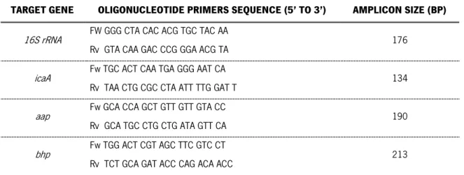
TARGET GENE OLIGONUCLEOTIDE PRIMERS SEQUENCE (5’ TO 3’) AMPLICON SIZE (BP)
16S rRNA FW GGG CTA CAC ACG TGC TAC AA
Rv GTA CAA GAC CCG GGA ACG TA 176 icaA Fw TGC ACT CAA TGA GGG AAT CA
Rv TAA CTG CGC CTA ATT TTG GAT T 134 aap Fw GCA CCA GCT GTT GTT GTA CC
Rv GCA TGC CTG CTG ATA GTT CA 190 bhp Fw TGG ACT CGT AGC TTC GTC CT
Rv TCT GCA GAT ACC CAG ACA ACC 213 bp – base pairs, Fw – forward, Rv – reverse
qPCR run was performed on a CFX96 (BioRad, Hercules, CA, USA) with the following cycle parameter: 94 ºC for 10 min, 39 cycles of 94 ºC for 15 s, 58 ºC for 20 s and 72 ºC for 25 s. qPCR products were analysed by melting curves to confirm the amplification of the desired product and detect possible unspecific products or primer dimer formation. Each experiment was performed in triplicate and a no template control (nuclease-free water) for each primer mixture was included to assess reagent
26
contamination. The expression of icaA, aap and bhp was normalised in relation to the housekeeping gene expression 16S rRNA through the n∆Ct method (Livak method), where n stands for the reaction
efficiency (n = 1.89 for icaA gene, n = 1.94 for aap gene and n = 1.96 for bhp gene) and ∆Ct = Ct16S rRNA
– Cttarget gene. The data analysis was based at least on 3 independent experiments.
2.7. Confocal laser scanning microscopy (CLSM)
Biofilms were prepared as described in the biofilm formation section although in a 6-well microtiter plate (Orange Scientific, Braine-l’Alleud, Belgium) with 20x20 mm cover glass (Labbox). Cultures were diluted 1:100 in fresh TSBG and incubated at 37 °C with shaking at 120 rpm for 12 and 48 h. At 48 h, the growth medium was replaced with an equal volume of fresh TSBG, every 24 h. After incubation time, the medium was removed andthe biofilms stained for confocal microscopy analysis.
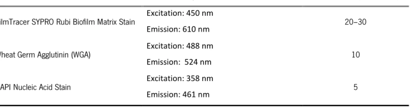
Biofilms staining was performed in the dark using three stains (Invitrogen, Table 2.3) to analyse the biofilm structure and composition. The DAPI stain was used to mark the cells by binds to the DNA; while SYPRO and WGA were used to detect the presence of proteins and polysaccharides, respectively. Two combinations were used: DAPI and WGA, SYPRO and WGA. Two independent biofilms of each isolate and time-point were analysed for DAPI+WGA while for SYPRO+WGA was only one biofilm at each time-point. Once the incubation time was completed the stain was removed and then washed with sterile water. The biofilm images were acquired in an OlympusTM FluoView FV1000 (Olympus, Lisboa,
Portugal) confocal scanning laser microscope. Biofilms were observed using a 60x water-immersion objective (60x/1.2 W).
Table 2.3 Information of the stains used in CLSM.
STAIN EXCITATION/EMISSION WAVELENGTHS INCUBATION (MINUTES)
FilmTracer SYPRO Rubi Biofilm Matrix Stain Excitation: 450 nm
Emission: 610 nm 20–30 Wheat Germ Agglutinin (WGA) Excitation: 488 nm
Emission: 524 nm 10
DAPI Nucleic Acid Stain Excitation: 358 nm
27
2.8. Statistical Analysis
Statistical significance of all results was determined by using Graphpad Prism version 6.02. Biomass quantification results of biofilm formation were analysed by two-way analysis of variance (ANOVA). The mean normalised gene expression was compared among isolates at different time-points by applying the student’s t test. All tests were performed with a confidence level of 95%.
CHAPTER 3
31
3.1. Isolates characterization
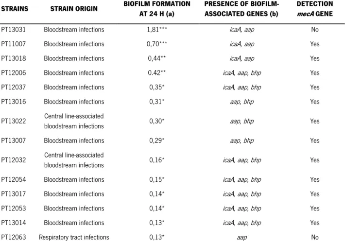
Clinical isolates were provided from the Laboratory of Microbiology of the Santo António General Hospital (Oporto, Portugal) from patients with Staphylococcus epidermidis infections. The isolates were streaked in plates with TSA growth medium and incubated at 37 ºC, until colonies were completely formed. After incubation, the colony morphology of all isolates was analysed and registered, such as colour, shape, elevation and pigment characteristics. Further characterization methods were performed to characterize the clinical isolates: a) quantification of the biofilm formation at 24 h of incubation, by OD measurement, b) presence of biofilm-associated genes, by PCR and c) presence of the methicillin gene (mecA), which is responsible for methicillin resistance, by PCR (Table 3.1).
Table 3.1 Characterization of S. epidermidis clinical strains.
STRAINS STRAIN ORIGIN BIOFILM FORMATION AT 24 H (a)
PRESENCE OF BIOFILM-ASSOCIATED GENES (b)
DETECTION
mecA GENE
PT13031 Bloodstream infections 1,81*** icaA, aap No PT11007 Bloodstream infections 0,70*** icaA, aap Yes PT13018 Bloodstream infections 0,44** icaA, aap Yes PT12006 Bloodstream infections 0.42** icaA, aap, bhp Yes PT12037 Bloodstream infections 0,35* icaA, aap, bhp Yes PT13016 Bloodstream infections 0,31* aap, bhp Yes PT13022 Central line-associated
bloodstream infections 0,30* aap, bhp Yes PT13007 Bloodstream infections 0,29* aap, bhp Yes PT12032 Central line-associated
bloodstream infections 0,16* icaA, aap, bhp Yes PT12054 Bloodstream infections 0,15* icaA, aap, bhp Yes PT13017 Bloodstream infections 0,14* icaA, aap, bhp Yes PT12053 Bloodstream infections 0,14* icaA, aap, bhp Yes PT13014 Bloodstream infections 0,13* icaA, aap, bhp Yes PT12063 Respiratory tract infections 0,13* aap No
(a) Biofilm formation was quantified by OD measurement. (b) The detection of the genes was determined by PCR using two independent primer sets. *** strong biofilm producer, ** moderate biofilm producer, * weak and no-biofilm producers.
32
According to our results, 71% of the clinical strains showed a weak biofilm formation, 14% were moderate biofilm producers and only 14% were strong biofilm producers. The aap and icaA genes were detected in all the clinical strains analysed. Regarding the bhp gene, it was detected in 71% of the clinical strains; however bhp gene was absent in the strong biofilm producers. Okee et al. (2012) analysed the presence of icaA, aap and bhp genes in 30 S. epidermidis clinical isolates, from which 70% of the isolates were icaA positives, 17% possessed the aap gene and only 10% had the bhp gene (Okee et al., 2012). Gad et al. (2009), characterized 35 S. epidermidis isolates from urinary tract catheterized patients regarding the capacity of biofilm formation and also the icaA gene presence. Their results demonstrated that 31 isolates were biofilms producers, from which 51,4% were strong biofilm producers and 37,1% were moderate biofilm producers. Additionally, all biofilms producers were positive for icaA gene, therefore enhancing that this gene is important in biofilm formation (Gad et al., 2009). Both authors had verified the absence of aap and bhp genes in most S. epidermidis isolates, although in our study, these genes were present in the majority of the strains. However, the role of these genes in biofilm formation of clinical strains is not yet fully understood.
Also important, the majority of the characterized strains are resistant to methicillin since approximately 86% revealed the presence of the mecA gene. Other authors verified the same high percentage of methicillin resistance in S. epidermidis clinical isolates, thus suggesting that its presence may be also related to the bacteria pathogenesis (Kitao et al., 2010; Iorio et al., 2011).
After the clinical strains characterization, one S. epidermidis strain was selected for further studies, taking into account the formation of a moderate biofilm and the presence of the three genes of interest, which are regarded as important in the biofilm accumulation stage.
Several studies have been performed to determine factors that may discriminate between invasive and commensal strains of S. epidermidis (Frebourg et al., 2000; Galdbart et al., 2000). The elucidation of the major differences between these two types of strains may help to better understand the S. epidermidis pathogenesis. Therefore, a S. epidermidis commensal strain was also selected from a previously characterized collection of CoNS (Oliveira, 2013) (appendix A), following the same considerations. Thus, S. epidermidis clinical strain PT12006 and the commensal strain SECOMO034.A1 were used in additional studies, in order to accomplish the objectives of this thesis.
33
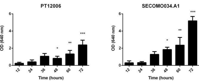
3.2. Biofilm formation
The biofilm formation, for both strains, was quantified by the measurement of the OD at 640 nm, for six timepoints: 12, 24, 36, 48, 60 and 72 h (Fig. 3.1). The TSB medium was supplemented with 0,4% of glucose and changed every 24 h. A sonication stage was applied, to reduce and eliminate the cell clusters present in the biofilms, improving the biofilm quantification by OD measurement, as previously described (Freitas et al., 2013).
Figure 3.1 Biofilm formation quantification determined by OD measurement of S. epidermidis PT12006 and SECOMO034.A1; * represents statistical significant differences between the strains at the different times of incubation (p < 0,05).
At an early stage (12-24 h) biofilm formation was not statistically different on both strains, although there was a notable statistical difference after 48 h. Biofilm formation of PT12006 seems to have a slight increase overtime, except at 72 h of incubation, which has almost double of the biomass. However, strain SECOMO034.A1 had significant differences only at a later stage of biofilm formation, demonstrating an evident twofold increase of biomass at 72 h in comparison with the 60 h incubation time.
In general, the commensal strain seems to have more biomass production, when compared with the clinical strain. However, this finding was only observed with the prolongation of the incubation time up to 72 h. According to our results, it is possible to conclude that screening assay for biofilm formation up to 24 h of incubation is not suitable to determine the real ability of a bacterial strain to form biofilm, not being able to distinguish between a strong or a moderate producer. On the other hand, increasing the incubation time allows a better discrimination of the biofilm formation ability.