Portugal
Teresa Maria Pereira Padrão Temudo
Dissertação de doutoramento em Ciências Médicas
Teresa Maria Pereira Padrão Temudo
Clinical and Genetic Study of Rett Syndrome in Portugal
Dissertação de Candidatura ao grau de Doutor em Ciências Médicas submetida
ao Instituto de Ciências Biomédicas de Abel Salazar
Universidade do Porto
Orientador - Professor Doutor António Jorge dos Santos Pereira de Sequeiros
Professor Catedrático do Instituto de Ciências Biomédicas de Abel Salazar,
Universidade do Porto
Co- orientadora - Prof. Doutora Patrícia Espinheira de Sá Maciel
Professora Auxiliar
De acordo com o disposto no nº 2 do artigo 8º do Decreto-lei nº 388/70, nesta dissertação
foram utilizados os resultados dos trabalhos publicados ou em preparação abaixo
indicados. No cumprimento do disposto no referido Decreto-lei, a autora desta
dissertação declara que interveio na concepção e execução dos trabalhos efectuados, na
interpretação dos resultados e na redacção dos resultados publicados ou submetidos
para publicação, sob o nome de Temudo T:
Based on the nº 2 of artigo 8º do Decreto-lei nº 388/70, in this dissertation were used
experimental results published or submitted to publication stated below. The author of this
dissertation declares that she participated in the planification and execution and data
interpretation of all the works stated below, under the name of Temudo T:
Temudo T., Maciel P., Rett’s syndrome. Clinical features and advances in genetics.
Rev de Neurol 2002, 34 (suppl.1): S54-8
T. Temudo. 2005. Movement Disorders in Rett Syndrome. Rev de Neurol 2005 ; 40
(suppl1): S167-71
Shi J, Shibayama A, Liu Q, Santos M, Temudo T, Maciel P, Sommer SS. Detection of
heterozygous deletions and duplications in the MECP2 gene in Rett syndrome by
Robust Dosage PCR (RD-PCR). Hum Mutat 2005;25:505-511
Temudo T, Maciel P (2005) “Síndrome de Rett. Características clínicas y avances
genéticos” In: Autismo Infantil. Eds : Fernando Mulas, Viguera Editores, S.L.,
Barcelona; pp 121-135
Temudo T (2007) “Movement Disorders in Rett Syndrome”. In : Movement Disorders
in Children: a clinical update with video recordings. Eds: Nardo Nardocci and Emilio
Fernandez-Alvarez. Editions John Libbey Eurotext, Montrouge; pp153-167
Temudo T, Oliveira P, Santos M J, Vieira J, Moreira A, Calado E, Carrilho I, Oliveira
G, Levy A, Barbot C, Fonseca M, Cabral A, Cabral P, Monteiro J, Borges L, Dias A,
Gomes R, Mira G, Barbosa C, Eusébio F, Santos M, Sequeiros J, Maciel P.
Stereotypies in Rett syndrome: analysis of 83 patients with and without detected
vi
Ramaekers VT, Sequeira JM, Artuch R, Blau N, Temudo T, Ormazabal A, Pineda M,
Aracil A, Roelens F, Laccone F, Quadros EV.Folate receptor autoantibodies and
spinal fluid 5-methyltetrahydrofolate deficiency in Rett syndrome. Neuropediatrics
2007; 38:179-83
Coutinho AM, Oliveira G, Katz C, Feng J, Yan J, Yang C, Marques C, Ataide A, Miguel
TS, Borges L, Almeida J, Correia C, Currais A, Bento C, Mota-Vieira L, Temudo T,
Santos M, Maciel P, Sommer SS, Vicente AM. MECP2 coding sequence and 3'UTR
variation in 172 unrelated autistic patients.Am J Med Genet B Neuropsychiatr Genet
2007;144:475-83
Temudo T, Maciel P, Sequeiros J. Abnormal movements in Rett syndrome are present
before the regression period: A case study. Mov Disord 2007;22:2284-2287
Temudo T, Freitas P, Sequeiros J, Maciel P, Oliveira G. Atypical stereotypies and
vocal tics in Rett syndrome: an illustrative case. 2008. Movement Disorders (in press)
Temudo T, Rios M, Prior C, Carrilho I, Santos M, Fonseca MJ, Monteiro JP, Sequeiros
J, Maciel P, Ormanbazal A, Artuch R. Is it worth to evaluate CSF neurotransmitters
and folate in Rett disorder? 2007 (submitted)
Temudo T, Santos M, Ramos E, Dias K, Vieira JP, Moreira A, Calado E, Carrilho I,
Oliveira G, Levy A, Barbot C, Fonseca M, Cabral A, Cabral P, Monteiro J, Borges L,
Gomes R, Mira G, Pereira AS, Santos M, Fernandes A, Epplen JT, Sequeiros J,
Maciel P . Rett Syndrome and Rett disorder: an attempt to redefine phenotypes, 2007
(submitted)
Prior C, Nunes A, Rios M, Sequeiros J, Maciel P, Gomes L, Temudo T. Nutritional and
gastrointestinal dysfunction in Rett disorder: importance of early intervention, 2007
(submitted)
Santos M, Temudo T, Kay T, Carrilho I, Gaspar I, Barbot C, Medeira A, Cabral H,
Gomes R, Lourenço MT, Venâncio M, Calado E, Moreira A, Oliveira G, Maciel P.
Mutations in the MECP2 are not a major cause of Rett-like phenotype in males
(submitted)
Santos M, Jin Yan, TemudoT, Jinong F, Sommer S, Maciel P. Analysis oh higly
conserved regions of the 3’UTR of the MECP2 gene in patients with clinical diagnosis
viii
Acknowledgments
To my family (including Jordi, of course), for unconditional love and support
To Jorge Sequeiros who “pruned” and improved the quality of this work
To Patricia Maciel who aided to concretize this project, shared my enthusiasm with the
results and incited me to divulge it to the world…
To the Portuguese Association of Rett Syndrome, in particular to Sandra Madeira, for
all her collaboration
To my Rett syndrome patients
To Dr Karin Dias who gave me the opportunity of study the Rett Syndrome
To Emilio Fernandez-Alvarez my first movement disorders teacher
To Isabelle Rapin who invited me to do a project
To Sonia Figueiroa, my colleague and friend, for her loyalty
To Cidália Oliveira, for her technical support
To Cristina Garrido, Susana Tavares, Felisbela Rocha, Sandra Ramos, Cláudia
Pedrosa, Catarina Prior, Rui Almeida, Carla Zilhão, now young Pediatricians, who
worked with me during their fellowship in Pediatric Neurology - I learned a lot trying to
Preceitos legais
VAcknowledgments
viiiResumo
1Abstract
3Resumée
5Preface
71. GENERAL INTRODUCTION 9
1.1. History of the disease 11
1.2. Clinical, neuropathological and genetic characteristics of Rett syndrome
13
1.2.1. Clinical overview 13
1.2.2. Genetics 20
1.2.3. Neuropathological studies 23
1.2.4 Phenotype-genotype correlation studies 25
1.2.5 Main objectives of the study 26
2. GENERAL SUBJECTS AND METHODS 39
2.1. Strategy for identification of the cases 41
2.2. Clinical study of the patients 42
2.2.1. Clinical database 43
2.2.2. Scoring and scales 43
2.2.3. Evaluation by other medical specialities 44
2.3. Genetic study 44
2.4. Other blood analyses 44
2.5. Study of LCR 45
2.6. Statistical analysis 45
3. RESULTS 47
3.1. Redefining phenotypes and genotypic correlations 49
3.1.1. Introduction 51
3.1.2. Rett syndrome and Rett disorder: an attempt to redefine the
phenotypes (Article 1)
53
3.1.3. Nutritional and gastrointestinal dysfunction in Rett
disorder-importance of early evaluation and intervention (Article 2)
73
3.1.4. Mutations in the MECP2 gene are not a major cause of Rett-like
phenotype in male patients (Article 3)
xii
3.2. Movement disorders in Rett syndrome and Rett disorder 107
3.2.1. Introduction 109
3.2.2. Movement disorders in Rett syndrome (Article 4) 111
3.2.3. Movement disorders in Rett syndrome: analysis of 60 patients with
detected MECP2 mutation
(Article 5)
113
3.2.4. Stereotypies in Rett syndrome: analysis of 83 patients with and
without detected MECP2 mutations (Article 6)
131
3.2.5. Abnormal movements in Rett syndrome are present before the
regression period: a case study (Article 7)
133
3.3. Biochemical studies in Rett syndrome and Rett disorder 135
3.3.1. Introduction 137
3.3.2. Is it worth to evaluate CSF neurotransmitters and folate in Rett
disorder? (Article 8)
139
3.3.3. Folate receptor autoantibodies and cerebral folate deficiency
in Rett syndrome (Article 9)
155
3.4. Rett syndrome and autism 157
3.4.1. Introduction 159
3.4.2. Clinical overlap between Rett syndrome and autism: an illustrative
case (Article 10)
161
3.4.3. MECP2 coding sequence and 3'UTR variation in 172 unrelated
autistic patients (Article 11)
173
4. GENERAL DISCUSSION 175
4.1. The clinical picture and differential diagnoses 177
4.2. Explaining the symptoms 177
4.3. Neurodegenerative or neurodevelopmental disorder? 180
4.4. Does a genotype predict the phenotype? 181
4.5. Diagnostic criteria of Rett syndrome: are they useful? 182
4.6. Movement: the signature of Rett disorder? 185
4.7. What were the major contribuitions of this work? 186
5. ONGOING WORK AND PERSPECTIVES FOR FUTURE
RESEARCH
191
5.1. Ongoing work 193
5.1.1 Stereotypies in Rett syndrome and cognitively impaired children with
autism
193
5.1.2. CNS magnetic resonance volumetric studies in patients with Rett
disorder and idiopathic autism
5.2.2. Motion analysis and quantification of hand stereotypies in Rett
syndrome patients with and without detected mutations in the MECP2 gene
198
6. ATTACHMENTS 201
6.1. Clinical database and scales 203
6.2. Other articles published (not included in the previous chapters) 211
6.2.1 Rett’s syndrome. Clinical features and advances in genetics
(Article 12)
213
6.2.2 Movement Disorders in Rett Syndrome (Article 13) 215
6.2.3 Detection of heterozygous deletions and duplications in the MECP2
gene in Rett syndrome by Robust Dosage PCR (RD-PCR) (Article 14)
A síndrome de Rett (RTT) é uma doença neurológica do desenvolvimento e a segunda
causa de atraso mental grave no sexo feminino; é causada, na maioria dos casos, por
mutações de novo num gene situado no cromossoma X que codifica para a proteína de
ligação às metil-CpG (MECP2). Foram encontradas mutações neste gene em mais de
90% dos probandos com a forma clássica de RTT. Os estudos de correlação
genótipo-fenótipo na RTT clássica sugerem que o tipo de mutação e o padrão de inactivação do X
têm um efeito na determinação da apresentação clínica e gravidade da doença.
Os objectivos da tese foram: (1) identificar um grande número de novos casos de RTT,
em Portugal; (2) fazer o estudo genético molecular em doentes portugueses com RTT
clássico e não-clássico, e identificar a frequência e tipo de mutações do MECP2; (3)
estudar e classificar clinicamente os doentes identificados e, em particular, as alterações
do movimento; (4) comparar doentes com e sem mutação identificada; (5) estudar
correlações genótipo-fenótipo em doentes com mutação identificada; (6) estudar o papel
dos neurotransmissores e folatos nos doentes com mutação identificada; e (7) pesquisar
mutações no gene MECP2 em doentes com outras patologias.
Com o objectivo de identificar o maior número possível de casos, contactámos todos os
serviços de neuropediatria portugueses, de forma a identificarmos os doentes já
diagnosticados; fizemos palestras de divulgação nos serviços de pediatria do Norte e nas
reuniões anuais de pediatria, neuropediatria, neurologia e genética. Incentivámos a
formação da Associação de RTT em Portugal, com quem mantivemos contacto estreito.
Observámos e filmámos as crianças com suspeita de RTT nos hospitais onde foram
sinalizadas. As doentes observadas no HGSA foram seguidas por uma equipa
multidisciplinar (neuropediatra, gastroenterologista, nutricionista, ortopedista e fisiatra).
Estabelecemos um banco de DNA para todos os casos identificados. Analisámos as
sequências codificantes do gene MECP2 em todos os casos de RTT e em doentes com
patologias afins. Efectuámos também estudos bioquímicos no sangue e LCR.
Identificámos 88 casos com RTT em Portugal, tendo sido identificada mutação no gene
MECP2 em 60: 9 mutações ainda não estavam descritas.
Definimos como portadores de doença de Rett (RD) ou síndroma de Rett (RTT) os
2
Comparámos as características clínicas destes dois grupos de doentes, de forma a
rede-finir o perfil da doença e a seleccionar melhor, no futuro, os candidatos a estudo genético.
Estudámos a correlação genótipo-fenótipo e individualizámos 3 tipos clínicos de RD.
Estudámos, em particular, as alterações de movimento dos doentes com RD e RTT e
descrevemos vários sinais neurológicos que não tinham sido previamente descritos.
En-contrámos diferenças em relação à marcha independente, distonia e tipo de tremor,
quando comparámos doentes com mutações missense ou truncante; encontrámos
dife-renças em relação ao aparecimento, tipo e gravidade das alterações de movimento com
o tempo de evolução da doença nos dois grupos. Documentámos em vídeo que os
mo-vimentos anormais estão presentes na RD antes da fase de regressão da doença.
Ana-lisámos os metabolitos da dopamina, serotonina e folato no LCR e efectuámos uma
avaliação e intervenção gastrointestinal e nutricional em 25 doentes com RD.
Concluímos que um período de regressão nítido e a presença de 3 ou mais estereotipias
diferentes, rigidez e marcha atáxica-rígida são úteis na distinção entre RD e RTT.
Doentes com mutações truncantes diferem daqueles com mutações missense em relação
à aquisição de linguagem e marcha autónoma, antes do início da doença, e microcefalia,
crescimento, tamanho do pé, distonia e gravidade global, à data de observação. Os
doentes com mutação R168X apresentavam fenótipo mais grave e os portadores da
mutação R133C o menos grave. Doentes com mutação R294X tinham um
comportamen-to hiperactivo e maior número de estereotipias; aqueles com a mutação T158M pareciam
ser particularmente atáxicos e rígidos.
As alterações de movimento são a “assinatura” da doença de Rett, sendo fundamentais
para fazer o diagnóstico diferencial entre RD e RTT; para além disso, parecem reflectir a
gravidade e progressão da doença. Doentes com mutações truncantes têm mais vezes e
com maior gravidade distonia e síndrome rígido-acinético, quando comparados com
doentes com mutação missense e o mesmo tempo de evolução da doença.
Os nossos resultados apoiaram a hipótese de o folato não ter um papel na patogénese
da DR, mas reforçaram a opinião de os neurotransmissores terem um papel importante.
Os resultados da avaliação gastrointestinal demonstraram a necessidade de equipas
multidisciplinares no tratamento das doentes com RD.
Propusemos ainda a revisão dos critérios de diagnóstico de RTT clássico e a abolição
Rett syndrome (RTT) is a neurodevelopmental disorder, caused in most cases by
muta-tions in the methyl-CpG binding protein 2 (MeCP2) gene (MECP2), wich mapps to Xq28.
It is considered the second most frequent cause of severe mental retardation in females.
The reported detection rate for MECP2 mutations in patients with clinically defined RTT is
60 to 95%, depending on the clinical parameters used to select the patients. Larger
stu-dies point to some correlation between genotype and phenotype. Another source of
phenotypic variability seems to be X chromosome inactivation.
The aims of this thesis were: (1) to identify a large number of patients with RTT in
Portugal and to study its phenotypic variability; (2) to analyze the entire coding sequence
of MECP2 in all patients and identify the most frequent mutations in our population; (3)to
study in detail and classify clinically the patients identified, specially their movement
disorders; (4) to compare patients with and without identified mutations; (5) to analyze
genotype-phenotype correlation; (6) to study the role of cerebral folate and
neurotransmitters in the physiopathology of the disease and (7) to study MECP2
mutations in patients with diseases clinically overlapping Rett syndrome.
All paediatric neurology services were asked to refer their RTT patients, and conferences
supported by videos were presented at the paediatric services of the North of Portugal
and at the meetings of the national societies of paediatrics and neurology. A contribution
was made toward organizing the Portuguese Rett Association.
The clinical study was based on all the information collected between 1996 and 2007. All
patients referred by paediatric neurologists, neurologists or paediatricians were personally
examined, and their fulfilment of the revised clinical criteria for RTT was reviewed.
All patients were observed and videotaped at the hospitals where they had been
diagno-sed. Patients followed at HGSA were also observed by a multidisciplinary team.
All coding sequences and the exon-intron boundaries of MECP2 were analyzed in all
cases, as well as in patients with clinical overlap with Rett syndrome. We also performed
biochemical studies in blood and CSF, in 25 RTT patients with a MECP2 mutation.
We studied 88 cases of RTT; MECP2 mutations were identified in 60 (9 were novel).
Based on the presence or absence of a MECP2 mutation, we defined two groups: patients
with Rett disorder (RD) and with Rett syndrome (RTT). We compared these clinically, in
ana-4
lysis. We also studied in detail the phenotype-genotype correlation in RD and identified
three clinical presentations. Movement disorders were analyzed in detail in RD and RTT;
several neurological signs were described by the first time. Folate and neurotransmitters
were analyzed in CSF and gastro-intestinal dysfunction was assessed in 25 RD patients.
We concluded that a clear regressive period (with loss of prehension and language, and
deceleration of growth) and the presence of more than three stereotypies, rigidity and
ataxic-rigid gait seemed to be helpful in differentiating RD from RTT.
Patients with truncating differed from those with missense mutations, regarding acquisition
of propositive words and independent gait before the onset of disease, and microcephaly,
growth, foot length, dystonia and severity score, at the time of observation. Patients with
the R168X mutation had a more severe phenotype, whereas those with R133C showed a
less severe one. Patients with R294X had hyperactive behaviour, while those with T158M
seemed to be particularly ataxic and rigid.
Movement anomalies are the hallmark of RD and are essential to differentiate RD from
RTT; furthermore, they seem to reflect severity and rate of progression of RD. Patients
with truncating mutations presented more frequent and severe dystonia and rigid-akinetic
syndrome, when comparing patients with similar time of disease evolution.
Folate deficit is not contributing to the pathogenesis of RD. In contrast, we report discrete
and novel neurotransmitter anomalies, highlighting the need for further studies of CSF
neurotransmitters, in clinically and genetically well characterized patients.
Management of Rett disorder requires a multidisciplinary team, including
gastroenterolo-gists. Individually designed feeding strategies are essential to achieve a good nutritional
status. Early identification of nutritional and gastrointestinal disturbances and their correct
management contributes to improve quality of life in these patients.
We suggested, based on the clinical experience acquired, that the international diagnostic
criteria should be revised and defined in a stage-dependent manner, according to the
cli-nical signs and symptoms present at the different stages of the disease, in a large series
of patients with MECP2 mutations. We also suggested that the only variant form that may
need to be considered is the male form, as the others do not differ from the classical
La syndrome de Rett (RTT) est une maladie neurologique du développement et la
deuxième cause de retard mental chez le sexe féminin; elle est causée, dans la plupart
des cas, par des mutations de novo chez un gène situé au chromosome X, qui codifie
pour la protéine de liaison aux methyl-CpG (MECP2). Des mutations dans ce gène ont été
observées chez plus de 90% des sujets avec la forme classique de RTT. Les études de
corrélation genotype-phenotype chez la RTT classique suggèrent que le type de mutation
et le patron d'inactivation du chromosome X ont un effet sur la détermination de la
présentation clinique et sur la sévérité de la maladie.
Les objectifs de cette thèse ont été: (1) d’identifier un grand nombre de nouveaux cas de
RTT au Portugal; (2) de faire l’étude génétique moléculaire de malades portugais(es)
avec RTT classique et non-classique, et identifier la fréquence et le type de mutations du
gène MECP2; (3) d’étudier et classifier cliniquement les malades identifiés et, en
particulier, ses altérations du mouvement; (4) de comparer des malades avec et sans
mutation identifiée; (5) d’étudier des corrélations génotype-phénotype; (6) d’étudier le rôle
des neurotransmetteurs et des folates chez les malades avec mutation identifiée; et (7) de
chercher des mutations au gène MECP2 chez de malades avec d’autres pathologies.
Avec le but d’identifier le plus grand nombre de cas possible, nous avons contacté tous
les services de neuropédiatrie portugais; nous avons fait des exposés sur la maladie dans
plusieurs services de pédiatrie du nord du pays et dans les réunions annuelles de
pédiatrie, neuropédiatrie, neurologie et génétique nationales. Nous avons stimulé la
formation de l’Association de RTT au Portugal, avec qui nous avons maintenu un contact
proche. Nous avons observé et filmé les enfants avec soupçon clinique de RTT, dans les
hôpitaux où ils ont été identifiés. Les malades observés à L’HGSA ont été suivis par une
équipe multidisciplinaire. Nous avons établi une banque d’ADN pour tous les cas
identifiés. Les séquences codifiantes du gène MECP2 ont été analysées en tous les cas
de RTT et chez des malades avec des pathologies similaires. Des études biochimiques
du sang et LCR ont été faits.
Nous avons identifié 88 cas avec RTT au Portugal, et une mutation au gène MECP2 a été
identifié en 60: 9 de ces mutations n’avaient jamais été décrites. Nous avons défini
comme porteurs de la maladie de Rett (RD) ou syndrome de Rett (RTT) les malades avec
6
Nous avons comparé les caractéristiques cliniques de ces deux groupes de malades, de
façon à redéfinir le profil de la maladie et ainsi mieux sélectionner, au futur, les candidats
à une étude génétique. On a étudié la corrélation génotype-phénotype et on a
individualisé 3 types cliniques de RD. On a étudié en particulier les altérations de
mouvement des malades avec RD et RTT et on a décrit plusieurs signes neurologiques
qui n’avaient pas été préalablement décrits. Des différences ont été observées en ce qui
concerne la marche indépendante, dystonie et type de tremblement, par comparaison
entre patients avec des mutations missense et troncantes; on a observé des différences
en ce qui concerne l’apparition, le type et la sévérité des altérations du mouvement avec
le temps d’évolution des deux groupes. On a documenté en vidéo que les mouvements
anormaux sont présents dans la RD avant la phase de régression de la maladie. Les
métabolites de la dopamine, sérotonine et folate ont été analysés par LCR et une
évaluation et intervention gastro-intestinale et nutritionnelle ont été faites en 25 malades
avec RD. Nous avons conclu qu’une période de nette régression et la présence de 3 ou
plus stéréotypies différentes, la rigidité et la marche ataxique-rigide sont utiles pour la
distinction entre RD et RTT.
Les malades avec des mutations troncantes diffèrent de ceux avec mutations missense
en ce qui concerne l'acquisition du langage et de la marche autonome, avant le début de
la maladie ; ainsi que la microcéphalie, le grandissement, la taille du pied, la dystonie et la
sévérité globale, à la date de l’observation. Les malades avec la mutation R 1 6 8 X
présentaient un phénotype plus grave et les porteurs de la mutation R133C le moins
grave. Les malades avec la mutation R294X avaient un comportement hyperactif et un
nombre plus grand de stéréotypies; ceux avec la mutation T158M paraissaient
particulièrement ataxiques et rigides.
Les altérations du mouvement sont la « signature » de la maladie de Rett, et sont
fondamentales pour le diagnostique différentiel entre RD et RTT ; en plus, elles paraissent
réflecter la sévérité et la progression de la maladie.
Nos résultats ont appuyé l’hypothèse selon laquelle le folate n’a aucun rôle chez la
pathogénèse de la RD, mais ont renforcé l'idée de que les neurotransmetteurs y jouent un
rôle important.
Les résultats de l’évaluation gastro-intestinale ont indiqué le besoin d'équipes
multidisciplinaires pour le traitement des malades avec RD.
Nous avons aussi proposé la révision des critères de diagnostique de RTT classique et
Maybe those who will read this thesis will ask why I became interested by this particular
disorder.
Our life is commanded by conscious and unconscious decisions, and … by chance. What
we do with our lives depends in a big part on the ability to seize new opportunities.
In 1996, when I had recently finished my training in Neuropediatrics, Karin Dias decided to
organize a Pediatric Neurology meeting in Lisbon, and invited Bent Hagberg to give a
conference on Rett syndrome. With the goal of doing a presentation about Rett cases in
Portugal, she proposed me to make a questionnaire and send it to all the Paediatric
neurology centres. At that time I collected 50 Rett syndrome cases from all the country. I
studied all the scientific material and videotaped some girls with the disease.
As I presented my work in the morning and Bent Hagberg would present his conference
only in the afternoon, I begun my lecture with a video of a typical Rett syndrome girl,
followed by a very exhaustive introduction of the syndrome and, only after that, I showed
the data I collected on the Portuguese Rett syndrome patients. I was so excited with this
disorder that I exceeded the time scheduled for my presentation. Karin Dias was not very
happy, as I didn’t leave much for Bent Hagberg to tell about Rett syndrome… But, being a
great person as he is, he understood my naive enthusiasm and, in an elegant way, he
congratulated me by saying that, as I did most of his conference, it would then be easier
for him to talk only on some particularities of the syndrome… One week later, I received a
letter from him with some papers he wrote on RTT. He also encouraged me to continue…
In 2000, after the discovery of the MECP2 gene mutation as the cause of Rett syndrome, I
decided to talk with Jorge Sequeiros about the possibilities of doing the genetic study of
the Portuguese RTT girls. Some months later, he contacted me because he had a young
PhD student in his group who became interested in that work. In March of 2001, we
decided to ask for a grant to Fundação para a Ciência e Tecnologia and, in December of
1.1. History of the disease
“One day in the spring of 1965, two mothers holding their children on their laps were sitting in the waiting room. Both children were swaying and the mothers held their arms. Both children, who were treated for epileptic seizures, were well known to me. That morning I passed by them repeatedly and, incidentally, the mothers let go of their children’s arms. At once, the children put their hands together and started nearly identically-looking washing movements. I asked the mothers not to stop these movements again, and was startled at the similarities. It was the same gaze, same facial expression, same weak muscles and the same stereotyped movements of their hands. Moreover, they were both females. At first, I was amazed by this enormous coincidence and thought I must have came across several other children with these symptoms. I asked my head nurse to look at these children and she immediately recalled some more names of children who behave similarly. We invited them, placed them next to each other and found that all of them showed the same kinds of movements. Further, all patients were female.”
(Andreas Rett,
unpublished letter to the Rett Syndrome Association, June 7, 1985).
The original publication of Rett’s observations, which characterizes the
neuro-developmental disorder that now bears his name, appeared in 1966 in the Austrian
medical newsletter, Wiener Medizinisch Wochenshrift (1)
This clinical paper, like most of Andreas Rett’s publications was not widely circulated and
remained unnoticed to the English-speaking world. Realizing the limitations of publishing
in the German literature alone and having become convinced that this syndrome was a
different entity, he prepared a film and a booklet, “A special kind of atrophy”, carrying it to
pediatric meetings throughout Europe. The first English description of Rett syndrome
appeared in the Handbook of Clinical Neurology, in 1977 (2).
In 1978, independently from Rett, Ishikawa and collaborators from Japan described, in a
brief note, three girls with the same clinical symptoms and signs described by Rett (3).
This publication also passed unnoticed.
In 1980, Karin Dias, a Portuguese paediatric neurologist invited Bent Hagberg to give a
conference about cerebral palsy - a subject that was the main interest of this Swedish
paediatric neurologist. During the coffee-break of the meeting, they walked along the
gardens surrounding the conference building. Physicians go to congresses with the
General introduction
12
unlike children, they are looking for repeated “stickers” and, when they find them, they
describe a “new” disease. This was what happened then.
Karin (not knowing yet Rett’s chapter in the Handbook of Clinical Neurology) asked Bent
Hagberg if he had ever seen a special type of girls with cerebral palsy, who had the
particularity of continuous hand stereotypies. She had four cases, all similar. At that time,
he answered that he had seen that clinical picture in both sexes, but as Karin insisted that
only girls seemed to have such symptoms, he promised her that when he returned to
Sweden he would check their record files. Some time later, he wrote her a letter
confirming that he found only females with the above mentioned characteristics, and he
presented the clinical data of 16 girls, in 1980, at a Child Neurology meeting, in
Manchester.
Jean Aicardi, a famous French paediatric neurologist was in that meeting and he told
Hagberg that he also had several girls with the same clinical findings. In November 1983,
a paper was wrote, with 35 girls diagnosed as “Morbus Vesslan” (the nickname of the first
Rett syndrome girl observed by Hagberg: Anne-Marie Vesslund) in Sweeden, Portugal
and France.
It was a collaborator of Jean Aicardi, consulting one volume of the Handbook of Clinical
Neurology (2), who noticed that the “Morbus Vessland” had already been described by
Andreas Rett. Immediately, they changed the title’s name of the publication of this new
entity to “Rett syndrome”.
The paper was published in a well-known neurological journal (4), and the diagnostic
criteria for the syndrome were established at the second international Rett syndrome
conference, in Vienna. While only three medical papers had been published, between
1961 and 1983, almost 300 appeared in the eight years after the publication in the Annals
of Neurology. Rett syndrome (RTT) was recognized all over the world, seen in all ethnic
groups, and known to appear with an ever widening pattern of clinical variability.
Meanwhile, in the fall of 1983 a young woman called Houda Zoghbi was doing a
fellowship in paediatric neurology, in Houston. She had read the first account of Rett
syndrome in an American medical journal (4); within 2 weeks, she encountered two girls
who had the classical symptoms of the disorder. Two years after first encountering
patients with this disorder, Zoghbi sidelined her clinical career and formally trained as a
researcher in Genetics in the hope of understanding the causes of diseases like Rett
syndrome. In 1999, more than 30 years after the first clinical description, she discovered
1.2. Clinical, genetic and neuropathological characteristics of Rett syndrome
1.2.1. Clinical overview
Rett syndrome (RTT) occurs worldwide, in all populations and ethnic groups, with an
incidence between 1 in 10000 and 1 in 20000 female live births (6), and is considered the
second most frequent cause of severe mental retardation in females (7).
The unique clinical profile of this syndrome was well delineated by Hagberg et al., in 1986
(8). RTT affects almost exclusively girls, with an apparently normal initial psychomotor
development, during the first six months of life. Gestation and birth of the classic RTT
infant are usually uneventful. Head circumference is normal at birth but, deceleration of
head growth occurs later, approximately between 5 months and 4 years of age.
During the latter half of the first year, the first psychomotor anomalies appear, with a
non-specific slowing down in development. In particular, the crawling ability is seldom
achieved, and unsupported walk is usually delayed. Some patients never reach the stage
of independent gait.
After this slow development stagnation occurs, later with loss (usually by the first or
second year of life) of acquired fine motor, intellectual and communication abilities.
Though a period of days, weeks or months, affected children usually loose interest in their
surroundings, loose their ability to speak, manipulate objects and play, and repetitive
movements with the hands are first noticed by their families.
Also, behaviour disturbances can occur, as unexpected screaming attacks, laughing
spells and nocturnal insomnia. Through the following months of the regression phase,
hand stereotypies become more frequent, and each girl develops her own personal type
of movements (washing, clapping, wringing or tapping the hands). These stereotypies are
considered the signature of this syndrome.
The regression period, with more or less apparent autistic traits, lasts for months or years.
Following this phase – usually about 3 to 5 years later – most girls improve social contact
and a few regain some of their original abilities, as the use of words, that had been
acquired before the regression phase. Although profoundly mentally retarded, they usually
have a particularly good eye contact.
At this phase, the combination of a previous personal history of developmental regression
General introduction
14
stereotypies, inability to use the hands and a particularly good eye gaze make the
diagnosis of RTT obvious (8).
Bruxism, breathing dysfunction and peripheral vasomotor disturbances (9) are other
characteristic signs; 85 to 90% of these patients have growth impairment. Seizures
usually start during this stage, and the majority has epilepsy (9). Although considered a
stationary period of the disease, neuromotor functions slowly, but steadily, decline in most
patients: scoliosis, ataxia, limb wasting, dystonia, pyramidal signs, bradikinesia and rigidity
may appear, after a variable time of evolution; however, 15 to 20% of these patients
remain in an ambulatory stage until their middle age (7). Some RTT patients may never
learn to walk, as they have a severe neurological impairment since the beginning of the
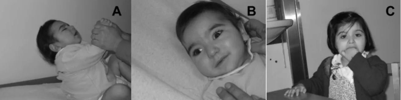
Figure 1. Clinical characteristics of patients with Rett syndrome with identified MECP2 mutation
A - C : intense eye contact in three children; D: the same visual attention in an adult patient; E: abdominal
distension, as a consequence of respiratory abnormalities; F - G: severe scoliosis; H: severe denutrition, in a
14 years old patient; I - K: small feet length in three adult patients; L: small hand length in an adult patient
The last stage (late motor deterioration stage) is exclusive of deteriorating girls, previous
walkers, who have lost that capacity, or of those who never acquired independent gait and
are older than ten years. At this stage, the frequency of hand stereotypies is usually lower,
in spite of the maintenance of the same pattern of movement; patients present wasting,
dystonia, and, in the older ones, Parkinsonian signs (hypomimia, bradikinesia, rigidity,
General introduction
16
Scoliosis is present in more than 50% of patients, and worsens with age, whereas
epilepsy (present in more than 70%) and breath dysfunction usually become less
disabling with disease evolution. Peripheral vasomotor disturbances, hypotrophic small
and cold feet and growth retardation are the rule.
Figure 2. RTT disease profile: a regression stage followed by a recovery of interaction contrasting with slow neuromotor regression
PMD: psychomotor development
The almost disappearance of stereotypies in some patients at this stage is probably the
cause of the under-diagnosis of RTT in adulthood, when the majority are
wheelchair-dependent. At this stage, only a good clinical history can confirm the suspicion of this
disorder. Although severely neurologically impaired, these patients can exceptionally last
decades in this stage, and their age of death is quite variable. In one third of the cases,
unexplained death occurs, during the first two decades of life.
On the hole, the above described symptoms and signs correspond to the diagnostic
criteria, as defined by Hagberg et al., to diagnose the classic form of RTT (10), which
were revised in 2002 (11) (Table 1):
PMD
Age 0. “Normal” I. Stagnation II. Rapid PMD
regression
III. Pseudostationary period
IV. Late motor deterioration Onset: 6M-1.5y
Deceleration of head growth dur: weeks to months
Onset 1-4 y
Loss of acquired skills /communication Mental retardation Duration: weeks to1y
Some communicative restitution Preserved ambulant ability
Manual apraxia/dispraxia Slow neuromotor regression
Table 1. Revised diagnostic criteria for classical Rett syndrome ( Hagberg et al. 2002) (11)
Necessary criteria
1. Apparently normal prenatal and perinatal history
2. Psychomotor development largely normal through the first 6 months or may be delayed from birth 3. Normal head circumference at birth
4. Postnatal deceleration of head growth for the majority
5. Loss of achieved purposeful hand skill between the ages of _ -2 _ years
6. Stereotypic hand movements such as hand wringing/squeezing, clapping/tapping, mouthing and washing/rubbing automatisms
7. Emerging social withdrawal, communication dysfunction, loss of learned words, and cognitive impairment
8. Impaired (dyspraxic) or failing locomotion
Supportive criteria
1. Awake disturbances of breathing (hyperventilation, breath-holding, forced expulsion of air or saliva, air swallowing)
2. Bruxism
3. Impaired sleep pattern from early infancy
4. Abnormal muscle tone successively associated with muscle wasting and dystonia 5. Peripheral vasomotor disturbances
6. Scoliosis/kyphosis progressing through childhood 7. Growth retardation
8. Hypotrophic small and cold feet; small, thin hands
Exclusion criteria
1. Organomegaly or other signs of storage disease 2. Retinopathy, optic atrophy, or cataract
3. Evidence of perinatal or postnatal brain damage
General introduction
18
Rett variants
Some patients present with a core of symptoms of classical RTT, but show considerable
variation in type and age of onset of the symptoms, severity of impairment and profile of
clinical course. Based on their clinical experience, Bent Hagberg et al. delineated the
clinical profile of five RTT variants (12),and also establisheddiagnostic criteria (11) (Table
2).
- Congenital form
In this form, patients are retarded from the very first months of life. By the
pseudostationary stage they have a complete clinical picture of RTT.
- Late - childhood regression
Usually, insidious psychomotor development regression occurs after the age of four years
in a child with clinical features of non-specific mental retardation through late infancy to
early school age.
- Infantile early onset seizure variant
Early - onset seizures are the first sign of the disease, and the most prominent one during
the first year of life. After that period, patients may develop the signs of classical RTT.
- Forme fruste
Hagberg et al. (12) reserved this term for the variants with a milder, incomplete and
proctrated clinical course. Hand use may be partially preserved, hand stereotypies
atypical or even absent, and there is only slight head growth deceleration.
- Preserved speech variant
Patients with this form, can speak some words, after the regression phase. It has been
emphasised that the words or phrases pronounced are exclusively those acquired before
the beginning of the disease.
- Male variant
Besides the five variant types described by Hagberg et al., RTT can also occur in males
(13-20). The cases reported in the literature correspond to males with a 47, XXY
encephalopathy (22, 23). Exceptionally, males can present as a non-lethal
encephalopathy with a clinical picture resembling RTT in females (24).
Figure 3 – Brother and sister with the same MECP2 mutation. The male (A, B) had a severe encephalopathy
since birth, with major hypotonia, microcephaly and dysmorphic features. The girl had a classical form of RTT.
Table 2. Revised delineation of variant phenotypes (Hagberg B et al. 2002) (11)
Inclusion criteria
1. Meet at least 3 of 6 main criteria 2. Meat at least 5 of 11 supportive criteria
Six main criteria
1. Absence or reduction of hand skills 2. Reduction or loss of babble speech 3. Monotonous pattern of hand stereotypies 4. Reduction or loss of communication skills 5. Deceleration of head growth from first years of life
6. RTT disease profile: a regression stage followed by a recovery of interaction contrasting with slow neuromotor regression
Eleven supportive criteria
1. Breathing irregularities 2. Bloating/air swallowing 3. Bruxism, harsh sounding type 4. Abnormal locomotion 5. Scoliosis/kyphosis 6. Lower limb atrophy
7. Cold, purplish feet, usually growth-impaired
8. Sleep disturbances including night screaming outbursts 9. Laughing/screaming spells
General introduction
20
1.2.2. Genetics
It was speculated for many years that genetic defects in the X chromosome were involved
in the pathogenesis of RTT, because almost only females were affected. It has been
assumed that there is male lethality in this condition; nevertheless, a de novo mutation
almost always occurs on the paternal X chromosome, what may explain the high
preponderance of females with RTT. As most cases are sporadic, linkage studies were
not possible until 1998, when a family identified with a maternal inheritance pattern of RTT
permitted exclusion mapping studies to be performed, then defining the chromosome
region Xq28 as the candidate one for the RTT gene (12). In 1999, Amir et al. (5) identified
the first mutations in MECP2 in 5 of 21 sporadic cases with RTT. MECP2 was not one of
the first candidate genes on chromosome Xq28 to be investigated,thus because it is
expressed in all tissues and has no known brain-specific function (26).
The MECP2 gene is thus located on the long arm of the X chromosome, at band q28, and
is subject to X-chromosome inactivation (27, 28). The gene consists of four exons, which
code for two different isoforms of the MeCP2 protein. Until early 2004, it was thought that
only one isoform existed. For this isoform, a start codon in exon two is used, while exon 1
is noncoding, explaning why, until recently, exon 1 was not analyzed in the routine
mutation screening; however, it has since been found that there is a second isoform of the
protein, in which exon 1 includes coding sequences and exon 2 is eliminated by
alternative splicing. The protein isoform that was first described was named MECPe2, and
the newly described isoform MECPe1 (29).
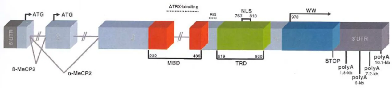
Figure 4. Schematic representation of the structure of the MECP2 gene. MBD – methyl – CpG binding
domain, TRD – transcription repression domain, NLS – nuclear localization signal, WW – group II WW
domain, poly A – polyadenylation site, kb – kilobase, 3’/5’ – untranslated region, ATG – start codon
The MeCP2 protein was first described in 1992 (30). It is one of five known proteins
sharing a methyl-CpG binding domain, which allows them to bind to methylated CpG
repression of genes in the area of these binding sites. MeCP2 dysfunction could thus
disrupt the normal development program of gene silencing, but how this might result in a
predominantly neurological phenotype has been a pressing question.
It is interesting that MeCP2 is more abundant in brain tissue than in most peripheral
tissues, is expressed in neurons but not in glia, and is localized to cell nuclei (31, 32, 33).
Even more interesting, MeCP2 levels increase in cortical neurons throughout
development (34, 35). This expression pattern suggests that MeCP2 might help maintain
or modulate neuronal maturity and plasticity.
Studies of MeCP2 expression in primate prefrontal cortex demonstrate increases during
development, with expression expanding from the deeper cortical layers in 110 day
embryos, to robust expression throughout the prefrontal cortex in adult monkeys (33).
Moreover, increased MeCP2 expression appears to be associated with neuronal
maturation, particular in the hippocampus, cortex and cerebellum, the brain regions
primarily affected in RTT (36, 37). These combined expression data suggest that MeCP2
has specific functions in neuronal cells of the central nervous system. Indeed, Aber et al.
(38) demonstrated that MeCP2 links the regulation of transcription to synaptic activity.
Recently, two papers have identified one possible MeCP2 target in this pathway in
mammals (39, 40). These authors found that MeCP2 binds specifically to BDNF (brain
derived neurotrophic factor) promoter III in rats and promoter IV in mouse, respectively,
thereby repressing BDNF transcription in resting neuronal cells.
There are two (possibly three) domains of the protein that are essential for its function (Fig
4). The methyl CpGs binding domain is encoded by exons 3 and 4, and binds to the
methylated DNA (41). The second important domain is the transcriptional repression
domain, which contains binding sites for the corepressor complex, and is therefore
involved in the repression process (42). The third domain is not yet well defined: it is
located in the C-terminus of the protein, and is thought to facilitate the binding both to
naked DNA and to the nucleosome core (43).
In RTT patients, more than 200 different MECP2 mutations have been described until now
(44). Eight MECP2 mutation hot spots are located at CpG dinucleotides, at transitions
from cytosine to thymine: they comprise 65% of all mutations (44). The other MECP2
mutations are less frequent and some have been found only once or twice.
Most missense mutations are located in the methyl-CpG binding domain and in the last
General introduction
22
between the methyl-CpG binding and the transcriptional repression domains, or in the first
part of the transcriptional repression domain.
It was noted early that there is a region at the C-terminus that harbours a hot spot for
deletions. The proximal breakpoint of these deletion, which can be found in about 10% of
the patients, is located in a section with repetitive sequence elements between
nucleotides 1050 and 1200 (45). Furthermore, many of the larger rearrangements have
one breakpoint in this region (46). They were detected in 16% of patients with classical
RTT, in whom sequencing had not revealed any mutations (46).
In the literature, the reported detection rate for MECP2 mutations, in patients with clinically
defined RTT, is between 60 and 95%, depending on the clinical parameters used to select
the patients for genetic analysis (47).
Despite advances in molecular diagnosis, the relationship between MECP2 mutations and
RTT remains complex. A small proportion of RTT patients do not have any detected
MECP2 mutation; conversely, males and females with mutations in this gene do not
always present with RTT features.
The clinical spectrum of phenotypes in girls with mutations in MECP2 is wide: some
patients had never been normal since birth, but others maintain language and hand use
after the regression stage of the disorder; however, by the pseudostationary stage,
patients accomplish the majority of the necessary criteria for RTT.
The speculation that other neurodevelopmental disorders could be caused by mutations in
MECP2 has not proved to be conclusive. Although some autism cases have been
reported, large studies have ruled out MECP2 mutations as playing a major role in the
pathogenesis of autism (48, 49). The same was true for patients with an Angelman-like
syndrome (50).
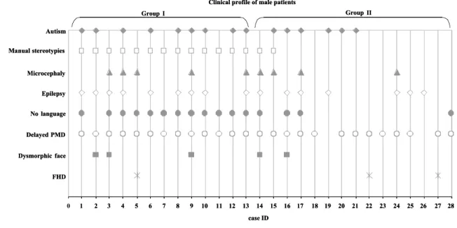
To this date, more than 60 male patients with mutations in the MECP2 gene have been
reported. They can be separated clinically into three groups: (1) patients with a severe
epileptic encephalopathy, who have the MECP2 mutations found in female patients with
RTT; (2) patients with a phenotype that resembles female patients with RTT and have
mosaic form mutations or the Klinefelter syndrome; (3) patients with a clinical picture that
does not resemble RTT and have MECP2 mutations infrequently found in female RTT
patients (51, 52).
Sporadic cases of RTT are the rule, with 99.5% being single occurrences within the family
explain the high preponderance of females with RTT; however, rare cases of germline
mosaicism of MECP2 (54) and of asymptomatic mothers passing on MECP2 mutations
(55) occur, accounting for familial cases. Genetic counselling should thus be offered to all
families regarding recurrence risk, including prenatal testing if so desired by the families.
1.2.3. Neurophathological studies
In spite of a predictable sequence of clinical symptoms that suggests a progressive
pathology, the processes characterizing degeneration associated with a progressive brain
disorder have not been recognized in RTT: there is no continuing deterioration in serial
clinical examinations (56), no progressive alteration in the magnetic resonance imaging
(57), no evident progressive deterioration in the electroencephalogram (EEG) (58), nor
progression of brain atrophy as defined by brain weight (59). Also, in the central nervous
system there is no recognizable malformation, degeneration or inflammatory process, nor
any consistent evidence of a cellular disorder involving the cytoskeleton, lysossomes or
the myelin. The alterations appear rather to be a deficiency of the dendritic and synaptic
apparatus of selected neurons, their neurotransmitters and, possibly, some cellular
proteins.
In RTT, the early decrease in head growth rate is followed by a decrease in height and
body weight, with extremely small feet. At autopsy, all organs (excluding the lungs and
adrenal glands) weight less than those of age-matched controls. The brain weight is
reduced by 12 to 34%, being lower than the 95% confidence limits of age-matched
controls (60). This is not related to age, since there is no progressive decrease in brain
weight, after the age of 2.5 years (61). There is a generalized decrease of neuronal size
with increased cell-packing density in the cerebral cortex, thalamus, basal ganglia,
amigdala and hippocampus (62).
Other studies revealed a decline in neuronal numbers in the frontal and temporal cortex
without decrease in cortical thickness, primarily involving the large pyramidal cells, more
prominent in layers II and III than in deep layers, and preservation of the visual cortex
(63). These changes were associated with a lack of area specialization in the orientation
patterns of dendrites and axons, decreased dendritic branching (61), small neurons with
increased neuronal packing (64), loss of dendrites of pyramidal cells in the frontal, motor
and subicular areas (65), shortening of the apical and basilar dendritic branches in layers
3 and 5 of the frontal, motor and inferior temporal cortex (66), and “naked dendrites”
General introduction
24
afferent neurons (67). The Golgi studies have identified a deficiency of dendrites in frontal,
motor and temporal cortex, brain regions that are associated with many of the functions
deficient in RTT. Similar abnormalities characterize the medial temporal lobe structures in
infantile autism (68).
There is atrophy in the frontal and temporal regions, with a reduction of up to 30% of the
corpus callosum (2). Some RTT brains show a pale substantia nigra, and tyrosine
hydroxylase staining has been reported to be diminished (69, 70, 71).
Jellinger observed gliosis of the basal ganglia, in his first study of RTT brains (72). Gliosis
in caudate, striatum, claustrum and nucleus basalis has been observed inconsistently
(73).
Certain symptoms and signs in RTT suggest a dysfunction of the autonomic nervous
system: small, cold blue feet; breathing irregularities; sleep disturbances; prolonged QT
intervals; altered heart rate variability; constipation and bloating. Julu (74) has
documented abnormalities of breathing patterns and of vagal tone, which he relates to an
immaturity of the integrative processes in the brainstem autonomic centers. These include
the nucleus of the solitary tract, the nucleus ambiguous, and the bulbar reticular formation.
Several chemo-architectonic studies of these regions suggest that the brainstem is
immature; a study of substance P immunoreactivity revealed an infant-like pattern of
staining in the spinal cord and brain stem with a consistent reduction of substance P
immunoreactivity, in the nucleus of the solitary tract and the reticular formation (75). In
another study, serotonin receptor binding was increased, approaching that of the normal
infant, in many brain stem nuclei involved in motor control, including autonomic centers
(76).
Oldfors et al. reported a progressive loss of neurons in the cerebellum of five RTT brains,
from patients aged 5 to 20 years. They observed a gross atrophy of all lobules of the
vermis, in older patients, and suggested that there was a progressive loss of Purkinje cells
(77). In 3 cases, Bauman and Kemper reported a reduction of Purkinje cells and simplified
principal olives, suggesting that these changes begin before birth (78).
Degeneration of the corticospinal tracts and lower than normal numbers of anterior
horncells in two RTT patients aged 20 and 30 years, were also reported. In younger
cases, gliosis in the corticospinal tracts has also been observed (73). A mild distal axonal
neuropathy was reported in RTT cases by Haas (79) and Jelinger (80). Muscle biopsies
In conclusion, the neuropathology of RTT points to arrested neuronal development rather
than neurodegeneration or severe malformation of nervous tissue. It should be noted,
however, that the majority of these data result from small samples of post-mortem tissue
and none of these changes are diagnostic or occur in every patient. Furthermore, the
majority of these studies were performed before the discovery of MECP2 mutations as the
cause of RTT, and the neuropathological studies in humans were all performed well after
the onset of the symptoms. It would be interesting to determine, in the existing animal
models, if alterations exist prior to the onset of symptoms.
1.2.4. Phenotype-genotype correlation studies
Since 1999, after the description of mutation in the MECP2 gene as the cause of RTT (5),
several series of patients with genetic studies have been published and tentative
phenotype-genotype correlations made. The results have not been conclusive, because
different methodologies were applied in the various studies, concerning clinical and
mutation classification, and severity scoring systems. For example, the scores of Percy
and Kerr (81, 82) increase with age, but this is not so evident with the scale of Pineda,
which was more based on developmental data than on the clinical features at the date of
observation (83); however, the larger studies point to a positive correlation between
mutation type and location with phenotype (84).
Two different genetic factors are likely to influence the phenotype in RTT: the type and
location of the mutations and the X inactivation pattern. Some research has found that
patients with missense MECP2 mutations may have a milder phenotype than those with
truncating mutations (83,85). Weaving et al. (86) found that age at onset of hand
stereotypies was higher, whereas speech and height (but not head growth) were slightly
closer to normal, in patients with missense mutations. In contrast, Nielsen et al. (87) found
no difference in severity between mutation types. In the study of Amir et al. (82), breathing
abnormalities were found to be more commonly associated with truncating mutations, and
scoliosis with missense mutations. Hoffbuhr et al. (88) concluded that patients with
missense mutations in the methyl binding domain (MBD) and mutations truncating the
entire transcription repression domain (TRD) were more severely affected than those with
missense and nonsense mutations in the TRD and C terminal segment. Similarly, in
another study, a milder phenotype was associated with late rather than early truncating
mutations (89). In a recent publication, Huppke et al. (84) considered mutations in the
nuclear localization signal (NLS) as a separate category from the other truncating
General introduction
26
truncation of the NLS were more severe than those with mutations downstream of the
TRD.
Examining common individual mutations, several works (90-93) showed that patients with
the R133C and R306C mutations have better function, overall. In general, the most
severe outcomes were found in patients with the R270X and R255X mutations (92).
Mutations toward the N terminus, including T158M and R168X, were also associated to a
more severe phenotype (93). However, until now, no specific neurological or behavioural
findings were identified in these groups of mutations, with the exception of an association
of fear and anxiety in those with R133C and R306C, and mood difficulties in those with
R294X (94).
1.2.5. Main objectives of the study
A - Identification of the RTT patients in Portugal
The incidence of RTT in females is 1:10000 to 1:20000 worldwide. Our goal was not to
perform an epidemiological study, but to identify a high number of patients with RTT in
Portugal, in order to obtain a sample large enough to study in detail the phenotypic
variability of this disorder.
B -To perform a detailed clinical study of the selected patients
The goal was to observe all suspected cases and to perform:
a general medical examination with particular attention to dysmorphisms and
anthropometry;
a neurological examination with a register of frequency and severity of
neurological signs;
a videotape register (10-15 minutes);
to offer all patients the possibility to be assessed by an orthopaedist, physiatrist
and a gastroenterologist;
an evaluation of nutritional status and of their phospho-calcic metabolism; and
Our goal was also to establish a DNA bank from all cases identified, in order to obtain
material for molecular studies.
C - To analyze the entire coding sequence of MECP2 gene in all patients and families; to identify the most frequent MECP2 mutations in the Portuguese population
We aimed to analyze the entire coding sequence (four exons that code for two different
isoforms of the MECP2 protein) of the MECP2 gene, for all RTT patients and families
selected for the study. (1) Initially, testing of most coding areas of MECP2, by DNA
sequencing and mutation scanning, for point mutations, should detect mutations in more
than 80% of the cases with classical RTT; (2) then, testing for large delections, which
detects an additional 10-16% of cases; and (3) later, to sequence mutations in exon 1,
which should detect mutations in about 1% of RTT patients.
D - To be able to offer counselling and prenatal diagnosis for subsequent pregnancies of mothers of patients with an identified MECP2 mutation
Though sporadic causes of RTT are the rule, some rare cases may be recurrent. Cases of
germline mosaicism of MECP2 and asymptomatic mothers passing on MECP2 mutations
may be responsible for familial cases. Genetic counselling should be offered to all families
regarding recurrence risks, and prenatal testing may be offered to those willing to have it.
E - To redefine more restrictive diagnostic criteria, based on the comparison of the groups of patients with and without MECP2 mutations
F - To analyze phenotype-genotype correlations in the group of patients with detected mutations
Our aim was to try quantify the phenotype severity using a severity scale, and also to
identify specific neurological signs, and to correlate these findings with type and
localization of the mutation. We also studied the X inactivation pattern in all the patients
with an identified MECP2 mutation.
General introduction
28
H - To perform the study of MECP2 in subsets of patients with clinical overlap with Rett syndrome
Although other neurodevelopmental disorderswith phenotypic overlapping with RTTare
rare, they provide important insight into the phenotypic consequences of mutations in
MECP2.
We decided to study the MECP2 also in the following groups of patients:(1) children with
References
1. Rett A.Uber ein eignartiges hirnatrophes syndrome bei hyperammonaemie im
kindesalter. Wiener Medizinisch Wochenshrift. 1966. 116: 723-26.
2. Rett A. Cerebral atrophy associated with hyperammonaemia. In Handbook of Clinical
Neurology. Vol 29: Metabolic and Deficiency Diseases of The Nervous System,
305-29. Ed. P. J. Vinken and G. W. Bruyn. Amsterdam: North Holland.
3. Ishikawa A, Goto T, Narasaki M, Yokochi K, Kitahara H, Fukuyama Y. A new
syndrome (?) of progressive psychomotor deterioration with peculiar stereotyped
movement and autistic tendency: a report of three cases. Brain and Development.
1978. 3: 258
4. Hagberg B, Aicardi J, Dias K, Ramos O. Progressive syndrome of autism, dementia,
ataxia and loss of purposeful hand use in girls: Rett’s syndrome: report of 35 cases.
Annals of Neurology 1983; 14:471-479
5. Amir RE, Veyver IB, Wan M, Tran CQ, Francke U, Zoghbi HY. Rett syndrome is
caused by mutations in X-linked MECP2, encoding methylCpG-binding protein 2.
Nature Genetics. 1999; 23: 185-1887.
6. Hagberg B.Rett’s syndrome: prevalence and impact on progressive severe mental
retardation in girls. Acta Paediatr Scand 1985; 74 (3): 405-408
7. Hagberg B. Rett syndrome: clinical particularities and biological mysteries. Acta
Paediatrica 1995; 84:971-976
8. Hagberg B, Witt-Engerstrom I. Rett Syndrome: a suggested staging system for
describing impairment profile with increasing age towards adolescence. American
Journal of Medical Genetics 1986; 24: 47-59
9. Hagberg B. Clinical criteria, stages and natural history. In: B. Hagberg, M Anvret, J
Wahlstrom (Eds) Rett Syndrome-Clinical and Biological Aspects. London: MacKeith
Press, 1993:4-20
10. The Rett syndrome Diagnostic Working Group. Diagnostic criteria for Rett Syndrome.