UNIVERSIDADE FEDERAL DO CEARÁ
FACULDADE DE FARMÁCIA, ODONTOLOGIA E ENFERMAGEM PROGRAMA DE PÓS-GRADUAÇÃO EM ODONTOLOGIA
CURSO DE MESTRADO
CARLOS EDUARDO DE ALBUQUERQUE GOMES
“FOTOSSENSIBILIZAÇÃO DE BIOFILMES FORMADOS EM RESINAS PARA BASE DE PRÓTESE – ESTUDO IN VITRO”
CARLOS EDUARDO DE ALBUQUERQUE GOMES
“FOTOSSENSIBILIZAÇÃO DE BIOFILMES FORMADOS EM RESINAS PARA BASE DE PRÓTESE – ESTUDO IN VITRO“
Dissertação apresentada à comissão examinadora aprovada pela coordenação do Programa de Pós-graduação em Odontologia, como pré-requisito para obtenção do título de Mestre.
Orientadora: Profa. Dra. Karina Matthes de Freitas Pontes.
Dados Internacionais de Catalogação na Publicação Universidade Federal do Ceará
Biblioteca de Ciências da Saúde
G613f Gomes, Carlos Eduardo de Albuquerque.
“Fotossensibilização de biofilmes formados em resinas para base de prótese – estudo in vitro” / Carlos Eduardo de Albuquerque Gomes. – 2012.
45 f. : il. color.
Dissertação (Mestrado) – Universidade Federal do Ceará, Faculdade de Farmácia, Odontologia e Enfermagem, Departamento de Clínica Odontológica, Programa de Pós-Graduação em Odontologia, Mestrado em Odontologia, Fortaleza, 2012.
Área de Concentração: Clínica Odontológica.
Orientação: Profa. Dra. Karina Matthes de Freitas Pontes.
1. Resinas Acrílicas. 2. Fotoquimioterapia. 3. Azul de Metileno. 4. Desinfecção. 5. Esterilização. I. Título.
CARLOS EDUARDO DE ALBUQUERQUE GOMES
“FOTOSSENSIBILIZAÇÃO DE BIOFILMES FORMADOS EM RESINAS PARA BASE DE PRÓTESE – ESTUDO IN VITRO“
Este trabalho é um estudo in vitro cujo objetivo é testar a eficácia da terapia fotodinâmica antimicrobiana aplicada a determinados grupos de microorganismos sobre espécimes de resina acrílica.
Programa de Pós-Graduação em Odontologia.
Universidade Federal do Ceará.
NOTA ____________
DATA DE APROVAÇÃO ______.______._______.
KARINA MATTHES DE FREITAS PONTES (orientadora) Faculdade de Farmácia, Odontologia e Enfermagem – UFC ______________________________________________________
HELENA DE FREITAS OLIVEIRA PARANHOS Faculdade de Odontologia de Ribeirão Preto – USP
______________________________________________________
MARCUS AURÉLIO RABELO LIMA VERDE
AGRADECIMENTOS
À DEUS, PELA FORÇA QUE ME FAZ SEGUIR SEMPRE EM FRENTE.
À MINHA FAMÍLIA, POR TODO O APOIO E AMOR DISPENSADO.
À MINHA NOIVA, PELA SUA COMPREENSSÃO E COMPANHEIRISMO.
À MINHA ORIENTADORA, POR TODA SUA DISPONIBILIDADE, COMPETÊNCIA E AJUDA INCONDICIONAL.
ÀS DOUTORAS LIDIANY KARLA AZEVEDO RODRIGUES E NÁDIA ACCIOLY PINTO NOGUEIRA POR SUA PRECIOSA CONTRIBUIÇÃO.
AOS AMIGOS DO NOSSO GRUPO DE PESQUISA, POR TODA A PACIÊNCIA E COLABORAÇÃO.
AOS TITULARES E SUPLENTES DA BANCA DE DEFESA POR SUA DISPONIBILIDADE E APOIO.
RESUMO
O Objetivo deste estudo foi avaliar o efeito do TFD (Terapia Fotodinâmica) em biofilmes formados por Streptococcus mutans, Staphylococcus aureus, Escherichia coli, Pseudomonas aeruginosa e Candida albicans em espécimes de resina acrílica. Métodos:
A TFD consistiu na aplicação de azul de metileno a 0,05% sobre os espécimes, associada à irradiação com um diodo emissor de luz (LED) de 630nm e 150 mW. As doses de energia previstas pelo LED foram definidos para 10 (a) e 30 (b) J/cm2. Para cada microorganismo, a formação de biofilme ocorreu durante a imersão dos espécimes num caldo de meio de cultura contendo 106 unidades formadoras de colónias ( UFCs ) / ml durante 48 horas a 37 ° C. Mas, em primeiro lugar, os espécimes foram esterilizados em gás óxido de etileno e distribuídos aleatoriamente em grupos (n = 5): experimentais (TFD a, b), que estavam contaminadas, corados e irradiados com as diferentes doses de energia; controle 1, apenas contaminado; controle 2, contaminado, corados e não irradiado; controle 3 (a, b), contaminado, não corado e irradiado; controle 4, não contaminado, não corados e não irradiado. Depois disso, os espécimes foram colocados em contato com meio de cultura
à
gar durante 10 minutos, em placas de Petri separadas, as quais foram incubadas durante 48 horas a 37 ° C. O número de UFCs foi obtido e os dados foram expressos de acordo com os escores (1 = 0; 2 = 1-10; 3 = 11-100; 4 = 101-1000; 5> 1000) e analisados através dos testes de Kruskal-Wallis e Dunn (α = 0,05). Resultados: S. mutans foi sensibilizado apenas pelo corante; P. aeruginosa e C. albicans também foramsensibilizados pelo corante, mas mostraram uma redução de UFCs para o escore 2 no grupo TFDb; S. aureus e Escherichia coli, apresentaram-se no escore inicial da 3 e alcançaram uma redução de UFC para o escore 2 nos grupos TFDb (p <0,05). A TFD foi eficaz na desinfecção de amostras de resina acrílica e a sua efetividade variou de acordo
com a dosagem de energia dos LEDs.
PALAVRAS-CHAVE: Resinas acrílicas, Terapia fotodinâmica, Azul de metileno,
SUMÁRIO
1. INTRODUÇÃO………..7 2. PROPOSIÇÃO...9 3. CAPÍTULOS – Artigo para publicação...10
CAPÍTULO 1 - “PHOTOSENSITIZATION OF IN VITRO BIOFILMS FORMED
ON DENTURE BASE RESIN “ 4. DISCUSSÃO………31 5. CONCLUSÃO………..34 6. REFERÊNCIAS BIBLIOGRÁFICAS...35 7. ANEXOS
ANEXO 1 ...41
1. INTRODUÇÃO
A terapia fotodinâmica ( TFD ) é uma modalidade terapêutica que tem se mostrado promissora na inativação de microorganismos patogênicos, podendo também ser chamada de inativação fotodinâmica (IFD) ou quimioterapia fotodinâmica antimicrobiana (Wilson, Dobson, 1993; Wainwright, 1998; Teichert et al., 2002; Bliss et al., 2004; Perussi, 2007; Donelly et al., 2007; Dovigo, 2007)
1,2,3,4,5,6,7. Há relatos datando mais de 100 anos sobre efeitos fotodinâmicos de
compostos químicos contra microorganismos (Raab, 1900; Tappeiner, 1904; Policard, 1924 apud Perussi, 2007)8,9,5, que foram suprimidos pela popularização
das sulfonamidas e da penicilina. Esta técnica foi primeiramente utilizada na Oncologia, tratando-se eficazmente lesões cancerosas, por meio da indução de danos irreversíveis nas células de tecidos neoplásicos (Dougherty et al., 1975; Woodburn et al, 1996; Sibata et al., 2000; Machado, 2000; Tardivo et al., 2006)10,11,12,13,14. Diante das dificuldades impostas pelo surgimento da
resistência microbiana, muitas pesquisas vêm sendo realizadas buscando soluções alternativas para o tratamento das infecções orais. Uma modalidade terapêutica promissora na inativação de microorganismos é a terapia fotodinâmica. Alguns estudos têm demonstrado o potencial da terapia fotodinâmica na inativação de microorganismos, incluindo alguns tipos de vírus, bactérias e fungos (Dovigo, 2007; Machado, 2000; Miyabe et al., 2011; Costa et al., 2011) 7,13,15,16.
absorve os fótons da fonte de luz e seus elétrons passam a um estado excitado. Na presença de um substrato como, por exemplo, o oxigênio, o fotossensibilizador transfere energia ao mesmo quando retorna ao seu estado natural, formando moléculas de vida curta e altamente reativas, como o oxigênio singleto, capaz de proporcionar sérios danos aos componentes celulares de microorganismos (Yamada, 2004)17. Devido ao mecanismo de ação da TFD, é
muito improvável que um microorganismo desenvolva resistência a este tipo de terapia (Machado, 2000; Wood et al., 2006; Perussi, 2007) 13,18,5.
O azul de metileno, utilizado para este estudo, demonstrou-se um eficiente fotossensibilizador contribuindo na redução de microorganismos como a candida albicans quando associado ao efeito da terapia fotodinâmica, ao contrário de seu efeito sozinho, o qual, não apresentou resultados satisfatórios segundo os estudos de Pupo, 2011 e Carvalho, 2009 32,33.
Outras vantagens também podem ser observadas com o uso da inativação fotodinâmica: seletividade do fotossensibilizador, focalização da luz apenas na região de interesse, possibilidade de repetição da terapia sem efeitos tóxicos cumulativos, não invasivo e de baixo risco (Teichert et al., 2002; Dovigo, 2007; Perussi, 2007)3,7,5.. A TFD tem sido aplicada em odontologia, nas áreas de
cariologia, periodontia, endodontia e patologia oral, com bastante eficácia (Giusti, 2005; Aghahosseini et al., 2006; Foschi et al., 2007; Oliveira et al., 2007; Garcez et al., 2008; Qin et al., 2008; Mima et al., 2011; Parker, 2011)19,20,21,22,23,24,25,26.
cirúrgicos para implantodontia, que devem ser utilizados em ambiente cirúrgico asséptico. As próteses dentárias removíveis ou guias cirúrgicos são compostos, basicamente, de resinas acrílicas. Este material é passível de esterilização por óxido de etileno, raios gama, imersão em solução de glutaraldeído 2% por 10 horas, ou irradiação por microondas. Os primeiros métodos são de uso hospitalar e apresentam alto custo. O segundo método tem como principal desvantagem o longo tempo de imersão necessário. Embora promissor, o terceiro método, não tem garantido 100% de eliminação microbiana, além de haver controvérsias quanto aos seus efeitos na estabilidade dimensional e dureza dos materiais envolvidos (Dixon et al., 1999; Campanha et al., 2005; Seo et al., 2007; )27,28,29. Além desses métodos, a utilização de hipoclorito de sódio
para a desinfecção é bem comum na prática clínica odontológica, embora que sua utilização esteja restrita ao processo de desinfecção e que seu mecanismo de ação possa afetar a coloração das peças de acrílico, sendo portanto mais indicado para outros fins odontológicos ( Estrela, 2002 )34.Há um interesse clínico
odontológico na procura de um método alternativo que seja simples e eficaz na desinfecção ou, até mesmo, esterilização de dispositivos confeccionados em resina acrílica, uma vez que problemas como infecções cruzadas entre laboratório e consultório, o uso de próteses imediatas e guias cirúrgicos não estéreis em cirurgias orais têm sido problemas recorrentes na odontologia. A TFD se mostra como uma opção eficaz e de baixo custo para a solução desses problemas.
O presente estudo in vitro teve como objetivo avaliar a eficácia antimicrobiana da TFD contra cinco espécies de microorganismos
(Streptococcus mutans, Staphylococcus aureus, Escherichia coli, Pseudomonas
aeruginosa e Candida albicans) inoculados em espécimes de resina acrílica, os
quais simulam peças protéticas ou guias de acrílico contaminados, utilizados na prática da reabilitação oral. Dessa forma, pôde-se testar a hipótese de que o azul de metileno a 0,05% seria eficaz como agente fotossensibilizador associado a um LED de comprimento de onda vermelho (630nm), nas fluências de 10 e 30 J/cm2.
3. CAPÍTULOS
CAPÍTULO 1 – artigo para publicação.
PHOTOSENSITIZATION OF IN VITRO BIOFILMS FORMED ON DENTURE BASE RESIN.
Carlos Eduardo de Albuquerque Gomes
DDS, graduate student, Faculty of Pharmacy, Dentistry and Nursing, Federal University of Ceará.
Rua Monsenhor Furtado S/N, Rodolfo Teófilo, CEP: 60430-350, Fortaleza, CE, Brazil.
Karina Matthes de Freitas Pontes
PhD, associate professor, Faculty of Pharmacy, Dentistry and Nursing, Federal University of Ceará.
Rua Monsenhor Furtado S/N, Rodolfo Teófilo, CEP: 60430-350, Fortaleza, CE, Brazil.
E-mail: kamatthes@yahoo.com.br Corresponding author.
Abstract
Aims: This study evaluated the effect of PDT (Photodynamic Therapy) against
aeruginosa and Candida albicans biofilms formed on the surface of acrylic resin
specimens. Methods: The PDT consisted in the application of 0.05% methylene blue on the specimens, associated to irradiation with a light-emitting diode (LED) of 630nm and 150mW. The energy doses provided by the LED were set to 10 (a) and 30 (b) J/cm2. For each microorganism, the biofilm formation occurred during
immersion of the specimens in a culture medium broth containing 106
colony-forming units (CFU)/ml for 48 hours at 37°C. But, at first, the specimens were sterilized in ethylene oxide gas and randomly divided into groups (n=5): experimental (PDT a,b), which were contaminated, stained and irradiated with the different doses of energy; control 1, only contaminated; control 2, contaminated, stained and not irradiated; control 3 (a,b), contaminated, not stained and irradiated; control 4, not contaminated, not stained and not irradiated. Afterward, the specimens were submitted to contact with culture medium agar for 10 minutes, in separated Petri plates, which were incubated for 48 hours at 37°C. The number of CFU was obtained, and the data were expressed according to scores (1=0; 2=1-10; 3=11-100; 4=101-1000; 5>1000) and analyzed by Kruskall Wallis and Dunn tests (α=0.05). Results: S. mutans was only sensitized by the dye; P. aeruginosa and C. albicans were also sensitized by the dye, but showed a CFU reduction to score 2 by PDTb; S. aureus and E. coli, presented an initial score of 3 and achieved a CFU reduction to score 2 by PDTb (p<0.05). Conclusion: PDT was effective in disinfection of acrylic resin specimens and the effectiveness varied according to the dose of energy of the LEDs.
Introduction
Photodynamic therapy is a therapeutic modality that has shown promise in the inactivation of pathogenic microorganisms and may also be called photodynamic inactivation (PDI) or antimicrobial photodynamic chemotherapy (Wilson, Dobson, 1993; Wainwright, 1998; Teichert et al., 2002; Bliss et al., 2004; Perussi, 2007, Donnelly et al., 2007; Dovigo, 2007)1,2,3,4,5,6,7. There are reports
dating back over 100 years on photodynamic effects of chemical compounds against microorganisms (Raab, 1900; Von Tappeiner, 1904; Policard apud Perussi, 2007)8,9,5, which were suppressed by the popularization of sulfonamides
and penicillin. This technique was first used in Oncology, effectively treating cancerous lesions through the induction of irreversible damage in the neoplastic tissue cells (Dougherty et al. 1975, Woodburn et al, 1996; Sibata et al., 2000; Machado 2000; Tardivo et al., 2006)10,11,12,13,14. Given the difficulties posed by the
emergence of antimicrobial resistance, many studies have been performed seeking alternative solutions for the treatment of oral infections. A promising therapeutic modality in the inactivation of microorganisms is photodynamic therapy. Some studies have demonstrated the potential of photodynamic therapy in the inactivation of microorganisms, including some types of viruses, bacteria and fungi (Dovigo, 2007, Machado, 2000, Miyabe et al., 2011, Costa et al., 2011)
7,13,15,16.
photons of light source and pass their electrons to an excited state. In the presence of a substrate such as oxygen, the photosensitizer transfers energy to the substrate to return to its natural state, forming short-living molecules, highly reactive, such as singlet oxygen, which can cause serious damage to cellular components of microorganisms (Yamada, 2004)17. Due to the mechanism of
action of PDT, it is very unlikely that an organism develops resistance to this kind of therapy (Machado, 2000; Wood et al, 2006; Perussi, 2007)13,18,5.
The methylene blue, that was used for this study, demonstrated be an efficient photosensitizer contributing to the reduction of microorganisms like as
Candida albicans when combined with the effect of photodynamic therapy, in
contrast to its effect alone, which did not provide satisfactory results according to the studies of Pupo, 2011 and Carvalho, 200932,33.
Others advantages may also be observed with the use of photodynamic inactivation: selectivity of the photosensitizer, focalization of light on the region of interest only, possibility of repeating the therapy without cumulative toxic effects, non invasive and low risk (Teichert et al, 2002; Dovigo, 2007; Perussi, 2007)3,7,5.
PDT has been used in dentistry, in the areas of cariology, periodontology, endodontics and oral pathology, quite effectively (Giusti, 2005; Aghahosseini et al., 2006; Foschi et al., 2007, Oliveira et al., 2007; Garcez et al., 2008; Qin et al., 2008; Mima et al., 2011; Parker, 2011)19,20,21,22,23,24,25,26.
composed of acrylic resins. This material is capable of being sterilized by ethylene oxide gas, gamma rays, immersion in a 2% glutaraldehyde solution for 10 hours, or microwave irradiation. The first two methods are used in hospitals and have a high cost. The main disadvantage of the third method is the long immersion time required. Although promising, the fourth method has not guaranteed 100% microbial elimination, in addition to the controversies in regard to its effects on the dimensional stability and hardness of the materials involved (Dixon et al., 1999; Campanha et al., 2005; Seo et al., 2007)27,28,29. Besides these
methods, the use of sodium hypochlorite for disinfection is a very common practice in the dental clinic, although its use is restricted to the disinfection process and its mechanism of action may affect the color of the acrylic pieces and is therefore more appropriate for other dental purposes( Estrela, 2002 )34.The
search for a simple alternative method of disinfection or sterilization of these devices made of acrylic resins is of great interest to clinical dentistry, since problems such as cross-infection between the laboratory and dental office, use of non sterile immediate dentures and surgical guides in oral surgeries, have been recurrent problems in dentistry. PDT has been shown to be an efficient, low cost option to solve these problems. This in vitro study aims to evaluate the antimicrobial efficacy of PDT against five different groups of microorganisms (Streptococcus mutans, Staphylococcus aureus, Escherichia coli, Pseudomonas
aeruginosa and Candida albicans) inoculated on acrylic resin substrates, which
Materials and methods
1. Preparation of specimens:
The heat cured acrylic resin selected for this study was Lucitone 550 (Dentsply Ind. Com. Ltd., Petrópolis, RJ, Brazil), which is representative of those used for the fabrication of total, partial removable dentures and surgical guides for implants. “Plex-glass” acrylic matrices, measuring 15mm long, 15mm wide and 4mm thick, were included in number 6 metallic flasks (Jon, São Paulo, Brazil), with Type III dental stone (Herodent, Vigodent S/A Ind. Com., Rio de Janeiro, Brazil).
After deflasking the matrices, the acrylic resin was manipulated with 21g of powder and 10 ml of liquid, pressed in the plastic phase and left waiting for 30 minutes, to be polymerized by the technique of immersion in water at 73°C for 90 minutes and at 100°C for 30 minutes, in accordance with the manufacturer’s instructions. The flasks were left to cool naturally after polymerization, in order to proceed with deflasking the test specimens and immersing them in distilled water at 50°C for 24 hours to eliminate the residual monomer. Two face of each specimen were finished using 150, 220, 400, 600, 1200, 2000 - grit sandpapers (Norton Indústria brasileira, São Paulo, Brazil). After these procedures, they were polished on a wet rag wheel (Invicta, São Paulo, Brazil), using felt cones and canvas polishing discs with pumice slurry followed by calcium carbonate.
2. Sterilization of specimens:
bonded to a 1 ml syringe attached to a Falcon tube cap with chemically polymerized acrylic resin (Clássico Art. Odontológicos Ltd., SP, Brazil). Afterwards, they were packed in surgical grade paper envelopes, duly sealed and submitted to sterilization with ethylene oxide gas. Falcon tubes without their lids were also distributed in groups of five and packaged in plastic envelopes and submitted to autoclave sterilization.
3. Selection of photosensitizers and light sources:
The photosensitizer selected for the experiment was methylene blue (Vetec Fine Chemicals LTD., Duque de Caxias, Rio de Janeiro, Brazil) at a concentration of 0.05%, recommended for biofilm staining on dental prosthesis surfaces (Lovato-Silva et al., 2002)30.
The light source selected was a red LED of 630 nm and output power of 150 mW (MM Optics Ltda., São Carlos, São Paulo, Brazil), with variations of energy intensity of 10 J/cm² and 30 J/cm².
The photosensitizer methylene blue was associated with the red LED due to the compatibility of the wavelength of this light source with the absorption band of the dye.
4. Preparation of microorganisms for inoculation of the specimens:
The microorganisms selected for the study were: Streptococcus mutans, provenance American Type Culture Collection (ATCC) 25175 and
Staphylococcus aureus ATCC 25923, both with the morpho-tintorial
characteristics of gram positive cocci; Escherichia coli ATCC 11205 and
characteristics of gram negative cocci; Candida albicans ATCC 001, yeast. These microorganisms underwent processes of activation, re-suspension, and readout in a spectrophotometer, in order to obtain a culture medium broth (Brain Heart Infusion – BHI - Acumedia, Michigan, USA) with a microbial inoculum of 1% at 0.5 McFarland scale, corresponding to 106 CFU/ml (Paranhos et al.,
2009; Miyabe et al., 2011; Costa et al., 2011)31,15,16.
For Streptococcus mutans, the absorbance used was 0.08, at a wavelength of 590 nm, whereas for Staphylococcus aureus, was used an absorbance between 0.08 and 0.10 at a wavelength of 652 nm. For Escherichia
coli the absorbance used was 1.43 at a wavelength of 600 nm, and for
Pseudomonas aeruginosa, an absorbance of 0.64 at a wavelength of 600 nm.
For Candida albicans, 0.28 absorbance at a wavelength of 530 nm.
Specimen contamination consisted of their immersion in 15 ml of contaminated culture medium broth in individual Falcon tubes. After this, the Falcon tubes containing the immersed test specimens were incubated at 37°C, for 48 hours.
5. Experimental design:
For each microorganism, the specimens were distribuited into groups (n = 5):
• Control 1 (C1): inoculated, but not stained, not irradiated;
• Control 2 (C2): inoculated, stained, but not irradiated;
• Control 3 (C3): inoculated, not stained, but irradiated by the light source (C3a –
10 J/cm2 and C3b - 30 J/cm2);
• Control 4 (C4): not inoculated, not stained and not irradiated;
• Experimental PDTa: inoculated, stained and irradiated at 10 J/cm2;
The C1 group was the negative control and aimed to test the effectiveness of the specimens’ contamination, while the C4 was the positive control and verified the effectiveness of previous sterilization with ethylene oxide gas.
6. Application of the photodynamic therapy:
Each contaminated specimen was stained in all its surfaces with the photosensitizer (0.05% methylene blue). They were then irradiated with the red LED for the time necessary to achieve the levels of energy: 10 J/cm2 and 30
J/cm2, according to the experimental design.
7. Analysis of antimicrobial activity:
After the treatments, each specimen was placed in contact with BHI Agar or Agar Sabouraud (Acumedia, Michigan, USA) in separated Petri plates (20 x 100 mm). After 10 minutes, the specimens were discarded and the plates were incubated at 37ºC for 48 hours.
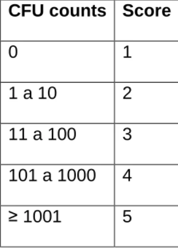
After the incubation period, the colony forming units (CFU) were counted and the results were expressed according to an ordinal scale (Table 1).
8. Statistical data analysis:
The scores attributed to each group were statistically compared by Kruskall Wallis and Dunn test (α = 0.05).
Results
Discussion
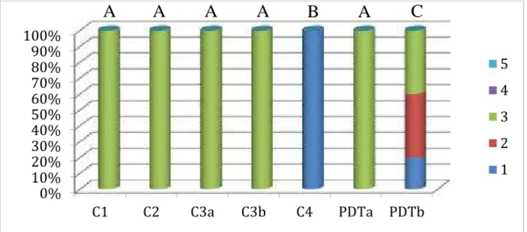
In this study, S. mutans was sensitive to 0.05% methylene blue dye, showing a reduction in counts in groups only stained, but not associated with light (p <0.05). For specimens inoculated with S. aureus, group PDTb showed a reduction in rates of bacterial growth. This bacteria was not sensitive to methylene blue. The group PDTa for E.coli showed a reduction in the number of CFU. The specimens inoculated with P. aeruginosa also showed a sensitivity of this microorganism to the methylene blue dye and the PDTb group showed great effectiveness, statistically similar to the C4 group. C. albicans presented sensitivity to methylene blue and the association with the red LED at 30 J/cm2
showed significant reduction in the count of CFU (p <0.05).
Some specimen groups presented intense microbial growth that resulted in colony aggregation, which made exact counting impossible. The sample size of this study demanded a nonparametric analysis. Therefore, an ordinal scale was used, as data do not allow a parametric description. This solution was taken with changes from a study of similar methodology performed by Paranhos et al., 200931.
Wood et al. (2006)18 reported that the action of erythrosine at a
concentration of 22μM when not subjected to irradiation had a negligible effect on S. mutans when compared to the control group, which was not stained and not irradiated. Miyabe et al. (2011)15 reported that only laser irradiation or 3mM
concentration of methylene blue, separately, did not reduce significantly the count of Staphylococcus spp. Costa et al. (2011)16 found equal results, using
erythrosine 0.39-200µM and laser 532 nm. However, Lovato-Silva et al. (2002)30
evaluated several disclosing agents, including erythrosine 5% and methylene blue 0.05% and reporting their antimicrobial activity. The concentration of the photossensitizer and the species of microorganism may promote variation in results.
As can be seen through the results of this study, almost all experimental groups, for almost all microorganisms, showed a reduction in microbial counts compared to control groups, in agreement with the results found by other authors (Wood et al., 2006; Dovigo, 2007; Miyabe et al., 2011; Costa et al., 2011)18,7,15,16.
It was also observed that this reduction was proportional to the energy applied by the light source. Dovigo (2007)7, using LEDs in the wavelength range of blue and
using the Photogem® as the photosensitizer, noted that the inactivation of
Candida spp. occurred in a concentration/ dose dependent way, which resulted
in complete inactivation of microorganisms in certain experimental conditions and that the concentration of the photosensitizer could be reduced by elevating the amount of energy provided by light source.
have avoided possible contamination coming from other areas not fully irradiated, providing, perhaps, values of microbial death possibly more effective; the red LED, with its maximum output power of 150 mW made it impossible the irradiation in higher fluence of energy, because it would demand a great time of exposure to light. - a more powerful machine, in the same wavelength, might have brought most effective results. Another factor to consider is that inoculation established for this study, although in agreement with other studies of similar methodology (Paranhos et al., 2009; Miyabe et al. 2011; Costa et al.,2011), is of great magnitude, 106 CFU / ml. Therapies introduced in this study to a lower microbial
load could have generated even better results.
More studies are necessary to establish a protocol for sterilization of acrylic resin specimens with the photodynamic therapy, by checking if higher amounts of energy in different wavelengths of light sources, and various photosensitizers, and preferably, in various lower concentrations. Therefore, this work represents the starting point for a promising line of research to be explored.
Conclusions
Based on the results and within the limitations of this study, it can be concluded that photodynamic therapy, using 0.05% methylene blue as photosensitizer, associated with a 630 nm LED, generated a reduction of microbial count in the biofilms formed on acrylic resin specimens, except for S.
mutans. Furthermore, inhibition of microorganism growth was directly
proportional to the amount of energy provided by light sources: 10 J/cm2 was not
References
1. Wilson M, Dobson J. Lethal photosensitization of oral anaerobic bacteria. Clin Infect Dis. 1993;16(4):414-415.
2. Wainwright M. Photodynamic antimicrobial chemotherapy. J Antimicrobial Chemother. 1998. 42: 13-28.
3. Teichert MC, Jones JW, Usacheva MN, Biel MA. Treatment of oral candidiasis with methylene blue-mediated photodynamic therapy in an immunodeficient murine model. Oral Surg Oral Med Oral Pathol Oral Radiol Endod. 2002; 93(2):155-160.
4. Bliss JM, Bigelow CE, Foster TH, Haidaris CG. Susceptibility of Candida species to photodynamic effects of photofrin. Antimicrob Agents Chemother. 2004;48(6):2000-2006.
5. Perussi JR. Inativação fotodinâmica de microrganismos. Quim Nova. 2007;30(4): 988-994.
6. Donnelly RF, Mccarron PA, Tunney MM, David Woolfson A. Potential of photodynamic therapy in treatment of fungal infections of the mouth. Design and characterisation of a mucoadhesive patch containing toluidine blue O. J Photochem Photobiol B. 2007;86(1):59-69.
7. Dovigo LN. Efetividade da terapia fotodinâmica na inativação de Candida spp. Dissertação de mestrado – Universidade Estadual Paulista Faculdade de Odontologia. Araraquara, 2007.
8. Raab O. Z Biol. (1900) ;39:524.
10. Dougherty TJ, Grindey GB, Fiel R, Weishaupt KR, Boyle DG. Photo radiation therapy II. Cure of animal tumors with hematoporphyrin and light. J Natl Cancer Inst. 1975;55(1):115-119.
11. Woodburn KW, Fan Q, Kessel, D.; Wright M, Mody TD, Hemmi G, Magda D, Sessler JL, Dow WC, Miller RA, Young SW. Phototherapy of cancer and atheromatous plaque with texaphyrins. J Clin Laser Med Surg. 1996; 14(5):343-348.
12. Sibata H, Colussi VC, Oleinick NL, Kinsella TJ. Photodynamic therapy: a new concept in medical treatment. Braz J Med Biol Res.2000;33(8):869-880.
13. Machado AEH. Terapia fotodinâmica: princípios, potencial de aplicação e perspectivas. Quim Nova. 2000;23(2):237-243.
14. Tardivo JP, Del Giglio A, Paschoal LH, Baptista MS. New photodynamic therapy protocol to treat AIDS-related Kaposi's sarcoma. Photomed Laser Surg. 2006;24(4):528-531.
15. Yamada Jr AM, Hayek RRA, Ribeiro MS. O emprego da terapia fotodinâmica na redução bacteriana em periodontia e implantodontia. RGO. 2004; 52(3):207-210.
16. Miyabe M, Junqueira JC, Costa ACBP, Jorge AOC, Ribeiro MS, Feist IS. Effect of photodynamic therapy on clinical isolates of Staphylococcus spp. Braz Oral Res. 2011; 25(3): 230-234.
17. Costa ACBP, Rasteiro VMC, Pereira CA, Hashimoto ESHS, Beltrame Jr. M, Junqueira JC, Jorge AOC. Susceptibility of Candida albicans and
Candida dubliniensis to erythrosine and LED mediated photodynamic
18. Wood S, Metcalf D, Devine D, Robinson C. Erythrosine is a potential photosensitizer for the photodynamic therapy of oral plaque biofilms. J Antimicrob Chemother. 2006;57(4):680-684.
19. Giusti JSM. Avaliação da terapia fotodinâmica utilizando diodo emissor de luz (led) na descontaminação de dentina bovina artificialmente cariada. Tese de Doutorado, Araraquara, 2005.
20. Aghahosseini F, Arbabi-Kalati F, Fashtami LA, Fateh M, Djavid GE. Treatment of oral lichen planus with photodynamic therapy mediated methylene blue: a case report. Med Oral Patol Oral Cir Bucal. 2006;11(2):126-129.
21. Foschi F, Fontana CR, Ruggiero K, Riahi R, Vera A, Doukas AG, Pagonis TC, Kent R, Stashenko PP, Soukos NS. Photodynamic inactivation of Enterococcus faecalis in dental root canals in vitro. Lasers Surg Med. 2007; 39(10):782-787.
22. Oliveira RR, Schwartz-Filho HO, Novaes AB Jr, Taba M Jr. Antimicrobial photodynamic therapy in the non-surgical treatment of aggressive periodontitis: a preliminary randomized controlled clinical study. J Periodontol. 2007;78(6):965-973.
23. Garcez AS, Nuñez SC, Hamblin MR, Ribeiro MS. Antimicrobial effects of photodynamic therapy on patients with necrotic pulps and periapical lesion. J Endod. 2008 Feb;34(2):138-142.
25. Mima EGO, Pavarina AC, Silva MM, Ribeiro DG, Vergani CE, Kurachi C, Bagnato VS. Denture stomatitis treated with photodynamic therapy – Five cases. Oral Surg Oral Med Oral Pathol Oral Radiol Endod. 2011;112:602-608.
26. Parker S. Photodynamic antimicrobial chemotherapy in dental practice. Dental Nursing. 2011; 7(7):380-385.
27. Dixon DL, Breeding LC, Faler TA. Microwave disinfection of denture base materials colonized with Candida albicans. J Prosthet Dent. 1999; 81(2):207-214.
28. Campanha N, Pavarina AC, Vergani C, Machado A. Effect of microwave sterilization and water storage on the Vickers hardness of acrylic resin denture teeth. J Prosthet Dent. 2005; 93(5):483–487.
29. Seo RS, Vergani CE, Pavarina AC, Compagnoni MA, Machado AL. Influence of microwave disinfection on the dimensional stability of intact and relined acrylic resin denture bases. J Prosthet Dent. 2007; 98(3):216-223.
30. Silva-Lovato CH, Paranhos HFO, Ito IY. Evidenciadores de biofilme em prótese total: avaliação clínica e antimicrobiana. Pesqui Odontol Bras 2002;16(3):270-275.
31. Paranhos HFO, Silva-Lovato CH, Souza RF, Cruz PC, Freitas-Pontes KM, Watanabe E, Ito IY. Effect of three methods for cleaning dentures on biofilms formed in vitro on acrylic resin. J Prosthodont. 2009;18:427-431. 32. Carvalho GG, Felipe MP, Costa MS. The photodynamic effect of
medium conditions. J. microbiol. 2009 Oct;47(5):619-23. Epub 2009 Oct 24.
33. Pupo YM, Gomes GM, Santos EB, Chaves L, Michel MD, Kozlowski VA Jr, Gomes OM, Gomes JC. Susceptibility of Candida albicans to photodynamic therapy using methylene blue and toluidine blue as photosensitizing dyes. Dentistry School, State University of Ponta Grossa (UEPG), Ponta Grossa, Paraná, Brazil. Acta Odontol Latinoam. 2011;24(2):188-9.
34. Estrela, Carlos ; Estrela, Cyntia R.A ; Barbin, Eduardo Luis ; Spanó, Júlio César E ; Marchesan, Melissa A ; Pécora, Jesus D. Mecanismo de ação do hipoclorito de sódio.Brazilian Dental Journal, 2002, Vol.13, p.113-117.
Table 1. Assigning scores, according to CFU counts.
CFU counts Score
0 1
1 a 10 2
11 a 100 3 101 a 1000 4
Figure 1. Percentage of specimens according to growth score of Streptococcus
mutans.
Different letters: p<0.05 0%
10% 20% 30% 40% 50% 60% 70% 80% 90% 100%
C1 C2 C3a C3b C4 PDTa PDTb
5
4
3
2
1
Figure 2. Percentage of specimens according to growth score of Staphylococcus
aureus.
Different letters: p<0.05 0%
10% 20% 30% 40% 50% 60% 70% 80% 90% 100%
C1 C2 C3a C3b C4 PDTa PDTb
5
4
3
2
1
Figure 3. Percentage of specimens according to growth score of Escherichia coli.
Different letters: p<0.05 0%
10% 20% 30% 40% 50% 60% 70% 80% 90% 100%
C1 C2 C3a C3b C4 PDTa PDTb
5
4
3
2
1
Figure 4. Percentage of specimens according to growth score of Pseudomonas
aeruginosa.
Different letters: p<0.05 0%
10% 20% 30% 40% 50% 60% 70% 80% 90% 100%
C1 C2 C3a C3b C4 PDTa PDTb
5
4
3
2
1
Figure 5. Percentage of specimens according to growth score of Candida
albicans.
Different letters: p<0.05 0%
10% 20% 30% 40% 50% 60% 70% 80% 90% 100%
C1 C2 C3a C3b C4 PDTa PDTb
5
4
3
2
1
4. DISCUSSÃO
A hipótese de que a terapia fotodinâmica com a utilização de azul de metileno 0,05% associado a um diodo emissor de luz de 630 nm seria eficaz contra os microorganismos selecionados neste estudo, foi confirmada. Porém, tal eficácia foi demonstrada nos grupos irradiados com a maior fluência de energia, ou seja, com 30 J/cm2. Não foi alcançada a esterilização dos espécimes,
mas os resultados foram promissores quanto à desinfecção, não descartando a possibilidade de que futuros estudos possam levar a este objetivo.
apresentou redução na contagem de UFC. Os espécimes inoculados com P.
aeruginosa mostraram sensibilidade deste microorganismo ao corante azul de
metileno, nenhuma à ação isolada das fontes de luz e o grupo TFDb apresentou grande eficácia, similar estatisticamente ao grupo C4. Para a C. albicans, também houve sensibilidade ao azul de metileno e a associação do azul de metileno ( 0,05% ) com o LED vermelho a 30 J/cm2 apresentou redução mais
significativa na contagem de UFC (p<0,05). Alguns grupos de amostras apresentaram crescimento microbiano intenso, o que resultou em agregação de colônias e fez com que a contagem exata de UFC se tornasse impossível. O tamanho da amostra deste estudo exigiu uma análise estatística não-paramétrica. Portanto, uma escala ordinal (escore) foi usada, já que os dados não permitiram uma descrição paramétrica. Esta solução foi tomada a partir de um estudo de metodologia semelhante realizada por Paranhos et al., 2009 31 .
Referindo-se aos controles, para todos microorganismos, os grupos apenas inoculados (C1) e apenas irradiados (C3) apresentaram significativo número de UFC, enquanto o grupo apenas esterilizado com óxido de etileno (C4) não apresentou nenhum crescimento microbiano. Isto comprovou que houve a verdadeira formação de biofilme nos espécimes contamindos, enquanto a esterilização em óxido de etileno foi também eficaz. A ação isolada das fontes de luz não causou nenhum efeito para as bactérias e leveduras, enquanto que os fotossensibilizadores causaram redução na contagem de alguns microorganismos em grupos apenas corados (C2).
Wood et al. (2006)18 relataram que a ação de eritrosina a uma
foi corado e nem irradiado. Miyabe et al. (2011)15 relataram que apenas a
irradiação a laser ou a concentração 3mM de azul de metileno, separadamente, não reduziu significativamente a contagem de Staphylococcus spp. Costa et al. (2011)16 encontraram resultados iguais, usando eritrosina 0,39 - 200μM e laser
de 532 nm. No entanto, Lovato-Silva et al. (2002)30 avaliando vários agentes
evidenciadores de biofilme, incluindo eritrosina a 5% e azul de metileno a 0,05% e encontraram atividade antimicrobiana neles. A concentração do fotossensibilizador e as espécies de microorganismos envolvidos nos diferentes estudos podem promover variação nos resultados.
Como pode ser visto através dos resultados do presente estudo, quase todos os grupos experimentais, de quase todos os microorganismos, mostraram uma redução nas contagens microbianas em relação aos grupos controle, assim como foi sugerido em estudos anteriores (Wood et al., 2006; Dovigo, 2007; Miyabe et al., 2011; Costa et al., 2011)18,7,15,16. Também foi observado que esta
redução foi proporcional à energia aplicada pela fonte de luz. Dovigo (2007)7,
Como limitações do estudo, alguns aspectos foram observados: a irradiação de luz ocorreu em apenas uma superfície do espécime, que entrou em contato com a placa de Petri - o tratamento de todo o espécime poderia ter evitado possíveis contaminações vindas das outras superfícies não totalmente irradiadas, proporcionando, talvez, valores de morte microbiana possivelmente mais eficazes; o LED vermelho, por apresentar potência máxima de saída de 150 mW tornou inviável a irradiação com maior fluência de energia, pois demandaria um grande tempo de exposição – um equipamento com LED mais potente, no mesmo comprimento de onda, poderia ter trazido resultados mais eficazes. Outro fator a ser considerado é que a inoculação estabelecida para este estudo, 106 UFC/ml, embora em concordância com outros estudos de
metodologia semelhante (Paranhos et al., 2009; Miyabe et al. 2011; Costa et al.,2011), é de grande magnitude, o que, provavelmente, pode não corresponder à rotina clínica. A terapia instituída neste estudo para uma menor carga microbiana poderia ter gerado ainda melhores resultados.
Mais estudos ainda são necessários para o estabelecimento de um protocolo de esterilização de espécimes de resina acrílica com a técnica da terapia fotodinâmica, verificando-se dosagens maiores de energia em diferentes comprimentos de onda de fontes de luz, além de diferentes fotossensibilizadores, em variadas e, de preferência, menores concentrações. Portanto, este trabalho representa o ponto de partida para uma promissora linha de pesquisa a ser explorada.
A terapia fotodinâmica cada vez mais é utilizada em diversas áreas da saúde e possui um potencial promissor para sua aplicação odontológica. Algumas áreas da odontologia têm desenvolvido técnicas para utlização clínica. Através dos resultados obtidos, e diante das limitações, pode-se concluir que a terapia fotodinâmica apresenta aplicabilidade na área da prótese dentária, pois ela propõe a descoberta de novos protocolos e meios de desinfecção para dispositivos acrílicos utilizados rotineiramente nos consultórios odontológicos. A utilização do fotossensibilizador azul de metileno a 0,05% associado a um LED 630 nm possibilitou redução na contagem da maioria dos microorganismos estudados, nos biofilmes formados sobre os espécimes de resina acrílica, com exceção para o S.
mutans. A inibição do crescimento de microorganismos foi dependente da
quantidade de energia fornecida pela fonte de luz. A irradiação com 30 J/cm2
apresentou melhores resultados do que os dos grupos irradiados com 10 J/cm2.
6. REFERÊNCIAS BIBLIOGRÁFICAS
1. Wilson M, Dobson J. Lethal photosensitization of oral anaerobic bacteria. Clin Infect Dis. 1993;16(4):414-415.
2. Wainwright M. Photodynamic antimicrobial chemotherapy. J Antimicrobial Chemother. 1998. 42: 13-28.
immunodeficient murine model. Oral Surg Oral Med Oral Pathol Oral Radiol Endod. 2002; 93(2):155-160.
4. Bliss JM, Bigelow CE, Foster TH, Haidaris CG. Susceptibility of Candida species to photodynamic effects of photofrin. Antimicrob Agents Chemother. 2004;48(6):2000-2006.
5. Perussi JR. Inativação fotodinâmica de microrganismos. Quim Nova. 2007;30(4): 988-994.
6. Donnelly RF, Mccarron PA, Tunney MM, David Woolfson A. Potential of photodynamic therapy in treatment of fungal infections of the mouth. Design and characterisation of a mucoadhesive patch containing toluidine blue O. J Photochem Photobiol B. 2007;86(1):59-69.
7. Dovigo LN. Efetividade da terapia fotodinâmica na inativação de Candida spp. Dissertação de mestrado – Universidade Estadual Paulista Faculdade de Odontologia. Araraquara, 2007.
8. Raab O. Z Biol. 1900;39:524.
9. Von Tappeiner H, Jesionek V. Dtsh Arch Klin Med. 1904; 80:427.
11. Woodburn KW, Fan Q, Kessel, D.; Wright M, Mody TD, Hemmi G, Magda D, Sessler JL, Dow WC, Miller RA, Young SW. Phototherapy of cancer and atheromatous plaque with texaphyrins. J Clin Laser Med Surg. 1996; 14(5):343-348.
12. Sibata H, Colussi VC, Oleinick NL, Kinsella TJ. Photodynamic therapy: a new concept in medical treatment. Braz J Med Biol Res.2000;33(8):869-880.
13. Machado AEH. Terapia fotodinâmica: princípios, potencial de aplicação e perspectivas. Quim Nova. 2000;23(2):237-243.
14. Tardivo JP, Del Giglio A, Paschoal LH, Baptista MS. New photodynamic therapy protocol to treat AIDS-related Kaposi's sarcoma. Photomed Laser Surg. 2006;24(4):528-531.
15. Yamada Jr AM, Hayek RRA, Ribeiro MS. O emprego da terapia fotodinâmica na redução bacteriana em periodontia e implantodontia. RGO. 2004; 52(3):207-210.
17. Costa ACBP, Rasteiro VMC, Pereira CA, Hashimoto ESHS, Beltrame Jr. M, Junqueira JC, Jorge AOC. Susceptibility of Candida albicans and
Candida dubliniensis to erythrosine and LED mediated photodynamic
therapy. Arch Oral Biol. 2011; 56:1299-1305.
18. Wood S, Metcalf D, Devine D, Robinson C. Erythrosine is a potential photosensitizer for the photodynamic therapy of oral plaque biofilms. J Antimicrob Chemother. 2006;57(4):680-684.
19. Giusti JSM. Avaliação da terapia fotodinâmica utilizando diodo emissor de luz (led) na descontaminação de dentina bovina artificialmente cariada. Tese de Doutorado, Araraquara, 2005.
20. Aghahosseini F, Arbabi-Kalati F, Fashtami LA, Fateh M, Djavid GE. Treatment of oral lichen planus with photodynamic therapy mediated methylene blue: a case report. Med Oral Patol Oral Cir Bucal. 2006;11(2):126-129.
22. Oliveira RR, Schwartz-Filho HO, Novaes AB Jr, Taba M Jr. Antimicrobial photodynamic therapy in the non-surgical treatment of aggressive periodontitis: a preliminary randomized controlled clinical study. J Periodontol. 2007;78(6):965-973.
23. Garcez AS, Nuñez SC, Hamblin MR, Ribeiro MS. Antimicrobial effects of photodynamic therapy on patients with necrotic pulps and periapical lesion. J Endod. 2008 Feb;34(2):138-142.
24. Qin YL, Luan XL, Bi LJ, Sheng YQ, Zhou CN, Zhang ZG. Comparison of toluidine blue-mediated photodynamic therapy and conventional scaling treatment for periodontitis in rats. J Periodontal Res. 2008;43(2):162-167.
25. Mima EGO, Pavarina AC, Silva MM, Ribeiro DG, Vergani CE, Kurachi C, Bagnato VS. Denture stomatitis treated with photodynamic therapy – Five cases. Oral Surg Oral Med Oral Pathol Oral Radiol Endod. 2011;112:602-608.
26. Parker S. Photodynamic antimicrobial chemotherapy in dental practice. Dental Nursing. 2011; 7(7):380-385.
28. Campanha N, Pavarina AC, Vergani C, Machado A. Effect of microwave sterilization and water storage on the Vickers hardness of acrylic resin denture teeth. J Prosthet Dent. 2005; 93(5):483–487.
29. Seo RS, Vergani CE, Pavarina AC, Compagnoni MA, Machado AL. Influence of microwave disinfection on the dimensional stability of intact and relined acrylic resin denture bases. J Prosthet Dent. 2007; 98(3):216-223.
30. Silva-Lovato CH, Paranhos HFO, Ito IY. Evidenciadores de biofilme em prótese total: avaliação clínica e antimicrobiana. Pesqui Odontol Bras 2002;16(3):270-275.
31. Paranhos HFO, Silva-Lovato CH, Souza RF, Cruz PC, Freitas-Pontes KM, Watanabe E, Ito IY. Effect of three methods for cleaning dentures on biofilms formed in vitro on acrylic resin. J Prosthodont. 2009;18:427-431.
32. Carvalho GG, Felipe MP, Costa MS. The photodynamic effect of methylene blue and toluidine blue on Candida albicans is dependent on medium conditions. J. microbiol. 2009 Oct;47(5):619-23. Epub 2009 Oct 24.
photosensitizing dyes. Dentistry School, State University of Ponta Grossa (UEPG), Ponta Grossa, Paraná, Brazil. Acta Odontol Latinoam. 2011;24(2):188-9.
ANEXO 1
ANEXO 2