RADIOPROTECTION, DOSES AND RISKS IN THE RADIOLOGICAL
ASSESSMENT OF PARANASAL SINUSES IN CHILDREN,
IN HOSPITALS OF BELO HORIZONTE, MG*
Marco Aurélio de Sousa Lacerda1
, Helen Jamil Khoury2
, Teógenes Augusto da Silva3 , Camila Maria de Sousa Lacerda4
, Alexandre Ferreira Carmo5
, Márcio Tadeu Pereira6
OBJECTIVE: The present study was aimed at evaluating the frequency of radiographic assessment of paranasal sinuses in pediatric patients in hospitals of Belo Horizonte, MG, Brazil. Additionally, aspects regarding radia-tion protecradia-tion condiradia-tions and radiographic parameters were evaluated, and entrance air kerma and organ doses were estimated. MATERIALS AND METHODS: Patients’ data and parameters of radiographic tech-nique employed in the assessment of children in the age range between 1 and 16 years were collected in five examination rooms of four hospitals in Belo Horizonte, also taking into consideration the radiation pro-tection aspects. Entrance air kerma calculation was based on the x-ray tubes output, and organ doses were estimated with the PCXMC software. RESULTS: Mean entrance air kerma values found in the five rooms were, respectively, 1398 µµµµµGy, 829 µµµµµGy, 877 µµµµµGy, 1168 µµµµµGy and 3886 µµµµµGy for patients with ages between 1 and 5 years. CONCLUSION: Most frequently, mento-naso and fronto-naso views are requested in con-junction in the majority of hospitals, which increases the radiation dose to the patients. It may be concluded that the risks for patients can be reduced by means of the utilization of an appropriate x-ray field, non-uti-lization of antiscattering grids, selection of high voltages and lower exposure times.
Keywords: Pediatric radiology; Patient dose; Organ dose.
Radioproteção, dose e risco em exames radiográficos nos seios da face de crianças, em hospitais de Belo Horizonte, MG.
OBJETIVO: Avaliar a freqüência das incidências radiográficas realizadas nos seios da face de pacientes pe-diátricos em hospitais de Belo Horizonte, MG, as condições de radioproteção, as técnicas radiográficas em-pregadas, o kerma no ar de entrada e as doses nos órgãos mais expostos. MATERIAIS E MÉTODOS: Foram coletados os dados dos pacientes e parâmetros de técnica radiográfica empregados em exames de crianças de 1 a 16 anos de idade, em cinco salas de quatro hospitais da cidade, observando, também, aspectos de proteção radiológica. O kerma no ar de entrada foi estimado a partir dos rendimentos dos tubos de raios-x e as doses nos órgãos utilizando o software PCXMC. RESULTADOS: Os valores médios do kerma no ar de entrada para as cinco salas foram, respectivamente, 1.398 µµµµµGy, 829 µµµµµGy, 877 µµµµµGy, 1.168 µµµµµGy e 3.886 µµµµµGy para pacientes entre 1 e 5 anos de idade. CONCLUSÃO: Foi constatado que as incidências mento-naso e fronto-naso são comumente solicitadas em conjunto, na maioria dos hospitais, o que confere dose signi-ficativa para os pacientes. Os riscos para os pacientes podem ser diminuídos mediante a utilização de cilin-dros de colimação, a não-utilização de grades antiespalhamento, o emprego de altos valores de tensão e bai-xos valores de tempo.
Unitermos: Radiologia pediátrica; Dose-paciente; Dose-órgão. Abstract
Resumo
* Study developed at Centro de Desenvolvimento da Tecnolo-gia Nuclear/Comissão Nacional de EnerTecnolo-gia Nuclear (CDTN/ CNEN), Belo Horizonte, MG, Brazil.
1. PhD of Nuclear Sciences, Assistant Researcher at Centro de Desenvolvimento da Tecnologia Nuclear/Comissão Nacional de Energia Nuclear (CDTN/CNEN), Belo Horizonte, MG, Brazil.
2. PhD of Physics, Titular Professor at Departmento de Ener-gia Nuclear da Universidade Federal de Pernambuco (DEN/UFPE), Recife, PE, Brazil.
3. PhD of Sciences – Nuclear Engineering, Titular Researcher at Centro de Desenvolvimento da Tecnologia Nuclear/Comissão Nacional de Energia Nuclear (CDTN/CNEN), Belo Horizonte, MG, Brazil.
4. Biochemist-pharmacist, Collaborator for Centro de Desen-volvimento da Tecnologia Nuclear/Comissão Nacional de Ener-gia Nuclear (CDTN/CNEN), Belo Horizonte, MG, Brazil.
5. Graduate Student of Physics, supported by Conselho Nacio-nal de Desenvolvimento Científico e Tecnológico (CNPq) scho-larship, Collaborator for Centro de Desenvolvimento da Tecnolo-gia Nuclear/Comissão Nacional de EnerTecnolo-gia Nuclear (CDTN/ CNEN), Belo Horizonte, MG, Brazil.
cells with high radiosensitivity and longer life expectancy, significantly increasing the stochastic effects from radiological exami-nations as compared with examiexami-nations in adults(1). Furthermore, innumerable pecu-liarities inherent in pediatric radiology re-quire specific procedures for the perfor-mance of radiographic examinations in children.
In this context, the Commission of Eu-ropean Communities has published a docu-ment establishing quality criteria for diag-nostic radiographic imaging of pediatric patients, defining the requirements for a 6. PhD of Biochemistry and Immunology, Titular Researcher
at Centro de Desenvolvimento da Tecnologia Nuclear/Comissão Nacional de Energia Nuclear (CDTN/CNEN), Belo Horizonte, MG, Brazil.
Mailing address: Dr. Marco Aurélio de Sousa Lacerda. Comis-são Nacional de Energia Nuclear, Centro de Desenvolvimento da Tecnologia Nuclear, Laboratório de Irradiação Gama. Avenida Prof. Mário Werneck, s/nº, Prédio 38, sala 02, Pampulha. Belo Horizonte, MG, Brasil, 30123-970. Cx. Postal 941. E-mail: [email protected]
Received January 23, 2007. Accepted after revision February 23, 2007.
INTRODUCTION
normal, basic radiograph, specifying ana-tomical image criteria and significant im-age details, providing guidance as regards the choice of the radiographic technique, and proposing reference values for radia-tion dose per patient in the most frequent radiographic examinations(2). Cook et al.(3) have developed similar guidelines, in ad-dition to reference criteria, patient prepa-ration, practical instructions, exposure fac-tors and doses related to four age ranges.
Computed tomography is the gold stan-dard for the diagnosis of sinusitis, one of the most frequent diseases in children(4,5). However, if acute sinusitis is suspected, conventional radiography is the investiga-tive method of choice(6).
The radiographic projections usually adopted for the diagnosis of acute sinusi-tis are(6,7): a) Waters or occipitomental view, for preferential evaluation of the maxillary sinuses; b) Caldwell or occipitofrontal view for evaluation of frontal and anterior ethmoidal sinuses; c) Hirtz or submental vertex view, where posterior ethmoidal and sphenoidal sinuses area evaluated; d) lat-eral view, allowing the evaluation of all the facial sinuses, particularly the frontal and sphenoidal ones, the maxillary sinuses pos-terior wall and floor, the nasal fossae and, also, the rhinopharynx.
Some clinical studies have demon-strated that a single Waters view (occipito-mental) presents a very high agreement with the four-view sinus series(8). With a single occipitomental view, 87% accuracy in the diagnosis of acute sinusitis in chil-dren can be achieved(9). In this context, the British Guidelines on best practice in the x-ray imaging of children(3) provides qual-ity criteria only for the occipitomental and lateral views.
The present study was aimed at evalu-ating the frequency of radiographic projec-tions, as well as radioprotection condiprojec-tions, radiographic techniques employed, entrance air kerma, and doses to the most exposed organs in pediatric patients of hospitals in the city of Belo Horizonte, MG, Brazil.
MATERIALS AND METHODS
The authors have collected patients’ data (sex, age, weight and height) and ra-diographic techniques parameters (kV,
mA.s, focus-skin distance and exposure time) adopted for radiographic assessment of facial sinuses in children between 1 and 16 years of age in four Belo Horizonte hospitals. The main features of the x-ray equipment evaluated in the present study are shown on Table 1.
During the radiographic procedures in the hospitals, radioprotection conditions were evaluated, by means of observation of the following items: utilization of lead pro-tective devices for the patient, utilization of antiscattering grids, collimation cylinder and added tube filtration.
The x-ray tube output in the five exami-nation rooms was determined for different kilovoltage values (kV) applied to the tube, with a fixed tube loading (Q). For measur-ing the air kerma rate, a previously cali-brated Radcal/MDH 10X5-6 ionization chamber connected to an also calibrated Radcal/MDH 9015 electrometer was uti-lized. The ionization chamber was posi-tioned on the examination table, on the center of the radiation field, at a 100 cm distance from the focus, and at 20 cm from the table. Based on the x-ray tube output and irradiation parameters utilized for each of the patients, it was possible to estimate the incident air kerma (INAK) and the en-trance air kerma (ESAK), by means of the following equations:
INAK = Ri.Q. (Dref./ DFP)² (1)
ESAK = INAK.BSF (2)
where: Ri is the x-ray tube output for the radiographic technique employed in the examination, interpolated from the output curve as a function of the kilovoltage [Ri =
a . (kV)b], a and b corresponding to the curve adjustment parameter(10); Q is the result of the tube current (I) by the expo-sure time (t), employed in the examination, in milliampere-second (mA.s); Dref. is the
distance adopted for the output measure-ment (1 m); DFP is the focus-skin distance, in meters; BSF is the non-dimensional backscattering factor related to the field size, equipment filtration and the radio-graphic technique employed. In the present study a fixed value (1.30) was adopted for
BSF(11).
The software PCXMC was utilized for estimating the doses to the most exposed organs based on the incident air kerma, the patients´ characteristics, and the radio-graphic techniques employed(12).
RESULTS
Figure 1 shows the number of examina-tions for each radiographic projection on facial sinuses of pediatric patients divided into three age ranges: 1–5 years, 5–10 years, and 10–16 years, in five examination rooms of four hospitals.
Figure 2 shows, respectively, for the same age ranges, the percentage of exami-nations utilizing antiscattering grid and col-limator of the x-ray tube in the four hospi-tals included in the present study.
Figure 3 shows, respectively, the num-ber of examinations of facial sinuses as a function of the x-ray tube voltage and ex-posure time in the four hospitals, for pa-tients in the same age ranges.
Figure 4 shows the distribution of esti-mated entrance air kerma rates for each ra-diographic projection for patients in the three age ranges, in each examination room of the hospitals included in the present study. This latest figure shows only the oc-cipitofrontal and occipitomental views which are most frequently requested by assisting physicians.
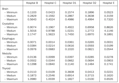
Table 2 shows minimum, medium and maximum values of doses to most exposed organs (brain, crystalline, esophagus,
me-Table 1 Features of the x-ray equipment of hospitals/examination rooms included in the present study.
Hospital/ examination room
Hospital B
Hospital C
Hospital D – Room 1 – Room 2
Hospital E
Hospital rating
General hospital
Children’s hospital
Children’s hospital
Hospital infantil
Equipment
Siemens – 3-phase, high frequency
Medicor – single phase, complete wave
VMI – single phase, complete wave Siemens – single phase, complete wave
Intecal – single phase, complete wave
Filtration (mmAl)
3.7
5.3
3.8 4.0
dulla, and thyroid) estimated for occipito-mental and occipitofrontal views. Consid-ering that the software PCXMC does not calculate the dose to the crystalline, this value was approximately estimated by the skin entrance dose (personally given infor-mation by Tapiovaara M).
DISCUSSION
Figure 1 shows that the projections most frequently utilized for facial sinuses assess-ment are the occipitofrontal and
occipito-mental ones. Only in the hospital E, a single occipitomental view was frequently re-quested. In the other institutions, occipito-frontal and occipitomental views were re-quested in conjunction. Lateral sinus view was observed only in the hospital D, al-though in a small part of the examinations (< 10%). For this reason, the lateral view has not been included in the other analyses of the present study.
As regards lead protective devices dur-ing the examinations, the authors observed that only the hospital D utilized thyroid
shielding in about 25% of examinations. In the other hospitals, the utilization of this type shielding was not observed. On the other hand, by the analysis of Figure 2, It can be observed that in the hospitals B and D collimation cylinders were not utilized according to the recommendations of the British guidelines on best practice in the x-ray imaging(3) and one of the main manu-als of radiological technique(7). The non-utilization of collimation cylinders in radio-graphic sinus evaluation may unnecessar-ily expose the thyroid (this is a highly ra-diosensitive organ) and esophageal re-gions, besides increasing the dose to other organs such as crystalline, brain and me-dulla. This fact may be observed on Table 2. For this reason, the utilization of a cy-lindrical collimator coupled with the x-ray equipment for studies of facial sinuses. An interesting fact observed in the hospital D is that a lead plate with a central orifice placed on the chassis, in a way that the appearance of the image obtained is simi-lar to the one if a collimator cylinder were utilized. This corroborates the absence of a culture of radiological protection, consid-ering that technicians try to meet the medi-cal requirements (visualization of a round-shaped image) with no concern regarding the protection of the patient.
Also, in Figure 2, it is possible to ob-serve that in almost all examinations antiscattering grids were utilized, contrar-ily to the recommendations included in the British guidelines on best practice(3), about the non-utilization of antiscattering grids in the examination of facial sinuses of chil-dren with < 10 years of age. The utilization of antiscattering grids is important for re-ducing the scattered radiation. However, the intensity of the radiation beam is re-duced, requiring higher tube loading (mA.s), and increasing the dose to the pa-tient. Considering that the scattered radia-tion intensity depends, among other fac-tors, on the patient thickness; and, consid-ering the lower thickness of pediatric pa-tients, antiscattering grids are deemed to play a non-significant role in the reduction of scattered radiation, so its utilization will only increase the dose to the patient.
Figure 3 shows the number of facial si-nuses studies (occipitofrontal and occipito-mental in conjunction) as a function of the Figure 2. Percentage of
studies with antiscatter-ing grid and collimator, performed in patients divided into three age ranges (1–5 years, 5–10 years, 10–16 years) in the four hospitals in-cluded in the present study.
Figure 1. Number of studies performed for each radiographic view of the facial sinuses in pediatric pa-tients divided into three age ranges (1–5 years, 5–10 years, 10–16 years) in five examination rooms of four hospitals included in the present study.
Fronto-naso
Mento-naso
tube voltage and exposure time. Hospital D employs higher kilovoltage (kV); and hospitals C and E, lower kilovoltage and longer exposure time. Along the develop-ment of the present study, the authors could observe that only the hospital B x-ray equipment was able to provide exposure times shorter than 10 ms, and, therefore this is the only hospital in the present study meeting the minimum best practices re-quirements for radiographic examination in
children(2,3). Notwithstanding, despite the equipment capability, the number of exami-nations with longer exposure time (more than 40 ms) is still considerably high in this hospital. As a result, the entrance air kerma rates found in this hospital are higher than those found in hospitals C and D, and lower than those found in the hospital E, as per Figure 4. This figure also demonstrates that hospital E presented a high variation in entrance air kerma rates.
Mean entrance air kerma values for five examination rooms of four hospitals were, respectively: 1398 µGy, 829 µGy, 877 µGy, 1168 µGy and 3886 µGy for patients in the age range between one and five years; 1561 µGy, 1107 µGy, 1184 µGy, 1327 µGy and 4400 µGy for patients in the age range be-tween five and 10 years; and 1613 µGy, 960 µGy, 1520 µGy, 1518 µGy and 5091 µGy for patients in the age range between 10 and 16 years. All of these values were above the reference levels proposed by the British guidelines(3). One can observe that mean values found at hospital E are three-fold higher than those found at the other hospitals. Considering that these values correspond to estimates of mean entrance air kerma/view, the two-view examinations (occipitofrontal and occipitomental) which are usual in the hospitals B, C, and D, ex-pose the pediatric patients to lower risks than those resulting from the single-view examination (occipitomental) performed in the hospital E.
Table 2 demonstrates that, despite the significantly highest entrance air kerma rates in the hospital E, the doses to thyroid and esophagus were lower. This can be explained by the extensive utilization, by this hospital, of a collimator cylinder coupled with the x-ray tube. The results found by the present study corroborate the relevance of the utilization of collimator cylinder in radiographic facial sinuses ex-aminations. The estimated values of dose to the crystalline of patients in the hospital E, probably, are lower than those on Table 2 because they were approximate by the entrance air kerma rates. Considering the frequent utilization of collimator cylinder in this hospital, this approximation tends to overestimate the value for dose to the crys-talline. Anyway, the high values for doses to this organ indicate the necessity of uti-lizing, if technically feasible, posteroante-rior projections instead of the anteroposte-rior projections usually adopted by all of the hospital included in the present study for children with less than six years of age.
CONCLUSIONS
Frequency, radioprotection, doses and risks in radiographic assessment of para-nasal sinuses of pediatric patients in hos-Figure 3. Number of studies of facial sinuses as a function of x-ray tube voltage and exposure time in the
four hospitals, for patients in three age ranges: 1–5 years, 5–10 years, 10–16 years.
pitals of Belo Horizonte, MG, Brazil were analyzed in the present study. The authors could observe that, in the majority of hos-pitals, occipitomental and occipitofrontal views are frequently requested in conjunc-tion. This implies a significant increase in the radiation dose delivered to the patient, calling into question the justification for a two-view examination, considering clini-cal studies published in the literature(8,9).
Risks for patients can be considerably reduced by means of an optimization of procedures, particularly regarding x-ray field collimation (cylinder), utilization of high x-ray tube voltage, low exposure times (and, consequently, lower tube loadings), and non-utilization of antiscattering grids.
Furthermore, the utilization of posteroan-terior projections instead of anteroposposteroan-terior projections, if technically feasible, also would contribute to a significant reduction in the radiation doses to the crystalline.
Acknowledgements
The authors express their gratitude to the physicians, radiology technicians, nurses, directors and other health profes-sionals in the institutions participating in the present study. The student Alexandre Ferreira Carmo thanks the Conselho Nacional de Desenvolvimento Científico e Tecnológico (CNPq), for the scholarship granted by the Probic – Programa de Bolsas de Iniciação Científica.
REFERENCES
1. International Commission on Radiological Pro-tection. Recommendations of the International Commission on Radiological Protection. ICRP Publication 60. New York, NY: Pergamon Press, 1991.
2. European Commission. European guidelines on quality criteria for diagnostic radiographic images in paediatrics. EUR 16261. Luxembourg: Office for Official Publications of the European Commu-nities, 1996.
3. Cook JV, Shah K, Pablot S, et al. Melhor prática em radiologia pediátrica: um manual para todos os serviços de radiologia. Rio de Janeiro, RJ: Edi-tora Fiocruz, 2006.
4. Araújo Neto SA, Souza AS, Pereira IMR, Bara-cat ECE. Alterações incidentais dos seios da face na tomografia computadorizada do crânio e órbi-tas em crianças. Radiol Bras 2005;38:245–250. 5. Gebrim EMMS. Alterações incidentais dos seios da face na tomografia computadorizada em crian-ças. Radiol Bras 2005;38(4):III–IV.
6. Jacomelli M, Souza R, Pedreira Júnior WL. Abor-dagem diagnóstica da tosse crônica em pacien-tes não-tabagistas. J Bras Pneumol 2003;29:413– 420.
7. Bontrager KL. Tratado de técnica radiológica e base anatômica. Rio de Janeiro, RJ: Guanabara Koogan, 1999.
8. Williams JW Jr, Roberts L Jr, Distell B, Simel DL. Diagnosing sinusitis by x-ray: is a single Waters view adequate? J Gen Intern Med 1992;7:481– 485.
9. Ros SP, Herman BE, Azar-Kia B. Acute sinusitis in children: is the Water’s view sufficient? Pediatr Radiol 1995;25:306–307.
10. American Association of Physicists in Medicine. Protocols for the radiation safety surveys of di-agnostic radiological equipment. AAPM Report No. 25. New York, NY: American Institute of Physics, 1988.
11. Petoussi-Henss N, Zankl M, Drexler G, Panzer W, Regulla D. Calculation of backscatter factors for diagnostic radiology using Monte Carlo methods. Phys Med Biol 1998;43:2237–2250.
12. Tapiovaara M, Lakkisto M, Servomaa A. PCXMC: a PC-based Monte Carlo program for calculating patient doses in medical x-ray exami-nations. Report STUK-A139. Helsinki, Finland: Finnish Centre for Radiation and Nuclear Safety, 1997.
Table 2 Minimum, medium and maximum values of radiation doses (in mGy) to the most exposed organs in children with < 10 years of age, in the five hospitals included in the present study.