Viral inactivation in hemotherapy: systematic review on inactivators with action on
nucleic acids
Patricia Marial Sobral1 Artur Emilio de Lima Barros2 Ayla Maritcha Alves Silva Gomes3 Cristine Vieira do Bonfim4
1 Empresa Brasileira de Hemoderivados e Biotecnologia, HEMOBRAS, Recife, PE, Brazil 2 Pronto Socorro Cardiológico de Pernambuco Professor Luiz Tavares - PROCAPE, Recife, PE, Brazil
3 Faculdade Associação Caruaruense de Ensino Superior - ASCES, Caruaru, PE, Brazil 4 Fundação Joaquim Nabuco - Fundaj, Recife, PE, Brazil
Conflict-of-interest disclosure:
The authors declare no competing financial interest
Submitted: 2/24/2012 Accepted: 4/4/2012
Corresponding author: Cristine Vieira do Bonfim
Empresa Brasileira de Hemoderivados e Biotecnologia, Hemobrás
Avenida Engenheiro Antônio de Góes nº 60, JCPM Trade Center, 11º andar, Pina 51.010-000 Recife, PE, Brazil [email protected]
www.rbhh.org or www.scielo.br/rbhh
DOI: 10.5581/1516-8484.20120056
Introduction
The current processes for pathogen inactivation in blood therapy components (red cell concentrate, platelets, plasma or cryoprecipitate) involve contact by the inactivation agent and subsequent removal
processes by systems integrated with the collection and processing procedure(1). These processes may
cause damage to the blood components, thereby resulting in shortening the in vivo lifespan of red cells or platelets, or diminished levels of coagulation proteins in fresh frozen plasma(2).
Photodynamics is a technological platform that uses a combination of a photosensitizer, light and oxygen to achieve selective destruction of a target(3). This method can be used to
target the viral genome and to perform hemotherapy. The following examples of treatments can be highlighted: phenothiazine stains such as methylene blue (MB), for viral inactivation of plasma components and derivatives; psorolens (amotosalen HCl ou S-59), for pathogen reduction in platelet and plasma units; S-303 FRALE (frangible anchor linker effector) compound and inactin (PEN 110), both used for viral inactivation in red cells; and riboflavin
(vitamin B12), with action on plasma, blood derivatives, platelets and red cells(4).
The MB molecule is relatively unstable when subjected to electron excitation, and can undergo an electron rearrangement known as a triplex excited state, thus facilitating the photodynamic action(5).This compound can bind to DNA in two ways: outside the DNA helix or intercalated
between its nucleotides. Its mechanism of action consists of guanine-specific cleavage(5).
Psorolens are small planar molecules that cross through cell membranes and viral capsids and intercalate between nucleic acids(6). During illumination with ultraviolet light (UVA), psorolen (S-59) reacts with the pyramidal bases of DNA or RNA to form covalent bonds within and between nucleic acids. Since the target of S-59 treatment is nucleic acid, it does not impede prion transmission. Prions do not contain these biomolecules and can cause a variant
of Creutzfeldt-Jakob disease and, possibly, serious chronic diseases(7).
The S-303 FRALE compound is capable of binding to nucleic acids after activation through changes in local pH. This compound is an alkylating agent derived from a quinacrine mustard that belongs to a class of compounds linked to drug delivery systems. The mechanism of action of S-303 consists of binding to nucleic acids. S-303 is used for pathogen inactivation in red blood cells(1).
In the treatment process using the inactin PEN 110, there is a covalent interaction with nucleophilic centers of nucleic acids (predominantly with N7 of the guanine residues), followed by methylation. This results in the spontaneous opening of the imidazole ring of the
guanine and produces a modified base (O6-methylguanine) that acts as a potent inactivator
of DNA polymerase(2). Modification of the base may also produce non-basic sites, which
represent a stop sign for DNA and RNA polymerase(8).
The aim of this study was to conduct a systematic review on the photoinactivators used in hemotherapy, with action on viral genomes. The SciELO, Science Direct, PubMed and Lilacs databases were searched for articles. The inclusion criterion was that these should be articles on inactivators with action on genetic material that had been published between 2000 and 2010. The key words used in identifying such articles were “hemovigilance”, “viral inactivation”, “photodynamics”, “chemoprevention” and “transfusion safety”. Twenty-four articles on viral photoinactivation were found with the main photoinactivators covered being: methylene blue, amotosalen HCl, S-303 frangible anchor linker effector (FRALE), riboflavin and inactin. The results showed that methylene blue has currently been studied least, because it diminishes coagulation factors and fibrinogen. Riboflavin has been studied most because it is a photoinactivator of endogenous origin and has few collateral effects. Amotosalen HCl is effective for platelets and is also used on plasma, but may cause changes both to plasma and to platelets, although these are not significant for hemostasis. S-303 FRALE may lead to neoantigens in erythrocytes and is less indicated for red-cell treatment; in such cases, PEN 110 is recommended. Thus, none of the methods for pathogen reduction is effective for all classes of agents and for all blood components, but despite the high cost, these photoinactivators may diminish the risk of blood-transmitted diseases.
Vitamin B12, or riboflavin, is an active compound for each of the main components of blood without causing significant damage to these structures, including for use in red cells(4,9). Joshi(10)
proved that the photodynamic reaction between riboflavin and
light generated the superoxide radicals O
-2 and HO2, thus causing
damage to nucleic acids. Since this is a substance found naturally in the organism (along with its metabolites, including those induced by photo-irradiation), it has so far not been considered to be mutagenic or toxic and its removal is unnecessary.
The photoinactivators mentioned above are the ones that have been studied most in relation to use in treating blood and its derivatives. When these inactivation techniques interact with proteins, this may cause cell signal transduction errors, deficiencies in cell respiration routes or structural abnormalities. Chemical bonding to nucleic acids or intercalation in the chains of these acids leads to transcription errors, translation errors or
replication of the microorganism(2).
Choosing an appropriate photoactivator is important for success in viral inactivation of blood and its derivatives. Therefore, it is relevant to investigate the mechanisms of action and chemical properties of inactivation agents, in order to improve this process. Reviewing the literature provides an excellent guide for observing the way in which this concept in hemotherapy has evolved. In this manner, the present study had the aim of conducting a systematic review on photoinactivators with action on viral genomes that are used in hemotherapy.
Methods
This was a systematic review of the literature, of descriptive and exploratory nature. The work was developed in a sequence of methodological stages as follows: definition of the problem; objectives of the study; inclusion criteria; search for studies; critical evaluation of studies; data gathering; and data synthesis.
The electronic search strategy made use of the following descriptors: viral inactivation; chemoprevention; inactivators (limited to those used in hemotherapy with action on viral genetic material); viral inactivation in hemotherapy; and photoinactivation of blood. These descriptors were cross-referenced with hemovigilance and transfusion safety. The search was conducted
in the SciELO, Science Direct, PubMed and Lilacs databases and was limited to the period from 2000 to 2010.
The inclusion criteria were that the articles should make reference to viral photoinactivation in hemotherapy and to transfusion safety, and should be published in Portuguese or English. From this, the following were selected for analysis: transfusion safety (studies dealing with means for ensuring safety in using blood and its derivatives); inactivation of blood and its derivatives (articles dealing with use of inactivators in hemotherapy); photoinactivators with action on viral genetic material (studies in which the inactivator acted on the viral genome); and chemistry and mechanism of action of viral photoinactivators (studies describing how the chemical structure of the inactivator interacted with the viral genome during the inactivation).
Articles were selected based on the titles and abstracts of indexed published papers in Brazilian and foreign periodicals that were accessed in full and were found to relate to viral inactivation in hemotherapy. Articles in which the inactivator studied did not have any action on the DNA/RNA, those in which the inactivator was not used on blood and its derivatives and those in which there was no clinical application were excluded.
To gather data, a form was created in order to transcribe the following identification data on the article: title, periodical (volume, number, page and year), authors (including country of origin) and language (English or Portuguese); and the following data relating to the research: photoinactivator studied, blood component treated, effectiveness of the inactivator, results obtained and conclusion.
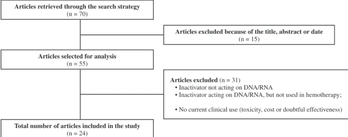
After reading the abstracts, a total of 70 studies were identified. Of these, 15 were excluded because of the title, abstract or date and 55 were potentially relevant to the investigation. Thirty-one other articles were excluded because they dealt with inactivators that did not act on DNA/RNA or were not used in hemotherapy, or furthermore, did not have any clinical use because they were considered to be toxic, too expensive or of doubtful effectiveness. Twenty-four articles were selected, and these were read carefully in full, given that they met the inclusion criteria and that their results were considered to be valid for inclusion in this study. The flow chart below (Figure 1) illustrates the selection sequence for the articles.
Figure 1 - Flow chart for study selection sequence
Articles retrieved through the search strategy
(n = 70)
Articles excluded because of the title, abstract or date
(n = 15)
Articles selected for analysis
(n = 55)
Articles excluded (n = 31)
•InactivatornotactingonDNA/RNA
•InactivatoractingonDNA/RNA,butnotusedinhemotherapy;
•Nocurrentclinicaluse(toxicity,costordoubtfuleffectiveness)
Total number of articles included in the study
Results
A total of 24 articles fulfilled the selection criteria for this study. The country with most published papers on this topic was the United States (fifteen), followed by the United Kingdom (five), Brazil (two), Canada (one) and Spain (one).
Regarding the years of publication, it was noted that the majority of the articles were published in the years 2001, 2002 and 2003. Amotosalen (S-59) was the most studied substance (15 studies), followed by PEN 110 (14 studies) and riboflavin (13 studies). Methylene blue was covered in 12 studies and S-303 FRALE in 11 studies, with data of interest for the present review. It should be noted that some of the studies presented reviews on several photoinactivators.
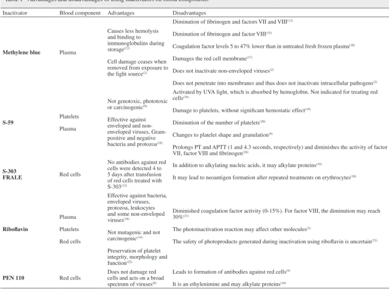
Table 1 presents the five photoinactivators studied here, the blood component treated and the advantages and disadvantages of using them. Three studies emphasized the advantages of using methylene blue and affirmed that this photoinactivator was not only
effective but also did not cause damage to plasma components(4,5,11).
Another study says it caused less hemolysis and bonding to immunoglobulins during storage and another states that it ceased
to cause damage to cells when the light source was removed(2,12).
Also on methylene blue, another four studies reported that it had the disadvantages of diminishing the quality of the treated product, with qualitative and quantitative losses in coagulation factors and fibrinogen and another study says that it damaged red cells, and yet another says that it did not inactivate non-enveloped viruses and did not penetrate into membranes, such that it was incapable of inactivating intracellular pathogens(2,13-17).
S-59, which was used on plasma and platelets, was not genotoxic, phototoxic or carcinogenic and was effective for enveloped and non-enveloped viruses, gram-positive and negative bacteria and protozoa(14,18). However, it could lead to structural damage and diminished platelet counts and was not indicated for treating red cells(9,19,20) (Table 1).
S-303 FRALE, which was used in treating red cells, could lead to the formation of neoantigens on the surface of erythrocytes after repeated treatments and, in addition to alkylating nucleic acids, could also alkylate proteins(16,18).
Riboflavin diminished the activity of coagulation factors, especially factor VIII, and the photodynamic reaction may affect other molecules, but it was effective against bacteria, enveloped viruses, protozoa, leukocytes and some non-enveloped viruses, as
Table 1 - Advantages and disadvantages of using inactivators on blood components
Inactivator Blood component Advantages Disadvantages
Methylene blue Plasma
Causes less hemolysis and binding to immunoglobulins during storage(12)
Cell damage ceases when removed from exposure to the light source(2)
Diminution of fibrinogen and factors VII and VIII(13)
Diminution of fibrinogen and factor VIII(15)
Coagulation factor levels 5 to 47% lower than in untreated fresh frozen plasma(18)
Damages the red cell membrane(17)
Does not inactivate non-enveloped viruses(2)
Does not penetrate into membranes and thus does not inactivate intracellular pathogens(2)
S-59
Platelets
Plasma
Not genotoxic, phototoxic or carcinogenic(9)
Effective against enveloped and non-enveloped viruses, Gram-positive and negative bacteria and protozoa(18)
Activated by UVA light, which is absorbed by hemoglobin. Not indicated for treating red cells(16)
Damage to platelets, without significant hemostatic effect(19)
Diminution of the number of platelets(20)
Changes to platelet shape and granulation(9)
Prolongs PT and APTT (1 and 4.3 seconds, respectively) and diminishes the activity of factor VII, factor VIII and fibrinogen(24)
S-303
FRALE Red cells
No antibodies against red cells were detected 4 to 5 days after transfusion of red cells treated with S-303(12)
In addition to alkylating nucleic acids, it may alkylate proteins(16)
It may lead to neoantigen formation after repeated treatments on erythrocytes(18)
Riboflavin
Plasma
Platelets
Red cells
Effective against bacteria, enveloped viruses, protozoa, leukocytes and some non-enveloped viruses(16)
Not mutagenic and not carcinogenic(14)
Preservation of platelet integrity, morphology and function(22)
Diminished coagulation factor activity (0-15%). For factor VIII, the diminution may reach 30%(21)
The photoinactivation reaction may affect other molecules(2)
The safety of photoproducts generated during inactivation using riboflavin is uncertain(21)
PEN 110 Red cells
Does not damage red cells and acts on a broad spectrum of viruses(8)
Leads to formation of antibodies against red cells(9)
PEN 110 has the advantage of not needing light for activation. Instead, it is activated through changes in pH(12). Other
advantages are that it preserves the morphological and functional integrity of platelets, does not damage red cells and has a broad spectrum of action on viruses(8,22). According to the study by
Seghatchian & Souza(9), it has the disadvantage that it may lead to
formation of antibodies against red cells.
Nevertheless, Bryant and Klein(6) reported that no clinical
consequences were observed in cases of antibody formation, and that the nature of the immune responses and the specificity of the antibodies formed by PEN 110 were unknown. Because riboflavin is a natural component, it has not been considered to be toxic, mutagenic or capable of diminishing coagulation factors or fibrinogen, and so far, nor have its photoproducts. Thus, removal of its photoproducts after inactivation has been completed is not considered to be necessary. It has been recommended in 90% of
the studies, but Shuyler(21) observed that riboflavin may decrease
the activity of coagulation factors by up to 15%. For factor VIII, this diminution may reach 30%.
Conclusion
The results show that methylene blue is currently the least studied of these agents because it decreases coagulation factors and fibrinogen. Riboflavin has been studied most because it is a photoinactivator of endogenous origin and has few collateral effects. Amotosalen HCl, which is effective for platelets and also used for plasma, may cause changes both to plasma and to platelets, but without significance for hemostasis. S-303 FRALE may lead to neoantigens in erythrocytes and is less indicated for treating red cells; PEN 110 is the agent of choice in such cases.
Thus, it can be concluded that the pathogen reduction technology consisting of photoinactivators is complementary to pre-transfusion triage. However, interference with certain blood constituents limits the use of some components, and the efficacy of inactivation also depends on the nature of the microorganism to be inactivated. In this way, none of these methods will be effective for all classes of agents and for all blood components.
Doubts remain regarding whether greater investments in these techniques might compensate for their high implementation cost and ensure the quality and quantity of safe hemotherapy products.
References
1. Wendel S. Quimioprofilaxia de doenças transmissíveis por transfusão em componentes lábeis hemoterápicos. Rev Soc Bras Med Trop. 2002;35(4):275-81.
2. Pelletier JP, Transue S, Snyder EL. Pathogen inactivation techniques. Best Pract Res Clin Haematol. 2006;19(1):205-42.
3. Jori G, Brown SB. Photosensitized inactivation of microorganisms. Photochem Photobiol Sci. 2004;3(5):403-5.
4. Perussi JR. Inativação fotodinâmica de microrganismos. Quim Nova. 2007;30(4):988-94.
5. Wainwright M. The emerging chemistry of blood product disinfection. Chem Soc Rev. 2002;31(2):128-36.
6. Bryant BJ, Klein HG. Pathogen inactivation: the definitive safeguard for the blood supply. Arch Pathol Lab Med. 2007;131(5):719-33.
well as being non-mutagenic and non-carcinogenic and preserving the morphological and functional integrity of platelets(2,14,16,21,22).
PEN 110 was used for treating red cells. It was the only one of these inactivators that did not require light for it to be
activated(12). It did not damage platelets or erythrocytes, but had
the disadvantages that saline washes to remove its residues were required after the treatment and that its use led to the formation of antibodies against red cells (Table 1)(2,8,9).
Discussion
The data showed that the USA was the country with the greatest number of published papers. The USA is a self-sufficient producer of blood derivatives, and this topic has been of interest there since 1980, the year when HIV was discovered. European countries such as France and Germany are important investors in photoinactivation within hemotherapy, but do not come close to the leading position of American-authored papers, where
methylene blue was first used in plasma in the 1990s(2).
In Brazil, the blood derivative industry has not yet become consolidated, and this topic remains underexplored. Dependence on external suppliers has also contributed towards a lack of investment in research on photoinactivation within hemotherapy. The three photoinactivators that have been studied the most are S-59, riboflavin and PEN 110. These three photoinactivators have a broad spectrum of action and practically do not have any toxicity, and this has led to increasing numbers of studies on them. Some comparative studies have presented methodologies of greater effectiveness and lower cost, such that clinical use of
these substances may become viable in the near future(2,4,6,17,18).
It has also been observed that methylene blue diminishes coagulation factors and fibrinogen in plasma and does not inactivate intracellular viruses because it does not cross membranes. According to Pelletier et al.(2), this diminution is secondary to the oxidation
of histidine residues and other amino acids, and the difficulty in crossing membranes is due to the hydrophobic nature of methylene blue at a concentration of 5 mM which impedes its penetration into plasma membranes. S-59 is inappropriate for treating red cells because it uses UVA light, which is absorbed by hemoglobin and causes qualitative and quantitative damage to platelets, but without hemostatic significance(9,16,19-20). It prolongs PT (prothrombin time)
and APTT (Active Partial Thromboplastin Time) and diminishes the activity of factor VII, factor VIII and fibrinogen, but it is advantageous in this process because lower doses can be applied to the plasma for shorter times, thus protecting the platelets from the high energy of UVA, without losing its antiviral activity(23).
S-303 FRALE was not greatly discussed by the authors of the studies examined here because this is a photoinactivator at an experimental stage. Nonetheless, some authors already highlighted the advantages of using this agent, with the affirmation that it does not lead to the formation of antibodies against red cells four to five days after transfusion of these cells(12).Two studies have shown that it has disadvantages: alkylation of proteins and formation of neoantigens after repeated treatments(16,18). In order to increase the
safety of the hemotherapy component that has been treated, this product is removed through fixation to an external matrix (which
7. Lin L, Hanson CV, Alter HJ, Jauvin V, Bernard KA, Murthy KK, et al. Inactivation of viruses in platelet concentrates by photochemical treatment with amotosalen and long-wavelength ultraviolet light. Transfusion. 2005;45(4):580-90.
8. Lazo A, Tassello J, Jayarama V, Ohagen A, Gibaja V, Kramer E, et al. Broad-spectrum virus reduction in red cell concentrates using INACTINE PEN110 chemistry. Vox Sang. 2002;83(4):313-23. 9. Seghatchian J, de Souza G. Pathogen-reduction systems for blood
components: the current position and future trends. Transfus Apher Sci. 2006;35(3):189-96.
10. Joshi PC. Comparison of the DNA-damaging property of photosensitized riboflavin via singlet oxygen and superoxide radical mechanisms. Toxicol Lett.1985;26(2-3):211-7.
11. Wainwright M. Pathogen inactivation in blood products. Curr Med Chem. 2002;9(1):127-43.
12. Corash L. Inactivation of infectious pathogens in labile blood components: meeting the challenge. Transfus Apher Sci. 2001;8(3):138-45.
13. Atance R, Pereira A, Ramírez B. Transfusing methylene blue-photoinactivated plasma instead of FFP is associated with an increased demand for plasma and cryoprecipitate. Transfusion 2001;41(12):1548-52.
14. Seghatchian J, Krailadsiri P. What’s happening? The quality of methylene blue treated FFP and cryo. Transfus Apher Sci. 2001;25(3):227-31.
15. Williamson LM, Cardigan R, Prowse CV. Methylene blue-treated fresh-frozen plasma: what is its contribution to blood safety? Transfusion. 2003;43(9):1322-9. Comment in: Transfusion. 2004;44(6):948-50; author reply 950.
16. Solheim BG. Pathogen reduction of blood components. Transfus Apher Sci. 2008;39(1):75-82.
17. Dodd RY. Pathogen inactivation: mechanisms of action and in vitro efficacy of various agents. Vox Sang. 2002;83 Suppl 1:267-70. 18. McCullough J. Progress toward a pathogen-free blood supply. Clin
Infect Dis. 2003;37(1):88-95.
19. Murphy S, Snyder E, Cable R, Slichter SJ, Strauss RG, McCullough J, Lin JS, Corash L, Conlan MG; SPRINT Study Group. Platelet dose consistency and its effect on the number of platelet transfusions for support of thrombocytopenia: an analysis of the SPRINT trial of platelets photochemically treated with amotosalen HCl and ultraviolet A light. Transfusion. 2006;46(1):24-33.
20. Wagner SJ, Skripchenko A, Myrup A, Awatefe H, Thompson-Montgomery D, Moroff G, et al. Evaluation of in vitro storage properties of prestorage pooled whole blood-derived platelets suspended in 100 percent plasma and treated with amotosalen and long-wavelength ultraviolet light. Transfusion. 2009;49(4):704-10. 21. Schuyler R. Use of riboflavin for photoinactivation of pathogens in
blood components. Transfus Apher Sci. 2001;25(3):189-90.
22. Li J, de Korte D, Woolum MD, Ruane PH, Keil SD, Lockerbie O, et al. Pathogen reduction of buffy coat platelet concentrates using riboflavin and light: comparisons with pathogen-reduction technology-treated apheresis platelet products. Vox Sang. 2004;87(2):82-90.
23. Singh Y, Sawyer LS, Pinkoski LS, Dupuis KW, Hsu JC, Lin L, et al. Photochemical treatment of plasma with amotosalen and long-wavelength ultraviolet light inactivates pathogens while retaining coagulation function. Transfusion. 2006;46(7):1168-77.
24. Chapman JR, Moore K, Butterworth BE. Pathogen inactivation of RBCs: PEN110 reproductive toxicology studies. Transfusion. 2003;43(10):1386-93.