Article
ISSN 0102-695X doi: 10.1590/S0102-695X2011005000026
Received 26 Sep 2009 Accepted 5 Aug 2010 Available online 25 Feb 2011
Gaurav V. Sanghvi, Rina D. Koyani, Vidya S. Patil, Kishore S.
Rajput
*Department of Botany, Faculty of Science, The Maharaja Sayajirao University of Baroda, India.
Abstract:Morpho-anatomical features in leaves, stems and unripe fruits of Solanum pseudocapsicum L., Solanaceae, were investigated by histological methods. Anatomically the plant may be characterised by the presence of uniseriate trichomes, anomocytic stomata, calcium oxalate needles in leaves while presence of oval to circular compound starch grains, angular vessels, vertically upright, uni-biseriate rays and intraxylary phloem with differentiation of internal cambium abutting marginal pith cells and protoxylem in transverse view. Development of distinct internal cambium may be considered as a characteristic feature for S. pseudocapsicum. Intraxylary secondary phloem was composed of sieve tube elements, companion cells and axial parenchyma cells.
Keywords:
Solanum pseudocapsicum
Solanaceae morpho-anatomy
Introduction
There has been increasing incidence of microbial infection in recent years, largely due to an increase in pathogenesis and emergence of resistant strains. Despite the increasing production of new antimicrobial agents by pharmacological industries over the last three decades, resistance to these drugs by microorganisms is still on the rise. Other factors like low drug potency, poor solubility of drugs, toxicity and also increase of undesirable situation like certain side effects of antibiotics and uncommon infections cause a serious medical problem (Marchese et al., 2001; Poole, 2001). One approach is to screen new, inexpensive and effective drugs from other sources, including plants, for possible antimicrobial properties (Dayang et al., 2005). The use of plant extracts and phytochemicals, both with known antimicrobial
properties, can be of great signii cance in therapeutic
treatments (Gislene et al., 2000). The family Solanaceae and genus Solanum, which is one of the plant sources is known to possess antimicrobial constituents.
Solanum pseudocapsicum L., Solanaceae, is profusely branched, compact subshrubs. Stem and branches glabourous terete. Leaves are narrowly elliptic and tapering at both ends, margin undulates. Flower axillary, solitary or 4-22 in clusters. Berries globose, green changing yellow to red at maturity. At maturity colour of berries are attractive, hence it is cultivated as an indoor ornamental. Number of seeds ranges from 50 to 100, while berries could be as many as 100 per plant (Bassett & Mundro, 1985).
It is a poisonous plant used in traditional medicine for the treatment of acute abdominal pain (Boericke, 1927), in the treatment of boils and gonorrhea and as tonic for men (Batten & Bokelmann, 1966). It contains solanocapsine and other alkaloids that are reported to be fatal to man and animals (Parisi & Francia, 2000). Phytomedical investigations have revealed that the plant possesses antiviral, cytotoxic, hepatoprotective, and antitumor properties (Shrishailappa et al., 2003; Vijayan et al., 2003).
Despite toxicological and volatile components from aerial parts as well as from roots have been documented, there is no information about morpho-anatomical features of S. pseudocapsicum. It is therefore, necessary to document the morpho-taxonomic characters
to avoid the falsii cation and adulteration of the drug.
Materials and Methods
Plant material collection
Acc. No.: KSR 13, 48 and 73. Collected plant is new report to the Gujarat State (Rajput & Raole, 2009) and its
identity was veriied by Prof. S. R. Yadav, Department of
Botany, Shivaji University, Kolhapur (MS), India.
Microtomy
Suitably trimmed samples of stem, root, leaves
and unripe fruits were ixed in FAA (Berlyn & Miksche,
1976). After 24 h they were transferred 70% alcohol and dehydrated with tertiary butyl alcohol series (30%, 50%, 70%, 90% followed by 3x 100% pure TBA) and
processed by routine method of parafin embedding. Thick
samples were also sectioned on a sliding microtome. Transverse, radial and tangential longitudinal sections of 12-15 µm thickness were obtained with rotary and sliding microtome and stained with safranin fast green combination (Johansen, 1940). Some of the sections were also stained with iodine/potassium iodide to study the morphology and depositions of starch.
To obtain the length and width of vessel elements,
ibriform vessel elements and xylem ibres, small pieces of
xylem adjacent to the cambium ring were macerated with Jeffrey’s solution (Berlyn & Miksche, 1976) at 55 to 60 ºC for 24-36 h, and stained with safranin to study general morphology and dimensional details. Length and width of
the sieve tube elements and ibre nuclei were measured
directly from the tangential longitudinal sections. One hundred measurements were chosen randomly to obtain mean and standard deviation for each cell type. Important results were microphotographed with Leica DM 2000 trinocular research microscope.
Results and Discussion
Solanum pseudocapsicum L., is a perennial, much branched, compact sub-shrub reaching 2 to 4 feet in height. Stem erect, terete, cylindrical, greenish when young and pale ash coloured at maturity. Leaves simple, mostly alternate, narrowly elliptic, tapering at both ends, margin undulate. Lamina 100 to 180 mm long and 43 to 67 mm wide, acute, acuminate, glabrous, minutely hairy beneath. Flowers axillary, solitary or 4-22 in clusters, small, pedicellate, pedicel 10 to 12 mm, smooth, hairy when young. Berries globose, dark green when young and yellow to orange at maturity, around 10 to 13 mm in diameter.
Microscopy
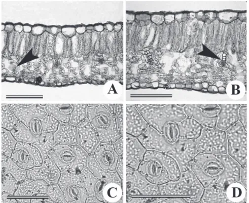
In transverse view cells of leaf epidermis were barrel shaped with slightly anticlinal walls and possessed
smooth thick cuticle (Figure 1A, B). Covering trichomes were not much numerous, uniseriate with 2-3 cells. Stomata were anomocytic (Figure 1C, D). The mesophyll reveals dorsiventral organisation, comprising one stratum of palisade parenchyma and about four layers of spongy parenchyma (Figure 1A, B), the latter occupying 50-60% of the mesophyll. Minor collateral vascular bundles were embedded in the mesophyll and are enclosed by a sheath of large parenchyma cells. Deposition of calcium oxalate needles were also observed in spongy parenchyma cells. In the young stems, the epidermis consists of single compact layer of isodiametric and thin walled parenchyma cells. A thin layer of cuticle covers the epidermis while 2-3 layered hypodermis composed of parenchyma cells differentiated beneath it. The bulk of cortex consisted parenchyma cells that showed heavy
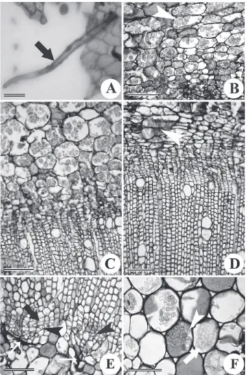
accumulation of oval to circular, compound starch grains (Figure 2B, C). As the secondary growth progressed further in the mature stems, medullary phloem rays away from the cambium underwent obliteration and became
tangentially lattened (Figure 2D).
Secondary xylem was diffused porous with
indistinct growth rings and was composed of ibres,
vessels, axial and ray parenchyma cells (Figure 2D). Vessels were mostly angular and possessed alternate bordered pits on their lateral walls. Perforation plates were simple and were arranged slightly oblique to transverse on the end walls. Vessel elements were 300 to 313 µm in
length and 48 to 73 µm in diameter whereas xylem ibres
were 550 to 623 µm in length and possessed simple slit like pits. However, differentiation of vessels remained restricted to certain portion of the cambium while rest
Figure 2. Leaf epidermal peel (A) and transverse (B–F) view of main stem of Solanum pseudocapsicum L. Solanaceae. A: Leaf peel showing warty trichome (arrow). B: Cortical parenchyma in mature stem with starch grains (arrowhead) note the tangentially
lattened parenchyma on upper side while lower side shows phloem parenchyma. C: Young stem showing cortical parenchyma illed
of the cambial cells exclusively differentiated into xylem
ibres and axial parenchyma cells (Figure 2D).
External and internal morphology studies on any plant used in the Ayurvedic medicine are extremely
important for the identiication. Although, some
sophisticated chemical and molecular methods are
available for the identiication of plant material,
morpho-anatomical methods are the simplest among the qualitative
methods to avoid the falsiication and adulteration of
the drug (Sharma et al., 2010). Family Solanaceae is characterised by the presence of intraxylery phloem (Metcalfe & Chalk, 1950, 1983). S. pseudocapsicum also showed presence of intraxylery phloem (Figure 2E). As the secondary growth progressed further, in mature stems the marginal pith cells acquired meristematic character and differentiate into internal cambium (Figure 2E). Vesque (1875) discovered such a cambium for the
irst time in the Solanaceae, the Asclepiadaceae and the
Apocynaceae. Although intraxylary phloem is reported in several other families but there are very few species that develop internal cambium at the margin of pith. This internal cambium was functionally unidirectional and differentiated only into phloem elements in centripetal
direction. Development of intraxylary secondary phloem exerted a pressure on the internal protophloem thus leading to obliteration and crushing of internal protophloem (Figure 2E). Pith cells were characterised by the presence of oval to circular and compound starch grains (Figure 2F) and calcium oxalate needles.
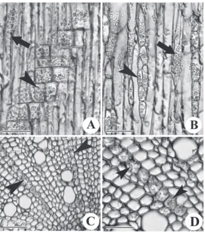
Both, in the stems as well as in roots, the xylem rays were mostly uni-biseriate with vertically elongated upright ray cells (Figure 3A, B). In the stem, deposition
of starch was observed in the xylem ibres and ray
parenchyma cells (Figure 3A, B). In roots, its deposition was restricted only to the axial and ray parenchyma cells
while ibres were free from the starch (Figure 3C, D).
Conclusion
As a characteristic feature of the family, Solanum pseudocapsicum L., Solanaceae, also shows presence of primary and secondary intraxylary phloem. In the maturing stems, the marginal pith cells formed intraxylery phloem but in mature plants, internal cambium was developed from them that gave rise to secondary phloem centripetally.
Figure 3. Radial (A), tangential (B) and transverse (C, D) view of xylem of Solanum pseudocapsicum L., Solanaceae. A: Xylem
ibres (arrow) and ray parenchyma (arrowhead) of stem showing distribution of starch in the xylem. B: Uni-biseriate rays in the stem showing starch (arrowhead). Note the lumen of the xylem ibres showing deposition of starch (arrow). C: Distribution of starch
Acknowledgement
The authors are thankful to the Department of Biotechnology, Ministry of Science and Technology,
Government of India for the inancial support. Thanks
are also due to anonymous reviewers for their valuable suggestions on the manuscript.
References
Batten A, Bokelmann H 1966. Wild lowers of the Eastern Cape
province. Cape and Transvaal Printers Ltd. Cape Town, p 129.
Bassett IJ, Mundro DB 1985. The biology of Canadian weeds: Solanum ptycanthum Dun, S. nigrum L. and S. sarrachoides Sendt. Can J Plant Sci 65: 401-414. Berlyn GP, Miksche JP 1976. Botanical microtechnique and
cytochemistry. The Iowa State University Press, Ames, Iowa, p.326.
Boericke W 1927. Pocket Manual of Homeopathic Materia Medica (9th ed.). Boericke and Runyon, New York, p.
598.
Dayang FB, Razinah S, Paden M 2005. Antimicrobial activities of ethanol and ethyl acetate extracts from the fruits of
Solanum torvum. Appl Biol (Malays) 34: 31-36. Gislene GF, Nascimento JL, Paulo CF, Guliana LS
2000. Antibacterial activity of plant extracts and phytochemicals on antibiotic resistant bacteria. Braz J Microbiol 31: 247-256.
Johansen DA 1940. Plant Microtechnique. Mcgraw hill, New
York.
Marchese A, Shito GC 2001. Resistance patterns of lower respiratory tract pathogens. in Europe. Int J Antimicrob Agents 16: 25-29.
Metcalfe CR, Chalk L 1950. Anatomy of the dicotyledons. Clarendon Press, Oxford.
Metcalfe CR, Chalk L 1983. Anatomy of dicotyledons. Vol IInd. Clarendon Press, Oxford.
Parisi P, Francia A 2000. A female with central anticholinergic syndrome responsive to neostigmine. Pediatr Neurol 23: 118-185.
Poole K 2001. Overcoming antimicrobial resistance by targeting resistance mechnisms. J Pharm Pharmacol 53: 283-84. Rajput KS, Raole VM 2009. Solanum pseudo-capsicum L.:
Addition to the Flora of Gujarat State. The Indian Forester 135: 1293-1295.
Sharma S, Hullatti KK, Prasanna SM, Sharma P 2010. Comparative morpho-anatomical and preliminary phytochemical studies of Cusucuta relexa and Cassytha iliformis. Int. J Pharm Pharmaceut Sci 2 (Suppl 1): 59-64.
Shrishailappa B, Reddy S, Kumar EP, Vijayan P, Suresh B 2003. Anti-tumour activity of total alkaloid fraction of Solanum pseudocapsicum. Phytotherapy 17: 1001-1004.
Vijayan P, Prashanth HC, Preethi V, Dhanaraj SA, Badami S, Suresh B 2003. Hepatoprotective effect of the total alkaloid fraction of Solanum pseudocapsicum leaves.
Pharm Biol 41: 443-48.
Vesque J 1875. Mémoire sur l’anatomie comparée de l’écorce.
Ann Sci Nat Bot 2: 82-198.
*Correspondence
Kishore S. Rajput
Department of Botany, Faculty of Science, The Maharaja Sayajirao University of Baroda, Vadodara 390 002, India [email protected]